Intervertebral Disk Degeneration: The Microenvironment and Tissue Engineering Strategies
←
→
Page content transcription
If your browser does not render page correctly, please read the page content below
REVIEW
                                                                                                                                                  published: 20 July 2021
                                                                                                                                         doi: 10.3389/fbioe.2021.592118

                                            Intervertebral Disk Degeneration:
                                            The Microenvironment and Tissue
                                            Engineering Strategies
                                            Yiming Dou 1† , Xun Sun 1† , Xinlong Ma 1 , Xin Zhao 2 and Qiang Yang 1*
                                            1
                                             Department of Spine Surgery, Tianjin Hospital, Tianjin University, Tianjin, China, 2 Department of Biomedical Engineering,
                                            The Hong Kong Polytechnic University, Kowloon, Hong Kong

                                            Intervertebral disk degeneration (IVDD) is a leading cause of disability. The degeneration
                                            is inevitable, and the mechanisms are complex. Current therapeutic strategies mainly
                                            focus on the relief of symptoms, not the intrinsic regeneration of the intervertebral
                                            disk (IVD). Tissue engineering is a promising strategy for IVDD due to its ability to
                                            restore a healthy microenvironment and promote IVD regeneration. This review briefly
                                            summarizes the IVD anatomy and composition and then sets out elements of the
                          Edited by:        microenvironment and the interactions. We rationalized different scaffolds based on
                  Chien-Wen Chang,
                                            tissue engineering strategies used recently. To fulfill the complete restoration of a
 National Tsing Hua University, Taiwan
                                            healthy IVD microenvironment, we propose that various tissue engineering strategies
                       Reviewed by:
                   Jennifer Patterson,      should be combined and customized to create personalized therapeutic strategies for
    Instituto IMDEA Materiales, Spain       each individual.
                    Nihal Engin Vrana,
              Spartha Medical, France       Keywords: intervertebral disk degeneration, microenvironment, tissue engineering, scaffold, regeneration, disk
                                            herniation, low back pain
                *Correspondence:
                        Qiang Yang
           yangqiang1980@126.com
     † These   authors have contributed
                                           INTRODUCTION
                    equally to this work
                                           Low back pain is prevalent in the society and is the number one cause of disability globally; 60–80%
                    Specialty section:     of adults experience varying degrees of low back pain (Murray et al., 2012; Hartvigsen et al., 2018).
         This article was submitted to     Although the etiology and pathology of low back pain are complex, evidence suggests that low back
                          Biomaterials,    pain is strongly associated with intervertebral disk degeneration (IVDD) (Luoma et al., 1976). The
                a section of the journal   intervertebral disk (IVD) is a fibrous cartilaginous tissue that connects two adjacent vertebral bodies
       Frontiers in Bioengineering and     consisting of three parts: the nucleus pulposus (NP), the annulus fibrosus (AF), and the cartilage
                        Biotechnology
                                           endplate (CEP). The NP is a centrally located highly hydrated gel-like tissue surrounded by the AF,
          Received: 06 August 2020         comprised of layer-by-layer collagen fiber lamellas. The CEPs are situated at the top and bottom of
            Accepted: 18 May 2021
                                           the vertebral bodies, and they interface the IVDs with the adjacent vertebrae (Lawson and Harfe,
            Published: 20 July 2021
                                           2017). This complex structure plays a vital role in transmitting and absorbing mechanical loading
                             Citation:     onto the spine and maintaining motor function.
     Dou Y, Sun X, Ma X, Zhao X and
                                              The IVD gradually degenerates due to aging and tissue damage caused by multiple stressors,
     Yang Q (2021) Intervertebral Disk
 Degeneration: The Microenvironment
                                           resulting in vertebral instability, spinal canal stenosis, and spinal segment deformity, causing low
   and Tissue Engineering Strategies.      back pain and mobility disability (Wang et al., 2016). There are three main events in IVDD:
 Front. Bioeng. Biotechnol. 9:592118.      (1) inflammation and catabolic cascades, (2) continuous loss of cells, and (3) decline in cellular
     doi: 10.3389/fbioe.2021.592118        functions and anabolic activities (Frapin et al., 2019). NP gradually degenerates under the influence

Frontiers in Bioengineering and Biotechnology | www.frontiersin.org                 1                                               July 2021 | Volume 9 | Article 592118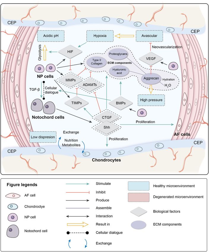
Dou et al.                                                                                                  IVDD and Tissue Engineering Strategies

of these events. The AF ruptures as the degree of degeneration            stress and inhibit inflammation; and (3) anabolic/catabolic
increases, while the degenerated NP is extruded and oppresses             balance should be mediated to ensure the quality and the
nerve roots due to abnormal stress, resulting in nerve                    quantity of the ECM.
compression symptoms. The CEP becomes more calcified,
which adversely affects the transport of nutrients and
metabolites, making it difficult to maintain sagittal stress              IVD AND THE MICROENVIRONMENT
balance. Inflammatory factors stimulate nerve roots, causing
or aggravating pain, and enhancing mobility disability. The               Anatomy and Composition of the IVD
deteriorating microenvironment further aggravates IVDD.                   The IVD is an articular cartilage structure located between the
    Traditional therapeutic strategies (surgical and non-surgical         vertebral bodies and accounts for 25–30% of the overall length
treatments) focus on solving the symptoms. Surgical treatments            (height) of the spine. An IVD consists of the NP, the AF, and the
focus more on physically relieving symptoms at the organ                  CEP. The primary function of the IVD is to provide mechanical
level, such as decompressing nerve roots (discectomy) and                 support for the vertebral body and allow movement of the spine
removing the degenerated disk (fusion and disk replacement).              (flexion, extension, and rotation) (Devereaux, 2007).
Non-surgical treatments (non-pharmacological therapy,                         The NP is a centrally located highly hydrated gel-like
pharmacological therapy, and interventional therapies)                    structure consisting of NP cells and the ECM. The cell density
perform limited intervention for the microenvironment                     in the NP is low (3,000/mm3 ), and the cell types are not
through anti-inflammatory, analgesic, and spasmolysant                    completely clear, mainly containing small chondrocyte-like
effects. Traditional therapeutic strategies do not promote disk           nucleopulpocytes (NPCs) and large vacuolated notochord cells
regeneration at the cell level and, therefore, do not reverse             (NCs) (Wang et al., 2014). The ECM of the NP is quite
the progress of disk degeneration (Harmon et al., 2020). Some             different comparing to that of hyaline cartilage and is mainly
patients relapse after treatment, and the disk degeneration               synthesized by the NPCs. It contains mostly aggrecan [a large
might even accelerate the degeneration of adjacent segments               proteoglycan (PG) aggregate], a high ratio of type II/type I
(Harper and Klineberg, 2019).                                             collagen, hyaluronic acid (HA), and secondary components (type
    In the early stage of IVDD, the enclosed environment                  IX/VI/V collagen and small proteoglycans). PGs are widely
makes it difficult for the external stem cells to exert potential         found in cartilage, the brain, IVDs, tendons, and corneal tissues.
regenerative ability on the NP. In the later stage when the               They provide viscoelastic properties, retain water, maintain
degeneration is severe, the AF will tear, and neovascularization          osmotic pressure, and arrange collagen tissue. Aggrecan in
and neoinnervation will occur in the NP. Inflammation                     the IVD contains more keratin sulfated (a highly hydrated
caused by newly colonized immune cells further harms the                  sulphated glycosaminoglycan). Thus, it provides more hydration
microenvironment and accelerates degeneration. Thus, the                  ability (Iozzo and Schaefer, 2015; Frapin et al., 2019; Harmon
deteriorating microenvironment and low self-regenerative                  et al., 2020). HA is a widely expressed glycosaminoglycan
ability of the IVD are primary obstacles for regeneration.                (GAG) located in the ECM, intracellular environment, and
How to customize suitable regenerative strategies for IVD                 the cell surface that interacts with specific proteins, such as
has become a hot topic of recent research. Tissue engineering             aggrecan, versican, lymphatic vessel endothelial HA receptor-
strategies combining material science, engineering, and life              1, tumor necrosis factor (TNF)-inducible gene-6 protein, and
science will become new clinical therapeutic approaches in                thrombospondin as well as membrane receptors, such as CD44,
the future. Tissue engineering strategies contain three key               HA-mediated motility receptor, and Toll-like receptor 4/2. Thus,
elements: cells, scaffolds, and biomechanical or biochemical              it plays a role in morphogenesis, cell migration, cell survival,
signals (Mhanna and Hasan, 2017). These elements have                     apoptosis, inflammation, and tumorigenesis (Dicker et al., 2014).
been applied and extended in IVD tissue engineering.                      HA binds to PGs to form aggrecan, which highly hydrates NP
Applications of stem cells in IVD and derivative methods                  and generates a hydrostatic pressure to effectively absorb stress,
(gene therapy and extracellular vesicle therapy) have shown               reduce vibration, and maintain the osmotic pressure and disk
promising therapeutic potential for IVDD. Illustrating the                height of healthy IVDs (Raj, 2008; Brown et al., 2012; Wang et al.,
interactive mechanisms between the components [IVD cells,                 2014). A small part of small leucine-rich proteoglycans (SLRPs)
biological factors, extracellular matrix (ECM) components,                in PGs, which mediate tissue order, cell proliferation, matrix
and environmental factors] in the IVD microenvironment                    adhesion, and the responses between cell and biological factors,
makes therapeutic strategies more rational. Scaffolds mimic               is an important signal transduction factor and receptor for
the microstructure of the IVD ECM and provide proper                      the development, morphogenesis, and immunization activities
structural support for IVD cells. Scaffolds can carry and                 of IVDs. Recent research has reported that SLRPs are related
release therapeutic biological factors. These findings contribute         to IVDD, where the asporin gene in the SLRP family rapidly
to IVD tissue engineering strategies. Tissue engineering                  upregulates after the age of 22. SLRPs also exhibit an increased
strategies should be devised, considering the pathological                expression in degenerated IVDs (Song et al., 2008; Gruber
changes of IVDD with the aim to reverse degeneration,                     et al., 2009). Type II collagen is the most critical collagen in
namely: (1) cell proliferation should be promoted and cell                the NP for forming irregular networks to support PGs and
apoptosis should be inhibited to ensure cell density; (2) the IVD         water. The distinctive arrangement equalizes the NP stress in
microenvironment should be ameliorated to mitigate cellular               different directions and, together with water, makes the NP elastic

Frontiers in Bioengineering and Biotechnology | www.frontiersin.org   2                                       July 2021 | Volume 9 | Article 592118
Dou et al. IVDD and Tissue Engineering Strategies
(Colombier et al., 2014). As IVD degeneration progresses, type One of the critical factors mediating cell metabolism is
II collagen is gradually replaced by low-elasticity type I collagen, hypoxia-inducible factor (HIF) (Semenza et al., 1994). HIF is a
and the fibrotic NP gradually loses its biomechanical function. transcription factor that initiates a coordinated cellular cascade
The AF is comprised of 15–25 layers of angle-ply collagen fiber in response to a low-oxygen tension environment, including the
lamellas containing PGs, arranged in concentric circles outside regulation of numerous enzymes in response to hypoxia (Li et al.,
the NP (Chu et al., 2018), and contains AF cells (AFCs) and ECM. 2013). The HIF-1 α-subunits remain stable in hypoxic conditions
The AF is divided into the outer AF and the inner AF. The outer but rapidly degrade under normoxic conditions. At the same
AF mainly consists of dense and organized type I collagen. Thus, time, HIF-1α and HIF-2α activities are stable in IVD, which
it has robust tensile strength, while the inner AF contains a lower illustrates that these two factors are positively related to IVD
ratio of type I collagen as the transition zone between the AF and activity (Wang et al., 1995; Agrawal et al., 2008; Li et al., 2013).
NP. The density of AFCs is about 9,000/mm3 and mainly consists Hypoxia-inducible factor is associated with most cell activities
of fibroblast cells (Raj, 2008). NP generates a hydrostatic pressure in the IVD. HIF-1α regulates the glycolytic activity of NPCs
when IVDs are under axial stress and releases fluid shear stress to ensure an energy supply under hypoxia, which results in
to the AF. The multi-lamellated AF structure effectively converts the acidic microenvironment in the IVD (Agrawal et al., 2007;
the axial stress to interlamellar stress and produces annular stress Kalson et al., 2008). HIF promotes the synthesis of PGs, type
to resist it (Liu et al., 2001; Iu et al., 2017). The AF can withstand II collagen, and GAGs by direct or indirect pathways (Agrawal
compression and tensile stress during movement of IVDs. et al., 2007; Li et al., 2013; Liu et al., 2017). It also inhibits
The CEP is a hyaline cartilage structure that connects the Fas/FasL-mediated apoptosis of NPCs by inducing the expression
vertebral bodies to the IVD. The CEP is comprised of hyaline of Galectin-3. The expression of HIF-1α increases significantly in
cartilage cells and chondrocytes that produce PGs and type II degenerated IVDs and is correlated with cell apoptosis (Ha et al.,
collagen to transport nutrients and metabolites to the IVD, which 2006; Li et al., 2013). HIF-2α regulates the expression of aggrecan
is avascular tissue. The blood supply ends in the CEP, making the and type II collagen by regulating the expression of catabolic
CEP a critical transportation junction. The CEP also bears the factors (MMPs-13 and ADAMTS-4). The expression of HIF-
stress from the IVD to protect the vertebral bodies (McFadden 2α, MMP-13, and ADAMTS-4 increases significantly, while the
and Taylor, 1989; Roberts et al., 1989; Fontana et al., 2015; Frapin expression of aggrecan and type II collagen decreases significantly
et al., 2019). in degenerated IVDs (Huang et al., 2019), indicating that HIF
As the core of this structure, the stable and enclosed plays an essential role in maintaining the IVD ECM. The hypoxic
microenvironment of the NP guarantees the expression of the environment is destroyed after IVDD occurs; the absence of HIF
ECM, which supports and separates the vertebrae, absorbs can further accelerate degeneration (Meng et al., 2018).
shock, permits transient compression, and allows for movement. Vascular endothelial growth factor (VEGF) is important
NPCs also affect the expression of the AFC ECM. A healthy during vasculogenesis and angiogenesis and mainly targets
ECM (mainly type 1 collagen in the outer AF) is comprised endothelial cells (Apte et al., 2019). Interestingly, the VEGF
of dense AF lamella, which provides protection for the NP protein and its receptors are expressed in the avascular NP
microenvironment. Besides bearing sagittal stress, the CEP is the (Fujita et al., 2008). VEGF-A has a strong angiogenic activity
sole pathway of metabolite exchange for the avascular NP. These and specific effects of mitosis and chemotaxis on endothelial
three parts together form a unique anatomical structure, which cells (Risau, 1997). The expression of VEGF-A can be induced
maintains homeostasis of the IVD microenvironment in unity by hypoxia, leading to a local vascular invasion, but, typically,
and maintains healthy IVD function. VEGF-A expressed by NPCs will not cause neovascularization.
The reason may depend on the inhibition of endothelial cell
Physiological Microenvironment adhesion and migration by the high aggrecan content in the IVD
The IVD is in a unique microenvironment: avascular, hypoxic, (Johnson et al., 1976). The complex of VEGF-A and its receptor
hyperosmotic, acidic, and with low diffusion of metabolites and VEGFR-1 also inhibit NP cell apoptosis (Fujita et al., 2008).
restricted by biomechanics (Roberts et al., 1996; Bartels et al., Nucleus pulposus cells stimulate NCs to secrete connective
1998; Wuertz et al., 2008). During early embryonic development, tissue growth factor (CTGF) and sonic hedgehog (Shh) by
a rod-like notochord is located in the central area of the embryo secreting transforming growth factor (TGF-β). CTGF and
and guides the ectoderm folds in on itself over the notochord to Shh together inhibit the expression of MMPs and stimulate
form neural tube mesenchymal cells (MSCs) to form vertebral the expression of tissue inhibitors of metalloproteinases
bodies and the AF. NCs are trapped inside and participate in the (TIMPs), which inhibit MMPs, thereby suppressing the
formation of the NP. NCs are considered to be involved in the degradation of the ECM (Erwin et al., 2006; Erwin, 2008;
regeneration of the NP through cellular dialogue with other cells. Frapin et al., 2019). CTGF and Shh also stimulate the
NCs disappear in most human adults before the bone matures, anabolism of the NP ECM and inhibit NP neovascularization
but signs of IVDD occur not long after their disappearance and apoptosis by inhibiting VEGF, interleukin (IL)-
(Hunter et al., 2003). 6, and IL-8. Shh also promotes the proliferation of
The cellular dialog between NCs and NP cells, which AF and CEP cells.
is triggered by multiple biological factors, maintains IVD The bone morphogenetic protein (BMP) family promotes
homeostasis. These biological factors can also mediate the IVD ECM synthesis by impacting the cellular dialog between
microenvironment. NCs and NP cells and regulates the production of MMPs
Frontiers in Bioengineering and Biotechnology | www.frontiersin.org 3 July 2021 | Volume 9 | Article 592118Dou et al. IVDD and Tissue Engineering Strategies
(Leung et al., 2017; Frapin et al., 2019). BMP-2 and osteogenic Davalos et al., 2010; Muñoz-Espín and Serrano, 2014; van
protein-1 (BMP-7) upregulate the expression of aggrecan Deursen, 2014; Sharpless and Sherr, 2015). Two intrinsic
and type II collagen, promote the synthesis of GAGs, and pathways are related to cellular senescence in IVDs: the p53-p21-
concomitantly inhibit the expression of profibrotic genes. BMP-2 RB pathway in a telomere-dependent manner and stress-induced
and BMP-7 are the most effective factors in the family stimulating premature senescence that activates the p16INK4a -RB pathway
the accumulation of PGs, while BMP-4 and growth differentiation independently of telomere length (Ben-Porath and Weinberg,
factor-5 GDF-5 (BMP-14) stimulate the accumulation of collagen 2005). A stimulus from the microenvironment can cause damage
(Zhang et al., 2006; Leung et al., 2017; Li et al., 2017). to IVD cells, resulting in early senescence.
The characteristics of high osmotic pressure and high Senescent cells in degenerated disks can form senescent
hydration ensure the biomechanical function of the IVD. As a cell clusters, which can cause inflammatory stress by secreting
weight-bearing organ, the metabolic and cellular activities of the pro-inflammatory cytokines and accelerate the senescence
IVD are closely related to the biomechanical microenvironment of neighboring cells (Wang et al., 2016). As a result,
(Neidlinger-Wilke et al., 2014). External loads on the spine cellular senescence, impaired self-repair capacity, increased
result in intense pressure on the disk. Intradiscal pressure inflammation, and enhanced catabolism gradually lead to
varies from 0.1 to 2.3 MPa in different locations (Wilke et al., deterioration of the microenvironment and cause disorder in the
1999; Neidlinger-Wilke et al., 2014). Severe degeneration can cellular dialogue and an imbalance of anabolism/catabolism.
unbalance sagittal/coronal stress, resulting in abnormal stress of
degenerated segments (Le Huec et al., 2011). NP cells in the Imbalance of ECM Anabolism/Catabolism
IVD are mainly under extensive hydrostatic pressure, AF cells The ECM provides mechanical support for the IVD and is
are under tensile strain, and the CEP is under compression due essential for maintaining the relatively avascular and aneural
to its location at the interface (Le Maitre et al., 2004; Setton nature of a healthy disk. Proteoglycans, which are a primary
and Chen, 2006). The cytoskeleton of the IVD cells responds to component of the ECM, retain water and contribute to the
the mechanical microenvironment. High osmotic pressure has osmotic pressure responsible for NP biomechanical properties. In
positive effects on metabolic activity and matrix gene expression the early stage of degeneration, proteoglycan content gradually
by IVD cells, and changes in hydrostatic pressure affect the decreases and is a sign of early degeneration. Type I collagen
synthesis of PGs by regulating the production of nitric oxide makes IVDs fibrotic, while type II collagen makes IVDs elastic.
(Liu et al., 2001). The proportions of collagen in the disk change with degeneration
Interactions between different components in the of the matrix. The absolute quantity of collagen changes little,
IVD microenvironment maintain the homeostasis of the but the type and distribution of the collagen can be altered.
microenvironment (Figure 1). Due to aging, impaired The ratio of collagen type II/type I decreases, and fibronectin
disk function, loss of cells, and imbalance of ECM content increases with increasing degeneration. As a result, the
anabolism/catabolism, the homeostasis of the microenvironment disk becomes more fibrotic and less elastic (Raj, 2008; Wang et al.,
breaks down, eventually resulting in pathological IVDD. 2016).
The balance between anabolism and catabolism is positively
Pathological Environment related to the IVD microenvironment, and the cellular dialogue,
The degenerative mechanisms are very complex and are related metabolic enzyme activity, and biomechanical changes affect this
to various causes, such as age, genetics, the microenvironment, balance. As the population of NCs decreases with age, the cellular
and biomechanics (Frapin et al., 2019). Early degeneration dialogue between NCs and NPCs gradually diminishes. Anabolic
may be asymptomatic. Signs may be detectable by radiography, activity is mediated by biological factor disorders, while catabolic
while the reduction in water content of NP can be visualized activity continuously progresses (Freemont, 2009).
by magnetic resonance imaging due to the reduced synthesis MMPs, disintegrin, and metalloproteinase with
of PGs and reduced disk height of the IVD on a computed thrombospondin motifs (ADAMTS) are important catabolic
tomography scan (Modic et al., 1988). Four main changes occur factors in disk degeneration. ADAMTS1, 4, 5, 9, and 15 are
during degeneration: (1) cell senescence, (2) imbalance of ECM aggrecanases that degrade aggrecan, while MMP1, 8, and 13
anabolism/catabolism, (3) inflammatory microenvironment, and are collagenases that cleave fibrillar collagen (Weiler et al.,
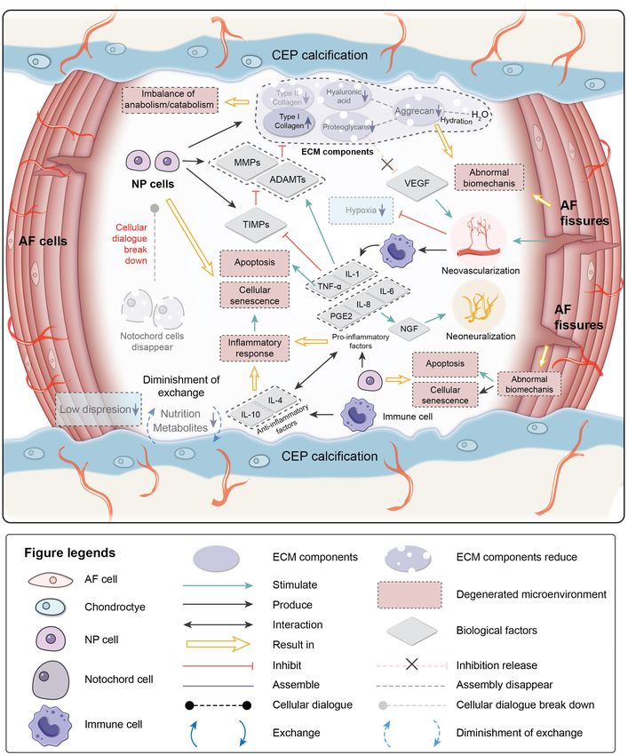
(4) abnormal biomechanics (Figure 2). 2002; Le Maitre et al., 2004; Bachmeier et al., 2009; Vo et al.,
2013). Upregulation of MMP and ADAMTS expression and
Cell Senescence enzymatic activity is implicated in the destruction of the disk
Cellular senescence, which is a fundamental mechanism ECM, leading to the development of IVDD (Bachmeier et al.,
that mediates age-related dysfunctions and chronic diseases, 2009; Pockert et al., 2009; Vo et al., 2013). TIMP, which is
accumulates in human, bovine, and rat degenerative IVDs during a specific inhibitor of MMPs, binds to the active forms of
aging (Roberts et al., 2006; Gruber et al., 2007; Kim et al., MMPs to suppress catabolic enzyme activity. However, the
2009; Muñoz-Espín and Serrano, 2014; van Deursen, 2014; activities of MMPs, ADAMTS, and TIMP are all regulated by
Sharpless and Sherr, 2015). Unlike apoptosis, senescent cells inflammation-related factors, particularly pro-inflammatory
are metabolically viable and arrest at the cell cycle transition, factors. PGs in the IVD are proposed as a barrier to vascular and
cease proliferation, and exhibit an altered expression of various neural ingrowth into the NP (Khan et al., 2017). As PG contents
catabolic cytokines and degrading enzymes (Campisi, 2005; decrease, the inhibiting effect for the neovascularization function
Frontiers in Bioengineering and Biotechnology | www.frontiersin.org 4 July 2021 | Volume 9 | Article 592118Dou et al. IVDD and Tissue Engineering Strategies
FIGURE 1 | Physiological microenvironment of intervertebral disk (IVD). AF, annulus fibrosus; NP, nucleus pulposus; CEP, cartilage endplate; ECM, extracellular
matrix; HIF, hypoxia-inducible factor; MMPs, metalloproteinases; ADAMTs, metalloproteinase with thrombospondin motifs; TIMPs, tissue inhibitors of
metalloproteinases; CTGF, connective tissue growth factor; Shh, sonic hedgehog; BMPs, bone morphogenetic protein; VEGF, vascular endothelial growth factor.
of VEGF weakens. Moreover, AF fissures caused by abnormal as an adaptive response to restore homeostasis. If the acute
biomechanics allow neovascularization and neoinnervation inflammatory response fails to eliminate the pathogen, the
from a lack of PG production. As a result, NP is invaded by inflammatory process persists and acquires new characteristics
immune cells transported by new blood vessels. Immune cells (Majno and Joris, 2004; Medzhitov, 2008). The IVD is an
respond to the deteriorating microenvironment, eventually avascular tissue until neovascularization occurs due to
leading to inflammation. degeneration. Crystals and ECM breakdown products may
be responsible for causing the IVD inflammatory response
Inflammatory Microenvironment (Medzhitov, 2008). When anabolism/catabolism becomes
Inflammation is an adaptive response triggered by noxious unbalanced, degradation products trigger inflammation.
stimuli and conditions, such as infection and tissue injury. In human IVD cells, HA fragments upregulate the mRNA
Regardless of the cause, inflammation presumably evolved expression levels of the inflammatory and catabolic genes IL-1β,
Frontiers in Bioengineering and Biotechnology | www.frontiersin.org 5 July 2021 | Volume 9 | Article 592118Dou et al. IVDD and Tissue Engineering Strategies FIGURE 2 | Pathological microenvironment of intervertebral disk (IVD). AF, annulus fibrosus; NP, nucleus pulposus; CEP, cartilage endplate. ECM, extracellular matrix; MMPs, metalloproteinases; ADAMTs, metalloproteinase with thrombospondin motifs; TIMPs, tissue inhibitors of metalloproteinases; VEGF, vascular endothelial growth factor; NGF, nerve growth factor; IL-1,4,6,8,10, interleukin-1,4,6,8,10; TNF-a, tumor necrosis factor; PGE2, prostaglandin E2. IL-6, IL-8, cyclooxygenase-2, MMP-1, and MMP-13 (Quero et al., Richardson et al., 2009; Wang et al., 2013), where they have 2013). After neovascularization occurs, immune cells migrate to been observed at increased levels along with other inflammatory the NP to respond to the microenvironment. NP embryologically mediators in degenerated IVDs (Le Maitre et al., 2005, 2007). develops in an enclosed structure, which makes NP an avascular TNF-α is a cytokine that stimulates the inflammatory response, and immune-privileged tissue. Hence, immune cells invade induces nerve swelling and neuropathic pain, and promotes when disk degeneration progresses to a certain stage, making cellular apoptosis via its cytotoxic effect (Pedersen et al., 2015). IVD inflammation a chronic process. Inflammatory factors play IL-1β induces a catabolic response by NP cells at the molecular essential roles in mediating IVD homeostasis and degeneration. level (Smith et al., 2011). Together they stimulate the production TNF-α, IL-1β, and IL-1α are expressed by healthy IVD of MMPs and ADAMTS by NPCs and suppress the expression cells and immune cells (macrophages, monocytes, dendritic of TIMPs, resulting in decreased synthesis of aggrecan and type cells, B cells, and natural killer cells) (Le Maitre et al., 2005; II collagen by NPCs (Frapin et al., 2019). Pro-inflammatory Frontiers in Bioengineering and Biotechnology | www.frontiersin.org 6 July 2021 | Volume 9 | Article 592118
Dou et al.                                                                                                  IVDD and Tissue Engineering Strategies

factors are also related to pain. IL-6, IL-8, and prostaglandin           affect daily life (el Barzouhi et al., 2013; Chiu et al., 2015).
E2 synthesis by NPCs stimulate nerve growth factor production,            Non-surgical       treatments    include      non-pharmacological
which induces abnormal nerve ingrowth and causes pain (Frapin             treatments, pharmacological treatments, and interventional
et al., 2019). IL-6 and IL-8 are both higher in severe sciatica           treatments. Non-pharmacological treatments, such as exercise,
patients, and IL-6 is correlates to low back pain frequency               traction, acupuncture, massage, physical therapy, and spinal
(Khan et al., 2017). A herniated disk impinging on the nerve              manipulation, are applied in daily life, but only for second-line
root is painless in 70% of patients, and it is likely that the            or adjunctive treatment options with insufficient evidence
secretion of products involved in the inflammation cascade in             for cure (Kreiner et al., 2014; Foster et al., 2018). More than
a torn AF sensitizes the nerve root or increases the number of            50% of patients with IVDD have used drugs. Non-steroidal
innervations, thereby causing pain (Sharifi et al., 2015). Anti-          anti-inflammatory drugs are most applied drugs for pain relief
inflammatory activity is also part of the IVD microenvironment.           and improved function. Oral glucocorticoids alleviate the
IL-4 and IL-10 are anti-inflammatory factors produced by                  inflammation of nerve roots. At the same time, muscle relaxants
activated macrophages and monocytes. They participate in                  can be useful to relieve muscle spasms, but these drugs are
pro-inflammatory/anti-inflammatory balance and inhibit the                discouraged for lack of sufficient evidence (Kreiner et al., 2014;
synthesis of pro-inflammatory cytokines (Khan et al., 2017).              Ramaswami et al., 2017; Foster et al., 2018; Benzakour et al.,
                                                                          2019; Lee et al., 2019).
Abnormal Biomechanics                                                        A meta-analysis reported that the therapeutic effect of an
Intervertebral disk degeneration can also be affected by                  epidural injection intervention treatment is better than an
the biomechanical environment. A positive dose-response                   intradiscal injection, percutaneous discectomy, traction, physical
relationship is observed between cumulative lumbar load and               therapy/exercise, radiofrequency therapy, or chemonucleolysis
early onset of symptomatic lumbar disk space narrowing: the               (Lewis et al., 2015). However, interventional treatments are
disk disease onset time of workers with the highest exposure to           discouraged in the guidelines (Foster et al., 2018).
heavy physical constraints is significantly advanced compared to             The goal of non-surgical treatments is to relieve symptoms
others (Petit and Roquelaure, 2015). Xing et al. (1976) designed          and improve function, but they cannot halt the degeneration.
an amputated-leg rat model. Due to the forelimb amputation, the           Surgical treatments are required when non-surgical treatments
IVDs underwent abnormal mechanical loading. The results of                are unable to relieve symptoms.
senescence-associated β-galactosidase-positive staining showed
that IVD cell senescence accelerates due to the abnormal loading          Surgical Treatments
of rat lumbar IVDs (Xing et al., 1976). Constant pressure on              Mixter and Barr (1934) reported that a tumor in the spinal
the disk can cause degeneration, which may be related to the              canal, which causes sciatica, was a herniated NP. Laminectomy,
upregulation of matrix degradation-related enzymes (Yurube                combined with excision of the NP, was performed, and
et al., 2012; Neidlinger-Wilke et al., 2014). The mechanical load         satisfactory results were obtained (Mixter and Barr, 1934).
can also induce IVD cell apoptosis through the mitochondrial              Since then, surgical treatments to treat disk herniation have
pathway (Rannou et al., 2004; Kuo et al., 2014).                          been developed. Four main types of surgical procedures
   Briefly, IVDs are continually adapting to changes in                   are used: (1) decompression for neurological problems, (2)
the microenvironment from embryogenesis to degeneration                   fusion to abolish motion at a functional spinal unit, (3)
under the regulation of numerous factors (Colombier et al.,               motion preservation/modifying surgery in the form of disk
2014). The avascular and immune-privileged NPs regulate cell              replacement/dynamic fixation devices, and (4) deformity
activity via cellular dialogue (Fontana et al., 2015; Henry               surgery to realign the biomechanics between a large number
et al., 2018). ECM synthesized by IVD cells ensures the                   of functional spinal units (Eisenstein et al., 2020). The
biomechanical function of IVD. The CEP ensures transport                  purpose of decompression surgery is to relieve pain and
of nutrients and metabolites (Zhang et al., 2018). When                   numbness caused by nerve compression, and the most common
degeneration begins, interactions between components in                   procedure is discectomy.
the microenvironment break down, resulting in cascades of                     A discectomy removes disk tissue that oppresses nerve roots
degeneration (Freemont, 2009). Therefore, therapeutic strategies          in the intervertebral space. An emergency discectomy is required
should first consider how to relieve pain and regain IVD                  for patients who already have cauda equina syndrome and a new
function in patients and, furthermore, correct the factors causing        motor disorder. Selective discectomy is required for patients with
degeneration to eventually achieve ideal IVD regeneration and             persistent neurological symptoms that cannot be relieved by non-
functional restoration.                                                   surgical treatments (Butler and Donnally, 2020).
                                                                              Discectomy can be open or minimally invasive. Open
                                                                          discectomy is performed in a wide range of operations but
CURRENT IVDD TREATMENTS                                                   with more tissue damage. Minimally invasive discectomy
                                                                          causes less tissue damage, but indications are limited.
Non-surgical Treatments                                                   A meta-analysis showed that the incidence of postoperative
Intervertebral disk degeneration is inevitable; 60–90%                    complications and reoperations is similar, while less blood
of patients can be treated non-surgically as long as no                   loss, shorter operating time, and shorter hospital stay are
symptoms occur or mild symptoms that occur do not                         common after minimally invasive discectomy (Alvi et al., 2018;

Frontiers in Bioengineering and Biotechnology | www.frontiersin.org   7                                       July 2021 | Volume 9 | Article 592118
Dou et al. IVDD and Tissue Engineering Strategies
Li et al., 2018a; Butler and Donnally, 2020). Discectomy relieves (Marcolongo et al., 2011). Annuloplasty is a minimally invasive
nerve root compression, relieves pain while retaining some method in which heat produced by electricity or radiofrequency
of the structure, and restores some of the biomechanical radiation strengthens the collagen fibers and seals fissures in
functions of the disk. However, discectomy cannot change a process similar to tissue soldering (Helm Ii et al., 2017).
the deteriorating microenvironment. Therefore, further Nucleoplasty releases the pressure on the outer AF, allowing the
progression of degeneration cannot be prevented, and disk to return to normal size, thereby decompressing the nerve,
postoperative complications may occur (12.5, 13.3, and with better therapeutic effects than non-surgical treatments. This
10.8% for open microdiscectomy, microendoscopic discectomy, procedure can be performed in the clinic, and the patient recovers
and percutaneous microdiscectomy, respectively) (Shriver et al., quickly after the procedure, but the indications of this procedure
2015). The AF must be broken to perform this surgery, and are limited (Eichen et al., 2014; Liliang et al., 2016; de Rooij et al.,
no new AF tissue is formed, so the opening remains open or 2017).
is closed with the formation of scar tissue, which might cause Patients who undergo surgery may have better pain relief,
reherniation (Sharifi et al., 2015). functional improvement, and satisfaction than those who
Spinal fusion is a classic procedure. Hibbs and Albee treated receive non-surgical treatments. Nevertheless, long-term follow-
Pott disease using fusion. Fusion abolishes pain by abolishing up shows that disability outcomes are similar regardless of which
the motion of adjacent segments (Hibbs, 1964). Spinal fusion treatment a patient receives (Atlas et al., 2001, 2005), suggesting
is widely used to treat many spinal diseases and is the that current strategies can only alleviate symptoms and that
gold standard for treating significant, chronic axial low back regenerative strategy is key to solving IVDD.
pain due to IVDD. About 60–65% of lumbar fusion procedures
are performed for degenerative disk disease (Lee and Langrana,
2004; Auerbach et al., 2009). Spinal fusion completely removes TISSUE ENGINEERING STRATEGIES
the IVD and entirely relieves the oppression on the nerve roots,
where the degenerated segments become integrated and lose Surgical treatments for IVDD have developed rapidly, but the
motor function. Although the pain symptoms are relieved, the limitations of surgical treatments are becoming apparent. Thus,
native IVD microenvironment is destroyed. The motor function researchers have turned their attention to tissue engineering
is abolished, which may cause long-term complications. The strategies. Tissue engineering is “an interdisciplinary field that
mechanical environment of adjacent segments is affected and applies the principles of engineering and life sciences toward the
may develop into adjacent segmental disease (Lee and Langrana, development of biological substitutes that restore, maintain, or
1984; Hilibrand and Robbins, 2004; Virk et al., 2014; Hashimoto improve tissue function or a whole organ,” as defined by Langer
et al., 2019). and Vacanti (1993). Mizuno et al. (2004) fabricated the first
Artificial disk prosthesis replacement surgery is performed to documented IVD scaffold of polyglycolic acid and polylactic acid.
restore disk height, biomechanical structure, and motor function The scaffold was seeded with AF cells. NP cells were injected
of the IVD and overcomes some deficiencies of spinal fusion. into the center after 1 day. Then, the scaffolds were implanted
This procedure was first applied in the 1950s. As more and more in the subcutaneous space of the dorsum of athymic mice. The
prostheses have been developed, disk replacement surgery has results showed that the engineered disks strongly resembled
developed quickly (Guyer and Ohnmeiss, 2003; Zhao et al., 2019). native IVDs and synthesized similar collagen components as
Disk replacement surgery effectively relieves pain and improves native NP and AF (Mizuno et al., 2004; Hukins, 2005). This
the quality of life, but it is not a superior substitute surgery attempt was initially intended to identify an alternative strategy
for fusion. The average reoperation rate is 12.1% during follow- for IVD replacement due to its long-term deficiencies. The flaws
up after lumbar disk replacement or cervical disk replacement. in that study included the cell source, immunological rejection,
Postoperative complications, such as prosthesis failure, infection, and fixation of the scaffold, but that study inspired researchers
adjacent segmental disease, and pain, can occur, yet no evidence to try tissue engineering strategies for IVDD. Various strategies
demonstrates that the surgical effect of disk replacement is better have been formulated based on different treatment principles. It is
than fusion (Thavaneswaran and Vandepeer, 2014; Cui et al., important to note that there are no separate strategies, only those
2018; MacDowall et al., 2019). The effect of disk replacement that are focused on in the study.
surgery greatly depends on the type of prosthesis and surgical
technique of the surgeon. An artificial disk prosthesis replicates Scaffold Strategy: From Natural ECM
the anatomical structure of the IVD and attempts to mimic Mimetic to Multifunctional Platform
its biomechanical properties. The risk of adjacent segmental Scaffolds were initially designed as cell substrates to mimic
disease is reduced compared with fusion (Findlay et al., 2018). the microenvironment where cells live. An ECM scaffold
However, it is similar to fusion surgery in that the native IVD is similar to the native microenvironment of cells and has
microenvironment is destroyed. Further follow-up studies are excellent biocompatibility and immunogenicity, which are
needed to investigate the long-term efficacy and safety of disk beneficial for cell proliferation and metabolic activities.
replacement surgery. Yang et al. (2008) fabricated a decellularized ECM scaffold
Several experimental surgeries have been developed. AF derived from human cartilage, and the scaffold successfully
sutures strengthen the intensity of AF; thus, they restrict NP generated cartilaginous tissue in nude mice. As the AF
from herniating but cannot prevent the progression of IVDD is a fibrous cartilage structure and has a similar ECM
Frontiers in Bioengineering and Biotechnology | www.frontiersin.org 8 July 2021 | Volume 9 | Article 592118Dou et al. IVDD and Tissue Engineering Strategies
composition to cartilage, an ECM scaffold might be feasible sufficient amount of specific ECM in vitro. The subcutaneous
to regenerate AF. McGuire et al. (2017) fabricated an ECM implantation results showed a negligible immune response
scaffold made of decellularized pericardial tissue. Collagen (Bhunia et al., 2018).
patches were obtained from treated pericardial tissue. Three-dimensional printing precisely fabricates the
These patches were assembled into a multi-laminate angle- scaffold structure. To generate a scaffold with angle-
ply scaffold. This scaffold mimicked the structure of AF, ply architecture similar to natural AF, Christiani
provided similar mechanical support, and supported cell et al. (2019) printed laminar constructs comprised of
viability, infiltration, and proliferation for bovine AF in vitro polycaprolactone (PCL) struts oriented at alternating
(McGuire et al., 2017). angles of ±30◦ . The mechanical characterization
Complete AF regeneration requires the recovery of results showed that the mechanical properties of the
biomechanical and structural properties of healthy AF and scaffolds were similar to native AF tissue. They also
the restoration of the biological behavior of resident cells in cultured bovine AFCs with smooth-surface PCL and
AF (Driscoll et al., 2013). The alignment and organization unidirectional channel-etched PCL to further study cell
of AF cells determine their biomechanical functions. As arrangement and ECM deposition, respectively. The
manufacturing technology develops, fabricating methods, such SEM micrographs of the scaffolds showed that cells
as electrospinning and 3-D printing, provide the possibility cultured on etched PCL had a tendency to align along
for microstructural scaffolds. Microstructural scaffolds guide the underlying surface, and the alignment of proteins
the cells to form a specific order and are widely applied in with the underlying surface texture can be observed
AF tissue engineering due to their unique structure. Ma et al. (Christiani et al., 2019).
(2018) developed a hybrid scaffold for AF tissue engineering. However, PCL materials have poor hydrophilicity, have
This hybrid scaffold consisted of traditional electrospun a long degradation time, and lack cell recognition sites.
aligned nanofibrous scaffolds (AFS) as a baseline scaffold and PCL materials bind poorly to surrounding host tissues
electrospun aligned nanoyarn scaffolds (AYS). Morphological after implantation in vivo (Cheng et al., 2017). Three-
measurements showed that this hybrid scaffold replicates dimensional bioprinting achieves precise bionics according
the tensile strength, axial compression, and anisotropic to the structure and size of native tissues and organs
properties of AF tissues to some degree. Mechanical testing (Kang et al., 2016). As a novel technology, 3D bioprinting
demonstrated that the tensile properties of AFS and AYS shows great potential and enormous advantages in the
were qualitatively similar to those of native AF tissue. In vitro repair of IVDs. Sun et al. (2021) fabricated a dual growth-
biocompatibility analyses demonstrated that the AYS and factor-releasing IVD scaffold using 3D bioprinting. They
HS yield improved bone marrow mesenchymal stem cell loaded CTGF and transforming growth factor-β3 (TGF-
(BMSC) proliferation (Ma et al., 2018). Gluais et al. (2019) β3) on polydopamine nanospheres (PDA NPs). Then,
fabricated electrospun aligned microfibrous scaffolds that CTGF@PDA and TGF-β3@PDA were mixed with rat
recruit neighboring healthy AF cells to migrate to the scaffold, BMSCs as the 3D bioprinting ink. A 3D model of the IVD
which promotes cell colonization and proliferation. The results scaffold was designed using AutoCAD software, and the
showed that, in addition to numerous dense collagen fibers support structures of CEP and AF were printed using a
in the aligned scaffolds, the fibers were arranged in the same PCL polymer, while the parts of the model corresponding
direction as the scaffold (Gluais et al., 2019). Kang et al. (2018) to AF and NP were printed using 3D bioprinting ink.
combined electrospinning and 3D printing techniques to In vitro experiments confirmed that the growth factors
fabricate a biomimetic biodegradable scaffold that consisted were released from the IVD scaffolds in a spatially
of multi-lamella nano/microfibers. Each lamella contained controlled manner and induced the corresponding BMSCs
one layer of aligned electrospun nanofibers and one layer to differentiate into NP-like cells and AF-like cells. After
of supporting 3D printed microfibers on each side, which being implanted subcutaneously into the dorsum of nude
were all aligned in the same direction, and the angle of the mice, the reconstructed IVDs exhibited a zone-specific
fibers between adjacent layers was 60◦ . The nano/microfibres matrix: primarily type II collagen and glycosaminoglycan
aligned as native collagen tissue of AF. These results show in the core zone and type I collagen in the surrounding zone
that these scaffolds can form and integrate collagen fibers (Sun et al., 2021).
(Kang et al., 2018). In addition to providing mechanical support and
However, electrospun scaffolds often face several topographic stimulus for cells, scaffolds can also be loaded
limitations, including low porosity that restricts uniform with therapeutic drugs or cells. Cheng et al. (2011) fabricated
cell infiltration and a discrepancy of mechanical properties an injectable thermosensitive chitosan/gelatin/glycerol
compared with native AF. To recapitulate the form and phosphate hydrogel as a controlled release system for
function of the complex anatomy of AF, Bhunia et al. ferulic acid (FA) delivery. FA is an excellent antioxidant
(2018) adopted a directional freezing technique to fabricate drug. The scaffolds were incorporated with FA, added to
silk-based multi-layered disk-like angle-ply (±30◦ of Transwells, and incubated with H2 O2 -induced NP cells
successive layers) bio-artificial scaffolds. The fabricated bio- in vitro. The results showed that these scaffolds achieved
discs supported the primary AF or human mesenchymal excellent antioxidant properties after loading with FA
stem cell proliferation, differentiation, and deposition of a (Cheng et al., 2011).
Frontiers in Bioengineering and Biotechnology | www.frontiersin.org 9 July 2021 | Volume 9 | Article 592118Dou et al. IVDD and Tissue Engineering Strategies
Scaffolds are a crucial element of this strategy, as they and differentiation factor-5 and act as a cell delivery vehicle
provide a similar microenvironment for IVD cells by mimicking for iPSC-derived NP-like cells. Then, they injected these
the structure of native IVDs. The scaffold strategy can cell-seeded polymeric microspheres into rat coccygeal IVDs,
also be combined with other strategies, such as delivery of and the results indicated that disk height was recovered,
therapeutic drugs or cells. water content was increased, and the NP ECM was partially
restored (Xia K. et al., 2019). This study utilized growth
factors to enhance the therapeutic efficacy of cells and
Cell Therapy Strategy and Derivative prolong the life of growth factors via a polymeric gelatin
Strategies microsphere as a sustained release platform. Multiple strategies
Cell Therapy Strategy complementing each other will become more important in IVD
Cell therapy is a classic strategy. An increasing number of tissue engineering.
studies have shown the efficacy of therapeutic cells in several Recent studies have also revealed the existence of
IVDD animal models (Hiyama et al., 2008; Jeong et al., endogenous stem/progenitor cells in the IVD (Hu
2009; Henriksson et al., 2019). One study reported that stem et al., 2018). Ying et al. (2019) found that stromal cell-
cells and stem-like cells are found in almost all adult tissues derived factor-1α expression is higher in the degenerative
(da Silva Meirelles et al., 2006). IVD microenvironment and showed a positive effect
Due to the potential of stem cells to differentiate, replacing on enhancing the proliferation and recruitment of
damaged cells in target tissues, stem cells are ideal therapeutic endogenous NP-derived stem cells into IVDs in vitro
cells for IVD tissue engineering (Krause, 2008; Meirelles and in vivo. These cells might be a promising source
Lda et al., 2009). Mesenchymal stem cells and induced for cell therapy.
pluripotent stem cells (iPSCs) are the most widely used in Cell-based therapies have the advantages of modulating
IVD cell therapy (Wei et al., 2014; Vickers et al., 2019). inflammation and concomitantly affecting the remodeling
Several studies have demonstrated the ability of BMSCs process, without presenting toxicity or immunosuppression
and adipose-derived stem cells to differentiate into an NP- (Lopes-Pacheco et al., 2016). These properties make cell therapy
like phenotype, and in vivo studies have demonstrated the an exceptionally advantageous therapeutic approach for IVD
ability of implanted MSCs to enhance matrix production, tissue engineering. Nevertheless, various complications occur
particularly GAG synthesis, and increase disk height and with stem cells, including tumorigenesis and immune reactions.
hydration (Richardson et al., 2006; Gantenbein-Ritter et al., Certain cases of tumorigenesis and immune reaction of iPSCs
2011; Stoyanov et al., 2011; Clarke et al., 2014). iPSCs and embryonic stem cells (which are also a cell source for
differentiate into NCs or NP-like cells and reduce IVDD after iPSCs) have been reported, and the application risks are always
transplantation in vivo (Sheyn et al., 2019; Xia K. et al., discussed (Pera et al., 2000; Taylor et al., 2011; Lee et al.,
2019). 2013; Kim, 2014; Jin et al., 2016). Compared to iPSCs and
Researchers have attempted different methods to embryonic stem cells, MSCs seem to be safer, as no case of
transport therapeutic cells to diseased areas, such as direct tumorigenesis has been reported after MSC transplantation
injection of therapeutic cells or loading of cells onto in vivo (Oryan et al., 2017).
scaffolds. Hiyama et al. (2008), Jeong et al. (2009), and Although the transplantation of stem cells may have
Henriksson et al. (2019) injected human MSCs into rat, risks, their efficacy cannot be denied. Risks may be hedged
dog, and pig disk degeneration models, respectively. The by enhancing the immune compatibility of stem cells.
results showed that transplantation of human MSCs has Immune rejection is caused by HLA mismatching. Xu
a positive repair effect on the xenogeneic animal disk et al. (2019) generated HLA pseudo-homozygous iPSCs
degeneration model. Sheyn et al. (2019) induced human through CRISPR-Cas9, with allele-specific editing of HLA
iPSCs to differentiate into notochordal cells in vitro and heterozygous iPSCs and HLA-C-retained iPSCs, which
proved their regenerative capacity in vivo in an annular evade T and NK cells in vitro and in vivo. HLA-C-
puncture pig model. retained iPSCs combined with HLA-class II knockout are
However, direct injection by needle puncture causes immunologically compatible with >90% of the world’s
damage to the AF, and implanted cells could leak out population, greatly facilitating iPSC-based regenerative
through annular fissures. AF damage can lead to further medicine applications (Xu et al., 2019). Cell strategies
degeneration and an increased risk of disk herniation have potential in tissue engineering, but improving
(Oehme et al., 2015). Scaffolds could be pivotal to provide safety and avoiding risks should be given more attention
transplanted cells with a supportive environment. Bhunia in future studies.
et al. (2018) seeded MSCs on AF structure-like scaffolds
and reported ideal results as mentioned above. GDF-5 Therapy Strategies Using Biological Factors
inhibits IVDD and promotes NP-like differentiation of Biological factors are promising therapeutic drugs for IVDD
stem cells (Taylor et al., 2011). However, such a factor as native mediators in the IVD microenvironment since they
could require multiple injections due to its short life are key signaling factors in the cellular dialogue. Unlike
in vivo (Jin et al., 2016). Xia C. et al. (2019) generated conventional drugs, biological factors are secreted by IVD
polymeric gelatin microspheres, which can release growth cells and have fewer side effects. Therapeutic biological factors
Frontiers in Bioengineering and Biotechnology | www.frontiersin.org 10 July 2021 | Volume 9 | Article 592118Dou et al. IVDD and Tissue Engineering Strategies
should be able to restore the healthy microenvironment of IVD. to normal height, similar to the healthy group. These results show
Therefore, biological factors should have at least one therapeutic that agomir874 regulates the balance of synthesis/decomposition
function as follows: (1) pro-anabolism/anti-catabolism, (2) anti- of the NP ECM and inhibits IVDD (Chen W. et al., 2020).
inflammation, and (3) regulate cell activity. The application Six gene therapy products have been approved since 2016
of biological factors is limited by their short life in vivo. (High and Roncarolo, 2019). Gene therapy is promising to be
Thus, biological factors are often used in conjunction with performed “one time,” with long-term and high-value therapeutic
other strategies. effects. However, there are still some problems to be solved, such
Researchers can customize biological factor scaffolds as safety issues and high treatment expense. Gene therapy is
according to different conditions, with the key concept of aimed at specific targets, which have been studied thoroughly,
restoring the balance of the microenvironment. Several widely to regulate cell activities. Thus, gene therapy cannot be used to
used factors are introduced in Table 1. regulate a series of therapeutic targets or pathways as in cell
therapy. Due to the encapsulated microenvironment of the IVD,
Gene Therapy Strategy gene therapy is still promising and not fully explored for IVDD
Biological factors intercellularly mediate cell activities, while treatment. Studies on safety and therapeutic mechanisms should
microRNAs (miRNAs) mediate intracellularly. Gene therapy is a be conducted in the future.
strategy to achieve the durable expression of a therapeutic gene
or “transgene” at a level sufficient to ameliorate or cure disease
symptoms with minimal adverse events. Two basic strategies Extracellular Vesicle Therapeutic Strategy
have been proposed. An integrating vector is introduced into a Studies have confirmed that the mechanism of action of
precursor or stem cell so the gene is passed to every daughter cell MSCs is predominantly paracrine (Lu et al., 2017; Zhu et al.,
or the gene is delivered in a non-integrating vector to long-lived 2020). Almost all types of cells secrete extracellular vesicles
post-mitotic or slowly dividing cells, ensuring the expression of (EVs). As the main components of paracrine activity, EVs
that gene for the life of the cell (High and Roncarolo, 2019). derived from MSCs achieve regenerative functions. The three
Although the application of gene therapy to the IVD has main kinds of EVs are exosomes, microvesicles, and apoptotic
lagged behind other tissues, gene therapy shows excellent bodies (El Andaloussi et al., 2013). Exosomes from MSCs
potential and safety for IVD tissue engineering. As an have various effects on IVD regeneration, such as antioxidant,
encapsulated and avascular tissue, the sealing property of IVD anti-inflammatory, anti-apoptosis, and proliferation-promoting
effectively prevents leakage of the disk contents to other sites effects (El Andaloussi et al., 2013; Lu et al., 2017; Xia C.
in the body (Levicoff et al., 2005). Some researchers are et al., 2019). Due to the low immunogenicity and high
using gene-editing techniques, such as CRISPR, to precisely efficiency of exosomes compared to MSCs, exosomes are
alter DNA sequences or genetically modify immune cells to promising substitutes for MSCs in cell therapy. The mechanism
imbue them with the ability to fight cancer. TNF-α and IL- of its regenerative ability remains unclear, but it is very
1β are inflammatory cytokines related to the inflammatory likely to be related to miRNAs. The level of miR-532-5p
microenvironment in IVDD through TNFR1/IL1R1 signaling. was observed to be decreased in TNF-α induced apoptotic
To regulate this signaling, Farhang et al. (2019) produced CRISPR NPCs but abundant in bone marrow mesenchymal stem cell
epigenome-edited lentiviral vectors based on TNFR1/IL1R1 (BMSC)-derived exosomes. Zhu et al. (2020) demonstrated that
targeted screens and delivered the genes into human NPCs by exosomes from BMSCs could deliver miR-532-5p and suppress
lentiviral transduction. The expression of the editing vectors was the IVDD via targeting RASSF5. Cheng et al. (2018) also
confirmed by qRT-PCR. Measurement of NF-κB activity (which reported that exosomes from BMSCs could deliver miR-21, which
is a downstream transcription factor that mediates TNFR1/IL1R1 could activate the PI3K/Akt pathway in apoptotic NPCs, and
signaling), apoptosis, and anabolic/catabolic changes in gene further inhibit IVDD.
expression demonstrated that the lentiviral vectors significantly Exosomes were also confirmed to be associated with
downregulated TNFR1 and IL1R1 and significantly attenuated pathological changes in IVDD. circRNA_0000253 has the
the deleterious microenvironment (Farhang et al., 2019). maximum upregulation in degenerative NPC exosomes.
Gene therapy provides a potentially ideal tool for many Song et al. (2020) found that exosomes in NPCs were
diseases by delivering synthetic miRNAs to regulate gene differentially expressed in degenerative and normal NPCs.
expression (Deverman et al., 2018). However, there are obstacles The circRNA_0000253 levels notably increased and the
to delivering miRNAs directly to target tissues due to their miRNA-141-5p levels markedly reduced in degenerative
inactivation, low transfection efficiency, and short half-life. Chen NPCs compared with normal NPCs. Further research
W. et al. (2020) synthesized agomir, which is cholesterol-, confirmed that the circRNA_0000253 could increase IVDD
methyl-, and phosphorothioate-modified miRNA, with good by adsorbing miRNA-141-5p and downregulating SIRT1
stability and enhanced transfection efficiency, in animals. Agomir in vivo and in vitro (Song et al., 2020). This study also
penetrates the barriers of the cell membrane and tissues in vivo demonstrated that, in a degenerative microenvironment of IVD,
to enrich target cells. Agomir874 downregulates the expression communication between NPCs by secreting exosomes may
of MMPs in NP by mimicking miRNA874. Chen et al. loaded aggravate degeneration. Utilizing or blocking degeneration-
agomir on an injectable polyethylene glycol hydrogel and injected related exosomes might become a promising therapeutic strategy
it into a rat IVD. After 8 weeks, the rat IVD was gradually restored for IVD degeneration.
Frontiers in Bioengineering and Biotechnology | www.frontiersin.org 11 July 2021 | Volume 9 | Article 592118You can also read