Infrared radiation from cage bedding moderates rat inflammatory and autoimmune responses in collagen induced arthritis - Nature
←
→
Page content transcription
If your browser does not render page correctly, please read the page content below
www.nature.com/scientificreports
OPEN Infrared radiation from cage
bedding moderates rat
inflammatory and autoimmune
responses in collagen‑induced
arthritis
Jasmina Djuretić1,6, Mirjana Dimitrijević2,6, Marija Stojanović1, Jelena Kotur Stevuljević3,
Michael R. Hamblin4, Ana Micov5, Radica Stepanović‑Petrović5 & Gordana Leposavić1*
The development of collagen type II (CII)-induced arthritis (CIA), a model of rheumatoid arthritis, in
rats housed in cages with bedding composed of Celliant fibres containing ceramic particles, which
absorb body heat and re-emit the energy back to the body in the form of infrared radiation (+IRF rats),
and those housed in cages with standard wooden shaving bedding (−IRF control rats) was examined.
The appearance of the first signs of CIA was postponed, while the disease was milder (judging by
the arthritic score, paw volume, and burrowing behaviour) in +IRF compared with −IRF rats. This
correlated with a lower magnitude of serum anti-CII IgG antibody levels in +IRF rats, and lower
production level of IL-17, the Th17 signature cytokine, in cultures of their paws. This could be partly
ascribed to impaired migration of antigen-loaded CD11b + dendritic cells and their positioning within
lymph nodes in +IRF rats reflecting diminished lymph node expression of CCL19 /CCL21. Additionally,
as confirmed in rats with carrageenan-induced paw inflammation (CIPI), the infrared radiation from
Celliant fibres, independently from immunomodulatory effects, exerted anti-inflammatory effects
(judging by a shift in pro-inflammatory mediator to anti-inflammatory/immunoregulatory mediator
ratio towards the latter in paw cultures) and ameliorated burrowing behaviour in CIA rats.
As a result of substantial advances in polymer chemistry, relatively recently, infrared radiation-emitting (IR) fibres
(filaments) have been developed. These fibres have a porous core-sheath and groove structure allowing various
optically active ceramic micron-sized particles to be incorporated into each fibre1,2. One type of these fibres is
Celliant (Hologenix, Santa Monica, CA, USA), a polyethylene terephthalate fibre that incorporates micron-sized
optically active ceramic particles exhibiting the property of temperature-dependent infrared emission. These
fibres have been woven to get high-performance functional t extiles1,2. When such functional textiles are used as
garments, bandages, or bed linen, the heat energy generated by the human body can be transferred by radiation,
conduction, convection to the ceramic p articles1–4. These ceramic particles act as black-body absorbers and re-
emit the absorbed energy as infrared radiation back to the body5. According to the classification of the Interna-
tional Commission on Illumination (CIE) and the classification provided in ISO 20473 standard (ISO 20473),
infrared radiation has three broad categories: near infrared (0.7–1.4 μm and 0.78–3 μm according to CIE and
ISO 20473, respectively), mid-infrared (0.4–3 μm and 3–50 μm according to CIE and ISO 20473, respectively)
and far infrared (3–100 μm and 50–1000 μm according to CIE and ISO 20473, respectively). Considering the
aforementioned data, IR fabrics are suggested to recycle the body’s natural energy5. At physiological skin tempera-
ture, much of the body’s emissive radiative power is centered between 7 and 14 μm6,7. Generally, infrared radia-
tion penetrates deeply through the layers of the skin to reach the muscles and bones8. This radiation moderates
1
Department of Pathobiology, Faculty of Pharmacy, University of Belgrade, Vojvode Stepe 450, Belgrade,
Serbia. 2Department of Immunology, Institute for Biological Research “Siniša Stanković”‑National Institute
of Republic Serbia, University of Belgrade, Bulevar despota Stefana 142, Belgrade, Serbia. 3Department of
Biochemistry, Faculty of Pharmacy, University of Belgrade, Vojvode Stepe 450, Belgrade, Serbia. 4Laser Research
Centre, Faculty of Health Science, University of Johannesburg, Doornfontein 2028, South Africa. 5Department of
Pharmacology, Faculty of Pharmacy, University of Belgrade, Vojvode Stepe 450, Belgrade, Serbia. 6These authors
contributed equally: Jasmina Djuretić and Mirjana Dimitrijević. *email: gordana.leposavic@pharmacy.bg.ac.rs
Scientific Reports | (2021) 11:2882 | https://doi.org/10.1038/s41598-021-81999-7 1
Vol.:(0123456789)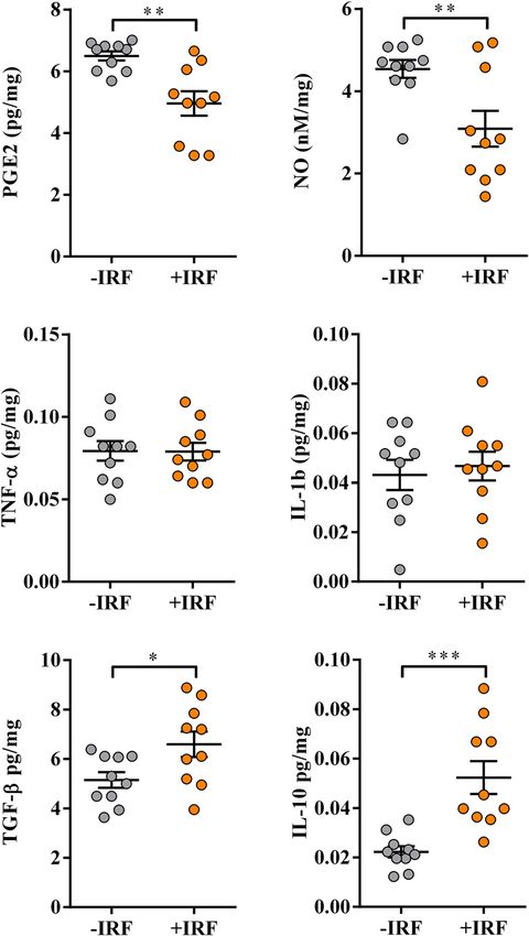
www.nature.com/scientificreports/
inflammation9,10 and pain4. Additionally, it promotes tissue regeneration and wound healing by improving
circulation11,12 and/or acting directly on cells to improve mitochondrial m etabolism13, and thereby a number of
cellular functions including energy generation, calcium signaling, and cell growth14. Given that infrared radiation
is non-invasive and painless, it could be a broadly applicable therapeutic option for moderating inflammation
and pain 2,8. However, when this radiation is delivered from standard electrically-powered sources (such as IR
heat lamps and IR saunas), it could cause a prolonged erythemal response due to excessive heating of the s kin2.
Additionally, when the radiation is delivered from these sources, it is difficult to delineate effects related to the
increase in the core body temperature (hyperthermia) from the direct biochemical effects on living cells2. It is
noteworthy that the infrared radiation that does not produce any detectable skin heating effects, such as the
infrared radiation emitted by the ceramic particles enriched IR f abrics2, can also produce biological effects15,16.
Indeed, IR fabrics that mainly rely on the energy emitted from the body have been found to reduce inflammation
and pain15, so their therapeutic use may be considered. In this context, it should be added that IR fabrics, may
be worn for extended periods in the form of clothing or bandages or used as bed linen to attain health benefits2.
Rheumatoid arthritis (RA) is a chronic inflammatory autoimmune disease primarily affecting the lining of the
synovial joints, and causing progressive disability, premature death, and high socioeconomic burdens17,18. The
clinical manifestations encompass symmetrical joint involvement including arthralgia (joint pain), swelling, red-
ness, and even a limited range of m otion17,18. Many immune and other cell types and their cytokines play roles in
the development of RA17,18. The synovial compartment is infiltrated with adaptive immune cells, including both
T cells and B cells, and innate immune cells (monocytes and macrophages), which interact between themselves
and with fibroblast-like synoviocytes to produce inflammatory m ediators17. Effector Th17 cells acting together
with arthritogenic autoantibodies are suggested to be the major driver of non-resolving joint tissue damage,
and therefore prolonged inflammation in RA17,19. Monocytes/macrophages massively infiltrating the synovial
membranes in R A20,21, are shown to be central to the joint i nflammation22. The imbalance between monocyte/
macrophages with pro-inflammatory secretory profile and monocyte/macrophages with anti-inflammatory/
immunoregulatory secretory profile is suggested to be particularly important for RA development, as a shift
towards the former contributes to osteoclastogenesis (i.e. production of osteoclasts, the cells specialized for bone
resorption), and thereby to bone loss that ultimately leads to the destruction of the subchondral bone and the
degeneration of the overlying articular cartilage 22,23. On the other hand, a shift towards monocyte/macrophages
with anti-inflammatory/immunoregulatory secretory profile has been suggested to contribute to the regression
of joint injury and inflammation in R A24.
While there is currently no long-term cure for RA, the treatment strategy aims to alleviate arthralgia and
rapidly achieve a lowering of the disease activity state17. The introduction of novel disease-modifying anti-
rheumatic drugs (DMARDs) has dramatically improved the prognosis of RA patients, but a significant propor-
tion of these patients fail to report long-term relief of arthralgia, reflecting the incomplete disease control17. In
this context, significant efforts have been made to show that is important to put inflammation under control in
the early phases of RA d evelopment25. Additionally, even if treatment with DMARDs does reduce arthralgia,
a proportion of RA patients is still dissatisfied with its management and continue to rate the pain relief as one
of their top requirements for improved health and quality of life17. Considering various side effects (including
gastrointestinal disorders, immunosuppression, and humoral disturbances) of analgesics, which are most com-
monly used in RA, i.e. nonsteroidal anti-inflammatory drugs and corticosteroids, research into new options to
control arthritis and arthralgia in RA is of great importance.
The most widely used RA model is rodent collagen type II (CII)-induced arthritis (CIA). This model has
gained acceptance since it is reproducible, well defined, and particularly because it has proven useful for the
development of new therapies for R A26. It has also been recommended to use rats for studying the anti-arthritic
effects of various agents, as rats are less variable than mice, their joints are bigger and the inflammatory changes
are more reproducible26.
The present study was primarily undertaken to examine the effects of exposure of rats to IR fibres used as cage
bedding (mimicking exposure to IR fabrics used as bed linen) on the development of autoimmune inflammation
of joints in the CIA model. The study included female Dark Agouti (DA) rats as compared with male rats, they
exhibit a substantially higher incidence of CIA and more severe disease27–29. In these rats, joint inflammation and
burrowing behaviour were examined. We decided to evaluate burrowing behaviour as it was suggested to be a
reliable tool to measure outcomes similar to those measured in the clinical trial evaluating effects of analgesics
in chronic pain conditions (viz. spontaneous pain and overall patient healthy status), as it is RA30. Additionally,
to elucidate the putative mechanisms standing behind the effects of infrared radiation on the development of the
inflammation of paw joints in CIA rats, the indicators of ongoing humoral (the serum levels of anti-CII-specific
antibodies) and cellular (the production levels of IL-17, Th17 signature cytokine, in inflamed paw cultures from
CIA rats) immune responses, and inflammatory response (the production levels of the key pro-inflammatory
cytokines, i.e.TNF-α, IL-1β and PGE2 and NO, and anti-inflammatory/immunoregulatory mediators IL-10 and
TGF-β in RA, in inflamed paw cultures)31–36 were examined. The main source of these mediators in RA are sug-
gested to be activated macrophages 31–36 Of note, macrophage activation is shown to be a dynamic process; the
same cells may initially take part in proinflammatory and cytotoxic reactions and later participate in the resolu-
tion of inflammation and wound healing, so in an inflammatory microenvironment, they are “blend” together
critically shaping the outcome of the i nflammation37. Given that the primary results showed that the exposure
to IR fibres as cage bedding moderated development of both humoral and cellular immune responses, and that
joint tissue-specific antibodies in RA/CIA may cause arthralgia in the absence of overt inflammation through
direct action on sensory neurons38, we extended our research to study the effects of exposure to IR fibres as
cage bedding to the development of carrageenan-induced paw inflammation (CIPI), a model commonly used
to assess the production of inflammatory mediators at sites of inflammation, the anti-inflammatory properties
of agents such as nonsteroidal anti-inflammatory drugs, and the efficacy of putative analgesic compounds to
Scientific Reports | (2021) 11:2882 | https://doi.org/10.1038/s41598-021-81999-7 2
Vol:.(1234567890)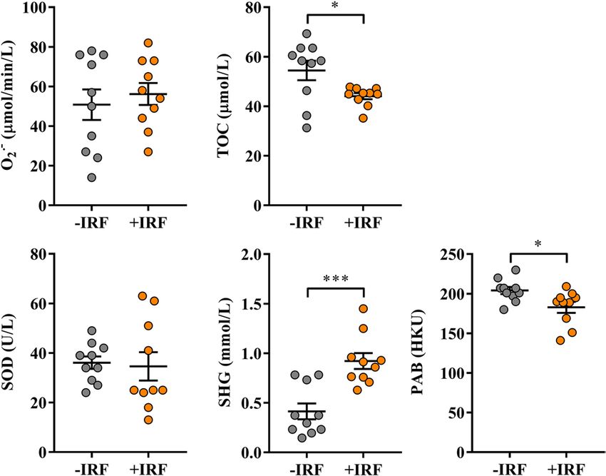
www.nature.com/scientificreports/
reverse cutaneous h ypersensitivity39. Given that rats from cages with bedding from IR fibres developed paw
inflammation of lower magnitude compared with those from cages with standard wood shaving bedding, the
effects of rat exposure to IR fibres in a treatment paradigm were examined, as well.
Materials and methods
Animals. In the present study four-month-old female Dark Agouti (DA) rats from a breeding colony in the
Immunology Research Centre “Branislav Janković” (Belgrade, Serbia) were used. Rats were maintained in a fully
controlled animal facility with a constant temperature (21–23 °C) and humidity (30–50%), a 12 h/12 h light/
dark cycle, and they were provided ad libitum access to water and standard pelleted food. All experiments were
performed in accordance with the Directive 2010/63/EU of the European Parliament and of the Council on the
Protection of Animals used for Scientific Purposes (revised Directive 86/609/EEC), and were approved by Labo-
ratory Animal Ethical Committee of University of Belgrade—Faculty of Pharmacy (Etička Komisija za ogledne
životinje, Univerzitet u Beogradu –Farmaceutski fakultet).
Induction and clinical evaluation of CIA. Rats were immunised intradermally at the base of the tail with
300 μg of bovine CII Sigma-Aldrich Chemie GmbH, Taufkirchen, Germany) emulsified in incomplete Freund’s
adjuvant (IFA), as previously described27,28. The immunisation emulsion was prepared by mixing equal volumes
of CII solution (2 mg/mL) in 0.1 M acetic acid and IFA. For immunisation 300 µL of this emulsion was injected
per rat. Before the immunisation, animals were anesthetized with an intraperitoneal injection of anesthetic cock-
tail [50 mg/kg/body weight (BW) of ketamine/5 mg/kg BW xylazine; Ketamidor, Richter Pharma AG, Wels, Aus-
tria; Xylased, Bioveta, Ivanovice na Hané, Czech Republic]. Clinical signs of arthritis were evaluated daily from
the 7th day post immunisation (d.p.i.) until the 22nd d.p.i., when the clinical severity of the disease reaches the
maximum 27,28. The severity of CIA was graded according to an arbitrary scale taking into account joint edema
and erythema of the hind and front paws (one point for each inflamed metacarpophalangeal/metatarsophalan-
geal or interphalangeal joint, and five points for the inflamed ankle)40 by two experienced researchers (MD and
MS), independently. Thus, each paw could receive the maximum score of 15 points giving the highest arthritic
score of 60. Additionally, paw volumes were measured before the immunisation (basal) and on the 22nd d.p.i.
Given that pathological changes predominantly occurred in the hind paws of rats, volumes of both hind paws
were measured. Of note, these measurements and all subsequent analyses were conducted by two investigators
blind to the rat cage bedding.
Induction and clinical evaluation of CIPI. Paw inflammation was induced by intraplantar injection of
1.5 mg carrageenan (λ-carrageenan, Sigma Aldrich, St.Louis, MO, USA) in 150 μL saline or 150 μL of saline
(controls) into the right hind paw starting from the midline near the heel and continuing toward the base of the
second or third toe as previously described39. The volumes of their right hind paws were measured before injec-
tion of carrageenan or saline (basal) and four times afterward at one hour intervals. The temperature of the right
hind paws was measured using a digital touch-free thermometer (Microlife AG, Widnau, Switzerland) at the
same time points. Of note, these measurements and all subsequent analyses were conducted by two investigators
blind to the rat cage bedding.
Experimental design. For the induction of CIA rats were randomly assigned into two groups (10 rats per
group). Five days before immunisation one group of rats was housed in cages with bedding composed of IR Cel-
liant fibres (+IRF rats), while the other group of rats was housed in cages with standard wood shaving bedding
(−IRF rats). Celliant is produced using a total of 13 naturally occurring, thermoreactive minerals. These include:
titanium dioxide, a photocatalyst that effectively absorbs light, silicon dioxide, which absorbs and reflects energy,
and aluminium oxide, which can help increase energy reflectivity. All 13 minerals are ground up into an ultra-
fine powder and then mixed with polyethylene terephthalate to create the ‘Celliant master batch’. Finally, a liquid
polyester resin is added to the master batch and synthesized into a stable fibre. The cage-bedding was con-
structed from 16 g Celliant (200 g/m2) of 42 mm fibres (thickness less than 10 μ, ceramic loading 1–1.25% by
weight) and standard wood shavings (1:10 weight/weight) (Fig. S1). According to the manufacturer, the ceram-
ics absorb the body’s heat, transforming it into full-spectrum infrared energy (https://celliant.com/how-it-works
) with a broad-peak cantered at a wavelength of 9.3 µm, whereas the power density depends on the proximity of
the heat source (living body), but is approximately 0.25 mW/cm2, so for each hour, an energy density of about
1 J/cm2 is delivered. The standard wood shaving and IR Celliant beddings were changed every second day. The
burrowing behaviour test was performed on the 21st d.p.i. Following the test animals were anesthetized with an
intraperitoneal injection of ketamine/xylazine anesthetizing cocktail (80 mg/kg BW ketamine/8 mg/kg BW xyla-
zine) and blood was taken by cardiac puncture. Additionally, from euthanized rats were removed lymph nodes,
spleens, and hind paws for further analysis.
To examine the effects of rat exposure to IR fibres on CIPI, two sets of experiments (each consisting of two
separate experiments) were performed. In the first set of experiments, rats were randomly assigned into three
groups (6 rats per group). Two groups were administered with carrageenan, whereas one group (6 rats) was
administered with saline (SAL rats). Five days before administration of rat hind paws with carrageenan one
group of animals was transferred to cages with Celliant bedding (+IRF rats), while the other groups remained in
cages with standard wooden shaving bedding (−IRF rats). In the second set of experiments, rats were randomly
assigned into three groups (6 rats per group). Following the administration of right hind paws with carrageenan
one group of rats was transferred to cages with Celliant bedding (+IRF rats), one group of rats was administered
with 5 mg/kg Diclofenac (Diklofen, Galenika AD, Belgrade, Serbia) per os (DIC rats), whereas one group of rats
Scientific Reports | (2021) 11:2882 | https://doi.org/10.1038/s41598-021-81999-7 3
Vol.:(0123456789)www.nature.com/scientificreports/
remained in cages with standard bedding (−IRF rats). The dose of Diklofen was chosen to correspond to that
used in humans41.
In one experiment from each set of experiments 240 min following the carrageenan/saline administration
burrowing test (lasting for 120 min) was started. At the end of this test, rats were euthanized by an intraperito-
neal injection of ketamine/xylazine anesthetizing cocktail (80 mg/kg BW ketamine/8 mg/kg BW xylazine), and
their right paws were retrieved for culturing. In the second experiment from each set of experiments, right paw
volume and mechanical hyperalgesia were examined at one hour intervals over 240 min from the carrageenan/
saline administration.
Paw volume measurement. Paw volume was measured using a plethysmometer (Ugo Basile, Gemonio,
reviously42. The average of two consecutive volume measurements for each rat
Lombardy, Italy), as described p
was used for further calculations. The results are expressed as the difference (dV) between post-immunisation
or CIPI induction and the basal paw volume according to the following formula42:
dV = volume of the inflamed paw (mL) − basal volume of the same paw (mL)
Next, the effect of treatment (Celliant fibres or Diklofen) on dV was calculated according to the following
formula
referent group ∗ average dV − dV of each rat from experimental group ∗ ∗
dV reduction (%) = ×100
referent group average dV
* CIA/CIPI rats from cages with standard bedding without any treatment; ** rats exposed to Celliant fibres
or administered with Diklofen/saline42.
Burrowing training and burrowing test. To assess influence of Celliant bedding on spontaneous joint
pain, burrowing test, the test developed with the goal of enhancing the translational potential of preclinical
findings in pain research43 was used. This test examines burrowing behaviour, an ancient adaptive behaviour
conserved across many rodent species, one in which various laboratory strains of mice and rats spontaneously
engage, and, more important, one which is depressed in both mice and rats experiencing inflammatory and
neuropathic pain44,45. It is noteworthy, that this behaviour is considered to represent a correlate of so-called
“activities of daily living” in humans—tasks that are essential to satisfactory quality of life and are often impeded
by pain46. For burrowing training and experiments, long plastic tubes (32 cm in length and 10 cm in diameter),
with the open-end elevated 6 cm from the cage bottom, were filled with 2500 g of gravel (2–6 mm diameter par-
ticles). The training was performed four days before housing in cages with Celliant bedding, and both training
and burrowing behaviour tests were performed during the dark phase of the daily cycle starting at 6 pm. The
training was conducted in social facilitation and individual training formats. For social facilitation, the rats from
one cage were placed in a cage-burrow setup for 120 min. To estimate burrowing behaviour, the weight of the
burrowed gravel was calculated (weight of gravel left in the tube after completion of burrowing training or test
was subtracted from the initial weight of gravel in the tube). Individual training was performed so that a single
rat was placed in a cage-burrow setup for 120 min per day for three consecutive days and the average amount of
burrowed gravel was determined as explained above. Rats burrowed less than 500 g (four out of 56) were classi-
fied as poor burrowers and they were excluded from further experiments as previously suggested46. The test was
performed before immunisation/inflammation induction (to assess basal burrowing activity) and at a certain
point following the immunisation/inflammation induction.
Following the test burrowing activity (BA) of each rat was calculated according to the following formula:
BA after immunisation/inflammation induction g
BA (%) = × 100
Basal BA
and then increase in burrowing activity was calculated according to the following formula:
BA of each rat from treated group ∗ − BA of referent group ∗ ∗ average
BA increase (%) =
×100
BA of referent group average
* rats exposed to Celliant fibres or rats administered with Diklofen; **rats from cages with standard bedding.
Electronic Von Frey test. The mechanical hyperalgesia following carrageenan injection was assessed by
measuring paw withdrawal thresholds (P, expressed in g) using an electronic Von Frey anesthesiometer (IITC
Life Science, Woodland Hills, CA, United States) as described previously47,48. The rats were placed in transpar-
ent boxes on the top of a metal grid and allowed to acclimatize for 30 min before testing. A plastic, semi-flexible
filament coupled with a force transducer was used to deliver the mechanical stimulus. The tip of the filament was
applied perpendicularly to the plantar surface of the right hind paw and the pressure was gradually increased
until the rat withdrew its paw (that pressure was recorded automatically on a digital screen). Basal Ps were
measured before inflammation induction. Basal and post-induction Ps were measured on the right hind paw.
The results are expressed as the difference (dP) between basal and post-induction Ps according to the following
formula47,48:
dP = P before inflammation induction g − P after inflammation induction g
Scientific Reports | (2021) 11:2882 | https://doi.org/10.1038/s41598-021-81999-7 4
Vol:.(1234567890)www.nature.com/scientificreports/
Treatment reducing dP was recognized as antihyperalgesic treatment. The percentage of the antihyperalgesic
activity (AHA) was calculated as previously suggested48:
dP of referent group ∗ average − dP of each rat in treated group ∗ ∗
AHA (%) =
× 100
dP of referent group average
* rats from cages with standard bedding; ** rats exposed to Celliant fibres or rats administered with Diklofen.
Isolation of mononuclear cells. To obtain mononuclear single cell suspensions from lymph nodes (LNs)
for the analysis of migratory capacity of innate immune cells in fluorescein isothiocyanate (FITC) painting test
and spleens for in vitro analyses of adherence and phagocytic capacity of innate immune cells, LNs draining the
site of FITC application (DLNs) and spleens were dissociated using a 70 µm nylon cell strainer (BD Biosciences,
Erembodegem, Belgium) and obtained cells were collected in phosphate buffered saline (PBS) supplemented
with 2% fetal calf serum (FCS, Gibco, Grand Island, NY, USA) and 0.01% NaN3 (Sigma-Aldrich Chemie GmbH)
(FACS buffer), respectively. The single cell splenocyte suspensions were subjected to NH4Cl lysis to remove red
blood cells and then washed in ice-cold FACS buffer. The mononuclear single cell suspensions from DLNs and
spleen of each animal were enumerated using an improved Neubauer hemacytometer and trypan blue dye to
exclude non-viable cells and adjusted to 1 × 10⁷ cells/mL.
Flow cytometry analysis (FCA). Briefly, for FCA single cell suspensions from DLNs and spleen were
etails49. After immunolabeling 50,000
subjected to direct or indirect immunolabeling as previously described in d
cell events per sample were acquired on a FACSCalibur flow cytometer (Becton–Dickinson, Mountain View, CA,
USA). Data were analysed using FlowJo software version 7.8. (TreeStar Inc, Ashland, OR, USA). Dead cells and
debris were excluded from the analyses by selective gating based on forward scatter (FSC) and side scatter (SSC).
Innate immune cell functional assays. To assess the migration capacity of innate immune cells FITC
painting test was used50. Briefly, rat left flank was shaved and painted with 300 μL of 5 mg/mL FITC dissolved
in equal volumes of acetone and dibutylphthalate. After 24 h, ipsilateral (the side subjected to FITC painting)
inguinal and axillary DLNs were extirpated and divided into two portions. From one portion single mononu-
clear cell suspensions were prepared as described above, whereas another portion was used to examine CCL19
and CCL21 expression using Reverse Transcription-Quantitative Polymerase Chain Reaction. Aliquots of 100
µL of DLN suspensions were processed for indirect immunolabeling with biotin-conjugated anti-CD11b and
PerCP-conjugated streptavidin as primary and second step reagent, respectively. Following washing, cells were
acquired for analysis using a FACSCalibur flow cytometer as described above. Results were expressed as % of
FITC + cells within the CD11b + LN cells.
Innate immunity cells isolated from spleen were tested for cell adherence ability using a modification of
previously described m ethod51. Aliquots of 500 µL of splenocyte suspension (1 × 10⁶ cells/ml) were placed in
an adherence column consisting of a 1 mL syringe packed with 50 mg of nylon fibres to a height of 1.25 cm.
After 10 min, the effluent containing the non-adherent cells was drained by gravity. Initial cell suspensions and
effluents were processed for immunolabeling with FITC-conjugated anti-CD11b (clone ED8; Serotec, Oxford,
UK) antibody. The cells were incubated with saturating concentrations of the fluorochrome-labeled antibody for
30 min, washed with FACS buffer, and subsequently subjected for FCA. Results were expressed as
% of total CD11b + cells − % of non-adherent CD11b + cells
adherence index = × 100%
% of total CD11b + cells
To assess the phagocytic capacity of innate immune cells a previously described method was u sed49. Briefly,
100 µl aliquots of splenocyte suspensions (1 × 10⁶ cells/ml) described, were incubated with sonicated (2 min at
room temperature) 1 µm sized yellow-green fluorescent carboxylated polystyrene latex beads (Sigma-Aldrich
Chemie GmbH) in complete RPMI-1640 culture medium supplemented with 5% FCS (bead:cell ratio of 50:1)
for 1 h at 37 ºC. To arrest phagocytosis, the cells were placed on ice for 5–10 min. Following this step, cells were
washed with ice cold PBS and incubated with biotin-conjugated anti-CD11b (BD Biosciences, Mountain View,
CA, USA) antibody for 30 min at 4 ºC and then again washed with FACS buffer. In the next step, cells were
incubated with PerCP-conjugated streptavidin (BD Biosciences) as the second step reagent for another 30 min,
washed with FACS buffer and acquired for analysis using a FACSCalibur flow cytometer. Cells incubated with
the latex beads at 4 ºC were used to set up the positive/negative cut-off for Latex + cells in FCA. Results were
expressed as % of Latex + cells within the CD11b + cell population.
Reverse transcription‑quantitative polymerase chain reaction. Total RNA was extracted by
the ABI Prism 6100 Nucleic Acid PrepStation system (Applied Biosystems, Foster City, CA, USA) using the
total RNA Chemistry Starter Kit (Applied Biosystems) and DNAse wash solution (Absolute RNA Wash Solu-
tion; Applied Biosystems). cDNA was synthesized using the High-Capacity cDNA Reverse Transcription Kit
(Applied Biosystems). Triplicate 25-μL RT-qPCR reactions were performed using the TaqMan Gene Expression
Master Mix (Applied Biosystems) and premade TaqMan Gene Expression Assays (Applied Biosystems) under
the default Applied Biosystems 7500 Real-Time PCR System conditions. All the procedures were described in
detail52. The following TaqMan Gene Expression Assays were used: CCL19 (Ccl19, Rn01439563_m1), CCL21
(Ccl21, Rn01764651_g1), and β-actin (Actb, Rn00667869_m1). Target mRNA expression was determined using
the comparative threshold cycle (dCt) method with β-actin as a reference and SDS v1.4.0. software (Applied
Scientific Reports | (2021) 11:2882 | https://doi.org/10.1038/s41598-021-81999-7 5
Vol.:(0123456789)www.nature.com/scientificreports/
Biosystems). Relative amounts of target mRNAs were shown as 2–dCt values, representing the ratio of target to
reference genes, where dCt = Ct target – Ct reference.
Anti‑CII antibody ELISA. Serum samples were obtained by centrifugation of blood at 2000g for 15 min at
4 °C. Aliquots of sera were de-complemented (56 °C, 30 min) and stored at -20 °C until analysis. The serum level
of CII-specific antibodies was assayed by ELISA in 96-well plates (MaxiSorp, Nunc). The plates were coated (50
μL/well) with 5 μg/mL of CII in 50 mM carbonate buffer (pH 9.6) by overnight adsorption at + 4 °C and then
incubated with 2% bovine serum albumin (BSA) in PBS (100 μL/well) for 1 h at room temperature. Serum sam-
ples were added to the plate (50 μL/well) in duplicate and incubated overnight at + 4 °C. The biotin-conjugated
anti-IgG antibody was added to the plate (50 μL/well) and incubated 1 h at room temperature followed by 1 h
incubation with a streptavidin–horseradish peroxidase. At each step, the plate was washed with 0.05% Tween
20/PBS (4 × 200 μL/well). Antigen–antibody interactions were visualized using the extrAvidin-peroxidase/o-
phenylenediamine system (Sigma, Steinheim, Germany). Dilutions of sera (1: 100), biotin-conjugated anti-IgG
antibody (1: 1000; Biolegend Inc., San Diego, CA, USA) and extrAvidin-peroxidase (1: 3000) were prepared in
2% BSA/PBS. The reaction was stopped by the addition of 1 M H2SO4 (50 μL/well) and absorbance was read
at 492/620 nm (A492/620). The cutoff value was defined according to the A492/620 value obtained from “negative
control” wells (2% BSA/PBS) plus 3 × Standard Deviation. Samples were considered positive when the A 492/620
value exceeded the cut off value.
Paw tissue culture. Inflamed paws collected by incising at the fur line. Paws from CIA rats were weighed,
carefully cut into small pieces and cultured in RPMI 1640 medium (Sigma-Aldrich Chemie GmbH) supple-
mented with 2 mM l-glutamine (Serva, Heidelberg, Germany), 1 mM Na pyruvate (Serva), 100 U/mL peni-
cillin (ICN, Costa Mesa. CA, USA), 100 μg/mL streptomycin (ICN) and 10% fetal bovine serum, at 37 °C, in
a humidified air atmosphere of 5% v/v CO2, for 4 h (for PGE2 assay) or overnight (for analyses of NO and
escribed28,29,53. Following extirpation of
cytokine production levels, and redox status parameters), as previously d
carrageenan-inflamed paws, soft paw tissues were carefully removed, weighed, cut into small pieces and cultured
as described above.
PGE2, cytokine and NO production. Paw tissue culture supernatants were examined for inflamma-
tory/pain mediators using the following commercial ELISA kits: IL-17A (BioLegend, San Diego, CA, USA),
IL-1β (Thermo Scientific, Pierce Biotechnology, Rockford, IL, USA), PGE2, IL-10 and TGF-β (R&D Systems,
Minneapolis, MN, USA), according to the manufacturer’s instructions. Standard curve was calculated for each
assay with limits of detection for IL-17 = 8 pg/ml, IL-1β = 6.5 pg/mL, TNF-α = 2 pg/mL, IL-10 < 10 pg/mL,
TGF-β = 4.6 pg/mL and PGE2 < 39 pg/mL. Cytokines and PGE2 concentrations were normalized to paw weight.
The concentration of nitrite, as the end-product of NO production, was measured in the paw tissue culture
supernatants using a method based on the Griess reaction54. The nitrite concentration was calculated using a
NaNO2 standard curve with a range from 1 to 40 μM and normalized to paw weight.
Assessment of redox status parameters. The parameters of redox status were measured in sera and
inflamed paw cultures. The levels of superoxide anion radical (O2• ‾) were estimated from the rate of reduction
of nitroblue tetrazolium (NBT) as described by Auclair and V oisin55. Results were expressed as μM of reduced
NBT/min/L.
Total oxidant capacity (TOC) was determined by a modified spectrophotometric method using o-dian-
isidinine56,57. The assay was calibrated with hydrogen peroxide (aqueous solution, concentration range
10–200 mmol/L) and the results were expressed in µmol/L hydrogen peroxide per liter (mmol H 2O2 eq./L).
Cu/Zn superoxide dismutase (SOD) activity was determined by a modified spectrophotometric method
based on the ability of the SOD enzyme to inhibit the autoxidation of epinephrine in an alkaline medium or
bicarbonate buffer 0.05 mmol/L pH 10.258. The SOD activity was calculated as the percentage inhibition of
epinephrine autooxidation.
The level of SH-groups (SHG) was determined by Ellman’s m ethod59 using 10 mM DTNB (dinitrodith-
iobenzoic acid) as a reagent. DTNB reacts with aliphatic thiol compounds in the base medium (pH 9.0) and this
reaction generates 1 mol of p-nitrophenol anion per mole of thiol. Calibration of the method was achieved using
reduced glutathione in the concentration range from 0.1 to 1.0 mM.
Pro-oxidative-antioxidant balance (PAB) was determined by a modified PAB assay using 0.6% 3.3 ’, 5,5′-tetra-
methylbenzidine (TMB) in dimethyl sulfoxide (DMSO) as a c hromogen60. The PAB test measures the concen-
tration of H2O2 in an antioxidant environment, as TMB can react at the same time with H2O2 (a peroxidase-
catalyzed reaction) as well as with reducing substances such as uric acid (chemical, non-catalyzed reaction). The
enzymatic reaction leads to the oxidation of TMB to a blue product and its reduction to a non-colored product.
The net reaction is the difference between the two opposite oxidative and reductive processes on the same sub-
strate. The reaction is calibrated with a mixture of H
2O2 and uric acid at different ratios, ranging from 0 to 100%.
All analyses were performed on ILAB 300 Plus analyzer (Instrumentation Laboratory, Milan, Italy) except
the PAB, which was measured on the Spectro star Nano ELISA reader (BMG Labtech, Ortenberg, Germany).
Statistical analysis. All statistical analyses were performed using GraphPad Prism Version 7 (GraphPad
Software, Inc., La Jolla, CA, USA) and SigmaPlot 11 (Systat Software Inc., Richmond, CA) softwares. Data are
expressed as mean ± SEM. Differences between groups were tested by Student’s unpaired t-test, except differ-
ences in paw volume, temperature, and hyperalgesia in carrageenan-induced paw inflammation that were ana-
Scientific Reports | (2021) 11:2882 | https://doi.org/10.1038/s41598-021-81999-7 6
Vol:.(1234567890)www.nature.com/scientificreports/
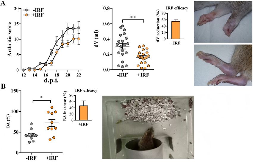
Figure 1. Influence of exposure to IR fibres on daily arthritis score, paw edema and burrowing behaviour in
CIA-affected rats. (A) Line graph shows daily arthritis score from the 12th–22nd day post-immunisation (d.p.i.)
in CIA rats housed in cages with IR-fibre bedding (+IRF rats) or in cages with standard wooden shaving bedding
(−IRF rats). Of note, rats were transferred to cages with IR fibre bedding five days before immunization. Scatter
plot indicates CIA-induced increase in hind paw volume (dV) in +IRF and −IRF rats. Bar graph shows the dV
reduction (%) in +IRF rats relative to −IRF rats (see Material and Methods). Photographs show representative
arthritic front and hind paw joints from rat with CIA. (B) Scatter plot indicates the burrowing activity (BA) of
each +IRF and −IRF rat expressed as the percentage of the BA before immunisation (basal activity). Bar graph
shows the increase in BA (%) in +IRF rats relative to −IRF rats (see Material and Methods). All graphs were
created using GraphPad Prism version 7.00 for Windows, GraphPad Software, La Jolla, California, USA (https
://www.graphpad.com). Photograph shows a cage-burrow setup and burrowed gravel. Results are expressed as
mean ± SEM. n = 10 rats/group. * p ≤ 0.05 and ** p ≤ 0.01.
lysed by two-way repeated measures ANOVA followed by Bonferroni test for post hoc comparisons. Values of
p ≤ 0.05 were considered significant.
Results
Modulatory effects of IR fibres on the autoimmune response, inflammation and burrowing
behaviour of CIA‑affected rats. CIA‑affected rats from cages with IR fibre bedding exhibit better burrowing
performance compared with their counterparts from cages with standard bedding. Generally, the kinetics of CIA
development in rats housed in cages with standard bedding followed that described in our previous studies27,28
(Fig. S2). In rats housed in cages with IR fibre bedding, the onset of the disease was slightly postponed (from the
13th to the 15th d.p.i. compared with −IRF controls (Fig. 1A). The analysis of the differences in the daily arthritic
score using repeated measures two-way ANOVA showed a clear tendency to significance (p = 0.051) in the re-
duction of the clinical severity of the disease in rats exposed to IR fibres compared with their −IRF counterparts
(Fig. 1A). Additionally, the cumulative score of the disease (the sum of daily clinical scores of each individual rat
during the observation period) was lower (p ≤ 0.05) in +IRF rats (72.1 ± 9.70) when compared with −IRF ones
(47.4 ± 5.56). Rats were also examined for the volumes of inflamed hind paws. In all CIA-affected rats the vol-
umes of hind paws were increased (Fig. 1A). This increase was less (p ≤ 0.01) pronounced in +IRF rats compared
with their −IRF counterparts, indicating that the exposure to infrared radiation from IR fibre bedding efficiently
reduces the inflammation-induced increase in paw volume (Fig. 1A).
Furthermore, rats were examined for burrowing behaviour. Rats housed in cages with IRF bedding showed
better (p ≤ 0.05) burrowing performance compared with −IRF rats (Fig. 1B, Fig. S3).
Lower serum levels of serum CII‑specific IgG antibodies in CIA‑affected rats from cages with IR fibre bedding and
diminished IL‑17 production in cultures of their hind paws. Given that (i) arthritogenic anti-CII antibodies are
essential for the development of CIA61,62 and (ii) particularly that the arthritogenic antibodies (including those
specific for CII) are shown to cause pain even in the absence of overt signs of inflammation38,63, serum levels
of CII-specific IgG antibodies in CIA rats were examined. Their levels were lower (p ≤ 0.01) in sera from rats
Scientific Reports | (2021) 11:2882 | https://doi.org/10.1038/s41598-021-81999-7 7
Vol.:(0123456789)www.nature.com/scientificreports/
Figure 2. Influence of exposure to IR fibres on the level of CII-specific IgG antibodies in sera and the
production of IL-17 in inflamed paws from CIA-affected rats. (A) Scatter plot indicates the level of anti-CII IgG
antibodies (OD492nm × 1000) in sera from CIA rats housed in cages with IR fibres bedding (+IRF rats) and with
standard wooden shaving bedding (−IRF rats). Of note, rats were transferred to cages with IR fibre bedding
five days before immunization. (B) Scatter plot shows IL-17 production level in the supernatants from hind
paw tissue cultures (normalized to the paw weight) from +IRF and −IRF rats. Scatter plots were created using
GraphPad Prism version 7.00 for Windows, GraphPad Software, La Jolla, California,USA (https://www.graph
pad.com). Results are expressed as mean ± SEM. n = 10 rats/group. * p ≤ 0.05 and ** p ≤ 0.01.
transferred to cages with IR fibres bedding (five days before CIPI induction) compared with their counterparts
housed into cages with standard bedding (Fig. 2A).
Considering the crucial role of Th17 cells in driving joint damage and consecutive i nflammation64, the pro-
duction levels of IL-17, Th17 cell signature cytokine, were examined in supernatants of inflamed paw tissue
cultures. The production levels of this cytokine were lower (p ≤ 0.05) in supernatants of inflamed paw tissue
cultures from +IRF rats compared with −IRF ones (Fig. 2B).
Impaired migration, but greater phagocytic capacity of innate immune CD11b + cells from CIA‑affected rats exposed
to IR fibres. Considering the role of innate immune CD11b + cells in the development of joint impairment and
inflammation in C IA65, and the significance of their infiltration into inflamed joint tissue, migratory, adherence
and phagocytic capacity of CD11b + cells from CIA rats housed in cages with IR fibre and standard bedding were
examined.
The analysis of the frequency of FITC-labelled CD11b + cells in DLNs in the FITC painting test revealed that
their frequency was lower (p ≤ 0.01) in +IRF rats than in −IRF controls, indicating impaired migration capacity
of CD11b + cells in rats from cages with IR fibre bedding (Fig. 3A). Considering that CD11b cells express CCR7
receptor so that their trafficking is substantially influenced by CCL19 and CCL21 c hemokines50,66, the expres-
sion of mRNAs for these chemokines in DLNs was examined, as well. Indeed, the exposure to IR cage bedding
reduced (p ≤ 0.01) the expression of mRNAs for both chemokines (Fig. 3A).
Next, considered that in the response to tissue inflammation CD11b + cells leave the spleen en masse to accu-
mulate in injured tissue, and participate in the control of inflammation by phagocyting dead cells and necrotic
tissue, and secreting anti-inflammatory/immunoregulatory m ediators67,68, the adherence and phagocytic capacity
of CD11b + cells from spleen were examined. We failed to show any statistically significant difference between
the adherence capacity of CD11b + splenocytes from +IRF rats and those from −IRF rats (Fig. 3B). The analysis
of Latex bead phagocytosis showed higher (p ≤ 0.001) phagocytic capacity of CD11b + splenocytes from rats
exposed to IR fibres had compared with those from rats housed in cages with standard bedding (Fig. 4).
Exposure of CIA‑affected rats to IR fibres decreases the production levels of pro‑inflammatory mediators, but
increases production levels of anti‑inflammatory/immunoregulatory mediators in cultures of inflamed paws. To
elucidate molecular mechanisms underlying less prominent paw swelling and better burrowing performance
of +IRF rats, the production of the key pro-inflammatory mediators with algogenic properties (PGE2, NO,
TNF-α and IL-1β)69,70 and anti-inflammatory/immunoregulatory mediators with analgesic properties (IL-10
and TGF-β)71,72 was examined in cultures of their hind paws.
Scientific Reports | (2021) 11:2882 | https://doi.org/10.1038/s41598-021-81999-7 8
Vol:.(1234567890)www.nature.com/scientificreports/
Figure 3. Influence of exposure to IR fibres on the migration and adherence capacity of innate immune
CD11b + cells from CIA-affected rats. (A) Representative flow cytometry dot plots show the frequency of FITC-
stained CD11b + cells in lymph nodes draining the site of FITC application (DLN) in FITC painting test (see
Material and Methods) in CIA rats housed in cages with IR fibre bedding (+IRF rats) and with wooden shaving
bedding (−IRF rats). Of note, rats were transferred to cages with IR fibre bedding five days before immunization.
Bar graphs show the expression of mRNA for CCL19 and CCL21 in DLNs from +IRF and −IRF rats, as
determined by RT-qPCR. Results are represented as 2−dCt relative to β-actin. (B) Representative flow cytometry
dot plots show CD11b expression on splenocytes from +IRF and −IRF rats before (upper dot plots) and after
(lower dot plots) passage through nylon wool in the adherence assay (see Material and Methods). Scatter plot
indicates the adherence index, i.e. the percentage of adherent cells of CD11b + cells from spleens of +IRF and −
IRF rats (calculated as indicated in Material and Methods). (A, B) Flow cytometry profiles were generated using
FlowJo Software for Windows, Version 7.8. FlowJo, LLC, Ashland, Oregon, USA (https://www.flowjo.com).
Bar graphs (A) and (B) scatter plot were created using GraphPad Prism version 7.00 for Windows, GraphPad
Software, La Jolla California, USA (https://www.graphpad.com). Results are expressed as mean ± SEM. n = 10
rats/group. ** p ≤ 0.01.
The production levels of P GE2, a pro-inflammatory lipid mediator present at high levels in the synovial fluid
of patients suffering from RA73–75, were lower (p ≤ 0.01) in paw cultures from +IRF rats when compared with
those from −IRF rats (Fig. 5).
Additionally, production levels of NO, the pro-inflammatory mediator with an important role in the develop-
ment of joint injury and inflammation76, were also lower (p ≤ 0.01) in inflamed paw cultures from rats exposed
to IR-emitting fibres than in those from rats housed in cages with standard bedding (Fig. 5).
On the other hand, we failed to show statistically significant differences in the production levels of pro-
inflammatory cytokines (TNF-α and IL-1β), which are shown to significantly contribute to the joint injury and
inflammation, and consequently arthralgia in R A77 in inflamed paw tissue cultures from +IRF rats and their −IRF
counterparts (Fig. 5).
Considering that the ratio between pro-inflammatory to anti-inflammatory/immunoregulatory mediators
(IL-10 and TGF-β) in inflamed tissue is more important for RA severity than their absolute levels37 and that both
IL-10 and TGF-β act as endogenous a nalgesics71,72, their production levels in hind paw cultures were explored,
as well. The levels of IL-10 and TGF-β were higher (p ≤ 0.05 and p ≤ 0.001, respectively) in cultures of inflamed
paws from +IRF rats compared with IRF rats (Fig. 5).
CIA‑affected rats from cages with IR fibres exhibit improved serum redox status compared with their counterparts
from cages with standard bedding. Given that oxidative stress has been also implicated in the development of
inflammation in chronic autoimmune diseases, including RA and its experimental m odels78, the redox status
was examined in both sera and inflamed joint tissue cultures.
Scientific Reports | (2021) 11:2882 | https://doi.org/10.1038/s41598-021-81999-7 9
Vol.:(0123456789)www.nature.com/scientificreports/
Figure 4. Influence of exposure to IR fibres on the phagocytic capacity of innate immune CD11b + cells from
CIA-affected rats. Representative flow cytometry histograms show latex particle phagocytosis at + 4° C and + 37°
C by CD11b + cells (gated as shown in representative flow cytometry dot plots generated using FlowJo Software
for Windows, Version 7.8. FlowJo, LLC, Ashland, Oregon, USA; https://www.flowjo.com.) from CIA rats housed
in cages with IR fibre bedding (+IRF rats) and with standard wooden shaving bedding ( –IRF rats). Rats were
transferred to cages with IR fibre bedding five days before immunization. Note that a significant difference
between the groups was apparent only at 37° C. Scatter plot (created using GraphPad Prism version 7.00 for
Windows, GraphPad Software, La Jolla, California, USA; https://www.graphpad.com) indicates the percentage
of phagocyting (latex +) cells among CD11b + cells from the spleen of +IRF and –IRF rats (see Material and
Methods). Results are expressed as mean ± SEM. n = 10 rats/group. *** p ≤ 0.001.
The exposure of rats to IR-emitting fibres had no effects on the serum level of O2• − and SOD activity in CIA
rats (Fig. 6). However, lower (p ≤ 0.05) TOC levels accompanied by higher SHG (p ≤ 0.001) levels were found in
sera from +IRF rats compared with control rats (Fig. 6). Consistently, lower (p ≤ 0.05) PAB value was detected
in sera from +IRF rats compared with −IRF rats (Fig. 6).
We failed to detect any statistically significant differences in the examined redox parameters in the inflamed
paw cultures (Fig. S4).
Housing of rats in cages with IF fibre bedding beginning 5 days before carrageenan adminis‑
tration moderates CIPI development. Given that rats from cages with IR bedding exhibited dimin-
ished antibody and Th17 cell responses, and that anti-CII antibodies are found to induce pain behaviour acting
directly on sensory neurons, viz. independently of their pro-inflammatory action as pathological a ntibodies38,
the study was extended to encompass CIPI model, a “classical” model of the non-autoimmune inflammatory
response and inflammatory pain.
Exposure to IR fibres reduces temperature, swelling and hyperalgesia of carrageenan‑inflamed rat paws and
improves burrowing behaviour of CIPI‑affected rats. In rats housed in the cages with IR fibre bedding the sur-
face temperature of carrageenan-injected inflamed paw was lower than in rats housed with standard bedding
at 120 min (p ≤ 0.05), 180 min (p ≤ 0.001) and 240 min (p ≤ 0.05) after the injection of carrageenan (Fig. 7). The
surface temperature of paws injected with saline did not change during the period of observation, and it was
lower when compared with the surface temperature of carrageenan-injected paws from −IRF rats at 120 min
(p ≤ 0.05), 180 min (p ≤ 0.001) and 240 min (p ≤ 0.001) post-injection (Fig. 7). Besides, it was lower when com-
pared with the surface temperature of carrageenan-injected paws from +IRF rats at 180 min (p ≤ 0.05) post-
injection (Fig. 7). Additionally, in rats housed in the cages with standard bedding, the intraplantar injection
of carrageenan elicited paw swelling, as one of the cardinal signs of inflammation (Fig. 8A). In −IRF rats this
increase in paw volume was at the maximum at 180 min post injection of carrageenan and remained at this level
until the end of observation (Fig. 8A). In the rats transferred to cages with IR fibre bedding five days before the
administration of carrageenan, the paw volume gradually increased until 240 min post the injection of carra-
geenan (Fig. 8A). Control SAL rats exhibited a transient increase in the affected paw volume 60 min after injec-
tion of saline, but even at this increase was lower (p ≤ 0.05) than in carrageenan-injected −IRF rats (Fig. 8A). In
rats exposed to IRF as cage bedding, the increase in the volume of the carrageenan-administered paw was lower
(p ≤ 0.05) at 60 min after the injection than in −IRF rats, and it remained lower (p ≤ 0.001) when examined at
later time points (Fig. 8A).
The analysis of burrowing performance showed that carrageenan markedly impaired (p ≤ 0.01) burrowing
activity of both −IRF and +IRF rats compared with SAL control rats (Fig. 8B, Fig. S5). However, this carrageenan-
induced decrease in burrowing performance was less (p ≤ 0.05) prominent in +IRF rats compared with their −IRF
counterparts (Fig. 8B, Fig. S5), indicating that infrared radiation from IRF cage bedding ameliorates burrowing
performance of CIPI rats (Fig. 8B, Fig. S5).
Scientific Reports | (2021) 11:2882 | https://doi.org/10.1038/s41598-021-81999-7 10
Vol:.(1234567890)www.nature.com/scientificreports/
Figure 5. Influence of exposure to IR fibres on the production of P
GE2, NO and cytokines by inflamed paws
from CIA-affected rats. Scatter plots (created using GraphPad Prism version 7.00 for Windows, GraphPad
Software, La Jolla, California, USA; https://www.graphpad.com) show P GE2, NO, TNF-α, IL-1β, TGF-β
and IL-10 production levels in the supernatants from 4 h (PGE2) and overnight hind paw tissue cultures
(normalized to the paw weight) (see “Material and methods”) from CIA rats were housed in cages with IR fibre
bedding (+IRF) and their counterparts housed in cages with standard wooden shaving bedding (−IRF rats). Rats
were transferred to cages with IR fibre bedding five days before immunization. Hind paws were excised from
CIA rats on the 22nd day post immunisation. Results are expressed as mean ± SEM. n = 10 rats/group. * p ≤ 0.05,
** p ≤ 0.01 and *** p ≤ 0.001.
Considering that differently from chronic pain conditions, in acute pain models reliability of tests evaluating
spontaneous behaviours or activities of rodents in their home environments, as it is burrowing test, have not been
systematically evaluated79, pain behaviour in CIPI model was also examined using classic mechanical stimulus-
evoked von Frey test (Fig. 8C). Using this test we found that, compared with the administration of saline, the
administration of carrageenan produced paw hyperalgesia (p ≤ 0.001) at all examined time points (Fig. 8C).
Additionally, at all examined time points following carrageenan administration rats housed in cages with IR fibre
bedding developed markedly less prominent (p ≤ 0.001) paw hyperalgesia compared with rats housed in cages
with standard bedding (Fig. 8C). The analysis of infrared radiation efficacy showed that its anti-hyperalgesic
efficacy decreased from 180 min onwards (Fig. 8C).
Exposure to IR fibres diminishes production of proinflammatory mediators, but increases production of anti‑inflam‑
matory mediators in cultures of carrageenan‑inflamed paws. The production of PGE2 and NO, pro-inflamma-
tory mediators implicated in the development of C IPI80, was examined in the cultures of carrageenan-inflamed
paws. The production levels of P GE2 were reduced (p ≤ 0.05) in cultures of inflamed paw tissues from +IRF rats
compared with their −IRF counterparts (Fig. 9). On the other hand, the production levels of NO were compara-
ble in inflamed paw tissue cultures from +IRF rats and −IRF ones (Fig. 9).
Considering the significant role of TNF-α and IL-1β in development CIPI and related a llodynia81,82, their
production levels in cultures of inflamed paws were also examined. The production of IL-1β by inflamed paw tis-
sues from +IRF rats was lower (p ≤ 0.01) when compared with its production by inflamed paw tissues from −IRF
Scientific Reports | (2021) 11:2882 | https://doi.org/10.1038/s41598-021-81999-7 11
Vol.:(0123456789)www.nature.com/scientificreports/
Figure 6. Influence of exposure to IR fibres on the redox status in sera from CIA-affected rats. Scatter plots
show pro-oxidant parameters: superoxide anion radical (O2• −) level and total oxidant capacity (TOC);
antioxidant parameters: superoxide dismutase (SOD) activity, and sulfhydryl groups (SHG) level; and pro-
oxidant-antioxidant balance (PAB) in sera from CIA rats housed in cages with IR fibre bedding (+IRF) and their
counterparts housed in cages with standard wooden shaving bedding (−IRF rats). Of note, rats were transferred
to cages with IR fibre bedding five days before immunization. Sera were obtained from CIA rats on the 2 2nd
day post immunisation. All graphs were created using GraphPad Prism version 7.00 for Windows, GraphPad
Software, La Jolla, California, USA (https://www.graphpad.com). Results are expressed as mean ± SEM. n = 10
rats/group. * p ≤ 0.05 and *** p ≤ 0.001.
Figure 7. Influence of exposure to IR fibre bedding on the paw temperature of rats with carrageenan-inflamed
paws. Rats housed in cages with IR fibre bedding (+IRF) or in cages with standard wooden shaving bedding
(−IRF) were injected with carrageenan. Of note, rats were transferred to cages with IR fibre bedding five days
before immunization. Rats housed in the cages with standard wooden shaving bedding were injected with
saline to serve as an additional control (SAL rats). Line graph (created using GraphPad Prism version 7.00 for
Windows, GraphPad Software, La Jolla California USA; https://www.graphpad.com) indicates the surface paw
temperature measured before and after carrageenan or saline injection in +IRF, –IRF and SAL rats. Of note,
rats were transferred to cages with IR fibre bedding five days before immunization. Results are expressed as
mean ± SEM. n = 6 rats/group.* p ≤ 0.05 and *** p ≤ 0.001 vs –IRF; and # p ≤ 0.05 vs +IRF.
rats (Fig. 9). Differently, the production levels of TNF-α in inflamed paw tissue cultures from +IRF rats did not
statistically significantly differ from those in inflamed paw cultures from −IRF rats (Fig. 9).
On the other hand, the production levels of TGF-β were higher (p ≤ 0.01) in cultures of inflamed paw tissues
from +IRF rats compared with those from −IRF ones, whereas those of IL-10 were comparable between these
cultures (Fig. 9).
Scientific Reports | (2021) 11:2882 | https://doi.org/10.1038/s41598-021-81999-7 12
Vol:.(1234567890)You can also read