Neutrophils self-limit swarming to contain bacterial growth in vivo - Microbiology, Immunology & Molecular ...
←
→
Page content transcription
If your browser does not render page correctly, please read the page content below
RES EARCH
◥ sensitization in neutrophil tissue navigation
RESEARCH ARTICLE SUMMARY and host defense.
IMMUNOLOGY RESULTS: We generated mouse strains whose
Neutrophils self-limit swarming to contain bacterial
neutrophils were deficient in GPCR kinases
(GRKs), critical enzymes for mediating the GPCR
growth in vivo
desensitization process. Of the four GRK isoforms
tested, in vitro experiments identified GRK2
as the kinase necessary to desensitize GPCRs
Korbinian Kienle, Katharina M. Glaser, Sarah Eickhoff, Michael Mihlan, Konrad Knöpper, activated by swarm-released attractants (LTB4
Eduardo Reátegui, Maximilian W. Epple, Matthias Gunzer, Ralf Baumeister, Teresa K. Tarrant, and CXCL2). When neutrophils sense high
Ronald N. Germain, Daniel Irimia, Wolfgang Kastenmüller, Tim Lämmermann* concentrations of swarm attractants in vitro,
GRK2 desensitizes the corresponding recep-
tors to induce migration arrest. Two-photon
INTRODUCTION: The collective behavior of cells RATIONALE: The stop signals for neutrophil intravital imaging of injured skin and infected
and insects often relies on self-organizing pro- swarming in mammalian tissues have not yet lymph nodes of mice showed that GRK2 and
cesses. By releasing attractant signals, a few been defined. They may be derived from cells GPCR desensitization play critical roles during
individuals can initiate the accumulation and of the surrounding inflammatory environment neutrophil swarming in physiological tissue. At
aggregation of a whole population. Neutrophils, or from neutrophils themselves. We reasoned sites where swarming neutrophils accumulate
key players in the innate immune response, in- that the attractants released by neutrophils and self-generate local fields of high swarm at-
filtrate inflamed and infected tissues in large may become highly concentrated at sites where tractant concentration, GPCR desensitization
numbers. These cells make use of such positive these cells cluster in larger numbers. It is well was crucial to stop neutrophil migration arrest.
feedback amplification to find and kill bacteria established that high chemoattractant concen- Desensitization-resistant neutrophils moved
in tissues. By secreting attractants that act trations can attenuate cellular responses by faster and explored larger areas of lymph node
through cell surface–expressed G protein– a process termed GPCR desensitization. We tissue infected with the bacterium Pseudomonas
coupled receptors (GPCRs) on neighboring cells, hypothesized a self-limiting mechanism for aeruginosa. Such behavior suggested more effec-
neutrophils use this form of intercellular com- swarming: The local accumulation of the same tive bacterial sampling throughout the infected
munication and coordinate their hunt for path- neutrophil-expressed attractants that amplify organ. Surprisingly, mice with GRK2-deficient
ogens as a swarm. How this swarming response swarming during early stages would cause neutrophils showed impaired rather than im-
is terminated to avoid uncontrolled neutrophil desensitization of their respective GPCRs at proved bacterial clearance. This finding could
Downloaded from https://www.science.org on September 03, 2021
accumulations and prevent excessive inflamma- later stages of neutrophil clustering. This not be explained by altered antibacterial effector
tion is currently unknown. led us to investigate the role of GPCR de- functions. In vitro assays for the detailed anal-
ysis of swarming behavior and bacterial growth
revealed that GPCR desensitization to swarm
attractants is required to induce neutrophil
arrest for optimal bacterial phagocytosis and
containment in swarm clusters.
CONCLUSION: We describe a cell-intrinsic stop
mechanism for the self-organization of neu-
trophil collectives in infected tissues, which is
based on sensing the local accumulation of the
same cell-secreted attractants that amplify
swarming during early stages. GPCR de-
sensitization acts as a negative feedback con-
trol mechanism to stop neutrophil migration
in swarm aggregates. This navigation mecha-
nism allows neutrophils to self-limit their dy-
Migration arrest
namics within forming swarms and ensures
optimal elimination of bacteria. Desensitiza-
tion to a self-produced activation signal as
a principle of self-organization is important
for immune host defense against bacteria,
and likely informs other categories of collec-
tive behavior in cells and insects.
▪
Bacterial containment
The list of author affiliations is available in the full article online.
*Corresponding author. Email: laemmermann@ie-freiburg.
mpg.de
Self-organization of neutrophil swarms. Top: Swarming neutrophils self-amplify their highly chemotactic
Cite this article as K. Kienle et al., Science 372, eabe7729
recruitment toward sites of tissue injury or bacterial invasion by releasing attractants that act on (2021). DOI: 10.1126/science.abe7729
neighboring neutrophils. Neutrophils are displayed as spheres with migration tracks (right). Bottom: The local
accumulation of the same cell-secreted attractants stops neutrophils when they accumulate and form READ THE FULL ARTICLE AT
clusters, a process important for the containment of bacteria in infected tissues. https://doi.org/10.1126/science.abe7729
Kienle et al., Science 372, 1303 (2021) 18 June 2021 1 of 1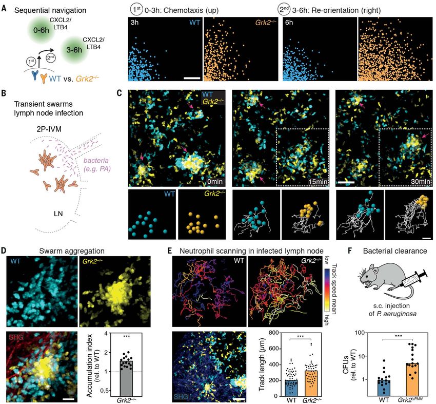
RES EARCH

                                                ◥                                                                    ral local increase of these attractants causes the
    RESEARCH ARTICLE                                                                                                 desensitization of the respective G protein–
                                                                                                                     coupled receptors (GPCRs) (13), potentially
IMMUNOLOGY                                                                                                           acting as an internal feedback control for

Neutrophils self-limit swarming to contain bacterial
                                                                                                                     swarming neutrophils. It is well established that
                                                                                                                     neutrophils can undergo GPCR desensitization

growth in vivo
                                                                                                                     and become unresponsive to repeated or con-
                                                                                                                     tinuous agonist stimulation in vitro (14–16).
                                                                                                                     However, it remains unresolved whether and
Korbinian Kienle1,2,3, Katharina M. Glaser1,2,3, Sarah Eickhoff4, Michael Mihlan1, Konrad Knöpper4,                  how this process contributes to neutrophil nav-
Eduardo Reátegui5,6, Maximilian W. Epple1,2,3, Matthias Gunzer7,8, Ralf Baumeister9,                                 igation and swarming in mammalian tissues,
Teresa K. Tarrant10, Ronald N. Germain11, Daniel Irimia5, Wolfgang Kastenmüller4, Tim Lämmermann1*                   or whether the anticipated desensitization
                                                                                                                     plays a role in their physiological host de-
Neutrophils communicate with each other to form swarms in infected organs. Coordination of this                      fense functions.
population response is critical for the elimination of bacteria and fungi. Using transgenic mice, we found
that neutrophils have evolved an intrinsic mechanism to self-limit swarming and avoid uncontrolled                   GRK2 controls GPCR desensitization and
aggregation during inflammation. G protein–coupled receptor (GPCR) desensitization acts as a negative                neutrophil arrest
feedback control to stop migration of neutrophils when they sense high concentrations of self-secreted               We began our study of these issues by first ex-
attractants that initially amplify swarming. Interference with this process allows neutrophils to scan               amining whether active desensitization occurs
larger tissue areas for microbes. Unexpectedly, this does not benefit bacterial clearance as containment             during swarming. To this end, we interfered
of proliferating bacteria by neutrophil clusters becomes impeded. Our data reveal how autosignaling                  with GPCR desensitization by genetically tar-
stops self-organized swarming behavior and how the finely tuned balance of neutrophil chemotaxis and                 geting GPCR kinases (GRKs). These critical
arrest counteracts bacterial escape.                                                                                 enzymes can phosphorylate cytoplasmic tails
                                                                                                                     of an activated GPCR, which ultimately leads

T
                                                                                                                     to the uncoupling of the GPCR from its sig-
       he collective behavior of eukaryotic cells               body, thereby playing a central role in host         naling cascade and often GPCR internalization
       and insects is often based on self-organizing            defense (4). They exit blood vessels and in-         as well (13) (fig. S1A). Because neutrophils ex-
       processes. The release of chemical sig-                  filtrate tissues to search for damaged cells and     press four GRK isoforms (GRK2, GRK3, GRK5,
       nals, such as chemoattractants or pher-                  invading pathogens when local surveillance by        and GRK6) (17–21), we crossed several mouse
       omones, is one mechanism that allows                     other tissue-resident immune cells fails to con-     strains to isolate primary mature neutrophils

                                                                                                                                                                          Downloaded from https://www.science.org on September 03, 2021
individual entities to attract neighboring in-                  trol inflammation or infection (5, 6). Therefore,    that were efficiently depleted of individual GRKs
dividuals, leading to the accumulation and                      neutrophil infiltrates and aggregates represent      or the complete GRK family (fig. S1, B to E). To
aggregation of a whole population of cells or                   major histopathological hallmarks of acute           identify the GRK isoforms that are functionally
insects. Examples of such self-amplifying posi-                 tissue inflammation and infection. Intravital        relevant for swarming, we imaged control and
tive feedback control to initiate phases of self-               imaging in mouse tissues has revealed that           Grk-deficient neutrophils side-by-side in chemo-
organization include the collective defensive                   collective-like swarming behavior underlies the      taxis assays and analyzed their migratory
behavior of honey bees that attack hornets by                   formation of neutrophil aggregates in many           response toward gradients of the primary
thermo-balling (1), the aggregation behavior of                 mouse models of sterile tissue injury and in-        swarm-mediating attractants LTB4 and CXCL2
locusts (2), and signal relay during the life cycle             fection with bacteria, fungi, and viruses (7, 8).    (Fig. 1A). In these experiments, control neu-
of Dictyostelium (3). However, the mechanisms                   During this population response, hundreds of         trophils performed highly directed chemotaxis
that stop self-organization are poorly under-                   individual neutrophils show coordinated se-          at the onset of the gradient before they slowed
stood for many of these processes.                              quential phases of highly directed chemotaxis,       down, rounded up, and stopped migrating when
   Neutrophils circulate in large numbers in                    intercellular signal relay, and cluster formation.   reaching areas of high attractant concentrations
the mammalian bloodstream to patrol the                         Reminiscent of the collective behavior of some       (Fig. 1, B to D, fig. S1F, and Movie 1).
                                                                insects and Dictyostelium, neutrophils self-            Among all single Grk-deficient cells, only
1                                                               amplify swarming in a feedforward manner             neutrophils lacking GRK2 (Grk2–/–) displayed
 Max Planck Institute of Immunobiology and Epigenetics,
Freiburg, Germany. 2International Max Planck Research           by secreting chemokines and chemoattrac-             clearly distinct behavior. Previous work has
School for Immunobiology, Epigenetics and Metabolism            tants, which allows intercellular communi-           shown that GRK2 in B and T cells selec-
(IMPRS-IEM), Freiburg, Germany. 3Faculty of Biology,            cation and provides the swarm with a level           tively controls the desensitization of the GPCR
University of Freiburg, Freiburg, Germany. 4Institute of
Systems Immunology, University of Würzburg, Max Planck          of self-organization in the complexity of an         sphingosine-1-phosphate receptor 1 (S1PR1),
Research Group, Würzburg, Germany. 5Center for                  inflammatory environment (9).                        but not of several other lymphocyte-expressed
Engineering in Medicine, Massachusetts General Hospital,           It remains unclear how this swarming re-          GPCRs (22). In gradients of combined LTB4
Harvard Medical School, Shriners Hospital for Children,
Boston, MA, USA. 6William G. Lowrie Department of
                                                                sponse ceases and thereby avoids uncontrolled        and CXCL2, Grk2–/– neutrophils showed twice
Chemical and Biomolecular Engineering, The Ohio State           neutrophil accumulation. The mechanisms for          the displacement of control cells from the start-
University, Columbus, OH, USA. 7Institute for Experimental      terminating this response in mammalian tissues       ing cell well (Fig. 1, A and B, and Movie 1). Max-
Immunology and Imaging, University Hospital, University
Duisburg-Essen, Essen, Germany. 8Leibniz-Institut für
                                                                have not yet been defined and may be controlled      imum displacement of knockout cells ranged
Analytische Wissenschaften–ISAS–e.V., Dortmund, Germany.        by external factors from the tissue environment      from 1.7 to 3.2 mm between independent ex-
9
  Bioinformatics and Molecular Genetics, Faculty of Biology,    or by neutrophils themselves, as suggested from      periments, whereas control cells reached 1 to
Centre for Biochemistry and Molecular Cell Research,
                                                                simpler model systems (10–12). On the basis          1.5 mm. At the onset of the gradient (early
Faculty of Medicine, Signalling Research Centres BIOSS and
CIBSS, University of Freiburg, Freiburg, Germany. 10Division    of our previous findings that neutrophils self-      phase, 0 to 30 min), Grk2–/– neutrophils showed
of Rheumatology and Immunology, Department of Medicine,         amplify swarming through the release of the          a slight increase in speed and y-straightness, a
Duke University School of Medicine, Durham, NC, USA.            chemoattractants LTB4 (leukotriene B4) and           measure of chemotactic behavior, in compari-
11
   Laboratory of Immune System Biology, National Institute of
Allergy and Infectious Diseases, Bethesda, MD, USA.             CXCL2 (chemokine C-X-C motif chemokine               son to control cells (Fig. 1C). However, the major
*Corresponding author. Email: laemmermann@ie-freiburg.mpg.de    ligand 2) (9), we hypothesized that the tempo-       effect of GRK2 depletion was observed in fields

Kienle et al., Science 372, eabe7729 (2021)         18 June 2021                                                                                               1 of 16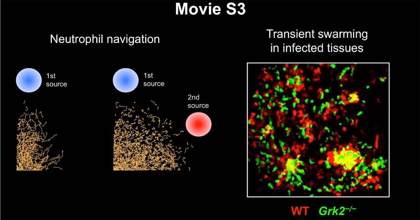
RES EARCH | R E S E A R C H A R T I C L E
A Neutrophil chemotaxis B Neutrophil arrest in gradient of swarm attractants (0-180 min)
WT (cells) WT (tracks) Grk2−/− (cells) Grk2−/− (tracks)
WT vs. LTB4/CXCL2
Grk−/− (swarm attractants)
3 **NS
Displacement ratio
**
LTB4/CXCL2
2
1
0
−/− −/− −/− −/− −/−
WT rk2 rk3 rk5 rk6 Grk
G G G G 4x
C Early phase (0-30 min)
40
D Late phase (90-180 min)
40
WT Grk2−/− *** 1
*** WT Grk2−/− ***
Speed (µm/min)
Speed (µm/min)
y-Straightness
30 30
y 20 y 20
0.5
10 10
0 0 0
Velocity angle Y WT Grk2−/− WT Grk2−/− Velocity angle Y WT Grk2−/−
180° 0° 180° 0°
reverse forward reverse forward
E F 4 G 1000
WT WT ** WT
Displacement (µm)
NS NS
** * ** *
Displacement ratio
40
Grk2−/− Grk2−/−
Instant. speed
Grk2−/−/ WT
3
** ***
(µm/min)
Mean
2 500
20
Grk2−/−
1
Downloaded from https://www.science.org on September 03, 2021
0 0
0 L2 B4 L2 tim tim C5a LTB4/ LTB4 CXCL2
0 50 100 150 (min) XC LT CXC R2 s R1 s
B 4/C FP FP CXCL2
LT
H 4
LTB4 WT I 4
CXCL2 WT
(Ratio to 1st stim.)
(Ratio to 1st stim.)
NS
(fold change)
(fold change)
3 Grk2−/− 1 3 Grk2−/− 1
Ca2+-signal
Ca2+-flux
Ca2+-flux
** Ca2+-signal
**
2 2
**
1 1
0.1 0.1
0 4 8 (min) 0 4 8 (min)
50nM 100nM 200nM 1st 2 nd 3 rd 50nM 100nM 200nM 1st 2 nd 3 rd
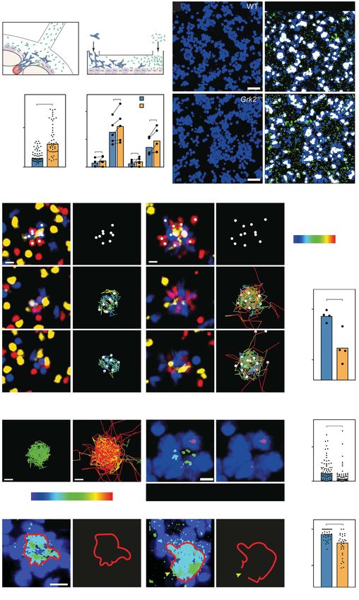
Fig. 1. GRK2-dependent neutrophil arrest in fields of highly concentrated means ± SD). (F) Comparative analysis of WT and Grk2–/– neutrophils migrating
swarm attractants. (A) Comparative analysis of wild-type (WT) and Grk–/– for 4 hours toward various attractive GPCR ligands, displayed as displacement
neutrophils migrating side by side in an under-agarose assay setup along a ratio of Grk2–/– cells to WT cells. Bars display means of n = 4 biological
combined gradient of the swarm attractants CXCL2/LTB4. Grk-deficient cells replicates performed as independent experiments for each comparison.
were lacking either an individual GRK or all four expressed GRKs (4×Grk–/–). *P < 0.05, **P < 0.01 (one-sample t test against 1). (G) Comparative analysis of
After 4 hours, migration endpoints were measured and are displayed as the ratio WT and Grk2–/– neutrophil mean displacement in gradients of LTB4 and
of Grk–/– to WT mean displacement. Bars display means of n = 3 biological CXCL2, combined and separately. Bars display means of n = 4 biological
replicates performed as independent experiments for each comparison. **P < replicates performed as independent experiments for each side-by-side
0.01 (post hoc after ANOVA); NS, nonsignificant. (B to E) Migration of WT and comparison. **P < 0.01, ***P < 0.001 (ratio paired t test). (H and I) Intracellular
Grk2–/– neutrophils toward CXCL2/LTB4 was recorded with live-cell microscopy calcium flux analysis as a measure of GPCR desensitization. WT and Grk2–/–
to obtain cell tracks for 3 hours. From one representative experiment, 50 cells neutrophils were stimulated sequentially with increasing concentrations (as
per genotype were tracked; cell displacement and full cell tracks after 3 hours indicated) of either LTB4 (H) or CXCL2 (I) (triangles). Left panels: Real-time
are displayed in (B). The same cells were analyzed for migration and chemotaxis calcium flux of one experiment representative of n = 5 to 7 biological replicates
parameters during the early phase (0 to 30 min) (C) and the late phase for each genotype. Right panels: Quantification of the decrease in calcium
(90 to 180 min) (D) of movement along the attractant gradient. The velocity signal after repeated attractant stimulation. Area under the curve (AUC) of the
angle Y is the angle between the velocity vector and the y axis (the axis of the calcium signal was measured for individual stimulation peaks. Desensitization
attractant gradient); the y-straightness is the ratio of the displacement along the was measured as the ratio of the second and third stimulation values to the first
y axis (Dy) to the total track length. In speed plot of (C), bars display means; stimulation in each independent experiment. The distribution of AUC values
***P < 0.001 (t test). In y-straightness plot of (C) and speed plot of (D), bars around the normalized average of the first stimulation is also displayed. Bars
display median values; ***P < 0.001 (U test). (E) Instantaneous velocities display means of n = 5 to 7 biological replicates for each genotype. **P < 0.01
with representative cell shapes at t = 120 min (N = 50 cells per genotype, (t test). Scale bars, 500 mm [(B) to (D)], 10 mm (E).
Kienle et al., Science 372, eabe7729 (2021) 18 June 2021 2 of 16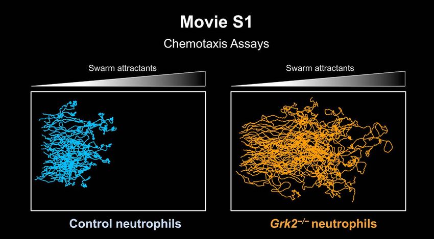
RES EARCH | R E S E A R C H A R T I C L E

of high attractant concentrations (late phase,            comparing the displacement of Grk2–/– relative            tion when we activated cells with the same
90 to 180 min). Grk2–/– neutrophils did not               to control neutrophils in gradients of attractants        triple rising stimulation that was used in the
arrest as did control cells, but they continued           binding to formyl peptide receptors (FPR1 and             calcium flux assay (fig. S3, A and B). Notably,
to move with polarized morphology at elevated             FPR2) or the complement component 5a an-                  repeated stimulation of control and Grk2–/–
speed (Fig. 1, D and E), before reaching an               aphylatoxin chemotactic receptor 1 (C5aR1),               neutrophils with the attractant complement
oscillating behavior with short alternating               which are GPCRs that do not show an im-                   component 5a (C5a) did not produce any
phases of forward and backward movement                   portant role during neutrophil swarming to                differences in calcium and MAPK signaling
(fig. S1G). Similar motility behavior was ob-             modest sterile injury (9), we could only mea-             measurements (fig. S3C). To test the effect
served in single gradients of LTB4 and CXCL2              sure minimal or statistically nonsignificant              of GPCR desensitization on neutrophil move-
alone (Fig. 1, F and G, and fig. S1G). We never           differences (Fig. 1F). These findings indicated           ment, we pretreated control and Grk2–/– cells
observed net reverse migration of Grk2–/– cells.          a particular role for GRK2 in controlling GPCRs           with concentrations of LTB4 and CXCL2 that
This persistent migration phenotype could not             that sense neutrophil-secreted LTB4 and CXCL2             caused receptor desensitization in the calcium
be attributed to gross alterations in the dif-            and contribute to the self-amplification of               flux assay, before analyzing GPCR-mediated
ferentiation and maturation of control and                neutrophil swarming. Because GRK2 can also                chemokinesis. In agreement with our results
Grk2–/– bone marrow neutrophils (fig. S2A).               act on non-GPCR substrates (23), we directly              on GPCR desensitization in calcium and MAPK
Notably, the same GRK2 dependency was also                tested GPCR desensitization by exposing neu-              signaling measurements, ligand-pretreated
evident with neutrophils isolated from blood              trophils to repeated stimulation with increasing          Grk2–/– cells were more chemokinetic than
(fig. S2, B and C), preactivated neutrophils (fig.        concentrations of agonist and measured the                control cells for both LTB4 (fig. S3D) and
S2D), and wild-type neutrophils upon acute                transient increase in intracellular calcium as            CXCL2 (fig. S3, E and F), as reflected by in-
chemical GRK2 inhibition (fig. S2E). Deple-               a readout for cellular responsiveness. Control            creased speed and track lengths.
tion of all four GRK isoforms in neutrophils              neutrophils became unresponsive upon se-                     Lastly, we tested whether GRK2-controlled
(4×Grk–/–) did not additionally increase the              quential stimulation, whereas Grk2–/– neutro-             desensitization is accompanied by receptor
migratory response in LTB4/CXCL2 gradients                phils remained responsive to a third GPCR                 internalization. In agreement with previous
(Fig. 1A, fig. S2F, and Movie 1), highlighting the        activation through LTB4 or CXCL2 (Fig. 1, H               reports that found little if any internalization
major role of GRK2 in this process.                       and I). As a consequence, Grk2–/– neutrophils             of the LTB4 receptor 1 (LTB4R1) (24–26), we
   Grk2–/– neutrophils did not show this sub-             showed increased activation of promigratory               did not observe any reduction in cell surface
stantial increase in responsiveness for all GPCR          mitogen-activated protein kinase (MAPK) sig-              expression of this receptor in wild-type and
ligands known to attract neutrophils. When                naling cascades downstream of GPCR activa-                Grk2–/– cells upon exposure to high concen-
                                                                                                                    trations of LTB4 (fig. S3G). There was a time-
                                                                                                                    dependent decrease in cell surface expression

                                                                                                                                                                         Downloaded from https://www.science.org on September 03, 2021
                                                                                                                    of the C-X-C motif chemokine receptor 2
                                                                                                                    (CXCR2) in both wild-type and Grk2–/– neu-
                                                                                                                    trophils, in agreement with previous reports
                                                                                                                    (26). However, cell surface levels of CXCR2
                                                                                                                    remained substantially higher in Grk2–/– neu-
                                                                                                                    trophils (fig. S3H). Thus, GRK2 plays a crucial
                                                                                                                    role in attenuating GPCR activation with swarm-
                                                                                                                    mediating attractants, which maintains neu-
                                                                                                                    trophil motility in fields of high attractant
                                                                                                                    concentrations.

                                                                                                                    Neutrophils and eosinophils self-limit
                                                                                                                    swarming
                                                                                                                    Neutrophil swarms come in a range of pheno-
                                                                                                                    types and can be categorized into persistent
                                                                                                                    and transient swarms (7). Persistent swarms
                                                                                                                    show sustained neutrophil recruitment to form
                                                                                                                    large cell clusters that can remain stable for
Movie 1. Grk2–/– neutrophils continue to migrate in areas of highly concentrated swarm attractants.                 hours, whereas transient swarms form smaller
First part: Wild-type (WT) and Grk2–/– neutrophils were differentially dye-labeled and filled in a 1:1 ratio into   clusters that last only for minutes before neu-
one well (left) of an under-agarose chemotaxis assay setup. The swarm-mediating chemoattractants LTB4               trophils leave the aggregate and join nearby
(1 mM) and CXCL2 (1 mM) were filled into an opposite well to establish a gradient of increasing attractant          competing swarms. To investigate the role of
concentrations (highest concentration at right). The representative video shows control (pseudo-colored in          GPCR desensitization for persistent swarm
blue; upper panel) and Grk2–/– (pseudo-colored in orange; lower panel) neutrophils migrating toward the             dynamics, we first used large microscale arrays
gradient (left to right). Graphic analysis of this experiment (Fig. 1, B to E, and fig. S1F) reveals that Grk2–/–   of fluorescent heat-killed bioparticle clusters, a
neutrophils do not arrest, but continue to migrate at high concentrations of swarm attractants. Spinning-disk       previously established experimental system to
confocal microscopy (x, y = 1070 mm, 870 mm; stitched from multi-tiled images), 10 frames/s. Time is                analyze neutrophil swarming in vitro (12). Upon
displayed as hours:min. Second part: WT, Grk2–/–, and 4×Grk–/– neutrophils were differentially dye-labeled          exposure to 400 mm–spaced micropatterns of
and filled in a 1:1:1 ratio into one well (left) of an under-agarose chemotaxis assay setup with combined LTB4/     heat-killed Staphylococcus aureus (HKSA), sen-
CXCL2 gradient as in the experiment before. The representative video shows control (top), Grk2–/– (middle),         tinel neutrophils sensed the bioparticles and
and 4×Grk–/– (bottom) neutrophils migrating toward the gradient (left to right). Graphic analysis of this           induced a recruitment wave of following neu-
experiment (fig. S2F) reveals comparable migration of 4×Grk–/– and Grk2–/– neutrophils in swarm-attractant          trophils, which then formed cell clusters in a
gradients. Spinning-disk confocal microscopy (x, y = 1119 mm, 959 mm; stitched from multi-tiled images),            LTB4- and CXCL2-dependent manner (12)
12 frames/s. Time is displayed as hours:min.                                                                        (Fig. 2A and fig. S4, A and B). In experiments

Kienle et al., Science 372, eabe7729 (2021)   18 June 2021                                                                                                    3 of 16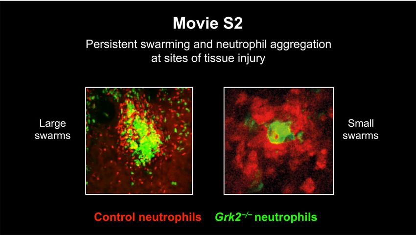
RES EARCH | R E S E A R C H A R T I C L E
Fig. 2. GRK2-dependent arrest in persistent A Persistent swarms HKSA 0h 3h 3h B
***
Accum. index
swarms. (A) In vitro microscale array of (micropatterns) 2
(rel. to WT)
patterned heat-killed S. aureus (HKSA) biopar-
ticles (blue) to study persistent neutrophil
swarms. Live microscopy of WT neutrophils 1
(yellow) and cell tracking (pink) exemplifies Neutrophils
swarming dynamics over 3 hours. (B) Analysis WT Grk2−/−
of neutrophil aggregation in clusters of two
mixed populations, quantified as the WT/WT C Persistent swarms (skin) E 30
and Grk2–/–/WT ratios of cells accumulating on Laser-induced NS
Speed (µm/min)
HKSA spots (accumulation index) after 2 hours. tissue injury
WT Neu and 20
Each dot represents one analyzed neutrophil Grk2–/– Neu
cluster (N = 30) pooled from n = 3 biological 2P-IVM
replicates. Bars display means; ***P < 0.001 10
(t test). (C to G) 2P-IVM on ear dermis of
anesthetized mice: Comparative analysis of B6.Albino host
0
persistent swarming after i.d. co-injection of WT WT Grk2−/−
–/–
and Grk2 neutrophils, which were differen-
tially labeled with fluorescent dyes, into D Persistent swarms skin injury: Recruitment to damage site
2
Tyrc-2J/c-2J (B6.Albino) mice. (C) Interstitial cell WT Grk2−/−
recruitment toward a laser-induced focal tissue NS
Grk2−/−/ WT
Speed ratio
injury. [(D) and (E)] For one representative
experiment, the full cell tracks toward the 1
damage site (dashed line) over the first 40 min
after the initiation of the tissue damage are
displayed (D) and the cell speed analyzed [(E),
top]. Each dot represents one tracked neutro- 0.5
phil (N = 62) from the side-by-side comparison F Persistent swarms skin injury: Aggregation
of WT and Grk2–/– cells in one experiment.
Downloaded from https://www.science.org on September 03, 2021
Bars display median values (U test). [(E),
bottom] Comparative analysis of WT and
Grk2–/– neutrophil speed during side-by-side
chemotactic migration, displayed as the ratio of
Grk2–/– to WT; n = 4 biological replicates (one-
sample t test against 1). (F) Neutrophil aggre- WT
gation was analyzed by 2P-IVM images at the Grk2−/− SHG WT Grk2−/−
endpoint of the clustering response when
neutrophil recruitment ceases. In the G H
Accumulation index
Accumulation index
2 ***
representative example (see Movie 2, first part), 2
(rel. to WT)
(rel. to WT)
neutrophil clustering is displayed 65 min after
the initiation of tissue damage. (G) Aggregation 1
in competitive clusters of Grk2–/– and WT cells 1
was also quantified over time. Accumulation
index was used as a quantitative parameter for Grk2−/−
neutrophil entry into the collagen-free wound 0.5 0.5
0 15 30 45 (min) Grk2−/− Grk3−/− Grk5−/− Grk6−/−
center [cyan dashed line in (F)], displayed as
the ratio of Grk2–/– to WT. Quantification began I Persistent swarms: Eosinophils / C. elegans larvae
(t = 0) when small aggregates have already
C. elegans WT
Accumulation index
formed, which commonly occurs 5 to 20 min larva Grk2−/− 4 **
after the initial tissue injury depending on the
(rel. to WT)
individual experiment. Time courses of neutro-
phil clusters from n = 4 biological replicates 2
(lines) are shown. (H) Quantification of endpoint
neutrophil clustering after i.d. co-injection of 1
WT cells and neutrophils lacking individual GRK
family members into mice. The accumulation WT Grk2−/− WT Grk2−/−
–/–
index (ratio of Grk to WT) as a measure of
aggregation was calculated when neutrophil recruitment had ceased and clusters stabilized. Each dot represents one analyzed neutrophil cluster (N = 4 to 6)
pooled from n = 2 or 3 biological replicates. ***P < 0.001 (post hoc after ANOVA). (I) Comparative analysis of WT and Grk2–/– eosinophils forming persistent
swarms around C. elegans dauer larvae (dotted line) in vitro. Confocal images illustrate endpoint eosinophil clusters after 2 hours. Each dot represents one
analyzed eosinophil cluster (N = 6 to 10) pooled from n = 3 biological replicates. **P < 0.01 (t test). Scale bars, 100 mm [(A) and (I)], 50 mm (D), 30 mm (F). Bars with
LUT color grading [(F) and (I)] display fluorescence signal intensities. SHG, second harmonic generation signal.
Kienle et al., Science 372, eabe7729 (2021) 18 June 2021 4 of 16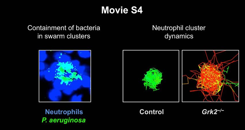
RES EARCH | R E S E A R C H A R T I C L E

with 1:1 mixtures of differentially dye-labeled               restricted tissue damage (9). After intradermal              quantify neutrophil clustering in the skin, we
control and Grk2–/– cells, we quantified neu-                 (i.d.) co-injection of differentially dye-labeled            defined the AI as a measure of cell entry into
trophil aggregation behavior in competitive                   control and Grk2–/– neutrophils, we used two-                the collagen-free zone, as previously described
clusters (fig. S4C). The accumulation index                   photon intravital microscopy (2P-IVM) to image               (9). In (9), we only identified gene knockouts
(AI), which is the ratio of Grk2–/– signal to wild-           the swarming response of the transferred cells               that showed impaired neutrophil aggregation
type signal on HKSA spots, was used as a                      to the induced skin wound for 1 to 1.5 hours                 behavior and mean AI values less than 1.
measure of neutrophil aggregation. Strikingly,                (Fig. 2C and fig. S5A). Neutrophils that swarmed             Remarkably, Grk2–/– neutrophils gravitated
Grk2–/– neutrophils showed pronounced ag-                     from the surrounding tissue toward the site of               more than wild-type cells toward the central
gregation and dominated over control cells in                 injury were tracked and migration parameters                 region of large competitive clusters, resulting
competitive clusters—a behavior that was re-                  analyzed (Fig. 2D and fig. S5B). Control and                 in mean AI values less than 1 (Fig. 2, F to H,
flected in a mean AI value greater than 1 (Fig. 2B            Grk2–/– neutrophils were recruited at compara-               and fig. S5D). Improved aggregation behavior
and fig. S4C). By comparison, competitive clus-               ble speed and straightness toward the injury site            of Grk2–/– cells was also observed in experi-
ters of two populations of control cells resulted             (Fig. 2E, fig. S5C, and Movie 2), demonstrating              mental setups that allowed the analysis of
in a mean AI of 1 (Fig. 2B).                                  that the minor measured effect on early-phase                small clusters (fig. S5E and Movie 2). In con-
   Next, we examined the role of GRK2 in                      chemotaxis in vitro was not relevant for migra-              trast to control cells, Grk2–/– neutrophils re-
regulating persistent swarms in vivo by using                 tion in native tissue (Fig. 1C).                             mained actively motile in a growing cluster and
an inducible model of sterile skin injury in                     We then analyzed the ensuing step of the                  continued to move toward the cluster center,
which a brief laser pulse causes focal, dermis-               swarming response, swarm aggregation. To                     where they outcompeted wild-type cells over
                                                                                                                           time (Fig. 2G, fig. S5D, and Movie 2). GRK3,
                                                                                                                           GRK5, or GRK6 deficiency had no measur-
                                                                                                                           able effect on central accumulation (Fig. 2H
                                                                                                                           and fig. S5F).
                                                                                                                             Finally, we studied persistent swarms of
                                                                                                                           eosinophils, another innate immune cell type,
                                                                                                                           that self-amplify their collective migration re-
                                                                                                                           sponse to worms by paracrine LTB4 signaling
                                                                                                                           (27). Like neutrophils, swarming Grk2–/– eosin-
                                                                                                                           ophils aggregated more closely than wild-type
                                                                                                                           cells around C. elegans larvae (Fig. 2I, fig. S5G,
                                                                                                                           and Movie 2), confirming the more general

                                                                                                                                                                                Downloaded from https://www.science.org on September 03, 2021
                                                                                                                           role for GRK2 in swarming responses beyond
                                                                                                                           neutrophil biology. Thus, GRK2 acts as negative
                                                                                                                           regulator of swarming in mammalian tissues,
                                                                                                                           and GPCR desensitization is integral at sites
                                                                                                                           where swarming granulocytes accumulate and
                                                                                                                           self-generate a local field of high attractant
                                                                                                                           concentrations.

                                                                                                                           Increased tissue scanning is not beneficial for
Movie 2. GRK2-dependent neutrophil arrest in cell clusters of persistent swarms. First part (large neutrophil              bacterial elimination
swarms): WT and Grk2–/– neutrophils were differentially dye-labeled and injected i.d. in a 1:1 ratio into the ventral      In many inflammatory conditions, neutrophils
ear skin of a Tyrc-2J/c-2J (B6.Albino) mouse 3 hours before laser-induced focal tissue damage (white circle at             respond to cell death at multiple locations
the start of the video). This representative video shows Grk2–/– (pseudo-colored in green) and control neutrophils         within a tissue, leading to several transient
(pseudo-colored in red) accumulating at the damage site in the skin dermis. Graphic analysis of the recruitment            swarms whose attractant release influences
phase of several experiments (Fig. 2, D and E, and fig. S5, B and C) and analysis of the clustering response in this       each other’s growth and disappearance (7). To
video (Fig. 2, F and G) reveal comparable recruitment of control and Grk2–/– neutrophils to the focal injury at early      address the role of GRK2 in situations where
swarming phases. Over time, Grk2–/– neutrophils remain actively motile in growing clusters and dominate over               migrating neutrophils sense multiple attract-
control cells in the neutrophil cluster center. Two-photon intravital microscopy (x, y, z = 512 mm, 512 mm, 12 mm;         ant sources, we preexposed wild-type and
merge of z-stack), 18 frames/s. Time is displayed in minutes. Second part (small neutrophil swarms): Primary               Grk2–/– neutrophils to LTB4/CXCL2 before
neutrophils were isolated from the bone marrow of Grk2DPMN Lifeact-GFP mice and injected i.d. into the ventral ear         they moved along a gradient of both agonists.
skin of a CAG-DsRed+/+ Tyrc-2J/c-2J mouse 3 hours before laser-induced focal tissue damage. This representative            In agreement with our calcium measurements
video shows the accumulation of Grk2–/– neutrophils (pseudo-colored in green) at a small cluster of endogenous             (Fig. 1, H and I), wild-type neutrophils de-
WT neutrophils (pseudo-colored in red) that formed at the laser damage site. Analysis of the clustering response of        sensitized and became unresponsive to the
several experiments, including comparison to control WT Lifeact-GFP neutrophil injection (fig. S5E) reveals that           subsequent attractant gradient in a concentration-
the continued motility of Grk2–/– neutrophils displaces control cells in the centers of small neutrophil clusters. Two-    dependent manner. By contrast, Grk2–/– neu-
photon intravital microscopy (x, y, z = 512 mm, 512 mm, 3 mm; merge of z-stack), 24 frames/s. Time is displayed in         trophils remained responsive at high attractant
minutes. Third part (eosinophil swarms): GRK2 controls the accumulation of swarming eosinophils around worm                concentrations and could still move along the
larvae. WT and Grk2–/– eosinophils from IL-5 cultures of WT and Vav-iCre Grk2fl/fl mouse bone marrow, respectively,        gradient of activating signals (fig. S6A).
were differentially dye-labeled and placed in a 1:1 ratio with 4-day-old C. elegans dauer larvae in Matrigel. This            Next, we analyzed the sequential navigation
representative video shows the recording of bright-field (top left) and fluorescent microscopy (right) in which            behavior of neutrophils in the presence of two
Grk2–/– (pseudo-colored in pink) and control (pseudo-colored in blue) eosinophils swarm and accumulate side by             spatiotemporally separated gradients of swarm
side around an individual larva. Analysis of eosinophil clustering of several experiments (Fig. 2I) reveals an increased   attractants (Fig. 3A) (28). As observed previously
clustering response of Grk2–/– eosinophils at the worm larva (dotted outline). Spinning-disk confocal microscopy           (Fig. 1, A to D), Grk2–/– neutrophils showed
(x, y = 269 mm, 365 mm; stitched from multi-tiled images), 10 frames/s. Time is displayed as hours:min.                    increased displacement from a starting cell well

Kienle et al., Science 372, eabe7729 (2021)     18 June 2021                                                                                                         5 of 16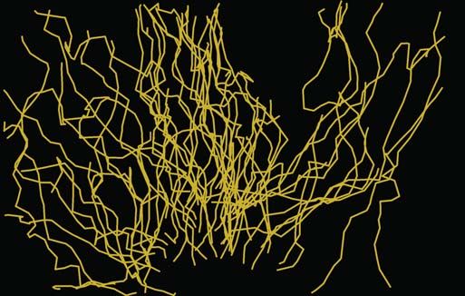
RES EARCH | R E S E A R C H A R T I C L E

toward an initial source of LTB4/CXCL2 in                clusters around locally proliferating bacteria        effector functions at sites of damaged tissue or
comparison to control cells. Grk2–/– neutro-             and dying cells (Fig. 4B and fig. S7D). In            pathogen invasion (7, 9). We have described
phils were redirected by an additional second            agreement with our in vivo findings, Grk2–/–          a cell-intrinsic stop mechanism for the self-
gradient at 90° angle, whereas control cells             neutrophils dominated over control cells in           organization of collective behavior, which is
were not (Fig. 3A, fig. S6, B and C, and Movie 3).       cluster centers in competitive experiments            based on sensing the local accumulation of the
Thus, neutrophils lacking GRK2-mediated                  (fig. S7E). When control and knockout cells           same cell-secreted attractants that amplify
GPCR desensitization increase their space                were studied individually, Grk2–/– cells formed       swarming during early stages. Our findings
exploration between competing gradients                  larger neutrophil aggregates around bacterial         highlight a crucial role of GPCR desensitization
of swarm attractants.                                    clusters than did wild-type cells (Fig. 4C and        in attenuating the self-organized swarming dy-
   To gain insight into the possible in vivo rel-        Movie 4). However, GRK2 deficiency resulted           namics of neutrophils in mammalian tissues
evance of our findings, we investigated the role         in higher counts of “free” bacteria outside of        (fig. S9). When neutrophils sense high concen-
of GRK2 during transient neutrophil swarming             neutrophil clusters and a significant increase        trations of swarm-secreted attractants (LTB4
in lymph nodes infected with Pseudomonas                 of bacterial growth (Fig. 4C), confirming the         and CXCL2), as found in growing neutrophil
aeruginosa or Salmonella typhimurium. We                 results of our mouse infection model. This            clusters, the GPCR kinase GRK2 desensitizes
previously showed that several bacteria spe-             could not be attributed to changes in neutro-         the corresponding GPCRs to induce migration
cies induce cell death in subcapsular sinus              phil maturation or major effector functions,          arrest. Because GRK2 has only minimal effects
(SCS) macrophages, subsequently leading to               as Grk2–/– and control cells were comparable          on the desensitization of GPCRs that detect
neutrophil recruitment and swarming in lymph             in standard phagocytosis assays (HKSA and             tissue- or bacteria-derived attractants (e.g.,
nodes (9, 29) (Fig. 3B). Neutrophil depletion            living P. aeruginosa) and in their release of         N-formyl peptides and C5a), neutrophils in
in these infection models leads to a substantial         reactive oxygen species, neutrophil elastase,         swarm aggregates remain responsive to new
increase of bacteria growth in lymph nodes               and myeloperoxidase (fig. S8, A to E). We also        tissue insults. This allows their redirection
(29). By imaging endogenous wild-type and                only rarely observed neutrophil extracellular         from neutrophil clusters to novel sites of cell
Grk2–/– neutrophils in SCS of mixed bone                 traps (NETs) around the small bacteria aggre-         death in tissues. Thus, our findings agree
marrow chimera, we found again that knock-               gates in our coculture system and depletion           with earlier in vitro studies that highlighted
out cells dominated over control cells in the            of GRK2 had no measurable effect on NET               the capacity of “end-target” attractants (N-formyl
central regions of newly forming clusters (Fig. 3,       formation (fig. S8F), a process that has pre-         peptides and C5a) to override “intermediary”
C and D, fig. S6, D and E, and Movie 3). This            viously been reported to occur in the presence        attractants (LTB4, CXCL2) and redirect neu-
was reflected in AI values less than 1 (Fig. 3D).        of P. aeruginosa biofilms (31). Establishing          trophils out of “intermediary” attractant fields
Moreover, Grk2–/– neutrophils moved faster               single-cell tracking of neutrophils in clusters       (28, 32). The described GRK2-mediated feed-
than wild-type cells between clusters and ex-            helped us to identify the GRK2-regulated cel-         back control to swarm attractants is particularly

                                                                                                                                                                       Downloaded from https://www.science.org on September 03, 2021
plored larger tissue areas (Fig. 3E, fig. S6F,           lular mechanism limiting bacterial growth.            critical in infected tissues, where it fine-tunes the
and Movie 3). Such behavior may be linked to             Control cells slowed down their persistent            local migratory arrest of neutrophils for optimal
more effective bacterial sampling throughout             migration and stopped in cell aggregates,             containment of proliferating bacteria (fig. S10).
the infected organ, leading us to undertake a            whereas many Grk2–/– neutrophils lacked such          This provides a potential mechanistic explana-
quantitative assessment of how GRK2 deficiency           arrest phases and migrated at high speed out          tion for earlier studies that implicated neutrophil
in neutrophils affects bacterial clearance. The          of clusters again (Fig. 4, D to F, fig. S7F, and      swarming in restricting microbial growth in vitro
draining lymph nodes of mice with neutrophil-            Movie 4).                                             (33) and in vivo (34). Although we identified
specific depletion of GRK2 (Mrp8Cre Grk2fl/fl,              This uncontrolled persistent movement of           GRK2 as a key molecular brake on GPCR ac-
“Grk2DPMN”) and controls were analyzed after             Grk2–/– neutrophils had two functional con-           tivation by neutrophil swarm attractants, its
subcutaneous (s.c.) infection with P. aeruginosa.        sequences. First, knockout cells were impaired        function may extend to other GPCRs in dif-
To our surprise, there were significantly higher         in picking up and ingesting microbes from             ferent inflammatory settings (35, 36).
bacterial counts in the lymph nodes of Grk2DPMN          bacteria clusters (Fig. 4G). Second, Grk2–/– cells       How migrating phagocytes coordinate cell
mice relative to control mice (Fig. 3F), although        were often unable to completely contain locally       movement and phagocytosis and negotiate
neutrophil recruitment into infected lymph               proliferating bacteria, allowing a breach in the      these two actin-dependent processes has been
nodes was comparable (fig. S6G). Thus, an                swarm-dependent barrier and subsequent path-          intensely studied in other phagocytic cells,
inverse relationship exists between persistent           ogen escape (Fig. 4H). Thus, GRK2-controlled          including dendritic cells, Dictyostelium, and
neutrophil interstitial movement and bacte-              neutrophil arrest is critical for bacterial phago-    Drosophila macrophages (37–43). Most of these
rial elimination.                                        cytosis and containment in swarm clusters.            studies were performed in vitro or in simpler
                                                         These findings emphasize that neutrophils have        model organisms and focused on the analysis
GPCR-controlled neutrophil arrest is critical            evolved a cell-intrinsic mechanism that self-         of individual cells and their uptake of inert
for restricting bacterial growth                         limits dynamic cell behavior within forming           elements. By contrast, we have addressed the
To understand in detail how GRK2-controlled              swarms and ensures optimal elimination of             population dynamics of phagocytes and how
neutrophil swarming counteracts bacterial                bacteria (figs. S9 and S10).                          these cells coordinate stop-and-go behavior
growth, we established an experimental in vitro                                                                within the population by self-secreting attrac-
mimic of a bacteria-infected SCS. By coculturing         Discussion                                            tants. We define a cell-intrinsic, GPCR-based
macrophages, neutrophils, and P. aeruginosa              Neutrophil navigation through inflamed and            mechanism for stopping the swarming behav-
bacteria, we were able to follow the dynamics            infected tissues has long been viewed from a          ior of neutrophils, which is functionally rele-
and major cellular events of SCS components              single-cell perspective, where cells were con-        vant for the containment of proliferating living
with live-cell microscopy (Fig. 4A). In this sys-        sidered to be individually guided by external         bacteria in infected mammalian tissues. This
tem, bacteria performed pack swarming and                signals released from the tissue environment          self-limiting mechanism does not rely on adap-
invasion of macrophages (30), causing cell               or directly from pathogens. It is now clear that      tations in gene regulation, as commonly found
death as previously shown in vivo (29) (fig. S7,         neutrophils autosignal to initiate a self-amplified   in the bacterial population responses of quorum
A to C, and Movie 4). Neutrophils showed                 population response, which accumulates these          sensing and quenching (44). Local attractant
swarming behavior and formed prominent                   cells in large numbers and concentrates their         degradation and the release of pro-resolving

Kienle et al., Science 372, eabe7729 (2021)   18 June 2021                                                                                                 6 of 16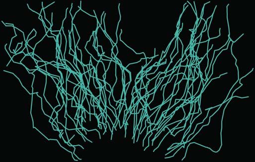
RES EARCH | R E S E A R C H A R T I C L E

                                                                                                                                                               Downloaded from https://www.science.org on September 03, 2021
Fig. 3. GRK2-controlled transient swarming restricts bacterial growth.         node SCS. Quantification of neutrophil accumulation in transient clusters is
(A) Left: Comparative analysis of sequential navigation behavior of WT and     displayed as the ratio of Grk2–/– to WT (see also materials and methods).
Grk2–/– neutrophils by exposing them to a first gradient of LTB4/CXCL2 and     Each dot represents one cluster (N = 16) pooled from n = 4 infected lymph
adding a second gradient of LTB4/CXCL2 at a 90° angle 3 hours later (right).   nodes. ***P < 0.001 (one-sample t test against 1). (E) Trajectories of
Right: Cell positions after initial chemotaxis (3 hours) and reorientation     individual neutrophils are shown as tracks color-coded for mean track speed
(6 hours) were obtained by live-cell microscopy. (B) Mice were infected with   of cells (top), which navigate in the interstitial space of lymph nodes
bacteria in the footpad, and 2P-IVM of transient neutrophil swarms was         between neutrophil clusters (bottom). The color code ranges from 2 to
then performed on draining popliteal lymph nodes. (C to E) Mice with mixed     12 mm/min. Cell track lengths were quantified over 60 min; each dot
bone marrow [cyan, WT (Ly6gCre/+ Rosa26LSL:Tom); yellow, Grk2–/– (Mrp8-Cre     represents one tracked neutrophil [N = 59 (WT), N = 48 (Grk2–/–)] from
Grk2fl/fl Lyz2Gfp/+)] were infected with P. aeruginosa (PA)–GFP before         one experiment. (F) Bacterial CFU counts of draining lymph nodes 8 hours
endogenous neutrophils were recorded 3 to 5 hours later. (C) Representative    after Grk2DPMN and littermate control (WT) mice were infected with
time-lapse sequence of neutrophil clusters (magenta arrows) in SCS. Bottom     P. aeruginosa. Each dot represents one lymph node (N = 16) pooled from
panels show migration tracks of neutrophils redirected to a second cluster     n = 8 mice for each genotype. In (E) and (F), bars display median values.
(dotted box) with dragontails from t = 0 to 30 min. (D) 2P-IVM images show a   ***P < 0.001 (U test). Scale bars, 1 mm (A), 50 mm [(C), top, and (E)], 20 mm
representative neutrophil cluster of a transient swarm in the infected lymph   [(C), bottom, and (D)]. SHG, second harmonic generation signal.

Kienle et al., Science 372, eabe7729 (2021)   18 June 2021                                                                                          7 of 16
RES EARCH | R E S E A R C H A R T I C L E

                                                                                                                     mediators are other potential self-limiting
                                                                                                                     processes that emerged from in vitro studies
                                                                                                                     (12, 45). However, it is still unclear whether
                                                                                                                     these processes also contribute to neutrophil
                                                                                                                     swarming in tissues and augment the mech-
                                                                                                                     anism described herein.
                                                                                                                        These findings provide insights into the
                                                                                                                     navigation strategies used by neutrophil pop-
                                                                                                                     ulations for the optimal elimination of bacteria
                                                                                                                     in infected mammalian tissues. This should prove
                                                                                                                     useful for an integrated view of self-limiting
                                                                                                                     processes and active anti-inflammatory programs
                                                                                                                     in controlling the resolution of neutrophil
                                                                                                                     swarms, which is a critical step for tissue repair
                                                                                                                     after infection. Our results also highlight the
Movie 3. GRK2 controls neutrophil arrest in transient swarm clusters and limits neutrophil space                     fact that desensitization to a self-produced acti-
exploration in infected tissues. First part (in vitro): GRK2 limits neutrophil space exploration between             vation signal acts as an important biological
competing gradients of swarm attractants. WT and Grk2–/– neutrophils were differentially dye-labeled and             principle of self-organization, which is likely
loaded in a 1:1 ratio into wells of a modified under-agarose assay setup that allows the analysis of neutrophil      relevant for other forms of collective behavior
sequential navigation behavior in response to multiple attractant sources. Neutrophils were exposed to               in cells and insects.
two spatiotemporally separated gradients of the swarm attractants LTB4 (1 mM) and CXCL2 (1 mM). First,
Grk2–/– (pseudo-colored in orange) and WT neutrophils (pseudo-colored in blue) respond to a first gradient           Materials and methods
of LTB4/CXCL2 (gradient direction from top to bottom). The movie sequence shows side-by-side                         Mice
migration of tracked cells and starts 2 hours after the attractants were added. Second, WT and Grk2–/–               Table S1 lists all mouse strains and crosses
neutrophils are redirected after 3 hours by an additional gradient of LTB4/CXCL2 at a 90° angle (gradient            used in this study. Mrp8-Cre (46), Grk2fl/fl (47),
direction from right to left). This second movie starts immediately after attractants were added. Cell               Grk3–/– (48), Grk5fl/fl (49), Grk6fl/fl (50), Lyz2gfp
migration was tracked using Imaris spot function. Each circle indicates an individual neutrophil with motion         (51), Ly6gcre/+ Rosa26LSL:Tom (52), Vav-iCre (53),
paths as dragontails over the last 10 min (first movie) or 30 min (second movie) in the corresponding                CAG-DsRed (54), Tyrc-2J/c-2J (B6.Albino) (55, 56),
pseudo-color. Graphic analysis of this video (Fig. 3A and fig. S6, B and C) reveals that Grk2–/– neutrophils, in     and Lifeact-GFP (57) mouse strains have been
contrast to WT cells, were not desensitized by the first gradient and could be redirected by an additional           described elsewhere. Mice were maintained in
gradient of the same attractants. Spinning-disk confocal microscopy (x, y = 1682 mm, 1391 mm; stitched from

                                                                                                                                                                             Downloaded from https://www.science.org on September 03, 2021
                                                                                                                     specific pathogen–free conditions at an Associ-
multi-tiled images), 12 frames/s. Time is displayed as hours:min. Second part (P. aeruginosa–infected lymph          ation for Assessment and Accreditation of Labo-
node): GRK2 controls neutrophil arrest in transient swarm clusters in vivo. Mice with mixed bone marrow              ratory Animal Care–accredited animal facility at
[Ly6gCre/+ Rosa26LSL:Tom (WT) pseudo-colored in red; Mrp8-Cre Grk2fl/fl Lyz2Gfp/+ (Grk2–/–) pseudo-colored in        NIAID and in a conventional animal facility at
green] were injected with P. aeruginosa (PA)–GFP (fluorescence not visible here) into the footpad before             the Max Planck Institute of Immunobiology and
endogenous neutrophils were recorded 3 to 4 hours later. Two-photon intravital microscopy of transient               Epigenetics according to local regulations. Mice
neutrophil swarms was performed on the SCS of draining popliteal lymph nodes. This representative video              were used for experiments at 8 to 16 weeks of
shows Grk2–/– (pseudo-colored in green) and control neutrophils (pseudo-colored in red) side by side                 age. Mice were age- and sex-matched in all ex-
during the formation and disappearance of transient neutrophil swarm clusters. Arrows indicate neutrophil            periments, and littermate animals were used as
clusters; the pink arrow highlights neutrophil migration out of one cluster to a newly developing cluster. Static    controls in most experiments. All animal proce-
images of this video are presented in Fig. 3C. Graphic analysis of several experiments (Fig. 3D) reveals that        dures were performed according to study pro-
Grk2–/– neutrophils dominate over control cells in the central regions of newly forming clusters. Moreover,          tocols approved by the German authorities and
Grk2–/– neutrophils also migrate rapidly out of clusters again and become redirected to the centers of newly         the Regional Council of Freiburg, the Animal
developing clusters. Two-photon intravital microscopy (x, y, z = 504 mm, 404 mm, 14 mm; merge of z-stack),           Care Commission of the state of North Rhine–
12 frames/s. Time is displayed in minutes. Third part (S. typhimurium–infected lymph node): Mice with mixed          Westphalia (LUA NRW), and the NIAID Ani-
bone marrow [Ly6gCre/+ Rosa26LSL:Tom (WT) pseudo-colored in red; Mrp8-Cre Grk2fl/fl Lyz2Gfp/+(Grk2–/–)               mal Care and Use Committee, respectively.
pseudo-colored in green] were injected with S. typhimurium into the footpad; endogenous neutrophils were
recorded 3 to 4 hours later. Two-photon intravital microscopy of transient neutrophil swarms was performed on        Neutrophil isolation and labeling
the SCS of draining popliteal lymph nodes. This representative video shows Grk2–/– (in green) and control            For all in vitro experiments, mouse neutrophils
neutrophils (in red) side by side during the formation and disappearance of transient neutrophil swarm clusters      were isolated from bone marrow (tibiae, femora,
(arrows). Static images of this video and graphic analysis of several experiments (fig. S6, E and F) reveal that     and os coxae) or peripheral blood using autoMACS
Grk2–/– neutrophils dominate over control cells in the central regions of newly forming clusters during              Pro Selector cell separator and MACS Neu-
S. typhimurium infection. Moreover, Grk2–/– neutrophils migrate rapidly out of clusters again, have increased        trophil Isolation Kit for negative selection ac-
interstitial speed, and become redirected to the centers of newly developing clusters. Two-photon intravital         cording to the manufacturer’s protocol (Miltenyi
microscopy (x, y, z = 512 mm, 512 mm, 10 mm; merge of z-stack), 12 frames/s. Time is displayed in minutes.           Biotec). For i.d. injection experiments, mouse
Fourth part (P. aeruginosa–infected lymph node): GRK2 limits neutrophil space exploration in infected lymph          neutrophils were isolated from bone marrow
node tissue. Cell tracking of endogenous WT and Grk2–/– neutrophils that migrate side by side in the interstitial    using a three-layer Percoll gradient of 78%, 69%,
areas of a P. aeruginosa–infected lymph node (image insert, tissue region as in second part of this video).          and 52% as described (9). Neutrophil purity was
This representative video shows the interstitial scanning behavior of Grk2–/– (pseudo-colored in green) and          >95% for both isolation procedures, as indi-
control neutrophils (pseudo-colored in red) with motion paths over the last 15 min as dragontails in the             cated by Ly6G+ phenotype in flow cytometry.
corresponding pseudo-color. At the end, the total trajectories of individual neutrophils after 60 min are shown as   When neutrophils required fluorescent cell label-
tracks color-coded for average speed. Graphic analysis (Fig. 3E) reveals that neutrophils lacking GRK2 show          ing in subsequent experiments, they were incu-
increased tissue scanning but impaired migration arrest during interstitial movement in infected lymph nodes. Cell   bated for 25 min at 2 × 10⁷ cells/ml with 0.5 mM
tracking based on two-photon intravital microscopy, 10 frames/s. Time is displayed in minutes.                       CellTracker Green (CMFDA), 10 mM CellTracker

Kienle et al., Science 372, eabe7729 (2021)   18 June 2021                                                                                                       8 of 16
You can also read