The evolving role of MUC16 (CA125) in the transformation of ovarian cells and the progression of neoplasia
←
→
Page content transcription
If your browser does not render page correctly, please read the page content below
Carcinogenesis, 2021, Vol. 42, No. 3, 327–343

                                                                                     doi:10.1093/carcin/bgab010
                                                                                     Advance Access publication February 20, 2021
                                                                                     Review

Review
The evolving role of MUC16 (CA125) in the

                                                                                                                                                      Downloaded from https://academic.oup.com/carcin/article/42/3/327/6145151 by guest on 22 November 2021
transformation of ovarian cells and the progression of
neoplasia
Panagiotis Giamougiannis1,2, , Pierre L.Martin-Hirsch1,3 and Francis L.Martin4,*
1
 Department of Gynaecological Oncology, Lancashire Teaching Hospitals NHS Foundation Trust, Preston PR2 9HT, UK,
2
 School of Pharmacy and Biomedical Sciences, University of Central Lancashire, Preston PR1 2HE, UK, 3Division of Cancer
Sciences, University of Manchester, Manchester M13 9PL, UK and 4Biocel UK Ltd, Hull HU10 7TS, UK
*To whom correspondence should be addressed. Tel: +44 (0) 7967727844; Email: flm13@biocel.uk

Abstract
MUC16 (the cancer antigen CA125) is the most commonly used serum biomarker in epithelial ovarian cancer, with
increasing levels reflecting disease progression. It is a transmembrane glycoprotein with multiple isoforms, undergoing
significant changes through the metastatic process. Aberrant glycosylation and cleavage with overexpression of a
small membrane-bound fragment consist MUC16-related mechanisms that enhance malignant potential. Even MUC16
knockdown can induce an aggressive phenotype but can also increase susceptibility to chemotherapy. Variable MUC16
functions help ovarian cancer cells avoid immune cytotoxicity, survive inside ascites and form metastases. This review
provides a comprehensive insight into MUC16 transformations and interactions, with description of activated oncogenic
signalling pathways, and adds new elements on the role of its differential glycosylation. By following the journey of
the molecule from pre-malignant states to advanced stages of disease it demonstrates its behaviour, in relation to the
phenotypic shifts and progression of ovarian cancer. Additionally, it presents proposed differences of MUC16 structure in
normal/benign conditions and epithelial ovarian malignancy.

Introduction
Ovarian cancer is the seventh most common cancer and the                       30 and 40% across the globe and has seen very modest increases
eighth most common cancer-related cause of death in women                      since 1995 (7).
worldwide, with approximately 300 000 new cases and 185 000                        Ovarian cancer is a non-specific term for a variety of tu-
deaths in 2018, according to the last report of the International              mours that involve the ovary and can be classified into three
Agency for Research on Cancer (1). It is predicted that, by 2035,              large groups: epithelial, germ cell and stromal cell, depending
there will be a 55% increase in incidence and a 67% increase                   on the different cell lineages they originate from (3). The vast
in deaths (2). Ovarian cancer consists the most fatal gynaeco-                 majority (approximately 90%) are epithelial ovarian cancers
logical malignancy, predominantly due to its generally vague                   (EOCs), included among the most complex of all human malig-
symptoms which preclude early recognition, and result in most                  nancies (8,9). One of their most unusual aspects is the change
cases (over 70%) being diagnosed when the disease has pro-                     in cellular differentiation that accompanies neoplastic progres-
gressed to advanced stages (3). The most commonly used sta-                    sion (8–11). The ovarian surface epithelium (OSE) is a single cell
ging system is the one proposed by the International Federation                layer primitive epithelium. During cycles of ovulation it under-
of Gynaecology and Obstetrics (FIGO) (4). In early ovarian cancer              goes rupture and repair with OSE cells switching between mes-
(FIGO stages I and II) the 5-year survival is high (80–90%) whereas            enchymal (migratory) and epithelial (proliferative) phenotypes
in advanced disease (FIGO stages III and IV) it drops to 20–30%                to restore its integrity, whereas in resting conditions they ex-
(5,6). The overall 5-year survival rate generally ranges between               hibit a predominantly mesenchymal phenotype (8,10,12). With

Received: October 23, 2020; Revised: January 19, 2021; Accepted: February 15, 2021
© The Author(s) 2021. Published by Oxford University Press. All rights reserved. For Permissions, please email: journals.permissions@oup.com.

                                                                                                                                                327
328   |   Carcinogenesis, 2021, Vol. 42, No. 3

Abbreviations                                                        invasion of peritoneal surfaces, following which they undergo
CTD                         carboxy-terminal domain                  MET to proliferate and form firm metastases (10,11,15,17,19).
EGFR                        epidermal growth factor receptor         The pattern of phenotypic cellular changes observed in EOC’s
EMT                         epithelial-to-mesenchymal transition     evolution (MET → EMT → MET) is unique, as other carcinomas
EOC                         epithelial ovarian cancer                follow EMT → MET in their metastatic process (11).
ERK                         extracellular-signal regulated kinase        Serous carcinomas are subclassified into high-grade (HGSOC)
MAPK                        mitogen-activated protein kinase         and low-grade (LGSOC), the former comprising 70–80% of all
MCA                         multicellular aggregate                  EOCs and the latter
P.Giamougiannis et al.    |   329

all over their surface (30). In this way, they become available to       MUC16 has been extensively studied in EOC and its
interact with several growth factor receptors typically located      overexpression has been associated with attenuated cellular
to the basolateral cell surface, and modulate their downstream       apoptosis, platinum chemotherapy resistance, tumour prolifer-
signalling in various malignancies (31–33). Additionally, the un-    ation and disease progression (60–64). It also has variable repre-
restricted expression of mucins leads to blocking of cellular        sentation among different histological EOC types. In the study
junctions, which facilitates detachment of cancer cells from         by Høgdall et al., involving 584 EOCs, MUC16 was present in 70%
the tumour mass. On the other hand, tumours use the adhesive         of cases. These included 85% of serous, 65% of endometrioid,
effects of mucins for metastasis, and both their adhesive and        40% of clear cell but only 12% of mucinous adenocarcinomas
anti-adhesive properties in order to evade immune cytotoxicity       (65). Similar results were reported by Rosen et al. who included
(30).                                                                296 EOCs (66). A significant correlation has been found between
    The aim of this review is to investigate the role of MUC16       MUC16 tissue expression and advanced FIGO stage but not
(CA125) in the complex milieu of ovarian cancer progression,         with tumour grade (65,66). Additionally, data analysis from The
with a demonstration of its different forms, functions and           Cancer Genome Atlas (TCGA) ovarian cancer project (including
interactions.                                                        316 FIGO stage II–IV high-grade serous ovarian cancers) showed

                                                                                                                                           Downloaded from https://academic.oup.com/carcin/article/42/3/327/6145151 by guest on 22 November 2021
                                                                     that tumours with the highest MUC16 expression resulted in
MUC16: functions and expression                                      significantly worse survival outcomes. However, no correl-
                                                                     ation was observed between MUC16 expression and resistance
Key advances have revealed the structure and functions of            to chemotherapy (67). In another study by de la Cuesta et al.,
MUC16 as well as the role it plays in fundamental processes,         including 50 EOC patients of all histological types, high MUC16
including protection of the epithelium and human carcinogen-         tissue expression doubled the risk of death compared with no
esis (34). MUC16 is the largest mucin identified to date (35). It    expression (68). Strikingly though, in the study by Høgdall et al.,
towers over other large mucins like MUC1 and MUC4 and, as            patients with advanced disease and MUC16 tissue expression
such, it likely represents the initial point of contact with other   had significantly longer disease-specific survival compared with
cells and matrices (36). Its gene is present on the short arm of     those with absent MUC16 (65).
the human chromosome 19 and spans around 179 kb of gen-
omic DNA (35). It encodes for a protein with a molecular mass
up to 2.5 MDa, estimated to be more than 5 MDa for the pre-
                                                                     Tumour marker CA125
dicted glycosylated mucin (37).                                      Shed MUC16 in the peritoneal fluid ultimately reaches the blood
    MUC16 forms with much lower molecular weights (ranging           circulation, where it is detected as the CA125 antigen, and as-
from 50 to 1500 kDa) have been reported by different groups          cites paracentesis in ovarian cancer leads to a substantial de-
and classified as splice variants (38–42). However, definitive       crease of serum CA125 levels (69,70). Høgdall et al. and Rosen
experimental validation is lacking to support their existence        et al. reported good correlation (though not always consistent)
and is regarded as possible degradation products from the use        between ovarian cancer tissue and serum CA125, but used
of experimental agents (27,43–45). On the other hand, MUC16          scoring patterns and not proper quantification of tissue levels
is known to be released from the cell following proteolytic          (65,66). However, other studies including ovarian and endo-
cleavage, and the shed fragment is not significantly different in    metrial carcinomas have found discrepancies, which were four
molecular weight than the intact mucin (26,27).                      times more pronounced in the latter group (71,72). In the studies
    In fetal tissues, MUC16 is detected in the amnion and deriva-    by Fleuren et al. and Harłozinska et al., exploring differences in
tives of the coelomic epithelium, i.e. the Müllerian epithelium      benign and malignant ovarian tumours, CA125 was expressed
and the lining cells of the peritoneum, pleura and pericardium       in almost all tissues; serum CA125 was elevated in the vast ma-
(46). In adults, MUC16 is normally expressed in lacrimal, cor-       jority of carcinomas (with higher concentrations as the stage of
neal, conjunctival and ocular epithelia. It is also expressed on     disease increased) but in almost none of the benign patients. In
the peritoneum, pleura and pericardium as well as respiratory        the majority of these cases tissue CA125 concentrations were
and female reproductive tracts (46–50). In the female repro-         tens to thousands of times higher compared with serum ones,
ductive tract, it is detected in fallopian tubes, endometrium and    whereas ascites levels were up to 130 times higher (73,74). It was
endocervix. MUC16 is expressed by the decidua in early preg-         suggested that basement membranes surrounding the tissues
nancy, contributing to the protection of the foetus from ma-         in which tumours arise, as well as peritoneal barriers, hinder
ternal immune cells. On the other hand, loss of MUC16 from           high molecular weight proteins such as CA125 from entering the
the endometrium facilitates the implantation of the trophoblast      circulation. Disruption of these barriers, either by local invasion
into the uterus (51,52). MUC16 is not expressed by normal OSE        in early or by metastatic lesions in advanced ovarian cancers, to-
cells but is present on areas of metaplasia and early neoplastic     gether with higher CA125 production by the latter, could explain
progression in inclusion cysts (13,46). OSE is the only region       the progressive increase in serum concentrations with disease
in the ovary that expresses mucins, with MUC1 (carrier of the        progression (73). However, these mechanisms cannot explain
cancer antigen CA15-3) being the best studied one (14). MUC16        the lack of elevated serum CA125 in a small (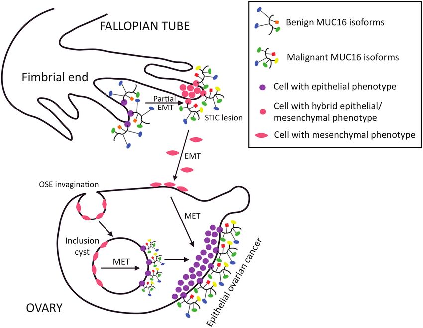
330   |   Carcinogenesis, 2021, Vol. 42, No. 3

screening algorithms for the early detection of ovarian cancer          The N-terminal region is composed of 12.068 amino acids (37).
(78–81); (ii) as part of diagnostic algorithms to distinguish be-   This domain is dominated by its capacity for O-glycosylation,
tween benign and malignant disease in women presenting              which may allow for extracellular matrix interactions. There is
with a pelvic mass (82,83); (iii) to monitor response to treat-     one short area with little or no glycosylation, which could me-
ment (84) and (iv) to detect disease recurrence (84). Setting       diate protein-to-protein interactions in the extracellular matrix
the benchmark cut-off of 35 U/ml the antigen is elevated in         (94).
more than 80% of women with non-mucinous EOC and its                    The extracellular tandem repeat domain also occupies a
levels correlate with disease progression and poorer survival       large part of the molecule, locating downstream from the amino-
outcomes (69,76,85,86). However, it soon became apparent            terminal domain (94). It is composed of a variable number of 45
that CA125 is not a very sensitive and specific marker for          to up to more than 60 repeats, which are arranged in tandem
screening and diagnosis of ovarian cancer, since many other         array (variable number tandem repeats—VNTR) and have a
non-gynaecological malignancies and non-malignant condi-            highly conserved nature (35,41,94). The repeat unit structure in-
tions lead to elevated serum levels (34). Non-gynaecological        creases the potential for multivalent interactions with extracel-
malignancies are almost invariably in advanced widely dis-          lular matrix neighbours. Each repeat has 156 amino acids (94).

                                                                                                                                         Downloaded from https://academic.oup.com/carcin/article/42/3/327/6145151 by guest on 22 November 2021
seminated stage and include lung, liver, bladder, breast, bil-      The length of the tandem repeat region is a unique feature of
iary tract, stomach, colorectal and pancreatic tumours, as well     MUC16, which has a longer repeat sequence compared with
as lymphomas and leukaemia (87,88). Benign entities include         MUC4 (16 amino acids) and MUC1 (20 amino acids) (31,101). The
ovarian cysts, endometriosis, adenomyosis, uterine fibroids,        primary amino acid sequence in each repeat is not identical but
pelvic inflammatory disease, non-gynaecological inflamma-           is homologous (26). The tandem repeat region features both O-
tory conditions, hepatic cirrhosis, heart and renal failure, etc.   and N-glycans (29,37).
(87,89–93). In a large study by Bast et al. it was found that 1%        The SEA domains form a major part (approximately 120
of normal women, 6% of those with benign disease and 28% of         amino acids) of those tandem repeats that contain them (102).
those with non-gynaecological cancers have elevated serum           They are supposed to play an important role in interactions with
levels (69). Furthermore, CA125 appears predominantly in the        other glycoproteins and have also been suggested as poten-
circulation of patients with advanced ovarian cancer but is         tial carriers of the CA125 epitope (35,95,102). The high number
raised in only 50% of those with FIGO stage I malignancy, ren-      of MUC16’s SEA sequences is unique among other tethered
dering it a weak biomarker for the screening of asymptomatic        mucins, which only have one SEA domain (with the exception
women with early disease (75).                                      of MUC4 that has none). It is also believed that MUC16 SEA do-
    Another reason for this low sensitivity and specificity could   mains have evolved separately from other mucins (99).
be weaknesses in the serum CA125 assay. The two monoclonal              The CTD is the smallest part of the molecule, composed of
antibodies currently used for this purpose (OC125 and M11) are      284 amino acids (26). It anchors MUC16 at the cell surface and
known to attach in a multivalent fashion to CA125. It is there-     is divided into three major regions: a small extracellular portion
fore believed that these antibodies identify regions within the     consisting of 229 amino acids, the transmembrane domain with
tandem repeat domains (26,94). However, our knowledge of the        23 amino acids and the cytoplasmic tail with 32 amino acids
exact molecular characteristics and location of the epitopes they   (61). Its extracellular part contains the last SEA domain and a
recognize on these domains remains elusive (26,95). The same        small number of O- and N-glycosylation sites (26,94,103). The
applies for the role of MUC16’s glycans, although it has been       cytoplasmic tail contains one serine, two threonine and three
recently suggested that binding epitopes of monoclonal anti-        tyrosine amino acid residues that can be potential phosphor-
bodies are dependent on the molecule’s conformation and not         ylation sites (48,57). An antibody against the cytoplasmic tail of
on its glycosylation (95). Additionally, because these antibodies   MUC16 has recently been developed (104).
detect released CA125 in the serum, they cannot detect the prox-        The variable number of tandem repeats contained in each
imal residual MUC16 fragment remaining on the cell surface fol-     molecule and differential glycosylation gives rise to several
lowing cleavage (34). Finally, another reason may be the rapid      MUC16 isoforms, probably conferring functional heterogeneity
clearance of mucin fragments by the hepatic reticuloendothelial     (41,105). The presence of multiple MUC16 isoforms was sup-
system, that may potentially skew the measured serum levels         ported by Reinartz et al., who found diverse binding patterns
(96). Combined, these effects suggest that the CA125 assay sig-     of OC125 and M11 antibodies in ovarian and breast cancer cell
nificantly underestimates both the serum concentration of this      lines with variable MUC16 expression, suggesting the existence
biomarker and its total quantity in the body (26). At the other     of different MUC16 binding epitopes (63).
end of the spectrum, a cross-reactivity of OC125 and M11 anti-
bodies with other proteins has been observed. This could lead       MUC16 glycosylation
to falsely raised levels of measured CA125, if the serum concen-
tration of these proteins is increased, thus also impacting the     As mentioned previously, MUC16 is abundant in O- and N-linked
accuracy of the CA125 assay (45).                                   glycans. The process by which mucin O-linked glycosylation
                                                                    occurs is well characterized, though we know relatively little
                                                                    about its regulation. It involves complex post-translational
MUC16 structure                                                     modifications on the protein backbone, whose initiating step
MUC16 is a transmembrane mucin composed of up to 22 152             is the addition of N-acetylgalactosamine carbohydrates; this
amino acids and consists of 3 major domains: an extracellular       forms the so-called Tn antigen (106). The Tn antigen can be
amino-terminal (N-terminal) domain, a large tandem repeat           further extended to form core 1 (T-antigen), 2, 3 and 4 struc-
domain interspersed with 16 sequences that are also found in        tures, based on the identity of linked carbohydrates and type
sea urchin sperm protein, enterokinase and agrin (called SEA        of linkage (107). In normal cells, these structures are elongated
domains) and a carboxy-terminal domain (CTD) (37,94,97–             further and often become branched (108). In contrast, cancer
99). MUC16 exhibits two types of glycosylation: O-linked and        cells express only their early biosynthetic intermediates (109).
N-linked (94,100).                                                  For example, Tn and T can be capped immediately with sialic
P.Giamougiannis et al. | 331
acid by sialyltransferases, producing the sialyl-Tn (STn) and (116). A possible explanation for this finding could be high levels
sialyl-T (ST) antigens, respectively. This occurs only rarely of T-synthase in patients with advanced metastatic ovarian
in normal tissues but is common in cancer (110,111). Cores cancer. Indeed, in the study by Davidson et al., the expression of
1 and 2 are the major O-glycans present on MUC16, both in Tn and STn was significantly up-regulated in pleural and peri-
normal conditions and ovarian cancer (100,112). Cores 3 and 4 toneal effusions compared with both primary and metastatic
O-glycans are synthesized in healthy states (mainly by colonic ovarian solid lesions, confirming that truncated O-glycans are
and respiratory tissues) but are down-regulated in malignancy more frequently observed at the migratory front and less often
(113). in metastases. The authors hypothesized that this up-regulation
Many tumours exhibit aberrant O-glycans and the aberrant is transient if a metastatic sequence of primary tumour → effu-
expression of truncated O-glycans is a hallmark of epithelial sion → solid metastasis is assumed. They concluded that Tn and
cancers (114). In many instances, expression of these truncated STn may have a role in the progression from primary tumour to
structures is driven by alterations to the expression of enzymes malignant effusions (132). Therefore, it can be speculated that
involved in the glycosylation process. For example, the exten- the expression of truncated core 1 O-glycans facilitates the de-
sion of core 1 structures relies on a single enzyme, T-synthase tachment of individual cells from the primary ovarian tumour,
Downloaded from https://academic.oup.com/carcin/article/42/3/327/6145151 by guest on 22 November 2021
(115). T-synthase is known to regulate O-glycan expression on but does not favour their settlement to metastatic sites (131).
glycoproteins of ovarian cancer cells, and loss of its activity On the other hand, in a study with pancreatic cancer cells, trun-
leads to increased Tn and STn levels (115,116). Another poten- cated O-glycans promoted their growth and invasive properties
tial factor is deregulation of enzymes that extend or terminate (114). These differences may reflect varied behaviours of trun-
extension of O-glycans (e.g. sialyltransferases), which has also cated O-glycans in different tumour types.
been observed in a variety of cancers, including serous ovarian With regard to N-glycosylation, changes in cancer involve in-
carcinomas (117–120). creases in size, branching and sialylation (133). In the study by
The presence of truncated core 1 O-glycans in ovarian tu- Saldova et al., CA125 from ovarian cancer patients’ sera displayed
mours has been investigated in several studies. T, Tn and STn are increased sialylated biantennary N-glycans and decreased bi-
almost absent in benign ovarian tumours and normal ovarian secting biantennary ones compared with healthy controls (112).
tissues but are clearly expressed in most ovarian carcinomas On the other hand, in the study by Nagata et al. predominantly
and the serum of ovarian cancer patients. Increasing expres- biantennary and triantennary bisecting type glycans were pre-
sion of these epitopes could correlate with the progression sent on CA125 isolated from serum and ascites of ovarian cancer
from benign ovarian cysts to invasive carcinomas (121–127). In patients (134). Of note, the bisecting type biantennary chains
most cases, more than one antigen is expressed, indicating that can inhibit the cytotoxic responses of NK cells (135). Complex
mucin glycoproteins exist at different stages of glycosylation in multi-antennary chains on N-glycans of glycoproteins have
a given tumour (125). In the study by Akita et al., MUC16 and STn been implicated in tumour metastasis (136).
co-localized in ovarian serous and endometrioid carcinomas, MUC16’s core 1 O- and N-glycans bind with two members of
whereas STn was mostly carried on mucins other than MUC16 the family of galectins, respectively galectin-1 and galectin-3
in mucinous ovarian carcinomas (possibly MUC1 or MUC4) (128). (137,138). Galectins consist part of the group of lectins, acting as
The same study discriminated the level of STn expressed on ligands between glycoproteins and glycolipids at the cell surface
MUC16 (STn/MUC16) in peritoneal fluid from patients with EOC (139). Both galectin-1 and galectin-3 are up-regulated in malig-
and endometriosis with 44% sensitivity and 100% specificity. nancies (including EOC) and can contribute in cancer progres-
Remarkably, the elevation of STn/MUC16 with advancing stages sion by enhancing transmembrane signalling, with subsequent
of ovarian cancer was more evident than that of MUC16 (128). activation of oncogenic transduction pathways (137,138,140,141).
Chen et al. developed a glycoprofiling microarray assay, in which Finally, extended core 1 O-, core 2 O- and N-MUC16 glycans
the combined serum levels of STn/MUC16, ST/MUC16 and STn/ display sialofucosylated oligosaccharides, such as sialyl Lewisx
MUC1 glycoforms distinguished benign ovarian tumours from (sLex) and its isomer, sialyl Lewisa (sLea—also known as the
EOCs with a specificity of 61%, whereas the serum CA125 assay tumour marker CA19-9) (100,142). These epitopes bind with
specificity was 41% (127). Finally, truncated core 1 O-glycans selectins, a family of lectins acting as vascular cell adhesion mol-
can protect tumours from immune system attack, by inhibiting ecules (143). Selectins promote lymphatic and haematogenous
cytotoxic effects of Natural Killer (NK) cells and T lymphocytes metastasis, by mediating interactions between their ligands on
(129,130). Of note, core 2 O-glycans can also suppress immune tumour cells and selectin-expressing endothelial cells in the
responses by blocking the binding of NK to cancer cells (113). microvasculature (142,143). In the study by Chen et al., MUC16
A very interesting hypothesis is that truncated O-glycans on pancreatic cancer cells was found to be the main ligand to
exhibit diverse properties at different stages of ovarian cancer selectins, through the sLea and sLex antigens (142). Aberrant
with an impact on tumours’ biological behaviour. In the study by expression of these epitopes on glycoproteins is associated
Coelho et al., an EOC cell line with induced expression of homo- with pancreatic cancer progression (144). In another study by
geneously truncated Tn and STn O-glycans exhibited decreased Ricardo et al., MUC16 was also found to be one of the main car-
cell proliferation and increased apoptosis compared with its riers (together with MUC1) of sLea and sLex in ovarian serous
parental cell line (derived from a peritoneal metastasis). Cells adenocarcinomas. Although these epitopes were present in the
with truncated O-glycans exhibited increased cell migration but majority of benign cystadenomas as well, they were mainly car-
decreased invasiveness and capacity to form metastases (131). ried by unidentified proteins and not by MUC16 or MUC1 (145).
Partially similar findings were reported by Chou et al., where Combined, the aforementioned findings suggest that the in-
T-synthase knockdown led to decreased growth but also de- creased metastatic potential inferred by sLea and sLex is mostly
creased migration, whereas its overexpression had opposite re- associated with their expression on particular carrier proteins,
sults (116). This discrepancy in cell migration might be due to with MUC16 being a major representative of these. Indeed, the
different ovarian cancer cell lines used in these two studies. binding affinity of selectins for isolated sLex and sLea antigens is
Interestingly, Chou et al. reported significantly lower overall sur- markedly low (142). They also suggest that MUC16 could play a
vival for ovarian cancer patients with overexpressed T-synthase significant role in distant lymphatic and haematogenous spread332 | Carcinogenesis, 2021, Vol. 42, No. 3
in ovarian cancer, a speculation that needs to be validated by role of extracellular factors (such as proteases, cytokines, growth
future studies. factors and hormones) or post-translational modifications (such
Figure 1 demonstrates differences in MUC16 structure be- as glycosylation) around the cleavage site, and intramembrane
tween normal/benign conditions, solid tumour growths and proteolysis has been ruled out (49,103,149–153). An endogenous
migratory EOC, focussing on glycosylation changes. Figure 2 pre- protease activity inherent to the molecule appears to exist,
sents these changes in the context of early EOC tumourigenesis leading to autoproteolysis (103,154,155). This takes place at the
and in correlation with cellular phenotypic shifts. Golgi apparatus en route to the cell membrane, dictated by a
change in MUC16 structure as it encounters Golgi’s acidic pH,
and not by a primary amino acid sequence (103,137). Finally,
MUC16 cleavage although it has been argued that cleavage is independent of
Cell surface expression and release of MUC16 proteolytic frag- intracellular cues, it is triggered by the phosphorylation of the
ments appear to be associated with the conversion of ovarian serine, threonine and tyrosine residues present on MUC16’s
cells from benign to cancer (137,146). The penultimate and final cytoplasmic tail (94,103,156,157).
SEA domains have been proposed as putative MUC16 cleavage As previously mentioned, EOC classically spreads through
Downloaded from https://academic.oup.com/carcin/article/42/3/327/6145151 by guest on 22 November 2021
sites (94,103,147). Potential cleavage locations as close as only peritoneal dissemination, with EMT being a hallmark event in
12 amino acids distal to the transmembrane domain have also the metastatic process. Two important molecules in EMT are
been suggested (94,103,147,148). The kinetics and dynamics of epithelial (E)-cadherin and neural (N)-cadherin. Cadherins are
MUC16 cleavage/shedding from the tumour cells is not well a family of transmembrane glycoproteins, which have a fun-
understood, part of the problem being the enormous size and damental role in the formation of cell-to-cell adhesions. Their
complexity of this glycoprotein (27). In non-migratory EOC, most intracellular domain binds with another group of proteins called
MUC16 remains intact on the cells while a small proportion is catenins, which are mainly located at the cytoplasmic part of
cleaved, leaving a carboxy-terminal fragment (consisting of a the membrane, and stabilize cadherin-mediated cell contacts
small extracellular portion of various lengths, the transmem- through linkage with the actin cytoskeleton. Among catenins,
brane domain and the cytoplasmic tail) on the cell membrane. β-catenin also plays a significant role in EMT (158). Even more
The rest of MUC16 ectodomain is released from the cells, while importantly MUC16 associates with E-cadherin/β-catenin
a fraction of the two fragments remains non-covalently associ- junctional complexes, being bound to the former extracellurarly
ated on the cells (i.e. these fragments remain associated with and to the latter intracellularly (159,160). E-cadherin is a feature
each other) (103,147). Antibodies directed to the extracellular of the epithelial phenotype (as well as the expression of pre-
portion of MUC16 membrane-bound CTD have been developed, dominantly uncleaved MUC16) and generates tight cell-to-cell
confirming its presence on the surface of ovarian cancer cells contacts in the normal fallopian tube, ovarian inclusion cysts,
(147,148). primary ovarian tumours and solid metastases. N-cadherin
Regulation of MUC16 cleavage has been extensively investi- is a marker of mesenchymal differentiation, expressed in the
gated, although most studies have included non-ovarian cancer normal OSE and metastatic lesions; its up-regulation is asso-
cell lines. Yet no firm conclusions can be drawn about what fac- ciated with tumour progression and produces loose bonding
tors potentiate it. For example, there is controversy about the between cells, which enhances their migratory and invasive
Figure 1. MUC16 structure. Demonstration of MUC16 domains (N-terminal, VNTR and carboxy-terminal), glycosylation patterns and putative cleavage sites. The
tandem repeat domain consists of 45 to more than 60 repeat amino acid sequences, interspersed by 16 SEA domains. The N-terminal domain is O-glycosylated whereas
the tandem repeat domain has both O- and N-glycans. The extracellular part of the CTD also has a few sites for O- and N-glycosylation. In normal and benign gynae-
cological conditions, MUC16 features extended and often branched core 1 and core 2 O-glycans, whereas N-glycans are short with limited branching. In EOC, there is
aberrant expression of truncated core 1 based O-glycans and core 2 ones are likely shorter. Truncated core 1 structures are mostly up-regulated in the migratory front
of EOC compared with solid primary and metastatic tumours, which maintain expression of extended core 1 O-glycans. N-Glycans become larger, extensively branched
and poly-antennary. Extended core 1 O-, core 2 O- and N-glycans express sialofucosylated oligosaccharides (such as the sialyl Lewis epitopes). TR, tandem repeat.P.Giamougiannis et al. | 333
Downloaded from https://academic.oup.com/carcin/article/42/3/327/6145151 by guest on 22 November 2021
Figure 2. MUC16 in EOC tumourigenesis. Resting cells in normal OSE exhibit mesenchymal phenotype and do not express MUC16. In contrast, normal fallopian tube
cells have predominantly epithelial features and express MUC16 in its benign isoforms. Inclusion cysts formed from OSE invaginations can develop early neoplastic
changes and progress to EOC. OSE cells trapped in inclusion cysts undergo MET and produce malignant MUC16 isoforms. Alternatively, epithelial cells at the fimbrial
end of the fallopian tube undergo partial EMT converting to a hybrid epithelial/mesenchymal phenotype, and form STICs that express malignant MUC16. Cells in STIC
lesions undergo further EMT to acquire a purely mesenchymal phenotype and migrate to the ovarian surface, with subsequent MET and EOC formation. Colours and
shapes of MUC16 glycans follow the labelling in Figure 1.
properties (11,16,17,161–163). Both cadherins contribute to the for MUC16 CTD overexpression (103,160,168). Indeed, Src phos-
formation of MCAs but increased N-cadherin expression multi- phorylation of a tyrosine residue at MUC16’s cytoplasmic tail
plies their aggressiveness and resistance to apoptosis (162,163). has been demonstrated in ovarian, colon and breast cancer cells,
As a rule cadherin subtype-specific junctions are formed, which leads to deregulation of E-cadherin/β-catenin junctional
whereas cells expressing both cadherins (such as the hybrid complexes and increases cellular migration (62,103,160). Src is
ones) can create contacts with epithelial and mesenchymal a non-receptor tyrosine kinase, whose activity enhances me-
cells (163). The cadherin/β-catenin complex is expressed on tastasis. (169). It is also known that Src activity negatively regu-
cells even when they are not involved in cellular adhesions or lates the E-cadherin/β-catenin cell adhesion system (170). More
aggregates (i.e. on single cells) (158). specifically Src-mediated phosphorylation of E-cadherin in-
E-cadherin down-regulation with concomitant N-cadherin duces its endocytosis and degradation, while Src inhibition in-
up-regulation is a key step in EMT, described as the ‘cadherin creases E-cadherin levels and can induce EMT reversal (171,172).
switch’ phenomenon (162). The loss/reduction of E-cadherin β-Catenin is also phosphorylated by Src, which negatively regu-
expression or function is associated with progression from lates its binding to E-cadherin (173). Taken together the above
well to poorly differentiated EOC and promotes metastasis suggest that Src phosphorylation initiates a cascade of events,
(10,11,164,165). It can be triggered by soluble factors present including MUC16 shedding with overexpression of MUC16 CTD
in the ascites (such as proteases, cytokines and growth factors and breakdown of E-cadherin junctional complexes (174). It is
that cause shedding of its extracellular part) but is also associ- also evident that increased MUC16 cleavage occurs in meta-
ated with overexpression of MUC16’s CTD (17,62,166). The latter static EOC, which would contribute to the higher serum CA125
consists a MUC16 isoform that promotes migration and inva- levels detected in advanced compared with early stage disease.
sion of tumour cells, with increased N-cadherin expression and MUC16 CTD is sufficient to increase the metastatic poten-
acquisition of mesenchymal phenotype (62,167). Additionally, tial of ovarian cancer cells (175). Following MUC16 cleavage
it attenuates cellular apoptosis and sensitivity to platinum the CTD fragment can remain on the cell membrane to exert
chemotherapy agents (61,64). Ectodomain shedding triggered its actions, or undergo endocytosis to affect signalling in other
by extrinsic factors (such as proteases and cytokines), enhanced cellular compartments (103). The latter is confirmed through
autoproteolytic MUC16 cleavage and phosphorylation of its binding antibodies to MUC16 CTD ectodomain, demonstrating
cytoplasmic tail have been proposed as potential mechanisms internalization by ovarian cancer cells (148). MUC16 CTD can334 | Carcinogenesis, 2021, Vol. 42, No. 3
also translocate to the nucleus and is present within chro- increase in cytoplasmic β-catenin levels, MUC16 CTD increases
matin, suggesting its function as a transcriptional co-regulator its cell membrane pools as well (166). It is therefore likely that
(103,167). N-Glycosylation at its ectodomain appears to be MUC16 CTD recruits β-catenin at its cytoplasmic tail to stabilize
associated with an altered gene-expression profile and in- junctional complexes with N-cadherin, leading to MCA forma-
creased expression of critical invasion genes, through activa- tion with other N-cadherin expressing cells. At the same time it
tion of the PI3K (phosphatidylinositol 3-kinase)/Akt (protein increases MCAs migratory and invasive properties, by enhancing
kinase B) and MAPK (mitogen-activated protein kinase)/ERK β-catenin’s transcriptional activity through the Wnt/β-catenin
(extracellular-signal regulated kinase) cell survival signal pathway. Overall the interactions with β-catenin indicate the
transduction pathways (175,176). Both of them induce EMT importance of MUC16 CTD’s cytoplasmic tail in intracellular
in carcinomas (159,177–179). The mechanisms by which signalling. This has been confirmed in experiments that induced
MUC16 CTD activates these pathways include interactions its deletion, leading to a reduction of MUC16 CTD’s oncogenic
of its N-glycans with galectin-3, which in turn binds with potential and its ability to form MCAs (62,176).
N-glycans expressed by two transmembrane glycoproteins, Figure 3 demonstrates the EMT mechanisms induced by
the epidermal growth factor receptor (EGFR) and β1-integrin MUC16 CTD overexpression in EOC.
Downloaded from https://academic.oup.com/carcin/article/42/3/327/6145151 by guest on 22 November 2021
(part of the integrins family of cell adhesion receptors). The
bonds in the formed tripartite heteromolecular junctions are
prerequisites for activation of both agents (138). For EGFR
MUC16 knockdown
this occurs after binding of its ligands (such as the epidermal MUC16 knockdown has been associated both with increased
growth factor—EGF) whereas for β1-integrin through intra- and decreased ovarian cancer aggressiveness. When induced at
cellular recruitment and phosphorylation of Focal Adhesion the surface of cancer cells it inhibits tumour growth (either by
Kinase (FAK—a non-receptor protein tyrosine kinase) and Src cellular apoptosis or by arrest in proliferation) and reduces their
complexes. These events trigger signalling cascades that can metastatic potential (62,63,167). Additionally, it increases their
eventually activate both PI3K/Akt and MAPK/ERK (180–182). sensitivity to platinum chemotherapy agents (64). However,
Disruption of MUC16 CTD’s N-glycan-mediated binding with ovarian cancer cells inherently lacking MUC16 expression
galectin-3 abrogates these effects, demonstrating its crucial do not undergo apoptosis (63). It has also been reported that
role for their conduct (138). MUC16 knockdown leads to decreased cell surface expression
Another example of MUC16 CTD’s role in intracellular of E-cadherin. Binding of MUC16 to E-cadherin results in the
signalling is its interaction with β-catenin (167). β-Catenin is a latter’s surface localization whereas, in the absence of MUC16,
dual function protein involved in regulation of intracellular ad- E-cadherin re-localizes to the cytoplasm. Ovarian cancer cells
hesion (as highlighted previously) but also in gene transcrip- with down-regulated MUC16 display a mesenchymal pheno-
tion (183,184). It accumulates in the nucleus of many malignant type with increased N-cadherin expression, cell motility and
cell types (including ovarian cancer), acting as an intracellular invasion. Therefore, MUC16 knockdown could be another EMT-
signal transducer in the Wnt (Wingless-related integration site) promoting mechanism in EOC metastasis (159). On the other
pathway (183,185). The Wnt/β-catenin transduction is known hand, MUC16 knockdown ovarian cancer cells do not form MCAs
to enhance EMT in carcinomas, up-regulating transcriptor and exhibit increased β-catenin degradation with attenuation of
factors that decrease E-cadherin and increase N-cadherin ex- its transcriptional activity (159,166). These effects are contrary to
pression, along with mesenchymal genes’ activation. In the what is observed in MUC16 CTD overexpression, strengthening
presence of E-cadherin, β-catenin locates at the cell membrane the assumption that MUC16 CTD participates in MCAs through
to participate in the formation of their junctional complexes. enhanced β-catenin recruitment in N-cadherin complexes.
Loss of E-cadherin leads to β-catenin release to the cytoplasm It is also apparent that oncogenic pathways other than
and its quick degradation. Binding of Wnt ligands to their re- the Wnt/β-catenin signalling are up-regulated by MUC16
ceptor stops this turnover, leading to increased levels of cyto- knockdown. Indeed, EGFR activation has been demonstrated
plasmic β-catenin, which is then transferred to the nucleus even in the absence of MUC16. More specifically, E-cadherin
(158). In ovarian cancer, overexpressed MUC16 CTD also inhibits extracellularly interacts with the EGFR and negatively regulates
β-catenin clearance hence up-regulating Wnt downstream the ligand-dependent activation of this receptor (189). MUC16
genes transcription, which enhances tumour proliferation and knockdown (with subsequent loss of extracellular E-cadherin)
migration (166,167). Of note, MUC16 CTD can increase the cyto- allows for EGF binding to EGFR, thus initiating the PI3K/Akt and
plasmic levels of another catenin participating in E-cadherin MAPK/ERK pathways transduction relay (159). Therefore, MUC16
complexes, the p120 catenin, with the same impact on tumour down-regulation triggers EMT-associated signalling, consistent
behaviour, but its mechanism of action is unknown (186). with the finding that MUC16 tissue loss is associated with poor
Similar effects have been described through the interaction prognosis in EOC (65).
of MUC16 CTD with a non-receptor tyrosine kinase called
Janus kinase 2 (JAK2). In pancreatic cancer cells, MUC16 CTD
MUC16 and formation of peritoneal
overexpression leads to JAK2 translocation from the cytoplasm
to the nucleus, with increased transcription of its target genes
metastases
(187). Activation of the JAK2 signalling has been reported in The primary target of the metastasizing EOC is the mesothelial
EOC as well (188). This could therefore be another pathway that cell monolayer, which covers the abdominal peritoneum (149).
MUC16 CTD activates to enhance ovarian cancer cells’ meta- MUC16 binds with very strong affinity to mesothelin, a meso-
static properties. thelial cells’ membrane-bound glycoprotein, which also has
Finally, MUC16 CTD contributes in the formation of MCAs a soluble form produced by proteolytic cleavage (77,190–192).
(166). Although not experimentally validated, it can be pre- Importantly it is expressed on EOCs of all histological types
sumed that this occurs through interactions with N-cadherin, (particularly serum ones), in significantly higher levels com-
as MUC16 CTD induces a relocation of cell surface E-cadherin pared with benign ovarian tumours (77,193–195). Of note, it
to the cytoplasm. Additionally, apart from the aforementioned may be able to trap MUC16 in the tissue, thus providing anotherP.Giamougiannis et al. | 335
Downloaded from https://academic.oup.com/carcin/article/42/3/327/6145151 by guest on 22 November 2021
Figure 3. EMT mechanisms induced by MUC16 CTD overexpression. The hallmark event in EMT is the switch from E- to N-cadherin expression, which enhances cancer
migration and invasion. In EOC cells with epithelial phenotype most MUC16 is expressed in its uncleaved form, participating in junctional complexes with E-cadherin
and β-catenin. E-cadherin’s interaction with EGFR inhibits binding of the latter’s activating ligands (such as EGF). MMPs and cytokines promote E-cadherin shedding
and MUC16 cleavage. Src phosphorylation of E-cadherin, β-catenin and MUC16 deregulates their complexes, leading to E-cadherin degradation, β-catenin release and
MUC16 cleavage. These mechanisms initiate EMT with MUC16 CTD overexpression. The latter is enhanced by increased autoproteolytic MUC16 cleavage at the Golgi
apparatus. MUC16 CTD can stay on the cell surface or translocate to the nucleus, to act as a transcriptional co-regulator. On the cell-surface MUC16 CTD interacts
with EGFR and β1-integrin through N-glycan-mediated tripartite junctions with each of these two glycoproteins and galectin-3. These bonds activate EGFR and β1-
integrin, which subsequently up-regulate EMT-enhancing gene transcription through intracellular signalling pathways (MAPK/ERK, PI3K/Akt, FAK/Src). Furthermore,
MUC16 CTD reinforces the Wnt/β-catenin signalling, by promoting β-catenin’s nuclear transfer to exert transcriptional activity. The Wnt/β-catenin transduction also
enhances EMT. MUC16 CTD recruits pools of β-catenin at the cell membrane as well, which contribute to the formation of MCAs, possibly through stabilizing junctional
complexes with N-cadherin. Dashed lines indicate not experimentally validated molecular interactions. Colours and shapes of MUC16 glycans follow the labelling in
Figure 1.
possible explanation for the discrepancies noted in tissue and not be adequate to maintain this heterotypic adhesion (190).
serum CA125 levels in ovarian cancer patients (77). Indeed, it has been found that other adhesion molecules me-
MUC16 is the only ligand for mesothelin on ovarian cancer diate ovarian cancer and mesothelial cells binding as well. For
cells and this binding is not shared by other mucins (77,190). example, β1-integrin expressed by ovarian cancer cells binds to
Both single cells and MCAs attach to the peritoneal mesothe- fibronectin expressed by mesothelial cells. However, the ovarian
lium (196). The ovarian cancer cell-to-mesothelin binding oc- cancer cells–mesothelin binding is only partially inhibited by
curs via MUC16’s N-glycans (190–192). It is questionable whether anti-β1-integrin antibodies (196–198). Interestingly, when β1-
cleaved MUC16 could inhibit or decrease binding of cell-bound integrin is blocked, cells with low surface MUC16 do not ad-
MUC16 to mesothelin. Mesothelin interacts with both shed here to mesothelin, whereas adhesion of cells with high surface
and cell-surface MUC16 but its affinity is higher for the latter. MUC16 remains unaffected (149). It is therefore likely that the
This could be due to proteolytic processing after MUC16’s tu- mesothelin–MUC16 interaction provides the necessary first step
mour release or digestion of N-glycans by glycosidases in the for metastasis transitioning to β1-integrins or other molecules,
ascites (190). Additionally, mesothelial cells produce and shed either in the absence of MUC16 or to stabilize contacts (149,190).
MUC16 in the peritoneal fluid, in benign and malignant con- The interaction between mesothelin and tumour cell-
ditions (70,151). It has been argued that the co-expression of surface MUC16 represents the initial adhesive event. Once at-
MUC16 and mesothelin in both ovarian cancer and mesothe- tached, ovarian cancer cells undergo EMT to start clearance
lial cells could competitively inhibit their binding. However, the of the mesothelial cell layer and spread to the submesothelial
multivalent nature of mesothelin’s binding on MUC16 likely matrix. Subsequently, invasion through degradation of the
promotes rather than inhibits the attachment of ovarian cancer submesothelial matrix collagen occurs, in order to form firm
cells to mesothelium, even involving expressed MUC16 CTD metastases through MET (11,15,149,161,165). These events
in these contacts (77). Nevertheless, co-expression of MUC16 seem to be strongly linked to the mesenchymal phenotype
and mesothelin on ovarian cancer cells leads to recruitment of and N-cadherin expression or acquisition, as cells maintaining
additional tumour load at metastatic sites and MCAs. On the E-cadherin exhibit reduced mesothelial retraction, migration
other hand, it has been argued that tumour cell attachment and collagen invasion (161,163,165). MUC16 participates in this
to mesothelium via the MUC16–mesothelin interaction may invasive process as well, through interactions with members of a336 | Carcinogenesis, 2021, Vol. 42, No. 3
family of proteolytic enzymes called matrix metalloproteinases cells antitumour functions. Of note, ascites NK cells are hypo-
(MMPs). Examples of metalloproteinases in ovarian cancer are responsive to tumour targets compared with NK cells from per-
the membrane type 1 matrix metalloproteinase (MT1-MMP), ipheral blood of EOC patients (209).
MMP-2 and MMP-9, all degrading interstitial collagen in the The role of MUC16 in EOC’s interactions with immune factors
submesothelial matrix. MMP-2 and MMP-9 are activated by has been investigated in several studies. MUC16 protects ovarian
MT1-MMP and MMP-9 is involved in ovarian cancer’s EMT, as cancer cells from complement attack by trapping effectors of the
it catalyses the shedding of E-cadherin’s extracellular domain complement cascade (210). Cytokines (interferon-gamma and
(199–201). tumour necrosis factor-alpha) can increase the expression and
An inverse relationship between MT1-MMP and MUC16 has shedding of MUC16 from ovarian cancer cells (36,151). MUC16
been demonstrated by Bruney et al. Ovarian cancer cell lines was also found to possess the highest number of HLA class I lig-
which express high MUC16 levels do not exhibit surface ex- ands compared with all other antigens present on the surface of
pression of MT1-MMP. On the other hand, cells overexpressing EOC cells (204), which as mentioned previously inhibit NK activa-
MT1-MMP exhibit loss of surface MUC16 but increased levels tion. The role of ovarian cancer cell-surface MUC16 in interactions
of soluble forms. These findings suggest that MT1-MMP may with NK cells was investigated by Gubbels et al. Low MUC16 ex-
Downloaded from https://academic.oup.com/carcin/article/42/3/327/6145151 by guest on 22 November 2021
catalyse MUC16 cleavage. Increased peritoneal adhesion in the pression was associated with significantly increased NK conju-
absence of MT1-MMP has been shown and, inversely, a loss gation compared with medium or high MUC16 levels. Variable
of adhesion in MT1-MMP expressing cells with reduced cell- expression of activating NK receptors’ (DNAM-1 and NKG2D) lig-
surface MUC16 (149). Therefore, it is likely that ovarian cancer ands was ruled out for this effect, as all ovarian cancer cells had
cells express MUC16 for mesothelin attachment, but subse- comparable levels. Following this observation, MUC16 knockdown
quent invasion and metastasis formation is achieved through cells were employed and compared with MUC16-positive ones.
MT1-MMP-mediated MUC16 shedding. The aforementioned These two lines were also similar in NKG2D and DNAM-1 ligands
relationship between MT1-MMP and MUC16 has been indir- expression. Although HLA class I antigens levels (presented by
ectly confirmed by Comamala et al., who found increased ac- molecules other than MUC16) were slightly elevated on MUC16
tivation of MMP-2 and MMP-9 in MUC16 knockdown ovarian knockdown cells, they were lysed to a significantly greater ex-
cancer cells (159). To that end, it is plausible to conclude that tent than the MUC16-positive ones. A similar effect was observed
MUC16 down-regulation enhances the activity of these three when KIR-negative NK cells were used as effectors, which mark-
metalloproteinases, thus potentiating the formation of solid edly enhanced their cytolytic capacity, as the inhibitory effect of
peritoneal metastases. HLA class I antigens was abolished (211).
However, partially contrary findings to the above were re- These findings suggest that MUC16 uses its HLA
ported by Reinartz et al. (63). Although ovarian and breast class I antigens to attenuate NK cells activation, but this is not
cancer lines with high MUC16 expression demonstrated loss its sole immunoprotective mechanism. In this respect, Gubbels
of invasiveness in collagen matrix, MUC16 knockdown did not et al. noted reduced NK binding not related to expression levels
alter this attitude. MUC16 knockdown also abolished MMP-2 of other mucins (MUC1, MUC4) present on the surface of ovarian
activation and this was accomplished by suppression of β1- cancer cells, which can block immune synapses (211–213). The au-
integrin expression. The discrepancies in the outcomes of these thors suggested that MUC16’s extensive O-glycosylation could be
studies could be explained by different cell lines and experi- at least partially responsible for this anti-adhesive effect. Indeed
mental approaches used. as mentioned before, MUC16’s core 2 O-glycans offer protection
Finally, MUC16 expression correlates with pancreatic cancer from NK immune attack. As MUC16 is much larger and more
cells binding capacity to mesothelin, and this interaction activates heavily glycosylated than MUC1 and MUC4, it could use its size
matrix metalloproteinase 7 (MMP-7) promoting metastasis (202). and abundant longer O-glycan chains to inhibit NK binding, irre-
Interestingly, Chang et al. found that mesothelin-overexpressing spective of the presence of those two other mucins (29,174,211).
ovarian cancer cells demonstrate enhanced migration and in- Finally, MUC16 overexpression protects pancreatic cancer cells
vasion, also through the induction of MMP-7 (203). It is there- not only from NK but also from cytotoxic T lymphocytes (130). It
fore possible that MMP-7 activation by mesothelin is mediated was highlighted previously that HLA antigens are strong stimu-
through MUC16 in ovarian cancer as well. lants of T-cell activation, but also that subsets of them possess
inhibitory receptors for HLA class I ligands. Therefore, it can be
speculated that MUC16 uses its HLA class I antigens to regulate
MUC16 and immune interactions T-cell binding too, favouring a mostly inhibitory effect.
Immune regulation plays an important role in controlling Apart from the aforementioned mechanisms, cell-surface
ovarian cancer growth and EOCs have been recognized as highly MUC16 binds with T cells and monocytes through siglec-9, an
immunogenic tumours. They exhibit strong expression of HLA inhibitory receptor present on the surface of T cells, B cells,
(Human Leukocyte Antigen) class I and class II molecules, which monocytes and NK cells (214–216). Siglecs (sialic acid-binding
are major prerequisites for effective T-lymphocyte-mediated immunoglobulin-type lectins) bind with sialic acid residues on
killing (204). EOCs also express ligands that can activate or in- proteoglycans, glycolipids and glycoproteins (217). As already men-
hibit NK cells receptors and ovarian carcinoma cells are sensi- tioned, these residues are abundantly present on MUC16’s glycan
tive to NK-mediated lysis (205). However, it is well established chains in ovarian cancer, particularly on truncated core 1 O- and
that ovarian cancer patients exhibit immune suppression (206). N-glycans. In EOC, shed MUC16 following cleavage binds with NK
With regard to NK cells, expression of HLA class I antigens cells, B cells and monocytes through siglec-9 as well. With regard
on ovarian tumour cells abrogates their cytotoxic responses, to NK cells, siglec-9 is selectively expressed on a subset that pos-
as these antigens are strong inhibitory ligands for several re- sesses the highest cytotoxicity, and MUC16 binding may inhibit
ceptors on their surface, such as the Killer Immunoglobulin- their activation (209,215). Exposure of healthy donors’ peripheral
like Receptors (KIR) (207). These inhibitory receptors are also blood NK cells to ovarian tumour-derived MUC16 induces a robust
present on subsets of T lymphocytes (208). Additionally, the inhibition of even their most potent forms, an effect that as pre-
malignant microenvironment, including ascites, inhibits NK viously stated can be exerted through MUC16’s truncated core 1P.Giamougiannis et al. | 337
O- and N-glycans. Additionally, it induces a severe down-regulation the submesothelial matrix collagen. These cells overexpress
of the activating receptor CD16, which is present on the highly MUC16 CTD or exhibit MUC16 knockdown, which both enhance
cytotoxic NK cells, and makes them acquire a less cytotoxic pheno- their invasive properties. Invasion is also facilitated by MMPs,
type (218,219). This phenotypic shift is also noted when peripheral which promote MUC16 cleavage and are up-regulated in the ab-
blood NK cells from healthy donors are treated with peritoneal sence of MUC16. Finally, these early metastatic lesions undergo
fluid from EOC patients (209). Indeed, ascites NK cells obtained MET to proliferate and form solid peritoneal metastases, with
from ovarian cancer patients show significant reduction in CD16 up-regulated uncleaved MUC16 expression promoting their
expression compared with NK cells derived from healthy donors’ growth and stabilizing intercellular contacts.
peripheral blood (220). This change in phenotype is not caused by MUC16’s participation in the evolution of ovarian cancer has
increased apoptosis of the more cytotoxic NK group or selective attracted attention to its potential use as a therapeutic target.
proliferation of the less cytotoxic one (209,218). Monoclonal antibodies against MUC16 (221–223) and agents
It is therefore plausible to assume that shed MUC16—siglec-9 targeting the MUC16–mesothelin interaction (224–226) have
binding, mediated mainly through the former’s truncated core 1 been developed, but have shown limited benefit in clinical trials
O- and N-glycans, inhibits NK cytolysis and induces a phenotypic (227–230). This could be due to MUC16 cleavage, leading to in-
Downloaded from https://academic.oup.com/carcin/article/42/3/327/6145151 by guest on 22 November 2021
shift to the less cytotoxic group in ascites. Competent siglec-9- creased binding of therapeutic agents by shed MUC16 in the
positive T cells and monocytes enter the tumour microenviron- serum and reducing the amount of intact MUC16 on tumour
ment and adhere via cell-surface MUC16 glycans, but this binding cells (27). The recently developed antibodies against MUC16
also suppresses immune responses. Finally, the inhibitory (through CTD could help overcome these hurdles (104,147,148). Better
HLA class I antigens) and anti-adhesive (through core 2 O-glycans) understanding of MUC16’s structural diversity and aberrant
cell-surface MUC16 properties promote hindering and evasion, re- glycoforms, as well as targeting other molecules involved in its
spectively, of siglec-9-negative immune cells. By using these mech- interactions (e.g. galectins, selectins, siglec-9) or the oncogenic
anisms, MUC16 could substantially protect ovarian cancer from signalling pathways it activates, could also contribute in the de-
immune responses and potentiate its progression. velopment of more effective treatment strategies (27,34).
Conclusion Funding
We propose a model for MUC16’s evolving role in relation to EOC No funding was received for this work.
progression and ovarian cancer cells’ phenotypic shifts (Figure
4). At early stages, predominantly uncleaved MUC16 contrib-
utes to tumour growth and stabilizes contacts between cells
Key points
with complex epithelial phenotypes. At the same time, it pro-
vides immunoprotection through its O- and N-glycans, thus sup-
• CA125 corresponds to the transmembrane glycoprotein
porting tumour survival. Increased MUC16 cleavage participates
MUC16, the largest mucin identified to date.
to disruption of cells’ adhesions and initiates their release in
• In normal conditions, MUC16 is not present on OSE but it is
the peritoneal cavity. This process is facilitated by up-regulated
overexpressed in EOC.
expression of truncated core 1 O-glycans, which is maintained
• The variable number of tandem amino acid repeats in
throughout cell migration, whereas solid tumour growths mainly
MUC16’s structure, the differential glycosylation and the
express their extended forms. Detached cells circulate in the as-
expression of its CTD give rise to a high number of MUC16
cites and undergo EMT as well as differentiation shifts between
isoforms.
epithelial, mesenchymal and hybrid (mixed epithelial/mesen-
• MUC16 exhibits a wide range of interactions with other
chymal) phenotypes. Mesenchymal cells overexpress MUC16’s
molecules, which contribute to EOC’s neoplastic potential,
CTD, which is retained on the cell membrane following cleavage,
growth and metastasis.
and increases their migratory potential as well as their resistance
• MUC16 undergoes proteolytic cleavage proximal and up-
to chemotherapy. The released MUC16 fragment and MUC16 pre-
stream to its transmembrane domain.
sent on free-floating ovarian cancer cells continue inhibiting im-
• In solid tumours, MUC16 is mostly expressed in its
mune cytotoxicity through their glycan chains. Sialofucosylated
uncleaved form, stabilizing contacts between cells with
oligosaccharide epitopes may bind to blood and lymph vessels
complex epithelial phenotypes.
to promote EOC’s haematogenous and lymphatic metastasis.
• Enhanced MUC16 cleavage leads to disruption of intercel-
Some mesenchymal cells undergo complete MUC16 knockdown,
lular junctions and overexpression of its CTD, associated
which might enhance their aggressiveness, but can also increase
with mesenchymal cellular phenotype as well as increased
their susceptibility to chemotherapy. Uncleaved MUC16 and
EOC migration and invasion.
MUC16 CTD contribute to the stability of MCAs, formed by con-
• Only the released MUC16 fragment following cleavage par-
tacts between single circulating cells, which enhance their sur-
tially enters the circulation and gets detected by the serum
vival in the ascitic environment. MCAs and single cells attach to
CA125 assay, which therefore underestimates the total
the mesothelial layer covering the peritoneal surfaces through
amount of MUC16 present in the body. Increased MUC16
binding of MUC16 and MUC16 CTD’s N-glycans with mesothelin,
cleavage in metastatic EOC contributes to higher serum
expressed by mesothelial cells. MUC16 knockdown cells attach
CA125 levels measured in advanced stages of disease.
through β1-integrin, which also stabilizes the initial mesothelin-
• Both uncleaved MUC16 and MUC16 CTD contribute to
mediated contacts in MUC16 expressing cells. Mesothelin is pre-
the stability of adherens junctions in MCAs, which help
sent on ovarian cancer cells as well, and its interactions with
migrating ovarian cancer cells survive in ascites.
MUC16 lead to further tumour recruitment on MCAs and meta-
• MUC16 knockdown is associated with the aggres-
static sites. Following anchoring to peritoneal surfaces, mesen-
sive mesenchymal phenotype but can increase cellular
chymal cells or cells undergoing further EMT induce mesothelial
chemotherapy-induced apoptosis.
clearance, spread to submesothelial matrix and start degradingYou can also read