Complement Evasion in Borrelia spirochetes: Mechanisms and Opportunities for Intervention - MDPI
←
→
Page content transcription
If your browser does not render page correctly, please read the page content below
Review
Complement Evasion in Borrelia spirochetes:
Mechanisms and Opportunities for Intervention
Jonathan W. Locke
 Department of Biology, University of New Mexico, Albuquerque, NM 87131, USA; jonathan.locke@unm.edu

 Received: 15 May 2019; Accepted: 11 June 2019; Published: 13 June 2019

 Abstract: Lyme disease (LD) is an increasingly prevalent, climate change-accelerated, vector-borne
 infectious disease with significant morbidity and cost in a proportion of patients who experience
 ongoing symptoms after antibiotic treatment, a condition known as post-treatment Lyme disease
 syndrome (PTLDS). Spirochetal bacteria of Borrelia species are the causative agents of LD. These
 obligate parasites have evolved sophisticated immune evasion mechanisms, including the ability to
 defeat the innate immune system’s complement cascade. Research on complement function and
 Borrelia evasion mechanisms, focusing on human disease, is reviewed, highlighting opportunities
 to build on existing knowledge. Implications for the development of new antibiotic therapies having
 the potential to prevent or cure PTLDS are discussed. It is noted that a therapy enabling the
 complement system to effectively counter Borrelia might have lower cost and fewer side-effects and
 risks than broad-spectrum antibiotic use and could avert the need to develop and administer a
 vaccine.

 Keywords: Lyme disease; spirochete; Borrelia; PTLDS; antibiotics; PLD; complement; CRASP

1. Introduction

1.1. Borrelia and Lyme Disease
     Spirochetes of the Borrelia genus are corkscrew-shaped, enzootic bacteria endemic in parts of
Europe, Asia and North America. Borrelia species are fully host-dependent for survival and growth
and possess a reduced genome, so this text will refer to the obligate endosymbiotic Borrelia as
parasites [1]. As parasites of mammals and birds, Borrelia bacteria move between hosts, transmitted
by tick and louse vectors (Figure 1) [2,3]. A number of Borrelia species are known to be pathogenic,
and six species are known to cause tick-transmitted Lyme borreliosis in humans, commonly known
as Lyme disease (LD): B. burgdorferi sensu stricto (s. s.), B. afzelii, B. garinii, B. spielmanii, B. bavariensis
and B. mayonii [4]. In addition, tick-borne relapsing fever (TBRF) is caused by Borrelia species,
including B. hermsii, B. parkerii, B. turicatae, B. duttoni and B. miyamotoi, and louse-borne relapsing
fever (LBRF) is caused by B. recurrentis (Supplementary Materials, Tables S1,S2).

Antibiotics 2019, 8, 80; doi:10.3390/antibiotics8020080                          www.mdpi.com/journal/antibiotics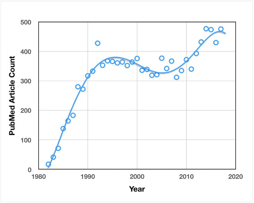
Antibiotics 2019, 8, 80                                                                                                          2 of 24

                                          Year 1                                                  Year 2

                   Spring      Summer                  Fall       Winter     Spring         Summer              Fall    Winter

         Stage      eggs         larva                                       nymph                              adult

                            Larval Acquisition of Borrelia spp.            Highest LD Risk in Humans

         Hosts                           Mice, Birds                                  Squirrels, Deer, Humans

      Figure 1. Tick Lifecycle and Host Interaction—The Ixodes scapularis tick lifecycle takes two years and
      requires three blood meals, one at the larval stage, one at the nymphal stage and one at the adult stage
      of development. Smaller mammals and birds serve as hosts for blood meals at the larval stage, while
      larger mammals are hosts during the nymphal and adult stages. Larvae which acquire Borrelia spp.
      from mice and birds in the summer of their first year can transmit these spirochetes as nymphs in the
      spring of their second year, infecting humans. For this reason, Lyme disease (LD) risk is highest in
      the spring and summer.

1.1.1. Pathogenesis
      Borrelia spirochetes exist through a complex enzootic cycle that requires them to be well-adapted
to tick, mammalian and avian environments. After a tick infected with Borrelia takes a blood meal
from a host, spirochetes in the midgut migrate through the midgut epithelium into the hemocoel and
follow chemotactic clues to locate and infect the salivary glands. Once in the salivary glands, there is
opportunity for Borrelia to be transmitted through tick saliva to the host [5,6]. Transmission typically
takes hours to achieve, but long attachment times are not uncommon as ticks have powerful
anesthetic compounds in their saliva, allowing them to feed unnoticed [7].
      Environmental clues from the vertebrate host, such as the presence of blood-related proteins and
an increase in ambient temperature to 37 °C, lead to changes in gene expression, including changes
in the expression of surface proteins, that prepare Borrelia for the host environment (Supplementary
Materials, Table S3) [8]. Once inside the host and after a period of incubation, Borrelia disseminates
away from the site of infection through use of its endoflagella and by adhering to host cells.
Movement away from the site of infection produces the classic erythema migrans (EM) “bullseye”
lesion that often accompanies the acute, localized stage of infection. If the infection is left untreated,
spirochetes will often disseminate widely throughout the body, resulting in more advanced stages of
infection [3]. The pathology of LD appears to be caused primarily by host immune response, as
Borrelia is not known to produce toxins or proteases that directly damage tissues [9]. The formation
of persister cells and biofilms harboring persisters and other microbes [10–16], as well as other
immune evasion mechanisms, likely play roles in pathogenesis and tissue damage through the
misdirection of host immune response.

1.1.2. Epidemiology
      LD is most common in the northerly latitudes of the Northern Hemisphere, including North
America, Europe, Russia, China and Japan. Since 1991, when LD became a reportable condition,
records of the incidence and geographic distribution of LD in the United States have increased
substantially and incidence in parts of Europe may also be increasing. In part, this increase may be
due to improvements in diagnosis and reporting, but cases are also increasing as a result of climate
change, which is expanding habitable tick territory and extending time intervals for tick breeding
[17,18]. Modeling studies from the Centers for Disease Control (CDC) estimate that there are
approximately 300,000 cases of LD per year in the US. These cases are concentrated in a relatively
small geographic area. Within the United States, 96% of confirmed cases in 2013 were reported from
just 14 states in the Northeast, Mid-Atlantic and Great Lakes regions [19].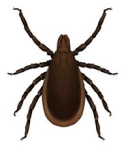
Antibiotics 2019, 8, 80                                                                          3 of 24

1.1.3. Clinical Presentation, Diagnosis and Treatment
      The presentation of LD in the early, acute phase of infection is often marked by flu-like
symptoms such as fatigue, headache, fever, chills, swollen lymph nodes, myalgia and arthralgia. In
some cases, an EM “bullseye” rash may be present as the infection disseminates away from the tick
wound site. Within a few days to weeks, the disseminated stage of infection can broaden to include
significant neurological symptoms, presenting as Lyme neuroborreliosis (LNB). At this stage,
patients may experience migratory pain, facial palsy, acute lymphocytic meningitis, heart block and
occasionally, vision problems. In late, disseminated infection, LD can go on to cause persistent
arthritis [3].
      It is believed that patients that receive early diagnosis and treatment mostly recover following
administration of antibiotics (typically doxycycline), but about 10–20% of these patients experience
ongoing symptoms, lasting for months to years, known as post-treatment Lyme disease syndrome
(PTLDS). The scope of this problem may be still wider as a recent large-scale study revealed that more
than 63% of LD cases develop at least one diagnosis associated with PTLDS [20]. Patients who receive
late diagnosis and treatment are even more likely to develop PTLDS [21]. The cause of PTLDS is
unknown and the subject of ongoing debate. Among the factors that might be involved are
autoimmunity, antigenic debris, persistent infection due to antibiotic failure, and tick-borne co-
infections such as babesiosis, anaplasmosis, erlichiosis, relapsing fever, tularemia and Rocky
Mountain spotted fever (RMSF).
      Correct diagnosis and timely treatment of LD can be complicated in a clinical setting by a
number of factors, increasing the chance of patients developing PTLDS. As mentioned above, the
classic presentation of LD after a tick bite is the EM “bullseye” rash. Unfortunately, patients often do
not recall tick bites, and may present either without a rash at all or with lesions that are not easy to
identify. In the absence of these textbook indicators, the diffuse symptom picture of LD can mimic a
wide range of illnesses, leading to frequent misdiagnosis or non-diagnosis. The difficulty of diagnosis
is further increased when the symptom picture becomes more complicated in late disseminated Lyme
or when it is clouded by symptoms caused by other tick-borne co-infections, such as those listed
above. Even when LD is suspected, false-negative ELISA and Western blot (IgM and IgG) antibody
test results are common and can further delay the correct diagnosis [22].

1.1.4. Impact
     Within the United States, LD is the most common and fastest growing vector-borne infectious
disease, causing significant, long-term morbidity in the minority of patients with PTLDS who do not
respond well to antibiotic treatment [23,24] as well as occasionally causing mortality [25,26]. Due to
challenges with testing sensitivity and clinical diagnosis, the true number of annual LD infections in
the US is likely significantly higher [22]. Medical costs for treatment of LD alone are estimated to be
between US$712M and $1.3B per annum [20].

1.2. Immune System and Treatment Interactions

1.2.1. Immune Response
      Normal immune response to Borrelia bacteria introduced by tick bite begins with components of
the innate immune system. Constitutively expressed antimicrobial peptides and lysozyme at the site
of infection directly combat the pathogen, while complement factors opsonize bacteria to enable more
efficient phagocytosis and to serve as anchors that enable the formation of membrane-lysing attack
complexes. After this initial, direct chemical response, resident antigen presenting cells begin to
release cytokines that promote inflammation and chemokines that attract neutrophils, which exit the
bloodstream and migrate to the infection site. Monocytes arrive soon after and differentiate into
macrophages and dendritic cells. Finally, the adaptive immune response begins to take shape.
Antigen presenting cells endocytose bacterial peptides and present them to B cells and T cells, which
undergo receptor recombination, maturation and clonal selection. T cells produce cytokines and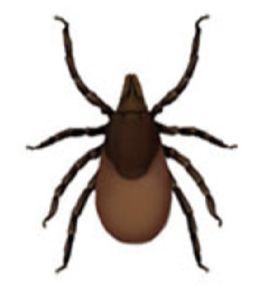
Antibiotics 2019, 8, 80                                                                             4 of 24

activate macrophages while B cells secrete highly specific antibodies that bind bacterial surface
proteins. In most murine species and natural reservoir hosts, host immune response eliminates
obvious objective symptoms. However, this response is insufficient to clear the infection, which then
becomes persistent [27]. In humans, there is evidence for asymptomatic infection [28]. It is not known
how often Borrelia infections are cleared in humans and this data is unlikely to become available in
the future as it would be unethical not to treat a human with definite evidence of borreliosis.

1.2.2. Borrelia Antibiotic Evasion and Tolerance Mechanisms
     Borrelia has mechanisms to both avoid and tolerate antibiotics. Borrelia biofilm likely restricts
antibiotic penetration, blocking antibiotics from reaching bacteria residing in the extracellular matrix
(ECM) through the binding of antimicrobials to ECM components as well as the inactivation of
antimicrobials by enzymes present in the ECM. Intracellular localization represents another strategy
to avoid contact with some antibiotics. When Borrelia does directly encounter antibiotics, it possesses
a non-specific RND type efflux pump that can eject a wide array of antibiotics [29]. If the antibiotic
attack is too strong, Borrelia’s “stringent response” to environmental stressors is triggered through a
RelA/SpoT homolog and the bacteria shift to a highly drug-tolerant persister form. Borrelia also forms
persister cells stochastically, which ensures that some individuals will survive sudden chemical
attacks [11,14].

1.2.3. Borrelia Immune Evasion Mechanisms
     To establish and maintain persistent infection in an immunocompetent vertebrate host, Borrelia
possesses a spectrum of mechanisms which it has evolved in order to evade or otherwise overcome
the innate and adaptive immune responses (Figure 2).

      Figure 2. Borrelial Counteractions to Immune Response During Cutaneous Invasion—Borrelia evasion
      mechanisms counter both innate and adaptive immune responses in order to establish persistent
      infection in the immunocompetent vertebrate host.

     Upon entering the vertebrate host, Borrelia spirochetes encounter lysozyme and antimicrobial
peptides such as defensin, cathelicidin and cecropins. Borrelia has only limited susceptibility to
lysozyme and is highly resistant to cathelicidin, although the precise mechanism of this resistance is
unknown [27,30].
Antibiotics 2019, 8, 80 5 of 24
Borrelia’s interference with cytokines and chemokines includes two especially interesting
mechanisms. Upon entering the vertebrate host, Borrelia is assisted in surviving by the tick vector, as
tick saliva contains a chemokine-inhibitory evasin protein that serves to reduce the migration of
immune cells to the site of infection [31]. When Borrelia does encounter macrophages, it stimulates
them to produce the anti-inflammatory interleukin, IL-10, which reduces phagocytosis and the
expression of MHC II and co-stimulatory receptors in antigen-presenting cells [32].
When professional phagocytes, such as fast-responding neutrophils, arrive at the site of
infection, they generate a “respiratory burst” of reactive oxygen species (ROS) including superoxide,
peroxide and hydroxyl radicals as well as reactive nitrogen species (RNS) including nitric oxide and
peroxynitrite. These chemicals are directly microbicidal but are also known to mobilize other
antimicrobials [33,34]. Borrelia utilizes a manganese superoxide dismutase (MnSOD) to scavenge
superoxide. Different types of SOD enzymes exist, each with unique requirements for metal cofactors
such as manganese, copper, zinc and iron. While some MnSOD enzymes are able to function with
either iron or manganese as a cofactor, Borrelia MnSOD specifically requires manganese, and is in fact
not functional when bound to iron [35,36]. The use of manganese makes Borrelia independent of iron,
which is tightly controlled during infection to prevent microbes from utilizing the resource [37].
However, the exclusive nature of this usage makes Borrelia dependent on manganese uptake, which
it achieves through Borrelia metal transporter A (BmtA) [38]. A notable experiment blocked the BmtA
transporter with microbicidal effect, demonstrating that a functioning BmtA transporter is required
for survival of the bacteria [39].
To physically evade phagocytes, other immune cells, immune chemicals and antibiotics, Borrelia
species generate biofilms. The construction of biofilms by Borrelia has been demonstrated both in vitro
and in vivo in human Borrelial lymphocytomas [12,40]. Another physical strategy used by Borrelia to
evade immune response is intracellular localization within human endothelial cells. The mechanism
for this invasion is partly understood and requires host integrins and Src kinase activity [41].
To combat the emerging adaptive immune response, Borrelia invades lymph nodes where it
skews the adaptive response to a B cell response that is T cell-independent. Borrelia also interferes
with class switching from IgM to IgG and causes germinal center (GC) “collapse” by altering lymph
node structures and inhibiting the development of memory B cells and plasma cells [42,43]. To evade
the effective binding of antibodies to its cell surface, Borrelia employs an elaborate antigenic variation
mechanism. The variable major protein-like sequence expression site (vlsE) on lp28-1 (GenBank
accession AE000794.2), is recombined through an unknown mechanism involving a Holliday junction
helicase, RuvAB, to produce a variable surface protein, VlsE1 (Figure 3). This mechanism is known
to be required for long-term survival, is up-regulated soon after infection, and likely produces
substantial diversity of surface epitopes by the time an adaptive response is mounted [44,45].
Figure 3. vlsE and Silent Cassettes on Linear Plasmid 28-1 of B. burgdorferi—Borrelia evasion
mechanisms counter both innate and adaptive immune responses in order to establish persistent
infection in the immunocompetent vertebrate host. Random segments of silent cassettes 2–16 are
recombined at the vlsE expression site near the end of lp28-1 to produce surface proteins with a high
degree of antigenic variation.
In addition to the immune evasion mechanisms listed above, Borrelia has evolved multiple
mechanisms to interfere with the complement system so as to avoid opsonization and subsequent
phagocytosis or complement-mediated lysis, including mechanisms that capture complement
regulatory proteins as well as mechanisms that directly interfere with molecules involved in theAntibiotics 2019, 8, 80 6 of 24
complement cascade [4]. These mechanisms and their significance are the subject of this paper and
are discussed in detail below.
2. Results
2.1. Research Overview
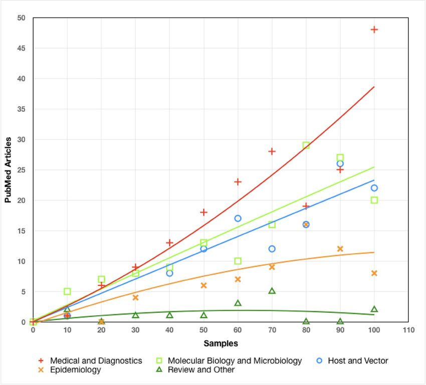
2.1.1. Quantitative Analysis of Borrelia Research
Research on Borrelia, as measured by articles published on PubMed, has trended up since its
discovery as the causative agent of LD in 1982, with a brief decline in the early 2000s (Figure 4a). The
maturation of initial, basic LD research is a likely reason for the decrease in interest. The considerable
increase in research interest in recent years is most likely due to the growing impact of LD as a result
of the dramatic, climate change-fueled expansion discussed above.
(a) (b)
Figure 4. Analysis of Borrelia Research: (a) Borrelia PubMed Article Count by Year—Number of
PubMed articles found for the query “Borrelia” plotted by year of publication. Curve fitting was
achieved by the application of a third order polynomial trendline in the Mac Numbers OS/X
application; (b) Borrelia Research Emphasis—Rarefaction curves indicating the richness of diversity
for broad categories of Borrelia research.
The emphasis of PubMed-accessible research has been primarily on medical and diagnostic
research, with a strong secondary emphasis on molecular and microbiological research, as well as
host and vector studies (Figure 4b). Epidemiological studies comprise a somewhat smaller portion of
the body of research, and review articles make only a minor contribution to the corpus. As with
Borrelia research as a whole, the heavy emphasis on medical and diagnostic research is likely a
reflection of the increasing prevalence and impact of LD.
2.1.2. Borrelia Genomics
The complete genome of Borrelia burgdorferi has been determined and its 911 kbp chromosomal
genome is available on GenBank (accession NC_001318.1). B. burgdorferi (s.s. strain B31-GB)
additionally possesses 12 linear and 10 circular plasmids, placing its genome among the most
complex bacterial genomes known [46]. Complete knowledge of the Borrelia genome, as well as
available proteomic (Uniprot) data and other “omics” data, can be expected to drive new research.Antibiotics 2019, 8, 80 7 of 24
2.1.3. Analysis of Borrelia Immune Evasion and Persistence Research
The pace of discovery of immune evasion and persistence mechanisms of Borrelia has been
relatively constant since 1991, indicating that this area of inquiry is still expanding and further novel
discoveries can be expected in the future (Figure 5).
Figure 5. Borrelia Immune Evasion and Persistence Research Timeline—A selection of novel immune
evasion and persistence discoveries since 1982.
2.1.4. Relative Importance of Evasion and Persistence Mechanisms
The immune evasion and persistence mechanisms shown in Figure 5 are likely all of some
importance to the establishment of persistent infection in an immunocompetent host. However, three
mechanisms in particular stand out. (1) In vivo biofilms are very likely to be of major importance as
they provide a safe, low-oxygen habitat for optimal spirochetal development, allowing nutrients in,
while restricting access to phagocytes and most likely to at least some antibiotics. (2) Drug-tolerant
persisters are also likely to be of major importance in LD treatment as all antibiotics tested and most
antibiotic combinations are not capable of eradicating persister populations [11]. (3) Complement
evasion mechanisms have a unique and important role because complement activation via the
alternative pathway (AP) and lectin pathway (LP) begin at the moment of infection and continue
through activation of the classical pathway (CP) and beyond (see background on the complement
system below). Since complement is a threat to Borrelia throughout the infectious process, restoration
of proper complement function represents a treatment opportunity applicable to all stages of
infection, possibly including PTLDS. This review will describe complement function, the current
status of Borrelia complement evasion research and possible avenues for the development of novel
complement-related therapies.
2.2. The Complement System
2.2.1. Opsonization
The human innate immune system includes an enzyme cascade, known as the complement
system, which serves to modulate inflammation, opsonize the cell membranes of pathogens for easier
phagocytosis and establish membrane attack complexes (MAC) that lyse pathogens (Supplementary
Materials, Table S4). Three separate pathways, the alternative pathway (AP), the lectin pathway (LP)
and the classical pathway (CP), converge on the opsonization of the pathogen’s cell membrane with
C3b (Figure 6). Opsonization occurs when C3 is cleaved to form C3b, exposing a highly reactive
thioester group which forms covalent bonds with hydroxyl and amine groups on cell surfaces.Antibiotics 2019, 8, 80 8 of 24
Alternative Pathway Lectin Pathway Classical Pathway
1 Antigen
MASP-2 C1r C1s
H2 O 2
1 C1 Complex C1q Antibody
MBL
1 C3
Mannose, NAG, C1r C1s
MASP-2 D-Glucose or
L-Fucose 3
C3(H2O)
Factor D 2
C4
2
3 Active
Anaphylatoxin C4a C1s
Factor B C4
3
C2 4
Inflammatory
C2b C4b
C3 Mediator 4 C3 Convertase 5
Ba C2a C4a C4b C4b
4 C3(H2O)Bb C3 Convertase C3 5
6 C2
C2
Anaphylatoxin C4b
Active
C3b Anaphylatoxin C3a C3b Opsonin C1s
Anaphylatoxin C3a 7 C2
5
Inflammatory
C2b
C3b Opsonin Mediator
C4b
C3 C3 Convertase
C2a
8
Anaphylatoxin C3a C3b Opsonin
Microbial Microbial Microbial
Membrane Membrane Membrane
1) C3 spontaneously hydrolyzes (1) Mannose Binding Lectin (MBL) or ficolin binds oligosaccharide (1) Antibody binds antigen on microbial membrane. Alternatively, C1q
2) C3(H2O) binds Factor B (FB) chain on microbial membrane, activating associated MASP-2 can directly recognize and bind patterns on microbial membranes.
3) Factor D cleaves Ba from FB forming C3(H2O)Bb C3 convertase (2) Activated MASP-2 cleaves C4a anaphylatoxin from C4 and C4b (2) C1 complex binds antibody Fc region
4) C3(H2O)Bb cleaves C3 into C3a anaphylatoxin and C3b opsonin remainder binds microbial membrane (3) C1r activates C1s
5) C3b (covalently) opsonizes microbial membrane (3) MASP-2 cleaves C2, releasing inflammatory mediator C2b (4) C1s cleaves C4 into C4a and C4b
(4) Fragment C2a binds C4b on membrane to form C4b2a C3 convertase (5) C4b binds microbial membrane
(5) C4b2a cleaves C3 into C3a anaphylatoxin and C3b opsonin, which (6) C2 binds C4b
(covalently) opsonizes microbial membrane (7) C1s cleaves C2b from C2, leaving C4b2a C3 convertase
(8) C4b2a cleaves C3 into C3a anaphylatoxin and C3b opsonin, which
(covalently) opsonizes microbial membrane
Figure 6. Microbial Opsonization via the Alternative, Lectin and Classical Pathways—To establish
infection in a vertebrate host, Borrelia must first overcome the constitutive alternative and lectin
pathways, and later the classical pathway. The initial stages of each complement pathway leading to
(covalent) opsonization by C3b (shown here) are unique, but then converge into a single common
pathway to form a membrane attack complex (Figure 7) [37].
Once C3b is established on the microbial cell membrane, the soluble factor B (FB) fragment Bb
can bind to C3b, forming the AP C3 convertase, which can cleave C3 to create still more C3b. Through
this amplification process, the target cell is extensively opsonized. Since opsonins are normally
cleared from host cell membranes by complement regulatory factors, the presence of opsonins on cell
surfaces enables the recognition of opsonized cells as non-self [37].
2.2.2. Membrane Attack Complex Formation
MAC formation via the terminal pathway (TP) begins when the C3 convertase of any pathway
is bound by C3b to form a C5 convertase. Soluble C5 is cleaved to form C5b, which is joined by C6
and C7 to form a C5bC6C7 complex with an exposed hydrophobic region at the C7 end. The
hydrophobic end embeds in the microbial membrane, and when C8 binds to this complex, a
polymerization reaction is triggered, binding multiple C9 proteins to form a porin channel through
the membrane. This channel is hydrophilic on the inside and cellular lysis begins as water exits
through the porin (Figure 7) [37].Antibiotics 2019, 8, 80 9 of 24
Alternative Pathway Lectin Pathway Classical Pathway
C3b C3 Opsonin C3b C3 Opsonin
C3
C3 C4b
C3 Convertase
C2a
C3b C3 Convertase C3b 1
C3b
1 Bb
1 C3b
C5
C5
2
2 C4b
C3b
C2a C5
C5 Convertase
Convertase
Bb C5 Convertase
C3b
C3b C5a
C5a
3
3 C5b
C5b C6
C6
5 5
C5b C6 C7
C5b C6 C7
C7
C7
4
4
C5b C6 C7
C5b C6 C7
C8 6
C8 6
C5b C6 C7
C5b C6 C7
C8
C8
7 C9
7 C9 C9 C9
C9 C9
C9
C9 C9
C9 C9 C9
C9 C9 C9
C9
Microbial Microbial
Membrane Membrane
(1) C3b binds to C3Bb C3 convertase, forming C3bBbC3b C5 convertase (1) C3b binds to C4b2a C3 convertase, forming C4b2aC3b C5 convertase
(2) C5 is cleaved into C5a and C5b by C3BbC3b C5 convertase (2) C5 is cleaved into C5a and C5b by C4b2aC3b C5 convertase
(3) C5b binds C6 (3) C5b binds C6
(4) C5bC6 binds C7 (4) C5bC6 binds C7
(5) C5bC6C7 inserts into microbial membrane (5) C5bC6C7 inserts into microbial membrane
(6) C8 binds to C5bC6C7 and initiates C9 polymerization (6) C8 binds to C5bC6C7 and initiates C9 polymerization
(7) C9 polymerization forms pore which begins cell lysis (7) C9 polymerization forms pore which begins cell lysis
Figure 7. Membrane Attack Complex Formation via the Alternative, Lectin and Classical
Complement Pathways—The formation of membrane attack complexes (MACs) is initiated when a
membrane-bound C5 convertase cleaves soluble C5 into C5a and C5b. C6 and C7 bind C5b and the
resulting C5bC6C7 complex, which is hydrophobic at the C7 end, embeds in the membrane. The MAC
polymerizes when C8 binds C7, which induces the addition of several C9 molecules, forming a pore
with a hydrophilic center that begins to lyse the cell. C5 convertases of the alternative pathway are
formed by an additional C3b molecule joining a membrane-bound C3bBb C3 convertase, forming
C3bBbC3b. In the lectin and classical pathways, the C3 convertase C4b2a is joined by C3b to form
C4b2aC3b, which serves as the C5 convertase [37].
2.2.3. Complement Regulation
Because opsonization of cell membranes with C3b happens indiscriminately and it can lead to
phagocytosis or lysis, it is necessary for host cells to regulate this activity by actively removing C3b
and C4b opsonins. C3b is removed when Factor H (FH) is acquired on the cell surface by binding
sialic acid (SA) or glycosaminoglycan (GAG) proteoglycans and cooperates with Factor I (FI) and
membrane cofactor of proteolysis (MCP) to cleave C3b bound by C3b/C4b complement receptor 1
(CR1). In a similar fashion, C4 binding protein (C4BP) is acquired and works with FI and MCP to
remove C4b bound to CR1 (Figure 8).Antibiotics 2019, 8, 80 10 of 24
Figure 8. Host Cell Regulation of C3b and C4b Opsonization—Mammalian host cells must be able to
degrade C3b (an opsonin) to prevent phagocytosis and C4b (a C5 convertase cofactor) to prevent
membrane attack complex (MAC) formation [37]. Borrelia burgdorferi has been able to co-opt this
regulatory system, avoiding phagocytosis by binding Factor H (FH) with complement regulator
acquiring surface proteins (CRASPs) expressed on the bacterial surface.
Host cells are also able to remove established C3 convertases. FH binds SA or GAG
proteoglycans on the cell surface and cleaves Bb from AP C3bBb C3 convertases. In a similar way,
C4BP binds proteoglycans on the cell surface and cleaves C2a from CP and LP C4b2a C3 convertases
(Figure 9). The remaining C3b and C4b opsonins can then be degraded by FI, as described above and
as shown in Figure 8.
Figure 9. Host Cell C3 Convertase Regulation—Mammalian host cells must be able to degrade the C3
convertases of all three complement pathways to prevent host cell opsonization from potentially
leading to phagocytosis [37].Antibiotics 2019, 8, 80 11 of 24
2.3. Complement Evasion by Borrelia
Borrelia species infect ticks by entering in the blood meal during feeding. Complement activation
relies on serum proteins and continues to act within the tick vector. Borrelia species vary in their
ability to survive complement attack, termed “serum resistance”. B. afzelii and B. burgdorferi strains
are strongly serum resistant while B. garinii strains are fairly sensitive to serum. However, even in
the most sensitive strains, some spirochetes do survive [47]. After feeding, Borrelia leaves the midgut
and migrates to the salivary glands where it can be transmitted to a new host during the next feeding
[48].
The array of surface proteins utilized by Borrelia spp. to evade vertebrate complement in the tick
vector during blood meals differs from the set of proteins employed during vertebrate infection, and
so the expression of these surface proteins changes accordingly during the transition from vector to
host [49]. The reason for expression of different complement evasion proteins with similar function
in different environments is unknown and puzzling, and deserves further research to determine
whether these proteins have physiological optima under the conditions present inside the tick vector
and in vertebrates.
The Borrelia surface proteins involved in complement evasion fall broadly into two categories,
each of which is explored below. The first category includes those proteins which directly interfere
with components of the complement cascade. These proteins inhibit the AP, LP or CP by inactivating
key pathway components. The second category of complement evasion proteins includes proteins
which subvert normal complement regulation mechanisms by acquiring host regulator proteins.
2.4. Category I: Direct Complement Interference
2.4.1. Classical Pathway Inhibition by C1r Binding: BBK32
The B. burgdorferi surface protein BBK32 (Table 1) has been known for some time to serve as an
adhesin to host cells by binding fibronectin found in the extracellular matrix (ECM) of vertebrates
[50]. Substantial portions of BBK32, and the fibronectin binding region in particular, are intrinsically
disordered, lacking a rigid 3D structure. More recently, BBK32 was found by Garcia et al. (2016) to
inhibit the CP by binding the C1 subunit C1r with high affinity (Kd = 3.9 nM), inactivating the serine
protease function of C1r and halting the CP cascade. The source of this binding was traced to the C-
terminal domain and the effectiveness of BBK32 at inhibiting the CP was demonstrated. An amount
of 1 μM of BBK32 provided nearly perfect protection against serum complement hemolysis to
sensitized sheep red blood cells and inhibition of the CP was found to be dose-dependent, with a
calculated IC50 value of 110 nM. The binding of C1 by spirochetes was found in this study to be
improved by BBK32, which was further found to confer serum resistance when added to a serum
sensitive strain [51].
Table 1. Proteins which interfere directly with complement. GenBank accession numbers are for B.
burgdorferi strain B31 except for BBA70, which is for B. burgdorferi strain 163b.
Protein Gene Plasmid GenBank Accession Species
BBK32 bbk32 lp36 AE000788.1 B. burgdorferi, B. afzelii, B. garinii, others
OspA ospA lp54 AE000790.2 B. burgdorferi, B. afzelii, B. garinii, others
OspC ospC cp26 AE000792.1 B. burgdorferi, B. afzelii, B. garinii, others
BBA70 bba70 lp54 AY696552.1 B. burgdorferi 163b, B. bavariensis
Since BBK32 inhibits the CP and the CP has been shown to kill B. burgdorferi independent of
antibody binding, it is possible that BBK32 is sufficiently necessary for infectivity that it could be
developed into a promising drug target [52]. This notion is supported by a knockout study that found
BBK32 mutants to have significantly reduced infectivity, but it is also somewhat tempered by
previous studies which have shown other complement components to have a relatively limited role
in controlling infection in mice.Antibiotics 2019, 8, 80 12 of 24
Garcia et al. (2016) note, however, that the direct microbicidal effects of complement may not be
the only issue at stake, suggesting that BBK32-driven complement inhibition caused by spirochetes
in infected lymph nodes might be responsible for an observed reduction in C4 deposited on follicular
dendritic cells (FDC) in lymph node germinal centers (GC), causing low levels of FDC antigen
presentation leading in turn to observed but presently unexplained GC “collapse” [51,53].
The phenomenon of GC collapse involves the development of short-lived, abnormal GCs that
fail to create sufficient numbers of memory B cells and plasma cells. The resulting
immunosuppressive effect is observed for months after infection and is not specific to Borrelia. GC
collapse is likely crucial to Borrelial persistence, but it may have even greater importance in complex
LD cases where co-infections are present or in PTLDS [43].
2.4.2. C3b Degradation by Binding Plasminogen
Activation of the zymogen precursor plasminogen forms the blood protein plasmin, which
dissolves fibrin clots. It is also known that plasmin inhibits complement by binding and cleaving C3b
and C5 [54]. The ability to dissolve fibrin and to regulate complement deposition makes plasminogen
an especially attractive molecule for pathogens to acquire toward interference.
Outer surface protein A (OspA), a prior vaccine target, has been known for quite some time to
bind plasminogen [55]. Unfortunately, expression of OspA is down-regulated upon passage of
spirochetes into the vertebrate host and so OspA would not make a good drug target if the goal is to
improve treatment of patients with active early or late infections.
Outer surface protein C (OspC), is up-regulated in Borrelia during vertebrate infection and is
known to bind plasminogen [56]. Anti-OspC antibodies were able to significantly reduce
plasminogen acquisition and wild type B. burgdorferi were found to bind plasminogen only if
expressing OspC, regardless of other surface protein expression. This finding appears to contradict
other studies enumerated in this section that indicate plasminogen binding also by other outer
surface proteins and follow-up studies seem required to elucidate the matter.
The B. burgdorferi protein BBA70 was found to bind plasminogen with high affinity and inhibits
the microbicidal effects of complement by cleaving C3b and C5. BBA70 was not able to bind
complement regulators and follow-up in vitro studies as well as in vivo studies need to be performed
[57]. For these reasons, BBA70 is not an attractive therapeutic target to improve complement function.
Borrelia species have evolved complement-regulator acquiring surface proteins (CRASPs) which
can bind complement regulators, in particular FH and FH-like proteins. CRASP genes have similar
function but two different nomenclatures for historical reasons. The csp family of CRASP genes
encode “conserved signature proteins” while the erp family of CRASP genes encode “OspE-related
proteins” (Table 2). In B. burgdorferi, B. afzelii, and B. spielmanii, CspA is able to bind plasminogen [58].
The genes cspZ, erpA, erpC, and erpP are only known to exist in B. burgdorferi and their expressed
proteins also bind plasminogen [4,59].
Table 2. Complement-regulator acquiring surface proteins (CRASPs) and related FH-binding proteins, gene names,
plasmid replicons, base pair intervals and the species in which they occur. The acronym csp stands for “conserved
signature protein” and erp stands for “OspE-related protein” [4]. GenBank accession numbers are for B. burgdorferi
strain B31 except for erpC, which is for strain B31_NRZ.
CRASP Gene Plasmid 1 Interval GenBank Accession Species
B. burgdorferi, B. afzelii, B.
CRASP-1 cspA lp54 46473-47228 2 AE000790.2
spielmanii
CRASP-2 cspZ lp28-3 2260-2970 2 AE000784.1 B. burgdorferi, B. afzelii
CRASP-3 erpP cp32-9 26210-26770 AE001581.1 B. burgdorferi
CRASP-4 erpC cp32-2 26834-27373 NZ_CP019757.1 B. burgdorferi
CRASP-5 erpA cp32-1 26235-26768 AE001575.1 B. burgdorferi
- BG0407 - 417734-418345 AAU07257.1 B. garinii, B. bavariensis
BafPKo_0408 ABH01676.1
- - 419301-419906 B. afzelii
(BAPKO_0422) (CP000395.1)
1 lp = linear plasmid, cp = circular plasmid. 2 Reverse strand.Antibiotics 2019, 8, 80 13 of 24
Serum resistance (insensitivity to complement-mediated lysis) is conferred by both CspA and
CspZ. CspA interacts with plasminogen and FH/FHL-1, but also with several additional complement
components: C7, C8, C9 and MAC [60,61]. The crystal structure of CspA has been determined and
the FH binding site has been characterized to a degree [62,63]. Of the known complement-interacting
mechanisms, CspA appears to be one of the best understood, although the Önder et al. (2012) result
appears to minimize the role of CspA in binding fibrinogen. Unfortunately, this well-developed
target is not a good candidate for antibiotic drug development because it is up-regulated in the tick
environment (to confer serum resistance during the blood meal), but becomes strongly down-
regulated during entry of the vertebrate host [64].
The OspE-related (Erp) CRASP proteins ErpP, ErpC and ErpA have highly similar amino acid
sequences (Table 3), while CspA and CspZ are not similar to each other, and neither CspA nor CspZ
is similar to ErpP. These differences indicate that the Erp family of proteins share an evolutionary
history, while CspA, CspZ and the Erp family of proteins evolved separately. When aligned with
Clustal Omega (version 1.2.4), the Erp protein amino acid sequences show similarity, including a
common OspE Pfam domain (Figure 10).
CLUSTAL O(1.2.4) multiple sequence alignment
Signal sequence
ErpA MEKFMNKKMKMFIICAVFILIGACKIHTSYDEQ-------------SNGEVKVKKIEFSE 47
ErpC ----MNKKMKMFIICFIFALISSCKNHT-------------LYDGQSNGEAKVKKIEFSE 43
ErpP ----MNKKMKMFIVCAVFILIGACKIHTSYDEQSSGEINHTLYDEQSNGELKLKKIEFSK 56
*********:* :* **.:** ** ** **** *:******:
ErpA FTVKIKNKNNSNNWADLGDLVVRKEKDGIETGLNAG--------GHSATFFSLEEEEINN 99
ErpC FTVKIKNKNNSNNWADLGDLVVRKEEDGIETGLNVGKGDSDTFAGYTATFFSLEESEVNN 103
ErpP FTVKIKNKDNNSNWTDLGDLVVRKEENGIDTGLNAG--------GHSATFFSLKESEVNN 108
********:*..**:**********::**:****.* *::******:*.*:**
ErpA FIKAMTEGGSFKTSLYYGYNDEESDKNVIKNKEIKTKIEKINDTEYITFLGDKINNSAGG 159
ErpC FIKAMTEGGSFKTSLYYGYKDEQSNANGIQNKEIITKIEKIDDFEYITFLGDKIKD--SG 161
ErpP FIKAMTKGGSFKTSLYYGYKYEQSSANGIQNKEIITKIESINGAEHIAFLGDKINNGVGG 168
******:************: *:*. * *:**** ****.*:. *:*:******:: .*
ErpA DKIAEYAISLEELKRNLK 177
ErpC DKVVEYAILLEDLKKNLK 179
OspE-like Pfam domain
ErpP DKTAEYAIPLEVLKKNLK 186
SMART Accession PF02471
** .**** ** **:***
Interpro Abstract IPR003483
Figure 10. OspE-related (Erp) CRASP Protein Alignment—Clustal Omega (version 1.2.4) alignment
for OspE-related (Erp) CRASP proteins shown in Table 2. The region highlighted in green is a shared
OspE Pfam domain (SMART accession PF02471, Interpro abstract IUPR003483).
Table 3. Clustal Omega (version 1.2.4) alignment identity, similarity and gaps for Erp amino acid
sequences.
Proteins Identity Similarity Gaps
ErpA vs ErpC 77% 85% 5%
ErpA vs ErpP 75% 83% 6%
ErpC vs ErpP 70% 78% 11%
2.4.3. MAC Interference
When C3b opsonizes Borrelia and direct degradation of C3b opsonins by Borrelia-acquired
plasminogen and complement regulator proteins fails to remove C3b, Borrelia species must prevent
the C5 convertase pathway from establishing lytic MAC complex pores on its surface (Figure 7).
Borrelia species possess CspA, which is described below (Category II) as interfering with
complement regulation. Hallström et al. (2013) discovered that CspA in B. burgdorferi has a second
function: interfering with MAC formation. CspA binds C7 and C9, inhibiting C9 polymerization and
blocking MAC assembly. Transfer of cspA by genetic modification to the serum-sensitive species B.Antibiotics 2019, 8, 80 14 of 24
garinii, caused it to gain serum resistance, indicating that surface expression of the protein is sufficient
for MAC inhibition. This discovery was the first of its kind in Gram-negative bacteria [65].
Unfortunately, as noted above, cspA expression is down-regulated during entry of Borrelia into the
vertebrate host such that it does not represent a worthwhile drug target for patients with active
infection.
B. bavariensis surface proteins BGA66 and BGA71, which have a moderate sequence similarity to
CspA, have since been found to inhibit MAC assembly as well and they further inhibit the AP
(BGA66), TP (both) and CP (both). Again, transformation of B. garinii with these proteins introduced
serum resistance [66].
In addition to CspA-mediated MAC inhibition, B. burgdorferi also expresses a (human) CD59-
like protein on its surface. Blocking this protein with anti-CD59 antibody components removed
serum resistance from a normally serum-resistant strain [67]. This protein is not likely to represent a
realistic therapeutic target due to its similarity to human CD59 in binding antigen, and it appears that
further investigation has not occurred.
2.5. Category II: Complement Regulation Interference
2.5.1. Factor H and FHL-1 Binding
In B. burgdorferi, B. afzelii, and B. spielmanii, surface-expressed CspA binds host complement
regulators FH and FHL-1 on the bacterial surface, where these proteins remove C3b opsonins,
preventing lysis or phagocytosis. In B. burgdorferi, CspZ has a similar function (Table 2). The
importance of FH acquisition for bacterial survival has been demonstrated by the induction of high
serum sensitivity via knock-out of cspA [64]. Unfortunately, while CspA conveys serum resistance in
vitro, the mechanism is ultimately not critical for infection in vivo. Woodman et al. (2007)
demonstrated this in an elegant experiment where they infected both wild-type (WT) and FH-
deficient mice with B. burgdorferi. The experiment found no detectable level of FH acquired on
Borrelial surfaces in the FH-deficient mice and yet found no quantitative difference in level of
infection between the two types of mice [68]. These two apparently contradictory findings need a
clear reconciliation through further research.
The OspE-related proteins (Erp) ErpA, ErpC and ErpP collectively bind FH, and augment the
serum resistance of OspA mutants modified to overexpress OspE proteins. This modification was
further shown to translate into a reduction in complement deposition and MAC formation [69]. The
crystal structure of OspE was recently determined, including the binding site for FH. Since OspE is
highly similar to ErpA, the structure and binding site of ErpA should be very similar. In addition,
other experiments have determined the crystal structures of ErpC and ErpP [70]. Unlike CspA and
CspZ, bacterial expression of OspE and related proteins (Erps) are up-regulated in the vertebrate
host.
In addition to lipoproteins, some OmpA-like (a porin-like integral membrane protein found in
E. coli) outer membrane proteins have been found to acquire FH. A screen of B. garinii whole-cell
sonicate against human sera identified an FH-binding homologue of BG0407, an unknown putative
protein sequence identified in B. bavariensis (Table 2) [47]. Follow-up work identified another FH-
binding homologue in B. afzelii, BafPKo_0408 (also known as BAPKO_0422), and provided evidence
that it forms an 8-stranded β-barrel similar to the membrane-spanning domain of OmpA (Table 2)
[71]. A homologue in B. burgdorferi, BB0405, was shown to be surface-exposed, but while required for
infection in mice, did not bind human FH [72]. The difference in FH binding between BB0405 and the
other homologues, as well as the reason that BB0405 is required for murine infection in spite of not
binding FH are both puzzles that seem to require further investigation and clarification. A similar
protein, BB0406, produced by a co-transcribed, paralogous gene, was found along with BB0405 to be
immunogenic in non-human primates and antibodies against the two proteins were found to be
borreliacidal, although BB0406 was not required for infection [73].
2.5.2. C4b Inactivation by Binding C4BPAntibiotics 2019, 8, 80 15 of 24
The CP was shown to be inhibited (B. burgdorferi, B. garinii) by the binding of C4BP by a protein
known as p43. When joined by FI, the captured C4BP protein was able to inactivate C4b, contributing
to serum resistance [74]. Unfortunately, this result was contradicted by other experiments that did
not observe C4BP binding, so it remains controversial [4].
3. Discussion
3.1. Lyme Disease Impact and Medical Response
LD is a widespread and rapidly expanding, emerging infectious disease with significant long-
term health consequences in the 10–20% of patients who develop post-treatment Lyme disease
syndrome (PTLDS). Additionally, 63% of all LD patients develop one or more PTLDS-associated
diagnoses [20,21]. The absence of tissue damage caused by known bacterial toxins or proteases may
seem to indicate to some that the immune-mediated damage caused by LD is not significant. On the
contrary, patients with PTLDS were more likely to report fair or poor health status than patients with
MS, lupus, diabetes, PTSD, stroke or congestive heart failure, and further reported significantly
higher fatigue, pain, sleep disturbance, and depression than controls [23,24].
The medical cost of LD treatment in the US is estimated to be between $712M and $1.3B per
annum [20]. Social costs are also high as LD causes significant disability. PTLDS patients between the
ages of 25 and 54 have a 45.9% employment rate, versus 81.0% in the general population, a 43% lower
rate. An additional 25% of LD patients have reduced work hours or changed occupations as a result
of PTLDS, while 24% receive disability [24].
In spite of the clear and urgent need for improved understanding leading to the development of
treatments for PTLDS, a viable strategic plan is lacking and National Institutes of Health (NIH)
funding for LD greatly trails other diseases on a per-case basis (Table 4). NIH funding for LD is just
1.8% to 3.2% of estimated direct annual medical costs. If allocated the same level of funding on a per-
case basis that was allocated to West Nile virus research in 2018 (which affected 2,544 individuals in
2018 or 0.8% of the estimated number of LD cases in the same year), the funding for LD would be
$5,306,700,000. It would make sense to prioritize LD research, particularly as it relates to PTLDS, and
to greatly increase funding for this costly and unmet medical need.
Table 4. In spite of a high annual cost for treatment and significant social costs, NIH funding for LD
significantly trails that appropriated for other diseases.
Disease NIH Funding (Millions, 2018) US Cases per Year Funding per Case
Malaria $202 1700 1 $118,824
HIV/AIDS $3000 38,739 2 $77,441
West Nile virus $45 2544 3 $17,689
PTLDS $23 45,000 4 $511.11
LD $23 300,000 5 $76.67
1CDC estimate (2018), 2 CDC (2017), 3 CDC preliminary (2018), 4 15% of CDC estimate (2015), 5 CDC
estimate, (2015).
3.2. PTLDS or PLD?
The cause of PTLDS is not known and is the subject of ongoing controversy. Hypotheses
regarding the mechanism(s) responsible for PTLDS have included autoimmunity [75,76], the post-
infectious persistence of spirochetal antigens [77] and permanent tissue damage. There is a growing
body of evidence that at least some cases (12 of 12 cases in [16]) involve persistent infection, with an
emerging understanding of the underlying mechanisms that may be responsible for antibiotic
treatment failure in LD, including immune evasion, intracellular localization, highly antibiotic-
tolerant persisters and biofilm formation in vivo [4,11,12,16,43].
The morphological state of inocula (MSI) applied to mice has very recently been shown by Feng
et al. (2019) to determine both disease severity and antibiotic treatment response. Three morphotypes
were found, each with unique properties, including sensitivity to different antibiotics. PlanktonicAntibiotics 2019, 8, 80 16 of 24
spirochetes caused the least severe pathology and were easily eradicated by standard antibiotic
treatment. Round-body antibiotic-tolerant persisters caused more severe illness and were more
difficult to treat. Biofilm aggregates caused the most severe disease and were fully resistant to the
currently preferred treatment recommendation of 21 days of doxycycline. The combination of
daptomycin, ceftriaxone and doxycycline antibiotics was found to eliminate the infection caused by
biofilm morphotype inocula in mice, as determined by ear punch culture [13]. However, this result
is unlikely to be definitive as it has been contradicted by in vitro studies that did not observe effective
bacterial killing [78,79]. In addition, follow-up testing at 12 months was not performed. Such follow-
up would be required to determine clearance of infection, as Borrelia infection was documented to
rebound after this interval by Hodzic, et al. (2014). In the Hodzic, et al. study, mice treated with
ceftriaxone in the same manner as previous studies reproduced the results of those studies, yielding
non-cultivable tissues with low and falling copy numbers of flaB DNA by qPCR. However, at 12
months, while spirochetes were still non-cultivable, flaB copy numbers had rebounded in multiple
tissues to nearly match those in the saline-treated control mice. This is an especially important result
that deserves close attention, because it demonstrates that treatment-resistant Borrelia infection is not
inconsistent with past observations of non-cultivability [15]. Even more interesting is the
reproduction by this study of low antibody titers observed in past studies. This may be the signature
of Borrelia immunosuppression (potentially via the BBK32 mechanism proposed by Garcia, et al. and
discussed above) rather than infection clearance [51,53]. Further, the similarity in cytokine regulation
between the sham-treated mice and the antibiotic-treated mice in this study provides a reasonable
explanation for the persistence of some of the symptoms present in PTLDS.
It is unknown how these morphotypes express virulence factors as a mixed infective population.
This discovery has produced a model system (MSI) for investigating the morphotypic variation of
treatment efficacy for what has been designated as persistent Lyme disease (PLD) [13]. The MSI
model is applicable to other persistent, inocula-dependent infections such as Staphylococcus aureus
[80], which underscores the need to advance this body of research.
3.3. Opportunities for Intervention
The impact of PTLDS and the growing body of evidence for PLD as a major contributing factor
in its pathogenesis makes it imperative that the search for more effective PLD treatments begins now,
even if it is not yet known what proportion of patients might respond to such treatments and what
proportion might not respond, having other unknown mechanism(s) involved in their PTLDS
pathology. There is a wide spectrum of possible interventions against PLD, including antibiotic
combination therapies, targeted small-molecule drugs, anti-biofilm agents, phage therapy, natural
medicines and antimicrobial peptides. From this range of approaches, it would be desirable to find a
single treatment that is highly effective against Borrelia alone, avoiding collateral treatment damage
to the microbiome. Ideally, this agent would be fully effective from the moment of initial, acute
infection through the end of the course of the illness, no matter how late the diagnosis. Due to a mixed
model of infection with planktonic spirochetes, round-body persisters, biofilm aggregates, and
intracellular bacteria, it will likely be difficult to effect a cure with a single treatment agent. An
alternative to directly combating Borrelia in its many forms may be to assist the immune system in
clearing the infection. Such an approach will require deeper knowledge of the biology and
pathogenicity of Borrelia and of its immune system interactions in particular.
3.3.1. Immune System Intervention
The functioning human immune system has a great capacity to combat infection. In the case of
Borrelia, there are asymptomatic individuals with positive LD serology, which indicates that the
human immune system is fully capable of controlling, if not eradicating LD infections [28]. Since
Borrelia can only persist in the human body due to its repertoire of immune-evasive strategies, the
successful inhibition of a strategy required for bacterial persistence would enable the immune system
to control or eradicate Borrelia.Antibiotics 2019, 8, 80 17 of 24
In this light, Borrelia’s interference with the complement system is highly interesting from a
therapeutics perspective. Complement is expressed constitutively and is therefore in effect from the
moment of initial infection onwards. It is directly microbicidal via MAC-mediated lysis and
phagocytosis, but also indirectly microbicidal as complement components serve as signal molecules
that shape and direct the immune response against the pathogen.
Any bolstering of complement function is expected to improve immune response. Theoretically,
direct, parenteral supplementation of LD patients with complement components is a possible
intervention, but this likely would be expensive and impractical. A better approach would be to
inhibit Borrelia’s complement interference to indirectly recover a higher level of natural complement
function. Since interference with aspects of the complement system confers serum resistance to
Borrelia, blocking such interference could restore immune function enough to permit control or
clearance of PLD. Since a drug that targets complement interference by Borrelia would be highly
specific, this approach might be superior to broad-spectrum antibiotic use with its associated risks
and damage to the microbiome, and it would be less expensive than the cost of developing a vaccine
(US $200 to $500 million) and administering it indefinitely to a large population [81]. Regardless of
approach, continued research is critical to understanding the pathobiology of Borrelia, the cause(s) of
PTLDS and PLD and to develop a cure.
3.3.2. Desirable Criteria for Complement Evasion Intervention
The research reviewed in this paper represents a starting point for intervening in PLD by
inhibiting Borrelia complement evasion. There are many interesting areas where this body of research
could be extended, but the areas which might benefit patients with active early or late infection,
potentially including those with PLD (or who are in danger of developing it), relate primarily to
Borrelia complement resistance mechanisms that meet the following proposed criteria:
1. The mechanism must be constitutively expressed, or up-regulated in the vertebrate host at or
soon after initial infection;
2. The mechanism must be required for infectivity and long-term maintenance of infection;
3. The mechanism should be required by all infective morphological forms of Borrelia, including
antibiotic-tolerant persister cells and biofilm aggregates;
4. The structure of the molecules involved in the mechanism must be known, including ligand
binding sites;
5. The structure of the molecules involved should be unique so that small molecule inhibition is
likely to avoid off-target effects that would impact microbiota or the patient.
3.3.3. BBK32
The deposition of complement factor C4 (Figure 6) exemplifies the crucial dual role of
complement in controlling infection. C4b plays an important role as an opsonin, but it also stimulates
antigen uptake and presentation in follicular dendritic cells (FDCs) in lymph nodes [37,82]. It is
logical then, that the inhibition of the CP by the B. burgdorferi surface antigen BBK32, would lead to
the observed reduction in C4b deposition on FDCs, and that this would lead in turn to reduced FDC
antigen presentation and finally to observed GC “collapse”, where short-lived germinal centers in
lymph nodes produce inadequate populations of memory B cells and plasma cells [51,53].
The potential ability of BBK32 to suppress antibody response may be directly involved in the
establishment of PLD as a chronic infection, as well as in B. burgdorferi Western blot antibody test
insensitivity observed in LD patients for decades. Because this immunosuppressive effect continues
for months after infection and because it limits the ability to produce antibodies against co-infections,
the issue is critical for patients who may be infected with many other tick-borne microbes, including
Babesia, Anaplasma, Erlichia and Tularemia. Without a functioning CP, it is not likely that the immune
system can mount an effective response against these organisms [42,43]. Further, the effect of CP
inhibition would be synergistic as each co-infecting organism likely has its own spectrum of
mechanisms for evading and suppressing the immune system.You can also read