T CELL AND ANTIBODY KINETICS DELINEATE SARS-COV-2 PEPTIDES MEDIATING LONG-TERM IMMUNE RESPONSES IN COVID-19 CONVALESCENT INDIVIDUALS
←
→
Page content transcription
If your browser does not render page correctly, please read the page content below
RESEARCH ARTICLES

                                                                                                                    Cite as: T. Bilich et al., Sci. Transl. Med.
                                                                                                                       10.1126/scitranslmed.abf7517 (2021).

CORONAVIRUS

T cell and antibody kinetics delineate SARS-CoV-2 peptides
mediating long-term immune responses in COVID-19
convalescent individuals
Tatjana Bilich1,2,3†, Annika Nelde1,2,3†, Jonas S. Heitmann1,3†, Yacine Maringer1,2,3, Malte Roerden2,3,4, Jens
Bauer1,2, Jonas Rieth1,2, Marcel Wacker1,2, Andreas Peter5, Sebastian Hörber5, David Rachfalski1, Melanie
Märklin1,3, Stefan Stevanović2,3,6, Hans-Georg Rammensee2,3,6, Helmut R. Salih1,3,6, Juliane S. Walz1,2,3,7*
1Clinical Collaboration Unit Translational Immunology, German Cancer Consortium (DKTK), Department of Internal Medicine, University Hospital Tübingen, 72076

Tübingen, Germany. 2Institute for Cell Biology, Department of Immunology, University of Tübingen, 72076 Tübingen, Germany. 3Cluster of Excellence iFIT (EXC2180)

                                                                                                                                                                     Downloaded from http://stm.sciencemag.org/ by guest on March 19, 2021
“Image-Guided and Functionally Instructed Tumor Therapies”, University of Tübingen, 72076 Tübingen, Germany. 4Department of Hematology, Oncology, Clinical
Immunology and Rheumatology, University Hospital Tübingen, 72076 Tübingen, Germany. 5Institute for Clinical Chemistry and Pathobiochemistry, Department for
Diagnostic Laboratory Medicine, University Hospital Tübingen, 72076 Tübingen, Germany. 6German Cancer Consortium (DKTK) and German Cancer Research Center
(DKFZ), partner site Tübingen, 72076 Tübingen, Germany. 7Dr. Margarete Fischer-Bosch Institute of Clinical Pharmacology and Robert Bosch Center for Tumor Diseases
(RBCT), 70376 Stuttgart, Germany.
†These authors contributed equally to this work.
*Corresponding author. Email: juliane.walz@med.uni-tuebingen.de

Long-term immunological memory to severe acute respiratory syndrome coronavirus 2 (SARS-CoV-2) is
crucial for the development of population-level immunity, which is the aim of vaccination approaches.
Reports on rapidly decreasing antibody titers have led to questions regarding the efficacy of humoral
immunity alone. The relevance of T cell memory after coronavirus disease 2019 (COVID-19) remains
unclear. Here, we investigated SARS-CoV-2 antibody and T cell responses in matched samples of COVID-19
convalescent individuals up to six months post-infection. Longitudinal analysis revealed decreasing and
stable spike- and nucleocapsid-specific antibody responses, respectively. In contrast, functional T cell
responses remained robust, and even increased, in both frequency and intensity. Single peptide mapping of
T cell diversity over time identified open reading frame-independent, dominant T cell epitopes mediating
long-term SARS-CoV-2 T cell responses. Identification of these epitopes may be fundamental for COVID-19
vaccine design.

INTRODUCTION                                                                         respiratory syndrome coronavirus (MERS-CoV), revealed
    The severe acute respiratory syndrome coronavirus 2                              early loss of humoral immunity (2, 3). So far, data on long-
(SARS-CoV-2) pandemic poses a serious threat to the world                            term immunity to SARS-CoV-2 is limited. Available reports,
population with dramatic socioeconomic consequences. Im-                             up to eight months after COVID-19, are partially conflicting,
munity after SARS-CoV-2 infection is crucial for individual                          but overall point toward a decrease and even loss of SARS-
long-term protection upon virus re-exposure, but even more                           CoV-2-specific antibody responses (4–9) and thus raise con-
important to reduce transmission rates and ultimately                                cerns regarding long-term humoral immunity. In contrast,
achieve population-level immunity. Moreover, elucidation of                          first reports suggest maintained cellular immunity (10, 11).
the immunological mechanisms underlying the potential de-                            However, the functionality of durable SARS-CoV-2-specific T
velopment of protective long-term immunity in the course of                          cells, as well as the exact epitopes mediating these long-term
coronavirus disease 2019 (COVID-19) will guide the design of                         T cell responses, remain unclear. In SARS-CoV-1, T cell im-
effective SARS-CoV-2 vaccines and treatment.                                         munity was identified as important determinant for recovery
    Long-term immunity is generally mediated by the adap-                            and long-term protection (12–15), with long-lasting memory
tive immune system. Memory B and T cells persist after in-                           T cell responses detected in convalescent individuals even 17
fection and enable more rapid and effective responses upon                           years after infection (16). Additionally, T cell immunity also
re-challenge with the same pathogen (1). However, the per-                           appears to play a key role in the immune response during
sistence of cellular and humoral immunological memory dif-                           COVID-19, with several studies reporting the presence of T
fers between pathogens, and experience with the other two                            cell responses in acute infection and up to eight months after
zoonotic coronaviruses, SARS-CoV-1 and Middle East                                   convalescence (5, 10, 11, 17–20). This is also supported by

First release: 15 March 2021                                           stm.sciencemag.org               (Page numbers not final at time of first release) 1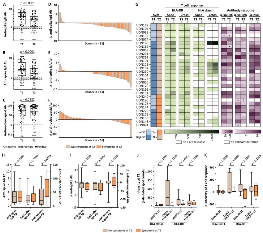
evidence for potential preexisting immunity mediated by in convalescent individuals after COVID-19 (specific EC) or
cross-reactive T cells to human common cold coronaviruses cross-reactive peptides recognized by both convalescent do-
(16, 21–23). We and others recently characterized the T cell nors and individuals never exposed to SARS-CoV-2 (cross-re-
epitopes mediating these specific and cross-reactive SARS- active EC, table S4), as described previously (24). The HLA
CoV-2 T cell responses in individuals during convalescence class I-restricted SARS-CoV-2 T cell epitopes included in this
and in unexposed individuals, providing evidence that the de- study for either specific or cross-reactive EC were restricted
velopment of immunity requires recognition of multiple to the 9 most common HLA class I allotypes covering at least
epitopes (16, 21–25). In light of the available data on immune one HLA allotype in more than 90% of the world’s population
responses against SARS-CoV-2, persistence of SARS-CoV-2- (26, 27). The number of convalescent individuals with detect-
specific T cell immunity may be crucial for long-term protec- able SARS-CoV-2 T cell responses was found to increase over
tion after COVID-19, which has additional consequences for time, from 93% at T1 to 100% at T2, as assessed by ex vivo
vaccine development. Here, we conducted a comprehensive interferon gamma (IFN-γ) ELISPOT assays (Fig. 1, C and D).
longitudinal analysis comparing T cell and antibody re- Specifically, the percentage of donors with detectable T cell
sponses in SARS-CoV-2 convalescent individuals up to six responses to SARS-CoV-2-specific EC increased from 45% to
Downloaded from http://stm.sciencemag.org/ by guest on March 19, 2021
months post infection. We report on the differential kinetics 69% for HLA class I and from 90% to 100% for HLA-DR (Fig.
of cellular and humoral immunity after COVID-19 and delin- 1C). The percentage of donors with detectable T cell re-
eate dominant peptides recognized by T cells that are essen- sponses to cross-reactive EC similarly increased (HLA class I:
tial for long-term immunity. 31% T1 versus 38% T2; HLA-DR: 93% T1 versus 100% T2; Fig.
1D).
RESULTS
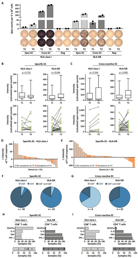
The intensity of SARS-CoV-2 T cell responses was main-
Longitudinal follow-up of COVID-19 convalescent do-
tained for CD8+ T cells and increased for CD4+ T cells
nors characterized post-infectious symptoms and iden-
over time.
tified sustained SARS-CoV-2-directed T cell responses.
We next performed longitudinal ex vivo IFN-γ ELISPOT
Clinical and immunological analysis of convalescent indi-
analysis of T cell responses from 29 individuals (group A) at
viduals after mild or moderate SARS-CoV-2-infection (n = 51,
T1 and T2 (Fig. 2A, fig. S2). These experiments revealed ro-
tables S1 and S2) was conducted 35 - 56 days (median 40 days,
bust intensities of HLA class I-restricted SARS-CoV-2-specific
time point 1, T1) and 141 - 183 days (median 159 days, time
and cross-reactive T cell response. In contrast, the intensities
point 2, T2) after positive SARS-CoV-2 polymerase chain re-
of T cell responses to HLA-DR-restricted SARS-CoV-2-specific
action (PCR) testing (fig. S1). Persistent or newly arisen post-
or cross-reactive EC significantly (p = 0.044 and p = 0.008,
infectious symptoms were reported by 27% of donors at T2,
respectively) increased over time (Fig. 2B and C). Ex vivo re-
with fatigue (64% of symptomatic donors) as well as anosmia
sponses to HLA class I-restricted or HLA-DR-restricted con-
and ageusia (64% of symptomatic donors) being most com-
trol peptide pools derived from other viruses, including
mon (Fig. 1A, table S1). Of the donors reporting post-infec-
cytomegalovirus, Epstein‐Barr virus, and adenovirus pep-
tious symptoms (n = 14), no PCR retesting data was available
tides, showed comparable T cell response intensities at T1
at T2. Five of 14 (36%) donors had been retested at different
and T2, with consistent intra-individual responses (n = 13, fig.
time points (12 - 98 days) after their initial positive PCR test,
S3, A to C). Accordingly, no correlation was observed in the
with one donor showing a positive PCR again two weeks after
variation of T cell responses (Δ intensity T2 - T1) to HLA-DR-
the initial test (table S3). Kinetics of SARS-CoV-2-directed T
restricted control peptide pools with SARS-CoV-2-specific or
cell immunity was determined longitudinally with regard to
cross-reactive EC over time (n = 12, fig. S3, D and E). A high
both (i) intensity (group A, n = 29) and (ii) diversity (percent-
inter-individual heterogeneity of the intensity of longitudinal
age of detected peptides per donor; group B, n = 23) of CD4+
SARS-CoV-2-directed T cell responses was observed. For HLA
and CD8+ T cell responses (Fig. 1B). To standardize determi-
class I-restricted SARS-CoV-2-specific EC 52% of donors
nation of changes in SARS-CoV-2 T cell response intensity
showed new or ≥ 2-fold increased T cell response intensities,
over time, we employed broadly applicable human leukocyte
24% showed stable (fold-change 0.6 - 1.9), and 24% showed ≥
antigens (HLA) class I- and HLA-DR-restricted SARS-CoV-2
2-fold decreased or lost T cell responses at T2 (Fig. 2D). For
epitope compositions (EC), as described previously (24).
HLA class I-restricted cross-reactive EC 45% of donors
These EC comprised multiple dominant and subdominant
showed new or ≥ 2-fold increased T cell response intensities,
SARS-CoV-2-specific and cross-reactive peptides, where dom-
45% showed stable (fold-change 0.6 - 1.9), and 9% showed ≥
inant peptides were recognized by ≥ 50% of HLA class I allo-
2-fold decreased or lost T cell responses at T2 (fig. S4A). For
type-matched donors and subdominant peptides were
HLA-DR, longitudinal increase of T cell response intensity in
recognized by < 50% of donors. We evaluated T cell responses
individual donors was even more pronounced, with 66% and
against SARS-CoV-2-specific peptides recognized exclusively
55% of donors with new or ≥ 2-fold increased T cell responses,
First release: 15 March 2021 stm.sciencemag.org (Page numbers not final at time of first release) 2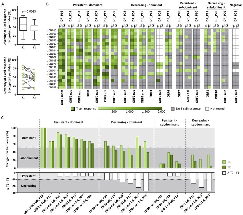
24% and 31% with stable T cell responses (fold-change 0.6 -         showed a comparable decrease in anti-nucleocapsid antibody
1.9), and 10% and 14% with ≥ 2-fold decreased or lost T cell        titers (Fig. 3, D to F). Anti-S1 IgG antibody responses moder-
responses to HLA-DR-restricted SARS-CoV-2-specific and              ately correlated with the intensity of T cell responses to HLA-
cross-reactive EC at T2, respectively (Fig. 2E, fig. S4B). Inter-   DR-restricted SARS-CoV-2-specific or cross-reactive EC, as
estingly, each of the three donors showing the most pro-            well as HLA class I-restricted cross-reactive EC at T2 (fig.
nounced decrease of T cell responses to the HLA-DR-                 S10). Longitudinal T cell and antibody responses, as well as
restricted SARS-CoV-2-specific EC still suffered from post-in-      symptoms during and after COVID-19, varied among the do-
fectious symptoms (Fig. 2E). Characterization of long-term          nors (Fig. 3G). Neither the intensity of SARS-CoV-2-specific
SARS-CoV-2-directed T cells at T2 using ex vivo flow cytome-        nor that of cross-reactive T cell responses to HLA class I- or
try-based assessment of surface markers and intracellular cy-       HLA-DR-restricted EC at T2 correlated with demographics
tokine staining (ICS) revealed that T cell responses to HLA         (table S5). High anti-nucleocapsid antibody titers at T2 were
class I-restricted cross-reactive EC were predominantly me-         associated with a higher prevalence of post-infectious symp-
diated by CD8+ T cells, whereas T cell responses to HLA-DR-         toms (Fig. 3, H and I). In contrast, neither intensity nor lon-
restricted SARS-CoV-2-specific and cross-reactive EC were           gitudinal kinetics of SARS-CoV-2 T cell responses were

                                                                                                                                         Downloaded from http://stm.sciencemag.org/ by guest on March 19, 2021
mainly mediated by CD4+ T cells (Fig. 2, F and G, fig. S5). The     associated with post-infectious symptoms (Fig. 3, J and K).
vast majority of T cell responses to HLA class I-restricted         Diversity of SARS-CoV-2 T cell immunity identifies pep-
SARS-CoV-2-specific EC were mediated by both CD8+ and               tides mediating long-term T cell responses.
CD4+ T cells (Fig. 2F), which is an often described phenome-            In various viral diseases, including COVID-19, diversity of
non especially in viral disease (28, 29). CD8+ T cells targeting    T cell responses, which means the recognition of multiple T
HLA class I-restricted SARS-CoV-2-specific EC were mainly           cell epitopes, has been implicated as a prerequisite for effec-
positive for CD107a, whereas CD4+ and CD8+ T cells targeting        tive immunity (24, 30). We longitudinally analyzed the diver-
HLA-DR-restricted SARS-CoV-2-specific EC displayed posi-            sity of SARS-CoV-2 T cell responses by single peptide
tivity for several of the markers interleukin (IL)-2, tumor ne-     mapping using dominant and subdominant promiscuous
crosis factor (TNF), IFN-γ, and CD107a (Fig. 2H). For HLA-          HLA-DR- (binding to several HLA-DR allotypes, n = 20) and
DR- and HLA class I-restricted cross-reactive EC multifunc-         HLA-A*24-restricted (n = 6) SARS-CoV-2-derived peptides, as
tional T cell responses (IL-2, TNF, IFN-γ, CD107a) could also       described previously (24). To enable detection of low-fre-
be observed (Fig. 2I). Longitudinal ICS and surface marker          quent peptide-specific T cell populations, we used an in vitro
analyses of T cell responses at T1 and T2 further validated         12-day pre-stimulation to expand SARS-CoV-2-specific T cells.
robust HLA class I-restricted (fig. S6) and HLA-DR-restricted       Longitudinal diversity of HLA-DR- and HLA-A*24-directed T
(fig. S7) SARS-CoV-2-specific and cross-reactive T cell re-         cell responses decreased across all donors and peptides over
sponses over time (fig. S8).                                        time (median T cell recognition per donor 59% and 50% at
Longitudinal SARS-CoV-2 antibody responses showed                   T1, 48% and 17% at T2, respectively; Fig. 4A, fig. S11). The de-
differential dynamics over time and correlation to post-            crease in HLA-DR-directed T cell diversity was confirmed in
infectious clinical status.                                         subgroup analyses for specific and cross-reactive peptides
    Two independent assays were employed to longitudinally          (fig. S12A) and dominant and subdominant peptides (fig.
assess SARS-CoV-2 antibody responses in convalescent do-            S12B). The decrease in diversity was also confirmed for pep-
nors (n = 51) at T1 and T2 to determine (i) ratios of IgG and       tides derived from structural or non-structural (fig. S12C) and
IgA antibodies targeting the S1 domain of the spike protein,        nucleocapsid versus non-nucleocapsid viral open reading
including the immunologically relevant receptor binding do-         frames (ORF, fig. S12D). T cell response intensity after in vitro
main (RBD; Fig. 3, A and B, fig. S9, A and B) as well as (ii)       12-day pre-stimulation showed high inter-individual and high
anti-nucleocapsid antibody titers (Fig. 3C, fig. S9C). Both         inter-peptide heterogeneity (fig. S13). For 88% of SARS-CoV-
anti-S1 IgG and IgA response significantly (p < 0.0001) de-         2 HLA-DR- and HLA-A*24-restricted peptides, expansion
creased over time (median 3.8 versus 2.6 and 2.6 versus 1.6,        ability of SARS-CoV-2 T cells did not differ significantly be-
respectively), whereas anti-nucleocapsid antibody titers re-        tween T1 and T2 (fig. S13).
mained stable from T1 to T2 (median 29 versus 25). Loss or ≥            Further, donor- and peptide-specific assessment identi-
2-fold decrease of anti-S1 IgG and IgA was observed in 31%          fied a subset of peptides derived from different ORFs that
and 44% of donors, respectively (Fig. 3, D and E), whereas          sustain a persistent T cell response (10/20 HLA-DR-restricted
loss or ≥ 2-fold decrease of anti-nucleocapsid antibody titers      peptides, Fig. 4, B and C, table S6; 2/6 HLA-A*24-restricted
was documented in only 13% of donors (Fig. 3F). Among               peptides, fig. S11, B and C, table S7). In particular, the seven
those still suffering from post-infectious symptoms at T2,          dominant HLA-DR-restricted peptides that mediated a per-
36% (5/14) and 50% (7/14) presented with ≥ 2-fold decrease          sistent T cell response in convalescent individuals appeared
or loss of anti-S1 IgG and IgA, respectively, whereas none          to be essential for long-term T cell immunity to SARS-CoV-2

First release: 15 March 2021                             stm.sciencemag.org      (Page numbers not final at time of first release)   3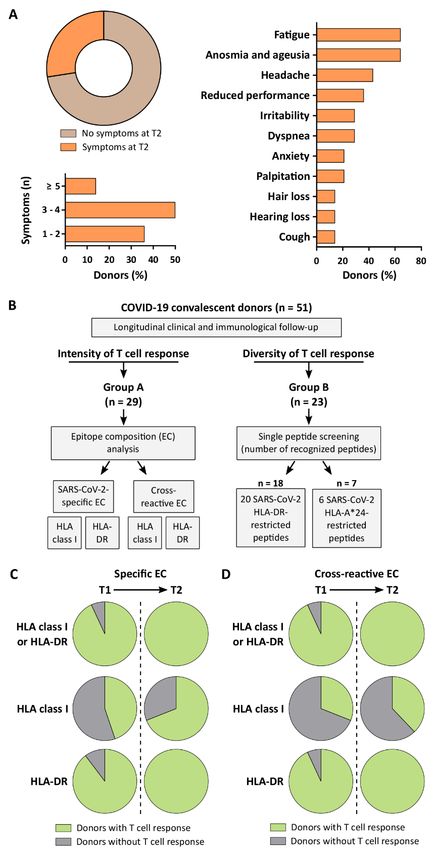
and may thus enable the development of effective vaccination        central role in cellular immunity to SARS-CoV-2. This is re-
approaches.                                                         flected by a higher frequency of convalescent donors with de-
                                                                    tectable SARS-CoV-2-specific CD4+ T cells, as well as an
DISCUSSION
                                                                    increased T cell response intensity and broader cytokine pro-
    The SARS-CoV-2 pandemic results in dramatic worldwide
                                                                    file of CD4+ T cells compared to CD8+ T cells (24, 37, 38). This
consequences for health care, the economy, and daily life. To
                                                                    important role of CD4+ T cells in the control of primary SARS-
enable the development of therapeutic and prophylactic in-
                                                                    CoV-2 infection might also continue for the development of
terventions for COVID-19, elucidation of the mechanisms un-
                                                                    long-term immunity, reflected by the here reported trend to
derlying SARS-CoV-2-directed immune responses is of the
                                                                    increased SARS-CoV-2-specific and cross-reactive HLA-DR-
utmost importance. This holds particularly true for the as-
                                                                    restricted T cell responses at six months post infection. This
sessment of immunological memory, which requires a de-
                                                                    is in line with several studies showing a correlation of the
tailed longitudinal analysis of cellular and humoral immune
                                                                    frequency of epitope-specific naïve CD4+ T cells with memory
responses. Accumulated evidence obtained from patients and
                                                                    repertoire for different pathogens (39, 40). T cell responses to
convalescents regarding frequency, intensity, and diversity of
                                                                    viral infections are considered to occur rapidly and peak
T cell responses and their correlation with SARS-CoV-2 anti-

                                                                                                                                         Downloaded from http://stm.sciencemag.org/ by guest on March 19, 2021
                                                                    about one week after acute infection before they decline (34,
body titers, as well as clinical characteristics, point to a cen-
                                                                    41). In contrast, data from the two previous coronavirus pan-
tral role of T cell immunity in COVID-19 (5, 16, 18, 19, 24, 31).
                                                                    demics/epidemics mediated by SARS-CoV-1 and MERS-CoV
Here, we present a comprehensive longitudinal analysis of
                                                                    revealed different kinetics of T cell responses. Strong ex vivo
donors with SARS-CoV-2 infection over a six-month follow-
                                                                    SARS-CoV-1-specific T cell responses were detected in conva-
up period comprising SARS-CoV-2 antibody and T cell re-
                                                                    lescent donors even seven to eight months after infection (36)
sponses as well as clinical symptoms of acute and post-
                                                                    and memory T cell responses could be detected after in vitro
COVID-19 disease.
                                                                    expansion up to 17 years after infection (16). This is in line
    We observed robust and increasing intensity of SARS-
                                                                    with first reports on persisting SARS-CoV-2 T cell responses
CoV-2 T cell responses targeting HLA class I- and HLA-DR-
                                                                    after three months (5) as well as our data in this six-month
restricted peptides over time. This observation is in line and
                                                                    follow-up analysis. Several studies have reported prolonged
extends the findings of a report on SARS-CoV-2 immune
                                                                    SARS-CoV-2 antigen persistence that might represent a con-
memory up to three months post-infection as well as data ob-
                                                                    tinuous trigger for T cell responses (42–46). Furthermore, a
tained from individuals following SARS-CoV-1 infection (3, 5,
                                                                    prolonged “inflammatory state” with persistent activation of
16). Consistent with previous work by our group (24), no dif-
                                                                    different components of the immune system has been de-
ference in frequency nor in intensity was observed between
                                                                    scribed after COVID-19 (47, 48). This might trigger an ongo-
T cell responses to cross-reactive or specific EC at the six-
                                                                    ing stimulation of T cells by different components of the
month follow-up time point. This suggests that there are no
                                                                    immune system, which is of particular importance for the de-
differences between SARS-CoV-2-derived peptides with and
                                                                    velopment and persistence of memory CD4+ T cells (49–53).
without similarities to human common cold coronaviruses in
                                                                    The interaction of B cells and T cells might play an essential
terms of mediating acute and long-term immune responses
                                                                    role and could explain the increase in SARS-CoV-2-specific
in COVID-19. The recognition of the SARS-CoV-2-derived
                                                                    and cross-reactive CD4+ T cell responses (50, 54). This is sup-
HLA-DR-presented peptides not only by CD4+ but, to a lesser
                                                                    ported by data from Dan and colleagues, showing a trend to-
degree, also by CD8+ T cells is due to several embedded SARS-
                                                                    ward an increase of SARS-CoV-2-specific CD4+ follicular
CoV-2 HLA class I-presented peptides within the HLA-DR-
                                                                    helper T cells (11), a specialized subset of CD4+ T cells re-
binding sequences. HLA-DR-restricted epitopes with embed-
                                                                    quired for B cell help (55), six months after infection.
ded HLA class I-binding peptides, to induce both CD4+ and
                                                                        In contrast to the kinetics of the T cell response, both IgG
CD8+ T cell responses, are widely used for anti-cancer as well
                                                                    and IgA antibody responses to the S1 domain of the spike
as anti-viral immunotherapy (32, 33). On the other hand, we
                                                                    protein declined during the six-month follow-up. In line with
could show that the SARS-CoV-2 HLA class I-restricted EC
                                                                    several reports on decreasing antibody titers after SARS-CoV-
could also be recognized by CD4+ T cells, which is an often
                                                                    2 infection (5–8, 34, 35), this finding raises concerns that hu-
described phenomenon, especially in viral diseases (28, 29),
                                                                    moral immunity against SARS-CoV-2 may not provide long-
as both HLA class I and class II molecules could bind to pri-
                                                                    term protection. It needs to be taken into consideration that
mary and secondary peptide anchor motifs covering the cen-
                                                                    the protective efficacy of the antibodies analyzed in our study
tral 9 - 10 amino acids.
                                                                    remains unclear, even if RBD antibody titers reportedly cor-
    Previous data on acute and chronic viral infection (34–36),
                                                                    respond to virus-neutralizing activity (56). Epidemiologic
including SARS-CoV-1 and MERS-CoV, as well as several
                                                                    studies employing neutralizing assays in large cohorts are re-
studies analyzing SARS-CoV-2 T cell responses during or
                                                                    quired to thoroughly unravel the relevance of long-term
early after COVID-19, have shown that CD4+ T cells play a

First release: 15 March 2021                             stm.sciencemag.org      (Page numbers not final at time of first release)   4
SARS-CoV-2 humoral immunity. Nevertheless, our finding This enabled the characterization of a first set of ORF-
that anti-S1 antibody responses decrease over time, whereas independent, dominant T cell epitopes that may govern long-
anti-nucleocapsid antibody titers persist, is important in the term SARS-CoV-2 T cell immunity. To expand this panel of
context of vaccine development, as several ongoing ap- highly promising candidate peptides for vaccine design, fu-
proaches are focusing on the induction of immune responses ture studies are warranted that broadly evaluate long-term
to the RBD of the spike protein (57, 58). SARS-CoV-2 T cell responses to T cell epitopes of further HLA
As more than 50 million people have recovered from class I allotypes. In contrast, the promiscuous HLA-DR-
COVID-19, increasing evidence for the prevalence and nature binding peptides identified to mediate long-term T cell re-
of post-infectious symptoms and secondary damages is aris- sponses in up to 100% of donors independently of their HLA-
ing (59–65). There are only limited datasets (66–68) available DR allotype represent broadly applicable candidates for vac-
reporting on the prevalence and nature of post-infectious cine design. The phenomenon that dominant HLA-DR-
symptoms after COVID-19, which was just recently claimed restricted epitopes are associated with promiscuous HLA
in a comprehensive review (69). This is especially true for class II binding, defined as the capacity to bind multiple HLA
mild COVID-19. Several large cohort studies are ongoing. Our allelic variants, is well described for other infectious diseases,
Downloaded from http://stm.sciencemag.org/ by guest on March 19, 2021
work provides insight into post-infectious symptoms in a co- including tuberculosis and malaria (75–77), and was just re-
hort of individuals with a mild to moderate course of COVID- cently also proven for a panel of dominant SARS-CoV-2 HLA-
19, showing post-infectious symptoms in 27% of the donors, DR-restricted T cell epitopes (78). The promiscuous HLA-DR-
which is in line with a very recent publication in an out-pa- presented SARS-CoV-2-derived peptides defined here thus al-
tient setting (70). The pathomechanism underlying persis- ready constitute the basis of a multi-peptide vaccine for in-
tence or development of these symptoms after SARS-CoV-2- duction of T cell immunity to SARS-CoV-2 to combat COVID-
infection is matter of investigation (62, 71). Microangiopathic 19, which is currently evaluated in a first-in-human clinical
cerebral lesions (72), effects directly mediated by the virus, trial (NCT04546841).
such as virus persistence (42, 43, 45, 46), and immune-medi- Caveats of this study include the limited follow-up time,
ated inflammatory syndromes (73, 74) are proposed to play a small sample size, and the focus on non-severe courses of
role. Previous studies reported on the correlation of high an- COVID-19. Sample size and follow-up time were limited by
tibody titers with more severe course of acute COVID-19 (6, expedience. The focus on non-severe cases of COVID-19 also
24). Here we could show that high nucleocapsid antibody ti- represents a strength, as long-term persistence of SARS-CoV-
ters at six-month follow-up also associate with an increased 2 immune responses in this large group of COVID-19 conva-
prevalence of post-infectious symptoms. No correlation of lescent donors is of utmost importance for the development
post-infectious symptoms with intensity or longitudinal dy- of population-level immunity. Completion of this data re-
namics of anti-SARS-CoV-2 T cell responses was observed. To- quires large cohort studies, including longitudinal sampling
gether with recent data providing evidence that the intensity of donors with severe COVID-19 over a longer period of time
of T cell responses does not correlate with acute COVID-19 to (i) delineate the mechanistic basis of SARS-CoV-2 long-
severity (21, 24), this finding is of high relevance for the de- term immunity and (ii) confirm presence of long-term pro-
sign of vaccines, as it provides evidence that disease-aggra- tective T cell immunity to SARS-CoV-2 based on monitoring
vating effects might not hamper the development of of convalescent individuals upon virus rechallenge. Addition-
vaccination approaches aiming to induce SARS-CoV-2- ally, in the light of further upcoming data on longitudinal
specific T cell responses. Future studies are needed to vali- SARS-CoV-2 immune responses (11), future analyses are re-
date these findings in larger cohorts and to delineate poten- quired to characterize and differentiate T cell subsets and
tial immune- or antibody-mediated mechanisms of post- their respective functionality state that mediated long-term T
infectious symptoms. cell immunity and define the T cell epitopes, epitope compo-
Previous work on viral diseases including SARS-CoV-2 im- sitions or peptide megapools that optimally enable their de-
plicates diversity of T cell responses, or the recognition of tection.
multiple T cell epitopes, as important prerequisite for effec- In conclusion, our data provides important insights into
tive immunity (24, 30). Identification of respective T cell the differential dynamics of SARS-CoV-2-directed antibody
epitopes that induce potent and long-lasting SARS-CoV-2- and T cell immune responses over time, their correlation to
specific responses is fundamental for both detection of im- post-COVID-19 illness, and the identity of SARS-CoV-2 pep-
munological memory and vaccine design. Our longitudinal tide targets for durable memory T cell responses after
analysis of T cell response diversity using a single peptide- COVID-19. Together, these data have broad implications for
based approach allowed for discrimination of HLA-DR- and - both detection and understanding of immunological memory
A*24-restricted peptides capable or incapable to induce per- as well as vaccine design.
sisting SARS-CoV-2-specific or cross-reactive T cell responses.
First release: 15 March 2021 stm.sciencemag.org (Page numbers not final at time of first release) 5MATERIALS AND METHODS addition, subjective post-infectious complaints and symp-
Study design toms were assessed at the follow-up sample acquisition (T2).
We performed a single-center study carried out at the Detailed donor characteristics as well as information on allo-
Clinical Collaboration Unit Translational Immunology, Uni- cation of the donors to the experimental groups are provided
versity Hospital Tübingen, Germany analyzing convalescent in tables S1 and S2.
adults after SARS-CoV-2 infection (n = 51). The study aimed SARS-CoV-2 peptides
to longitudinally determine the kinetics of the SARS-CoV-2- Synthetic peptides were provided by EMC Microcollec-
directed immune response in terms of intensity and diversity tions GmbH and INTAVIS Peptide Services GmbH & Co. KG.
of T cell response, as well as antibody response. Blood and The HLA class I- and HLA-DR-restricted peptides as well as
serum samples were collected and a questionnaire-based as- the applied EC were characterized in detail in a previous
sessment of donor characteristics and disease symptoms dur- work (24) analyzing T cell responses in convalescent individ-
ing and after COVID-19 from SARS-CoV-2 convalescent uals after COVID-19 as well as in healthy donors never ex-
donors was obtained between 4/2020 and 5/2020 (time point posed to the virus. T cell epitopes were defined as dominant
T1) and in 08/2020 (time point T2). Informed consent was if immune responses against these peptides were detected in
Downloaded from http://stm.sciencemag.org/ by guest on March 19, 2021
obtained in accordance with the Declaration of Helsinki pro- ≥ 50% of convalescent donors. For HLA class I-restricted pep-
tocol. The study was approved by and performed according tides, T cell responses and definition of dominance were only
to the guidelines of the local ethics committees assessed in HLA-matched donors. The SARS-CoV-2 HLA class
(179/2020/BO2). I-restricted peptides used in this study were predicted to bind
The donors were analyzed in two groups for (i) intensity to a specific HLA allotype and validated in COVID-19 conva-
of CD4+ and CD8+ T cell responses (group A, n = 29) and for lescent donors with this respective HLA allotype, whereas
(ii) diversity of T cell responses (the number of detected pep- promiscuous SARS-CoV-2-derived HLA-DR-restricted pep-
tides per donor; group B, n = 23). Donors analyzed in each tides binding to several HLA-DR allotypes were selected and
group were identical at T1 and T2 and were assigned to the validated independent of the HLA-DR allotype.
groups according to time of sample acquisition and available The term “cross-reactive peptide” is used for SARS-CoV-2
sample cell number as well as HLA allotype restriction (for peptides not only eliciting T cell responses in donors after
HLA-A*24 T cell diversity assessment). One donor was ana- SARS-CoV-2 infection, but also in unexposed individuals. To
lyzed in both group A and B (table S2). All analyzed donors delineate any differences in SARS-CoV-2 T cell responses to
and data were shown. Outliers were not excluded from the specific or cross-reactive peptides in the long-term follow-up,
analyses. we included the cross-reactive HLA class I- and HLA-DR-
SARS-CoV-2 infection was confirmed by PCR test after na- restricted EC in this longitudinal analysis.
sopharyngeal swab. Donor recruitment was performed by For the determination of ex vivo intensity of SARS-CoV-2
online- and paper-based advertising (homepage, flyer). Sam- T cell responses at T1 and T2, standardized and previously
ple collection was performed in a longitudinal manner ap- validated HLA class I- and HLA-DR-restricted SARS-CoV-2-
proximately 35 - 56 days (T1) and 141 - 183 days (T2) after specific and cross-reactive EC were applied (table S4) (24, 78).
positive PCR. Samples were processed in the Department of As the determination of T cell response diversity requires the
Immunology located at the same hospital site. Peripheral analysis of multiple peptides, we used HLA allotypes with
blood mononuclear cells (PBMCs) were isolated by density several validated SARS-CoV-2 peptides including HLA-DR
gradient centrifugation and used directly or stored at -80°C (20 peptides with multiple HLA-DR restrictions) and HLA-
until further use. Serum was separated by centrifugation for A*24 (6 peptides).
10 min and the supernatant was stored at -80°C. Flow cytom- IFN-γ enzyme-linked immunospot (ELISPOT) assays
etry- and T cell-based experiments were conducted at the De- HLA class I- and HLA-DR-restricted SARS-CoV-2-specific
partment of Immunology, aliquots were also shipped at -20°C and cross-reactive EC and single peptides were used for lon-
to the Department of Clinical Chemistry and Pathobiochem- gitudinal analysis of T cell response intensity and diversity,
istry for antibody analysis. respectively, comprising multiple dominant (recognized by ≥
HLA typing was carried out by Immatics Biotechnology 50% of donors) and subdominant (recognized by < 50% of
GmbH and the Department of Hematology and Oncology at donors) SARS-CoV-2-specific peptides recognized exclusively
the University Hospital Tübingen. Symptom score (SC) to as- in COVID-19 convalescent donors or cross-reactive peptides
sess severity of COVID-19 was determined by combining ob- recognized by both convalescent donors and individuals
jective (fever ≥ 38.0°C) and subjective disease symptoms never exposed to SARS-CoV-2 (table S4), as described previ-
(no/mild/moderate versus severe, reported by questionnaire) ously (21).
of individual donors. Donors with severe disease symptoms For the longitudinal analysis of T cell response intensity,
or fever were classified as “high SC”, all others as “low SC”. In freshly isolated or thawed PBMCs were pulsed with either
First release: 15 March 2021 stm.sciencemag.org (Page numbers not final at time of first release) 6SARS-CoV-2-specific or cross-reactive EC (HLA class I- or dilution of GolgiStop (BD Biosciences) for 12 - 14 hours. Stain-
HLA-DR-restricted) and analyzed directly ex vivo by IFN-γ ing was performed using Cytofix/Cytoperm solution (BD Bio-
ELISPOT assay in duplicates. Ex vivo responses to a control sciences), APC/Cy7 anti-human CD4 (1:100 dilution,
peptide pool, including peptides derived from cytomegalovi- BioLegend, Cat# 300518, RRID: AB_314086), PE/Cy7 anti-hu-
rus, Epstein-Barr virus, and adenovirus (table S8), served as man CD8 (1:400 dilution, Beckman Coulter, Cat# 737661,
controls. T cell diversity (percentage of recognized peptides) RRID: AB_1575980), Pacific Blue anti-human TNF (1:120 di-
was analyzed following 12-day in vitro pre-stimulation prior lution, BioLegend, Cat# 502920, RRID: AB_528965), FITC
to single peptide analysis (HLA-DR- or HLA-A*24-restricted anti-human CD107a (1:100 dilution, BioLegend, Cat# 328606,
peptides) by IFN-γ ELISPOT assay. For pre-stimulation, RRID: AB_1186036), and PE anti-human IFN-γ monoclonal
PBMCs were pulsed with HLA-A*24- (1 μg/mL) or HLA-DR- antibodies (1:200 dilution, BioLegend, Cat# 506507, RRID:
restricted peptide pools (5 μg/mL) and cultured for 12 days AB_315440). T cells exposed to phorbol myristate acetate
under addition of IL-2 (20 U/mL, Novartis) on days 2, 5, and (PMA, 5 μg/mL, Sigma-Aldrich) and ionomycin (1 μM, Sigma-
7. Expanded PBMCs were analyzed by single peptide readout Aldrich) served as positive controls. Viable cells were deter-
ELISPOT in duplicates. Up to 8 × 10⁵ cells per well were co- mined using Aqua live/dead (1:400 dilution, Invitrogen). All
Downloaded from http://stm.sciencemag.org/ by guest on March 19, 2021
incubated in 96-well plates with 1 μg/mL of HLA class I-re- samples were analyzed on a FACS Canto II cytometer (BD Bi-
stricted or 2.5 μg/mL of HLA-DR-restricted peptide pools di- osciences) and evaluated using FlowJo software version
rectly ex vivo (for EC) or of single peptides following the 12- 10.0.8 (BD Biosciences). The gating strategy applied for the
day T cell expansion. 96-well plates were coated with 2 μg/mL analyses of flow cytometry-acquired data is provided in fig.
anti-IFN-γ antibody (clone 1-D1K, MabTech, Cat# 3420-3-250, S14.
RRID: AB_907283). After a 24 hour incubation, spots were SARS-CoV-2 IgG and IgA ELISA (EUROIMMUN)
revealed with 0.3 μg/mL anti-IFN-γ biotinylated detection The 96-well SARS-CoV-2 IgG and IgA ELISA assay
antibody (clone 7-B6-1, MabTech, Cat# 3420-6-250, RRID: (EUROIMMUN, 2606A_A_DE_C03, as constituted on 22
AB_907273), ExtrAvidin-Alkaline Phosphatase (1:1,000 dilu- April 2020) was performed on an automated BEP 2000 Ad-
tion, Sigma-Aldrich), and BCIP/NBT (5-bromo-4-chloro-3-in- vance system (Siemens Healthcare Diagnostics GmbH) ac-
dolyl-phosphate/nitro-blue tetrazolium chloride, Sigma- cording to the manufacturer’s instructions. The ELISA assay
Aldrich). Phytohemagglutinin (PHA, Sigma-Aldrich) served detects anti-SARS-CoV-2 IgG and IgA directed against the S1
as positive control. Irrelevant HLA-matched control peptides domain of the viral spike protein (including the immunolog-
(HLA-A*24, AYVHMVTHF, BI1_HUMAN45-53 and HLA-DR, ically relevant RBD) and relies on an assay-specific calibrator
ETVITVDTKAAGKGK, FLNA_HUMAN1669−1683) or, in case of to report a ratio of specimen absorbance to calibrator absorb-
HLA class I-restricted EC, 10% dimethyl sulfoxide (DMSO) in ance. The final interpretation of positivity is determined by
double-distilled water (ddH2O) served as negative control. ratio above a threshold value given by the manufacturer: pos-
Spots were counted using an ImmunoSpot S5 analyzer (CTL) itive (ratio ≥ 1.1), borderline (ratio 0.8 - 1.0), or negative (ratio
and T cell responses were considered positive when the mean < 0.8). Quality control was performed following the manufac-
spot count was ≥ 3-fold higher than the mean spot count of turer’s instructions on each day of testing.
the negative control. The intensity of T cell responses is de-
Elecsys anti-SARS-CoV-2 immunoassay
picted as calculated spot counts, which represent the mean
The Elecsys anti-SARS-CoV-2 assay is an ECLIA (electro-
spot count of duplicates normalized to 5 × 10⁵ cells minus the
generated chemiluminescence immunoassay) designed by
normalized mean spot count of the respective negative con-
Roche Diagnostics GmbH and was used according to the
trol (as in Fig. 2 B and C, Fig. 3, G, J and K, fig. S3, fig. S8, C
manufacturer’s instructions (V1.0, as constituted in May
and D, fig. S10, fig. S13). The recognition frequency of T cell
2020). It is intended for the detection of high-affinity anti-
responses within a donor group indicates the relative number
bodies (including IgG) directed against the nucleocapsid pro-
of donors which can recognize the respective EC or peptide
tein of SARS-CoV-2 in human serum. Readout was performed
(positive donors/tested donors) (as in Fig. 1, C and D, Fig. 4C,
on the Cobas e411 analyzer (Roche Diagnostics). Negative re-
fig. S11C, tables S6 and S7). The diversity of T cell responses
sults were defined by a cut-off index of < 1.0. Quality control
for single donors represents the number of recognized SARS-
was performed following the manufacturer’s instructions on
CoV-2-derived peptides (positive peptides/tested peptides)
each day of testing.
(as in Fig. 4A, fig. S11A, fig. S12).
Software and statistical analysis
Intracellular cytokine and cell surface marker staining
Flow cytometric data was analyzed using FlowJo 10.0.8
Peptide-specific T cells were characterized by intracellular
(BD Biosciences). Data are displayed as mean with standard
cytokine and cell surface marker staining. PBMCs were incu-
deviation (for n ≥ 3), scatter dot plot with mean, box plot as
bated with 10 μg/mL per peptide of EC or negative control
median with 25th or 75th percentiles, min/max whiskers, or
peptide, 10 μg/mL Brefeldin A (Sigma-Aldrich), and a 1:500
First release: 15 March 2021 stm.sciencemag.org (Page numbers not final at time of first release) 7violin plots with median and quartiles. Descriptions of the recovered patients with severe acute respiratory syndrome: A six-year follow-up
statistical tests that were used for evaluation of the experi- study. J. Immunol. 186, 7264–7268 (2011). doi:10.4049/jimmunol.0903490
Medline
ments are provided within the respective figure legends. Con- 4. C. Kreer, M. Zehner, T. Weber, M. S. Ercanoglu, L. Gieselmann, C. Rohde, S. Halwe,
tinuous data were tested for distribution, and D’Agostino’s K2 M. Korenkov, P. Schommers, K. Vanshylla, V. Di Cristanziano, H. Janicki, R.
test was used as a normality test. Individual groups were Brinker, A. Ashurov, V. Krähling, A. Kupke, H. Cohen-Dvashi, M. Koch, J. M. Eckert,
tested by use of paired Wilcoxon or unpaired Mann-Whitney S. Lederer, N. Pfeifer, T. Wolf, M. J. G. T. Vehreschild, C. Wendtner, R. Diskin, H.
Gruell, S. Becker, F. Klein, Longitudinal Isolation of Potent Near-Germline SARS-
U tests and tests were two-sided. Spearman’s rho (ρ) was cal- CoV-2-Neutralizing Antibodies from COVID-19 Patients. Cell 182, 843–854.e12
culated for correlation between continuous data. Missing (2020). doi:10.1016/j.cell.2020.06.044 Medline
data were included in tables and in descriptive analysis. 5. L. B. Rodda, J. Netland, L. Shehata, K. B. Pruner, P. A. Morawski, C. D. Thouvenel, K.
K. Takehara, J. Eggenberger, E. A. Hemann, H. R. Waterman, M. L. Fahning, Y.
Graphs were plotted using GraphPad Prism 8.4.3. Statistical
Chen, M. Hale, J. Rathe, C. Stokes, S. Wrenn, B. Fiala, L. Carter, J. A. Hamerman,
analyses were conducted using GraphPad Prism 9.0.2 and N. P. King, M. Gale Jr., D. J. Campbell, D. J. Rawlings, M. Pepper, Functional SARS-
JMP Pro (SAS Institute Inc., version 14.2) software. P values CoV-2-Specific Immune Memory Persists after Mild COVID-19. Cell 184, 169–
of < 0.05 were considered statistically significant. 183.e17 (2021). doi:10.1016/j.cell.2020.11.029 Medline
6. Q. X. Long, X. J. Tang, Q. L. Shi, Q. Li, H. J. Deng, J. Yuan, J. L. Hu, W. Xu, Y. Zhang,
SUPPLEMENTARY MATERIALS
Downloaded from http://stm.sciencemag.org/ by guest on March 19, 2021
F. J. Lv, K. Su, F. Zhang, J. Gong, B. Wu, X. M. Liu, J. J. Li, J. F. Qiu, J. Chen, A. L.
stm.sciencemag.org/cgi/content/full/scitranslmed.abf7517/DC1 Huang, Clinical and immunological assessment of asymptomatic SARS-CoV-2
Fig. S1. Schematic overview and timeline of the experimental setting for the infections. Nat. Med. 26, 1200–1204 (2020). doi:10.1038/s41591-020-0965-6
longitudinal analysis of convalescent donors after SARS-CoV-2 infection Medline
Fig. S2. IFN-γ ELISPOT assays of longitudinal SARS-CoV-2 T cell responses in 7. T. J. Ripperger, J. L. Uhrlaub, M. Watanabe, R. Wong, Y. Castaneda, H. A. Pizzato, M.
convalescent individuals R. Thompson, C. Bradshaw, C. C. Weinkauf, C. Bime, H. L. Erickson, K. Knox, B.
Fig. S3. Control ex vivo IFN-γ ELISPOT assay for validation of T cell response Bixby, S. Parthasarathy, S. Chaudhary, B. Natt, E. Cristan, T. El Aini, F. Rischard,
intensities between T1 and T2 J. Campion, M. Chopra, M. Insel, A. Sam, J. L. Knepler, A. P. Capaldi, C. M. Spier,
Fig. S4. Kinetics of T cell response intensity to cross-reactive epitope compositions in M. D. Dake, T. Edwards, M. E. Kaplan, S. J. Scott, C. Hypes, J. Mosier, D. T. Harris,
individual convalescent donors B. J. LaFleur, R. Sprissler, J. Nikolich-Žugich, D. Bhattacharya, Orthogonal SARS-
Fig. S5. Flow cytometry-based characterization of long-term SARS-CoV-2-specific T CoV-2 Serological Assays Enable Surveillance of Low-Prevalence Communities
cell responses and Reveal Durable Humoral Immunity. Immunity 53, 925–933.e4 (2020).
Fig. S6. Flow cytometry-based characterization of CD8+ T cell responses at T1 and T2 doi:10.1016/j.immuni.2020.10.004 Medline
Fig. S7. Flow cytometry-based characterization of CD4+ T cell responses at T1 and T2 8. F. J. Ibarrondo, J. A. Fulcher, D. Goodman-Meza, J. Elliott, C. Hofmann, M. A.
Fig. S8. Longitudinal flow cytometry-based analysis of SARS-CoV-2 T cell response Hausner, K. G. Ferbas, N. H. Tobin, G. M. Aldrovandi, O. O. Yang, Rapid Decay of
Fig. S9. Longitudinal assessment of SARS-CoV-2-directed antibody responses Anti-SARS-CoV-2 Antibodies in Persons with Mild Covid-19. N. Engl. J. Med. 383,
Fig. S10. Correlation of SARS-CoV-2 antibody response with intensity of T cell 1085–1087 (2020). doi:10.1056/NEJMc2025179 Medline
responses at T2 9. C. Gaebler, Z. Wang, J. C. C. Lorenzi, F. Muecksch, S. Finkin, M. Tokuyama, A. Cho,
Fig. S11. Diversity of T cell responses to HLA-A*24-restricted peptides in convalescent M. Jankovic, D. Schaefer-Babajew, T. Y. Oliveira, M. Cipolla, C. Viant, C. O. Barnes,
donors over time Y. Bram, G. Breton, T. Hägglöf, P. Mendoza, A. Hurley, M. Turroja, K. Gordon, K. G.
Fig. S12. Longitudinal assessment of SARS-CoV-2 T cell response diversity in different Millard, V. Ramos, F. Schmidt, Y. Weisblum, D. Jha, M. Tankelevich, G. Martinez-
subgroups of HLA-DR-restricted peptides Delgado, J. Yee, R. Patel, J. Dizon, C. Unson-O’Brien, I. Shimeliovich, D. F.
Fig. S13. Longitudinal assessment of SARS-CoV-2 T cell responses to HLA-DR- and Robbiani, Z. Zhao, A. Gazumyan, R. E. Schwartz, T. Hatziioannou, P. J. Bjorkman,
HLA-A*24-restricted SARS-CoV-2-derived peptides S. Mehandru, P. D. Bieniasz, M. Caskey, M. C. Nussenzweig, Evolution of antibody
Fig. S14. Gating strategy for flow cytometry-based evaluation of surface marker and immunity to SARS-CoV-2. Nature (2021). doi:10.1038/s41586-021-03207-w
intracellular cytokine staining Medline
Table S1. Clinical characteristics of convalescent donors 10. J. Zuo, A. Dowell, H. Pearce, K. Verma, H. Long, J. Begum, F. Aiano, Z. Amin-
Table S2. Characteristics of individual convalescent donors Chowdhury, B. Hallis, L. Stapley, R. Borrow, E. Linley, S. Ahmad, B. Parker, A.
Table S3. PCR retesting of convalescent donors with post-infectious symptoms at T2 Horsley, G. Amirthalingam, K. Brown, M. Ramsay, S. Ladhani, P. Moss, Robust
Table S4. SARS-CoV-2-specific and cross-reactive HLA class I- and HLA-DR-restricted SARS-CoV-2-specific T-cell immunity is maintained at 6 months following primary
T cell epitope compositions infection. bioRxiv, 2020.2011.2001.362319 (2020).
Table S5. Intensity of T cell and antibody response at T2 according to gender, age, and 11. J. M. Dan, J. Mateus, Y. Kato, K. M. Hastie, E. D. Yu, C. E. Faliti, A. Grifoni, S. I.
BMI Ramirez, S. Haupt, A. Frazier, C. Nakao, V. Rayaprolu, S. A. Rawlings, B. Peters, F.
Table S6. Longitudinal characterization of SARS-CoV-2-derived HLA-DR-restricted Krammer, V. Simon, E. O. Saphire, D. M. Smith, D. Weiskopf, A. Sette, S. Crotty,
peptides Immunological memory to SARS-CoV-2 assessed for up to 8 months after
Table S7. Longitudinal characterization of SARS-CoV-2-derived HLA-A*24-restricted infection. Science 371, eabf4063 (2021). doi:10.1126/science.abf4063 Medline
peptides 12. J. Zhao, J. Zhao, A. K. Mangalam, R. Channappanavar, C. Fett, D. K. Meyerholz, S.
Table S8. Control peptides Agnihothram, R. S. Baric, C. S. David, S. Perlman, Airway Memory CD4(+) T Cells
Data file S1. Raw data Mediate Protective Immunity against Emerging Respiratory Coronaviruses.
Immunity 44, 1379–1391 (2016). doi:10.1016/j.immuni.2016.05.006 Medline
REFERENCES AND NOTES
13. J. Zhao, J. Zhao, S. Perlman, T cell responses are required for protection from
1. R. Ahmed, D. Gray, Immunological memory and protective immunity:
clinical disease and for virus clearance in severe acute respiratory syndrome
Understanding their relation. Science 272, 54–60 (1996).
coronavirus-infected mice. J. Virol. 84, 9318–9325 (2010).
doi:10.1126/science.272.5258.54 Medline
doi:10.1128/JVI.01049-10 Medline
2. W. C. Cao, W. Liu, P. H. Zhang, F. Zhang, J. H. Richardus, Disappearance of
14. R. Channappanavar, C. Fett, J. Zhao, D. K. Meyerholz, S. Perlman, Virus-specific
antibodies to SARS-associated coronavirus after recovery. N. Engl. J. Med. 357,
memory CD8 T cells provide substantial protection from lethal severe acute
1162–1163 (2007). doi:10.1056/NEJMc070348 Medline
respiratory syndrome coronavirus infection. J. Virol. 88, 11034–11044 (2014).
3. F. Tang, Y. Quan, Z. T. Xin, J. Wrammert, M. J. Ma, H. Lv, T. B. Wang, H. Yang, J. H.
doi:10.1128/JVI.01505-14 Medline
Richardus, W. Liu, W. C. Cao, Lack of peripheral memory B cell responses in
First release: 15 March 2021 stm.sciencemag.org (Page numbers not final at time of first release) 815. O. W. Ng, A. Chia, A. T. Tan, R. S. Jadi, H. N. Leong, A. Bertoletti, Y. J. Tan, Memory Stevanović, H. G. Rammensee, J. S. Walz, SARS-CoV-2-derived peptides define
T cell responses targeting the SARS coronavirus persist up to 11 years post- heterologous and COVID-19-induced T cell recognition. Nat. Immunol. 22, 74–85
infection. Vaccine 34, 2008–2014 (2016). doi:10.1016/j.vaccine.2016.02.063 (2021). doi:10.1038/s41590-020-00808-x Medline
Medline 25. D. Weiskopf, K. S. Schmitz, M. P. Raadsen, A. Grifoni, N. M. A. Okba, H. Endeman,
16. N. Le Bert, A. T. Tan, K. Kunasegaran, C. Y. L. Tham, M. Hafezi, A. Chia, M. H. Y. J. P. C. van den Akker, R. Molenkamp, M. P. G. Koopmans, E. C. M. van Gorp, B. L.
Chng, M. Lin, N. Tan, M. Linster, W. N. Chia, M. I. Chen, L. F. Wang, E. E. Ooi, S. Haagmans, R. L. de Swart, A. Sette, R. D. de Vries, Phenotype and kinetics of
Kalimuddin, P. A. Tambyah, J. G. Low, Y. J. Tan, A. Bertoletti, SARS-CoV-2-specific SARS-CoV-2-specific T cells in COVID-19 patients with acute respiratory distress
T cell immunity in cases of COVID-19 and SARS, and uninfected controls. Nature syndrome. Sci. Immunol. 5, eabd2071 (2020). doi:10.1126/sciimmunol.abd2071
584, 457–462 (2020). doi:10.1038/s41586-020-2550-z Medline Medline
17. T. Sekine, A. Perez-Potti, O. Rivera-Ballesteros, K. Strålin, J. B. Gorin, A. Olsson, S. 26. H. H. Bui, J. Sidney, K. Dinh, S. Southwood, M. J. Newman, A. Sette, Predicting
Llewellyn-Lacey, H. Kamal, G. Bogdanovic, S. Muschiol, D. J. Wullimann, T. population coverage of T-cell epitope-based diagnostics and vaccines. BMC
Kammann, J. Emgård, T. Parrot, E. Folkesson, O. Rooyackers, L. I. Eriksson, J. I. Bioinformatics 7, 153 (2006). doi:10.1186/1471-2105-7-153 Medline
Henter, A. Sönnerborg, T. Allander, J. Albert, M. Nielsen, J. Klingström, S. 27. R. Vita, J. A. Overton, J. A. Greenbaum, J. Ponomarenko, J. D. Clark, J. R. Cantrell,
Gredmark-Russ, N. K. Björkström, J. K. Sandberg, D. A. Price, H. G. Ljunggren, S. D. K. Wheeler, J. L. Gabbard, D. Hix, A. Sette, B. Peters, The immune epitope
Aleman, M. Buggert, M. Buggert; Karolinska COVID-19 Study Group, Robust T Cell database (IEDB) 3.0. Nucleic Acids Res. 43 (D1), D405–D412 (2015).
Immunity in Convalescent Individuals with Asymptomatic or Mild COVID-19. Cell doi:10.1093/nar/gku938 Medline
183, 158–168.e14 (2020). doi:10.1016/j.cell.2020.08.017 Medline 28. M. Wang, S. T. Tang, O. Lund, M. H. Dziegiel, S. Buus, M. H. Claesson, High-affinity
Downloaded from http://stm.sciencemag.org/ by guest on March 19, 2021
18. J. R. Habel, T. H. O. Nguyen, C. E. van de Sandt, J. A. Juno, P. Chaurasia, K. Wragg, human leucocyte antigen class I binding variola-derived peptides induce CD4+ T
M. Koutsakos, L. Hensen, X. Jia, B. Chua, W. Zhang, H. X. Tan, K. L. Flanagan, D. L. cell responses more than 30 years post-vaccinia virus vaccination. Clin. Exp.
Doolan, J. Torresi, W. Chen, L. M. Wakim, A. C. Cheng, P. C. Doherty, J. Petersen, Immunol. 155, 441–446 (2009). doi:10.1111/j.1365-2249.2008.03856.x Medline
J. Rossjohn, A. K. Wheatley, S. J. Kent, L. C. Rowntree, K. Kedzierska, Suboptimal 29. L. H. Boyle, J. C. Goodall, J. S. Gaston, Major histocompatibility complex class I-
SARS-CoV-2-specific CD8+ T cell response associated with the prominent HLA- restricted alloreactive CD4+ T cells. Immunology 112, 54–63 (2004).
A*02:01 phenotype. Proc. Natl. Acad. Sci. U.S.A. 117, 24384–24391 (2020). doi:10.1111/j.1365-2567.2004.01857.x Medline
doi:10.1073/pnas.2015486117 Medline 30. E. J. Yager, M. Ahmed, K. Lanzer, T. D. Randall, D. L. Woodland, M. A. Blackman,
19. Y. Peng, A. J. Mentzer, G. Liu, X. Yao, Z. Yin, D. Dong, W. Dejnirattisai, T. Rostron, Age-associated decline in T cell repertoire diversity leads to holes in the repertoire
P. Supasa, C. Liu, C. López-Camacho, J. Slon-Campos, Y. Zhao, D. I. Stuart, G. C. and impaired immunity to influenza virus. J. Exp. Med. 205, 711–723 (2008).
Paesen, J. M. Grimes, A. A. Antson, O. W. Bayfield, D. E. D. P. Hawkins, D. S. Ker, doi:10.1084/jem.20071140 Medline
B. Wang, L. Turtle, K. Subramaniam, P. Thomson, P. Zhang, C. Dold, J. Ratcliff, P. 31. L. Swadling, M. K. Maini, T cells in COVID-19 - united in diversity. Nat. Immunol. 21,
Simmonds, T. de Silva, P. Sopp, D. Wellington, U. Rajapaksa, Y. L. Chen, M. Salio, 1307–1308 (2020). doi:10.1038/s41590-020-0798-y Medline
G. Napolitani, W. Paes, P. Borrow, B. M. Kessler, J. W. Fry, N. F. Schwabe, M. G. 32. R. J. May, T. Dao, J. Pinilla-Ibarz, T. Korontsvit, V. Zakhaleva, R. H. Zhang, P.
Semple, J. K. Baillie, S. C. Moore, P. J. M. Openshaw, M. A. Ansari, S. Dunachie, E. Maslak, D. A. Scheinberg, Peptide epitopes from the Wilms’ tumor 1 oncoprotein
Barnes, J. Frater, G. Kerr, P. Goulder, T. Lockett, R. Levin, Y. Zhang, R. Jing, L. P. stimulate CD4+ and CD8+ T cells that recognize and kill human malignant
Ho, R. J. Cornall, C. P. Conlon, P. Klenerman, G. R. Screaton, J. Mongkolsapaya, A. mesothelioma tumor cells. Clin. Cancer Res. 13, 4547–4555 (2007).
McMichael, J. C. Knight, G. Ogg, T. Dong; Oxford Immunology Network Covid-19 doi:10.1158/1078-0432.CCR-07-0708 Medline
Response T cell Consortium; ISARIC4C Investigators, Broad and strong memory 33. C. J. Melief, “License to kill” reflects joint action of CD4 and CD8 T cells. Clin.
CD4+ and CD8+ T cells induced by SARS-CoV-2 in UK convalescent individuals Cancer Res. 19, 4295–4296 (2013). doi:10.1158/1078-0432.CCR-13-1432
following COVID-19. Nat. Immunol. 21, 1336–1345 (2020). doi:10.1038/s41590- Medline
020-0782-6 Medline 34. T. M. Wilkinson, C. K. Li, C. S. Chui, A. K. Huang, M. Perkins, J. C. Liebner, R.
20. Z. Chen, E. John Wherry, T cell responses in patients with COVID-19. Nat. Rev. Lambkin-Williams, A. Gilbert, J. Oxford, B. Nicholas, K. J. Staples, T. Dong, D. C.
Immunol. 20, 529–536 (2020). doi:10.1038/s41577-020-0402-6 Medline Douek, A. J. McMichael, X. N. Xu, Preexisting influenza-specific CD4+ T cells
21. J. Braun, L. Loyal, M. Frentsch, D. Wendisch, P. Georg, F. Kurth, S. Hippenstiel, M. correlate with disease protection against influenza challenge in humans. Nat. Med.
Dingeldey, B. Kruse, F. Fauchere, E. Baysal, M. Mangold, L. Henze, R. Lauster, M. 18, 274–280 (2012). doi:10.1038/nm.2612 Medline
A. Mall, K. Beyer, J. Röhmel, S. Voigt, J. Schmitz, S. Miltenyi, I. Demuth, M. A. 35. D. Z. Soghoian, H. Jessen, M. Flanders, K. Sierra-Davidson, S. Cutler, T. Pertel, S.
Müller, A. Hocke, M. Witzenrath, N. Suttorp, F. Kern, U. Reimer, H. Wenschuh, C. Ranasinghe, M. Lindqvist, I. Davis, K. Lane, J. Rychert, E. S. Rosenberg, A.
Drosten, V. M. Corman, C. Giesecke-Thiel, L. E. Sander, A. Thiel, SARS-CoV-2- Piechocka-Trocha, A. L. Brass, J. M. Brenchley, B. D. Walker, H. Streeck, HIV-
reactive T cells in healthy donors and patients with COVID-19. Nature 587, 270– specific cytolytic CD4 T cell responses during acute HIV infection predict disease
274 (2020). doi:10.1038/s41586-020-2598-9 Medline outcome. Sci. Transl. Med. 4, 123ra25 (2012). doi:10.1126/scitranslmed.3003165
22. A. Grifoni, D. Weiskopf, S. I. Ramirez, J. Mateus, J. M. Dan, C. R. Moderbacher, S. Medline
A. Rawlings, A. Sutherland, L. Premkumar, R. S. Jadi, D. Marrama, A. M. de Silva, 36. M. Zhou, D. Xu, X. Li, H. Li, M. Shan, J. Tang, M. Wang, F. S. Wang, X. Zhu, H. Tao,
A. Frazier, A. F. Carlin, J. A. Greenbaum, B. Peters, F. Krammer, D. M. Smith, S. W. He, P. Tien, G. F. Gao, Screening and identification of severe acute respiratory
Crotty, A. Sette, Targets of T Cell Responses to SARS-CoV-2 Coronavirus in syndrome-associated coronavirus-specific CTL epitopes. J. Immunol. 177, 2138–
Humans with COVID-19 Disease and Unexposed Individuals. Cell 181, 1489– 2145 (2006). doi:10.4049/jimmunol.177.4.2138 Medline
1501.e15 (2020). doi:10.1016/j.cell.2020.05.015 Medline 37. G. A. Poland, I. G. Ovsyannikova, R. B. Kennedy, SARS-CoV-2 immunity: Review
23. J. Mateus, A. Grifoni, A. Tarke, J. Sidney, S. I. Ramirez, J. M. Dan, Z. C. Burger, S. and applications to phase 3 vaccine candidates. Lancet 396, 1595–1606 (2020).
A. Rawlings, D. M. Smith, E. Phillips, S. Mallal, M. Lammers, P. Rubiro, L. doi:10.1016/S0140-6736(20)32137-1 Medline
Quiambao, A. Sutherland, E. D. Yu, R. da Silva Antunes, J. Greenbaum, A. Frazier, 38. A. Sette, S. Crotty, Adaptive immunity to SARS-CoV-2 and COVID-19. Cell 184,
A. J. Markmann, L. Premkumar, A. de Silva, B. Peters, S. Crotty, A. Sette, D. 861–880 (2021). doi:10.1016/j.cell.2021.01.007 Medline
Weiskopf, Selective and cross-reactive SARS-CoV-2 T cell epitopes in unexposed 39. R. Geiger, T. Duhen, A. Lanzavecchia, F. Sallusto, Human naive and memory CD4+
humans. Science 370, 89–94 (2020). doi:10.1126/science.abd3871 Medline T cell repertoires specific for naturally processed antigens analyzed using libraries
24. A. Nelde, T. Bilich, J. S. Heitmann, Y. Maringer, H. R. Salih, M. Roerden, M. Lübke, of amplified T cells. J. Exp. Med. 206, 1525–1534 (2009).
J. Bauer, J. Rieth, M. Wacker, A. Peter, S. Hörber, B. Traenkle, P. D. Kaiser, U. doi:10.1084/jem.20090504 Medline
Rothbauer, M. Becker, D. Junker, G. Krause, M. Strengert, N. Schneiderhan-Marra, 40. W. W. Kwok, V. Tan, L. Gillette, C. T. Littell, M. A. Soltis, R. B. LaFond, J. Yang, E. A.
M. F. Templin, T. O. Joos, D. J. Kowalewski, V. Stos-Zweifel, M. Fehr, A. Rabsteyn, James, J. H. DeLong, Frequency of epitope-specific naive CD4(+) T cells
V. Mirakaj, J. Karbach, E. Jäger, M. Graf, L. C. Gruber, D. Rachfalski, B. Preuß, I. correlates with immunodominance in the human memory repertoire. J. Immunol.
Hagelstein, M. Märklin, T. Bakchoul, C. Gouttefangeas, O. Kohlbacher, R. Klein, S. 188, 2537–2544 (2012). doi:10.4049/jimmunol.1102190 Medline
First release: 15 March 2021 stm.sciencemag.org (Page numbers not final at time of first release) 9You can also read