Distinct roles of BRCA2 in replication fork protection in response to hydroxyurea and DNA interstrand cross-links - Genes Dev
←
→
Page content transcription
If your browser does not render page correctly, please read the page content below
Downloaded from genesdev.cshlp.org on November 18, 2020 - Published by Cold Spring Harbor Laboratory Press

Distinct roles of BRCA2 in replication fork
protection in response to hydroxyurea and
DNA interstrand cross-links
Kimberly A. Rickman,1 Raymond J. Noonan,1 Francis P. Lach,1 Sunandini Sridhar,1 Anderson
T. Wang,1,5 Avinash Abhyankar,2 Athena Huang,1 Michael Kelly,3 Arleen D. Auerbach,4
and Agata Smogorzewska1
1
 Laboratory of Genome Maintenance, The Rockefeller University, New York, New York 10065, USA; 2New York Genome Center,
New York, New York 10013, USA; 3Tufts Medical Center, Boston, Massachusetts 02111, USA; 4Human Genetics and Hematology,
The Rockefeller University, New York, New York 10065, USA

DNA interstrand cross-links (ICLs) are a form of DNA damage that requires the interplay of a number of repair
proteins including those of the Fanconi anemia (FA) and the homologous recombination (HR) pathways. Pathogenic
variants in the essential gene BRCA2/FANCD1, when monoallelic, predispose to breast and ovarian cancer, and
when biallelic, result in a severe subtype of Fanconi anemia. BRCA2 function in the FA pathway is attributed to its
role as a mediator of the RAD51 recombinase in HR repair of programmed DNA double-strand breaks (DSB). BRCA2
and RAD51 functions are also required to protect stalled replication forks from nucleolytic degradation during re-
sponse to hydroxyurea (HU). While RAD51 has been shown to be necessary in the early steps of ICL repair to prevent
aberrant nuclease resection, the role of BRCA2 in this process has not been described. Here, based on the analysis of
BRCA2 DNA-binding domain (DBD) mutants (c.8488-1G>A and c.8524C>T) discovered in FA patients presenting
with atypical FA-like phenotypes, we establish that BRCA2 is necessary for the protection of DNA at ICLs. Cells
carrying BRCA2 DBD mutations are sensitive to ICL-inducing agents but resistant to HU treatment consistent with
relatively high HR repair in these cells. BRCA2 function at an ICL protects against DNA2–WRN nuclease–helicase
complex and not the MRE11 nuclease that is implicated in the resection of HU-induced stalled replication forks. Our
results also indicate that unlike the processing at HU-induced stalled forks, the function of the SNF2 translocases
(SMARCAL1, ZRANB3, or HLTF), implicated in fork reversal, are not an integral component of the ICL repair,
pointing to a different mechanism of fork protection at different DNA lesions.
[Keywords: BRCA2; Fanconi anemia; homologous recombination; DNA interstrand cross-link repair; ICL; replication
fork protection; RAD51; DNA2; WRN; MRE11]
Supplemental material is available for this article.
Received January 6, 2020; revised version accepted April 1, 2020.

DNA interstrand cross-links (ICLs) are a deleterious form                   variants in one of the 22 FANC genes (FANCA-W) whose
of DNA damage that covalently link the Watson and                           protein products are required for proper ICL repair (Kotte-
Crick strands of DNA. ICLs can be produced by exogenous                     mann and Smogorzewska 2013; Wang and Smogorzewska
compounds such as mitomycin C (MMC), diepoxybutane                          2015; Ceccaldi et al. 2016; Niraj et al. 2019).
(DEB), cisplatin, psoralen, and nitrogen mustards, or by                      When an ICL is encountered during DNA replication, it
naturally occurring biological metabolites such as alde-                    causes fork stalling and FA pathway activation (Garcia-
hydes (Langevin et al. 2011; Kottemann and Smogorzew-                       Higuera et al. 2001; Knipscheer et al. 2009). The removal
ska 2013; Garaycoechea and Patel 2014).                                     of an ICL is a multistep process requiring activation of
  The importance of the proper repair of ICLs is empha-                     the FA core complex and monoubiqutination of FANCD2
sized by the rare genetic disorder, Fanconi anemia (FA).                    and FANCI (Garcia-Higuera et al. 2001; Timmers et al.
FA is characterized by developmental abnormalities,                         2001; Smogorzewska et al. 2007). Monoubiquitinated
bone marrow failure (BMF), predisposition to solid tumors                   FANCD2 and FANCI form a heterodimer that is recruited
and leukemia, and cellular hypersensitivity to cross-link-                  to chromatin and is required for ICL processing, which en-
ing agents (Auerbach 2009). FA results from pathogenic                      tails nucleolytic unhooking of the cross-linked DNA

                                                                            © 2020 Rickman et al. This article is distributed exclusively by Cold
5
 Present address: Cancer Research UK Cancer Therapeutics Unit, The In-      Spring Harbor Laboratory Press for the first six months after the full-issue
stitute of Cancer Research, London SM2 5NG, United Kingdom.                 publication date (see http://genesdev.cshlp.org/site/misc/terms.xhtml).
Corresponding author: asmogorzewska@rockefeller.edu                         After six months, it is available under a Creative Commons License (Attri-
Article published online ahead of print. Article and publication date are   bution-NonCommercial 4.0 International), as described at http://creative-
online at http://www.genesdev.org/cgi/doi/10.1101/gad.336446.120.           commons.org/licenses/by-nc/4.0/.

                  GENES & DEVELOPMENT 34:1–15 Published by Cold Spring Harbor Laboratory Press; ISSN 0890-9369/20; www.genesdev.org                   1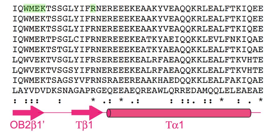
Downloaded from genesdev.cshlp.org on November 18, 2020 - Published by Cold Spring Harbor Laboratory Press

Rickman et al.

(Niedernhofer et al. 2004; Kim et al. 2011, 2013; Klein               Results
Douwel et al. 2014; Alcón et al. 2020; Tan et al. 2020;
Wang et al. 2020). Unhooking of the ICL enables transle-              Atypical presentation of Fanconi anemia in individuals
sion bypass on one-strand and double-strand break (DSB)               with BRCA2/FANCD1 DNA-binding domain variants
repair by homologous recombination (HR) on the second                 Two female siblings, enrolled in the International Fanconi
strand (Howlett et al. 2002; Litman et al. 2005; Xia et al.           Anemia Registry (IFAR), with unknown causative
2007; Long et al. 2011).                                              gene mutations, were born with a multitude of congenital
   A number of FA proteins, BRCA2/FANCD1, PALB2/                      abnormalities and had mildly elevated levels of chromo-
FANCN, FANCJ/BRIP1, RAD51C/FANCO, RAD51/                              somal breakage at birth (see Supplemental Table S1 for
FANCR, and BRCA1/FANCS are known for facilitating                     clinical presentation). Biallelic BRCA2/FANCD1 variants
HR (Howlett et al. 2002; Litman et al. 2005; Rahman                   (c.2330dupA and c.8524C>T) were identified by whole-
et al. 2007; Xia et al. 2007; Vaz et al. 2010; Sawyer et al.          exome sequencing (WES) and no other likely pathogenic
2015). BRCA2/FANCD1 is an essential gene and single al-               FA gene variants were observed. These results were sur-
lele pathogenic variants predispose to breast and ovarian             prising since neither sibling displayed the typical clinical
cancer and biallelic pathogenic variants result in a sub-             findings of the FA-D1 complementation group, with no
type of Fanconi anemia, FA-D1 (Howlett et al. 2002). FA               history of malignancy or bone marrow failure at the ages
is a heterogeneous disease, but even within the disease               of 20 and 23. There is no reported family history of FA,
spectrum, patients with biallelic pathogenic variants in              but there are cases of breast cancer that were diagnosed
BRCA2/FANCD1 are phenotypically distinct from the                     later in life (above 60 yr of age), individuals with skin can-
most common complementation groups, FA-A, FA-C,                       cer in the family, and early onset colorectal cancer in the
and FA-G. A higher proportion of FA-D1 patients have                  father (40 yr old) (Fig. 1A).
developmental abnormalities and nearly one hundred per-                  The frameshift c.2330dupA variant of exon 11 (maternal
cent have a malignancy by 5 yr of age (Alter et al. 2007),            origin) results in premature truncation of BRCA2
which is most likely due to HR deficiency.                            (p.Asp777Glufs∗ 11) and has been described previously in
   Functional analysis of BRCA2 has largely focused on ca-            hereditary breast and ovarian cancer (HBOC) (Supplemen-
nonical HR, and the role of BRCA2 in ICL repair has been              tal Fig. S1A). The c.8524C>T missense variant of exon 20
associated with the repair of DSBs generated by pro-                  (paternal origin) results in an p.Arg2842Cys residue chan-
grammed incisions at the ICL. Outside of their role in                ge in the highly conserved DNA-binding domain (DBD) of
HR and ICL repair, BRCA2 and RAD51, along with a num-                 BRCA2 and has previously been identified as a variant of
ber of other recently described proteins, function in repli-          unknown significance (VUS) in HBOC (Fig. 1B,C; Supple-
cation fork protection (Rickman and Smogorzewska                      mental Fig. S1B,C). At the protein level, the missense var-
2019). In the absence of replication fork protection, newly           iant results in the p.Arg2842Cys change at a highly
synthesized DNA is degraded at replication forks stalled              conserved residue at the base of the BRCA2 Tower domain
due to dNTP imbalance secondary to hydroxyurea (HU)                   of the DBD (Supplemental Fig. S1C). Sequencing of periph-
treatment, and a number of nucleases including MRE11,                 eral blood and lymphocytes demonstrated the presence of
CTIP, and EXO1 have been implicated in the process                    both variants and no evidence of somatic mosaicism.
(Lemaçon et al. 2017; Przetocka et al. 2018; Rickman                     A third individual with FA, biallelic BRCA2 variants,
and Smogorzewska 2019).                                               and an atypical presentation, was identified in the litera-
   Another nuclease, DNA2, has also been shown to resect              ture (Howlett et al. 2002). This individual was homozy-
DNA at ICLs in cells expressing the RAD51/FANCR sep-                  gous for the c.8488-1G>A variant (alias “IVS19-1G>A”)
aration of function mutant, p.T131P, identified in an indi-           that alters the splice acceptor site of exon 20. cDNA anal-
vidual with FA-like syndrome. The mutant RAD51                        ysis demonstrated the use of an alternate splice acceptor
p.T131P has a dominant-negative effect on RAD51 func-                 that results in the loss of 12 bp of exon 20 and translates
tion that does not seem to affect HR at cellular levels but           into p.Trp2830_Lys2833del (Fig. 1B,C; Howlett et al.
disrupts the function of RAD51 at ICLs, suggesting a fork             2002). Amino acid residues 2830–2833 are located within
protection role for RAD51 in ICL repair. The requirement              the DBD at the transition of the OB2 fold and the base of
for BRCA2 in the early steps of ICL repair to prevent aber-           the Tower domain (Fig. 1B,C; Supplemental Fig. S1C).
rant resection has not previously been determined. Here               This individual was 30 yr of age at last follow up, was
we investigated the requirements of BRCA2 with RAD51                  born with a thumb malformation, but had no history of
in fork protection at ICLs and demonstrate that the two               bone marrow failure or malignancy. Similar to the sibling
proteins are both required to prevent hyperresection by               pair, chromosomal breakage was modest (Howlett et al.
the DNA2-WRN nuclease-helicase complex, but not                       2002).
MRE11. These studies were performed using BRCA2
DNA-binding domain (DBD) mutants discovered in FA pa-
tients and these variants were determined to confer loss of
                                                                      BRCA2 DNA-binding domain variants identified in FA
replication fork protection but only moderate HR defi-
                                                                      patients confer defects in the response to replication
ciency. Our results indicate that the BRCA2 DBD is re-
                                                                      stress
quired for replication fork protection and that BRCA2
fork protection at HU-induced and ICL-induced stalled                 Lymphoblastoid cell lines (LCL) (RA3105 and RA3106)
forks are distinct processes.                                         were derived from the sibling pair with compound

2      GENES & DEVELOPMENT
Downloaded from genesdev.cshlp.org on November 18, 2020 - Published by Cold Spring Harbor Laboratory Press

                                                                                                Function of the BRCA2 DNA-binding domain

        A                                                      C                                           D

                                                                                                           E

        B

          F                           G                            H                    I

          J                               K                                        L

Figure 1. BRCA2 variants identified in individuals with atypical Fanconi anemia. (A) Family pedigree showing a sibling pair with Fan-
coni anemia (red circles) who are compound heterozygous for BRCA2 variants c.2330dupA (maternal inheritance) and c.8524C>T (pater-
nal inheritance). Family history of breast cancer (purple, all diagnosed in 60s and 70s), skin cancer (gray), and colon cancer (green;
diagnosed at 40 yr old). (B) Schematic of BRCA2 domain structure and key interacting proteins. (C ) Alignment of exon 20 BRCA2 DBD
peptide sequence demonstrating that it is evolutionary conserved across many species. In green are the amino acid residues modified
by the patient variants, p.W2830_K2833del (c.8488-1G>A) and p.R2842C (c.8524C>T). Purple arrows indicate amino acid residues that
contact DNA (Yang et al. 2005). (D) Immunoblot showing BRCA2 levels in WT (RA2985) control, FA-D1 (RA2525), and patient
RA3105 and RA3106 LCLs. (E) Quantification of chromosome breaks following DEB treatment of WT (RA2985), FA-A (RA2939), and pa-
tient RA3105 and RA3106 LCLs. (F,G) Cell survival assays of patient-derived lymphoblast cell lines (LCLs) RA3105, FA-A (RA2939), WT
(RA2985), and FA-D1 (RA2525) after MMC and PARP inhibitor olaparib (PARPi) treatment. Relative cell survival was normalized to un-
treated controls to give percent survival. Error bars indicate SD. (H) Quantification of chromosome breaks following MMC treatment of BJ
wild-type fibroblasts, FA-A patient fibroblasts, and HSC62 fibroblasts. (I) Cell survival of HSC62 (c.8488-1G>A) fibroblasts compared with
BJ WT fibroblast and complemented FA-A patient cells (RA3087) expressing wild-type FANCA (FA-A+A) or empty vector (FA-A+EV).
Cells were treated with increasing concentrations of MMC. Relative cell survival was normalized to untreated controls to give the percent
survival. Error bars indicate SD. (J) Cell survival of MMC-treated HSC62 uncorrected patient cell line (HSC62mut) compared with BJ WT
fibroblast and CRISPR/Cas9 corrected wild-type HSC62 (HSC62WT) clones 1-3. (K,L) Cell survival of BJ WT fibroblasts, and CRISPR/Cas9-
targeted BJ fibroblasts: BJ WT fibroblast clone (BRCA2 WT), c.8488-1G>A BJ clones (BRCA2 8488-1G>A), c.8524C>T BJ clones
(BRCA2 8524C>T), and exon 20 BRCA2 frameshift mutant (BRCA2 Trun.). Cells were treated with increasing concentrations of MMC or
PARPi. Error bars indicate SD. Kruskal-Wallis ANOVA, with Dunn’s post-test. (∗∗∗ ) P < 0.001; (∗∗∗∗ ) P < 0.0001.

heterozygous BRCA2 variants, c.2330dupA and c.8524C>T.                  MMC and DEB, but to a lesser degree than RA2939 (Fig.
FA pathway activation requires monoubiqutination of                     1F; Supplemental Fig. S1F). RA3105 was also hypersensi-
FANCI, which was observed in patient-derived LCLs (Sup-                 tive to replication stress-inducing agents including ola-
plemental Fig. S1D). Analysis of BRCA2 expression by                    parib, a PARP inhibitor (PARPi), and camptothecin
Western blot demonstrated a full-length (∼390-kDa)                      (CPT), a topoisomerase I inhibitor (Fig. 1G; Supplemental
band, the presumed product of the c.8524C>T allele, for                 Fig. S1G).
both patient cell lines (Fig. 1D). DEB-induced breakage                    Similarly, analysis of patient-derived fibroblasts,
analysis confirmed previous clinical data that breakage                 HSC62 (Howlett et al. 2002), from the individual with ho-
was elevated, but not to levels of the typical FANCA-defi-              mozygous c.8488-1G>A variant also revealed more mod-
cient (FA-A) LCLs (RA2939) (Fig. 1E). RA3105 LCL dis-                   erate chromosomal breakage to DEB and MMC and
played hypersensitivity to the cross-linking agents                     cellular hypersensitivity to cross-linking agents (Fig. 1H,

                                                                                                         GENES & DEVELOPMENT            3
Downloaded from genesdev.cshlp.org on November 18, 2020 - Published by Cold Spring Harbor Laboratory Press
Rickman et al.
I; Supplemental Fig. S1O). The cells were not hypersensi- that the BRCA2 DBD mutants are hypomorphic in their
tive to ionizing radiation (IR), but were sensitive to repli- mediator function.
cation stress induced by CPT and PARPi (Supplemental
Fig. S1J–L). In contrast, the cells were not sensitive to rep-
Increased ssDNA in BRCA2 DBD variants is dependent
lication stress produced by the agents aphidicolin and HU
on DNA2 and WRN
(Supplemental Fig. S1M,N).
We corrected the pathogenic variants in the HSC62 The previously described RAD51/FANCR p.T131P
patient fibroblast cell line to demonstrate that the patient-derived cell line that is proficient for HR but de-
c.8488-1G>A variant caused the observed defects. The ho- fective in ICL repair displays increased RPA phosphoryla-
mozygous c.8488-1G>A variant was corrected to wild tion and foci formation indicating an increase in ssDNA
type at the endogenous locus using CRISPR/Cas9 gene upon MMC treatment (Wang et al. 2015). Given that the
targeting. Both heterozygous and homozygous clones interaction of BRCA2 and RAD51 is required for their
were recovered (HSC62WT/MUT or HSC62WT/WT) (Supple- canonical function in HR and their noncanonical func-
mental Fig. S1P). cDNA analysis demonstrated that resto- tion in replication fork protection at HU-induced stalled
ration of the splice acceptor base (A>G) in HSC62WT/MUT forks, we investigated whether BRCA2 also functions
or HSC62WT/WT clones restored the cDNA exon 19-20 in preventing increased ssDNA generation at ICLs
junction (Supplemental Fig. S1Q). Both HSC62WT/MUT (Schlacher et al. 2011; Wang et al. 2015; Mijic et al.
and HSC62WT/WT clones rescued hypersensitivity to repli- 2017; Bhat et al. 2018). We observed an increase in
cation stress-inducing agents MMC, CPT, and PARPi (Fig. RPA foci formation in HSC62MUT cells compared with
1J; Supplemental Fig. S1S,T). wild-type fibroblasts upon MMC treatment (Fig. 2G;
For a direct comparison of the BRCA2 DNA-binding Supplemental Fig. S3A). Similar to RAD51/FANCR
domain variants, we generated isogenic cell lines by intro- p.T131P-expressing patient cells, the increased RPA
ducing the variants, c.8524C>T (p.R2842C) and c.8488- foci formation in HSC62 cells was also dependent on
1G>A (p.Trp2830_Lys2833del), into wild-type BJ fibro- DNA2 and WRN activity, but not MRE11, EXO1,
blasts with CRISPR/Cas9 gene editing (Supplemental CTIP, or BLM (Fig. 2H; Supplemental Fig. S3B–D). Code-
Fig. S2A,B). Knock-in of the BRCA2 c.8488-1G>A variant pletion of WRN with BLM did not further rescue the in-
in BJ fibroblasts conferred the same splicing defect ob- creased RPA foci in HSC62MUT cells after MMC (Fig. 2I;
served in HSC62 cells (Supplemental Fig. S2A). Western Supplemental Fig. S3E). Increased RPA foci and phos-
blot analysis of BRCA2 demonstrated an ∼390-kDa band phorylation following MMC was also observed for
for all mutants except for BRCA2 clones containing c.8524C>T and c.8488-1G>A mutants, with a greater in-
exon 20 frameshift variants obtained in parallel using crease for the c.8488-1G>A mutants (Fig. 2J; Supplemen-
CRISPR/Cas9 gene targeting (Supplemental Fig. S2C). tal Fig. S3F–J). These results suggest that BRCA2 is
The BRCA2 frameshift mutant is homozygous functioning with RAD51 to protect against aberrant pro-
c.8531dupA with a predicted p.R2845Kfs∗ 22 truncation cessing by DNA2 and WRN at ICLs, but not against the
(BRCA2Trun.). Analysis of cellular sensitivity of the other effectors of DSB end resection such as MRE11,
BRCA2 DBD mutants revealed that presence of both EXO1, or CTIP. Overexpression of RAD51 in the
DBD variants sensitize cells to MMC, PARPi, and CPT BRCA2 c.8524C>T- and c.8488G>A-expressing cells par-
but not aphidicolin, recapitulating phenotypes of patient tially rescued cellular sensitivity to MMC and RPA foci
HSC62 fibroblasts (Fig.1K,L; Supplemental Fig. S2D,E). formation after MMC (Fig. 2K,L; Supplemental Fig. S3K,
L). This data supports that RAD51 and BRCA2 function
interdependently at ICLs.
BRCA2 DNA-binding domain variants confer defects
To determine whether blocking ICL unhooking or
in RAD51 recruitment after IR and MMC
nuclease mediated fork collapse (McPherson et al. 2004;
To determine the impact of DBD variants on the ability of Niedernhofer et al. 2004; Dendouga et al. 2005; Kim
BRCA2 to load RAD51 onto ssDNA following DNA dam- et al. 2011; Bogliolo et al. 2013) would rescue RPA foci for-
age, we analyzed RAD51 foci formation after IR and mation in BRCA2 DBD mutant cells following MMC, we
MMC. Levels of RAD51 foci and focus size were reduced depleted SLX4 and MUS81. Depletion of either SLX4 or
after IR and MMC treatment in HSC62 cells, which was MUS81 did not rescue the increase in chromatin bound
rescued by the CRISPR/Cas9 gene editing (Fig. 2A–D; Sup- RPA in the BRCA28524C>T and BRCA28488-1G>A- express-
plemental Fig. S2H,I). Analysis of isogenic BJ cell lines ing cells (Supplemental Fig. S3M–O). SLX4 depletion fur-
with DBD mutations also demonstrated defects in ther increased the RPA foci formation, indicating further
RAD51 foci formation following IR and MMC (Fig. 2E,F; defects in ICL repair in its absence, which may be the re-
Supplemental Fig. S2J,K). The c.8488-1G>A variant had sult of loss of function of the associated nucleases.
a stronger impact on RAD51 foci formation, resulting in
fewer cells with RAD51 foci and reduced focus size. The
ICLs are a substrate of nucleolytic processing
c.8524C>T mutant did not show a significant reduction
in the absence of a functioning FA pathway
in the number of cells with RAD51 foci; however, the
foci were smaller in size (Fig. 2F; Supplemental Fig. Having demonstrated that BRCA2 and RAD51 share a role
S2K). By comparison, the BRCA2Trun. mutant had com- in protecting ICLs from overresection by DNA2 and
plete loss of observable RAD51 foci. These data indicate WRN, we investigated whether other FA proteins are
4 GENES & DEVELOPMENTDownloaded from genesdev.cshlp.org on November 18, 2020 - Published by Cold Spring Harbor Laboratory Press
Function of the BRCA2 DNA-binding domain
A B C D
E F
G H
K L
I J
Figure 2. Defective ICL repair in BRCA2 DBD mutants results in increased ssDNA that is WRN and DNA2 dependent. (A) Immunoflu-
orescence images of RAD51 foci, 8 h following 12 Gy ionizing radiation (IR) of BJ WT fibroblast and patient derived HSC62 fibroblast,
detected with anti-RAD51 antibody. Third row images are individual cells enlarged to better demonstrate differences in RAD51 focus
size. (B) Quantification of RAD51 foci 1 h, 8 h, and 24 h following 12 Gy IR of BJ WT fibroblast and HSC62 fibroblast. Error bars indicate
SD of two independent experiments (≥200 cells per experiment). (C) Quantification of RAD51 foci 8 h after 12 Gy IR of BJ WT fibroblast,
wild-type HSC62 (HSC62WT) clones 1–3, and HSC62 uncorrected patient cell line (HSC62mut). (D) Quantification of RAD51 foci 24 h fol-
lowing 1-h treatment with 3 µM MMC. Error bars indicate SD of three independent experiments (≥200 cells per experiment). (E) Quan-
tification of RAD51 foci in isogenic BJ fibroblasts clones at 1 h, 8 h, and 24 h following 6 Gy IR of BJ WT fibroblasts, BJ WT fibroblast clone
(BRCA2WT), BRCA2 8488-1G>A BJ clones 2–3, BRCA2 8524C>T BJ clones 1–2, and a BRCA2 homozygous truncation mutant, c.8531dupA
(BRCA2 Trun). Error bars indicate SD of three independent experiments (≥200 cells per experiment). (F) Representative images of
RAD51 foci in isogenic BJ fibroblasts clones, 8 h after 6 Gy IR, detected by immunofluorescence with anti-RAD51 antibody. Third row
images are individual cells enlarged to better demonstrate differences in RAD51 focus size. (G) Quantification of RPA foci 24 h following
1-h treatment with 3 μM MMC of BJ WT fibroblast, CRISPR/Cas9 corrected wild-type HSC62 clones (HSC62WT), and HSC62 uncorrected
patient cell line (HSC62mut). (H) Quantification of RPA foci 24 h following 1-h treatment with 3 μM MMC in HSC62mut cells depleted of
DNA2, MRE11, EXO1, CTIP, WRN, or BLM by siRNA compared with luciferase control (Luc). Error bars indicate SD of four independent
experiments. (I ) Quantification of RPA foci 24 h following 1 h treatment with 3 μM MMC in HSC62mut cells depleted of DNA2, WRN,
BLM, or codepleted of WRN and BLM by siRNA compared with luciferase control (Luc). Error bars indicate SD of three independent ex-
periments. (J) Immunoblot analysis of RPA phosphorylation in isogenic BJ fibroblasts clones 24 h after 1-h treatment with 3 μM MMC.
BRCA2 WT, BRCA2 8524C>T, and BRCA2 8488-1G>A BJ fibroblast cells were transfected with siRNA control luciferase (Luc) or siRNAs target-
ing DNA2 or WRN. (K,L) MMC cell survival of BJ BRCA2 WT, BRCA2 8488-1G>A, and BRCA2 8524C>T fibroblasts overexpressing (OE) WT
RAD51 or empty vector (EV) control. Relative cell survival was normalized to untreated controls to give percent survival. Error bars in-
dicate SD.
also required for protection against DNA hyperresection foci formation following MMC treatment for all comple-
at ICLs. Analysis of a panel of FA patient-derived cells mentation groups (Fig. 3A). To determine whether the ge-
with mutations in FANCA, FANCL, FANCD2, FANCI, netic requirement for RPA suppression was the same as in
FANCJ, and SLX4/FANCP demonstrated increased RPA BRCA2 and RAD51 mutant cells, DNA2 and WRN were
GENES & DEVELOPMENT 5Downloaded from genesdev.cshlp.org on November 18, 2020 - Published by Cold Spring Harbor Laboratory Press
Rickman et al.
A Figure 3. Proper ICL repair is required to prevent aber-
rant nuclease processing. (A) Quantification of RPA foci
8 h, 24 h, and 48 h following 1-h treatment with 3 μM
MMC of FA patient-derived fibroblasts compared with
BJ wild-type fibroblasts. Patient cells lines from FA
complementation group FA-R (RAD51/FANCR), FA-A
(FANCA), FA-L (FANCL), FA-D2 (FANCD2), FA-I
(FANCI), FA-J (FANCJ), and FA-P (SLX4/FANCP). FA-
A patient complemented cell lines were generated by
transducing WT FANCA cDNA or EV. Error bars indi-
cate SD of two independent experiments. (B) FA-A pa-
tient cells expressing WT FANCA (FA-A+FANCA) or
empty vector (FA-A+EV) were transfected with siRNA
B C D control luciferase (Luc) or siRNAs targeting DNA2
and WRN. Quantification of RPA foci 24 h following
1-h treatment with 3 μM MMC. Error bars indicate
SD of two independent experiments. (C ) FA-A+EV
were transfected with siRNA Luc or siRNAs targeting
DNA2 and BLM. Quantification of RPA foci 24 h fol-
lowing 1-h treatment with 3 μM MMC. Error bars indi-
cate SD of two independent experiments. (D) FA-G
patient cells expressing WT FANCG (FA-G+FANCG)
or empty vector (FA-G+EV) were transfected with
siRNA control luciferase (Luc) or siRNAs targeting
DNA2, WRN, and BLM. Quantification of RPA foci 24 h following 1-h treatment with 3 μM MMC. Error bars indicate SD of three
independent experiments.
depleted in a complemented pair of FANCA (Fig. 3B,C; readout of HR (Sonoda et al. 1999). SCEs were induced
Supplemental Fig. S3P,Q) and FANCG patient-derived by increasing concentrations of MMC or depletion of
cells (Fig. 3D). Interestingly, the dependence on DNA2 BLM. There was no significant difference in SCE levels ob-
was the same, but the helicase dependency was different, served in wild-type BJ fibroblasts and HSC62 cells (Fig. 4B;
as WRN did not rescue RPA levels but BLM depletion did Supplemental Fig. S4E–G); however, SCE levels were sup-
(Fig. 3C,D; Supplemental Fig. S3R–T). These data demon- pressed in BRCA2 Trun fibroblasts (Supplemental Fig.
strate a dependence on the FA core complex and pathway S4H). These observations suggest that the DNA-binding
associated proteins to prevent resection of ICLs by DNA2 domain defect in HSC62 cells, while decreasing RAD51
and BLM. They also suggest that different nuclease–heli- foci formation, does not significantly reduce HR as ob-
case pairs engage when ICL repair is halted at different served by normal resistance to IR and SCE levels in these
stages of the process. cells. Taken together, the variants moderately reduce HR
at Cas-9 targeted DSBs but do not impact cellular HR read-
outs, which is similar to the behavior of cells carrying the
Determination of homologous recombination efficiency RAD51 p.T131P mutation (Wang et al. 2015).
in DNA-binding domain mutants
To determine the HR proficiency of BRCA28488-1G>A and
The BRCA2 DNA-binding domain is required for
BRCA28524C>T -expressing cells, we used a HDR assay
replication fork protection at HU-induced stalled forks
that targets DSBs at the LMNA locus (Pinder et al. 2015;
Arnoult et al. 2017). The assay was performed in To determine the requirement for the BRCA2 DBD
HEK293T cells after CRISPR-Cas9 gene editing to engi- in replication fork protection after HU treatment,
neer either BRCA2 DBD variants or the exon 27 BRCA28524C>T- and BRCA28488-1G>A-expressing cells
p.S3291A variant, previously reported to have an effect were examined by DNA fiber analysis. Replication fork
on replication fork protection but not on HR (Supplemen- protection by BRCA2 has largely been attributed to the
tal Fig. S4A,B; Schlacher et al. 2011; Kim et al. 2014). C-terminal RAD51 interacting domain by analysis of the
Compared with wild-type cells, HR in all BRCA2 BRCA2 p.S3291A variant (Schlacher et al. 2011). Analysis
clones, including the S3291A mutant, was moderately de- of BRCA2 Trun-, BRCA28524C>T-, and BRCA28488-1G>A-ex-
creased (Fig. 4A). Cells with DBD BRCA28488-1G>A and pressing cells demonstrated defects in replication fork
BRCA28524C>T variants showed similar decreases in HR protection of HU-induced stalled forks as measured by
levels to approximately half that of wild-type cells but re- the degradation of nascent DNA tracks labeled with nu-
tained significantly more HR activity than cells depleted cleotide analogs, IdU and CldU. As previously reported,
of RAD51 and BRCA2 or BRCA2 Trun cells, consistent nascent strand degradation in the absence of BRCA2 was
with a previous study (Siaud et al. 2011). rescued by the MRE11 inhibitor mirin and MRE11
Given the normal resistance to IR in HSC62 fibroblasts, depletion (Fig. 4C,D). These data demonstrate that the
we assessed sister chromatid exchange (SCEs) levels as a BRCA2Trun-, BRCA28524C>T-, and BRCA28488-1G>A-
6 GENES & DEVELOPMENTDownloaded from genesdev.cshlp.org on November 18, 2020 - Published by Cold Spring Harbor Laboratory Press
Function of the BRCA2 DNA-binding domain
A B C
D
E F
Figure 4. BRCA2 DBD and C-terminal domain variants confer a moderate defect in HR and disrupt replication fork protection function.
(A) Levels of mClover-positive cells were normalized to WT HEK293T (siLuc). Error bars indicate SD of three independent experiments
performed in triplicate. P-values were determined by ANOVA and Tukey’s multiple comparison. (∗∗∗∗ ) P < 0.0001. (B) Sister chromatid
exchange (SCE) assay in BJ WT fibroblast and HSC62 patient derived fibroblast following treatment with 0.1 μg/mL or 0.2 μg/mL
MMC. (C ) Isogenic BJ fibroblast BRCA2 mutants, BRCA2 Trun., BRCA2 8524C>T, and BRCA2 8488-1G>A were analyzed for replication fork re-
section. Cells were labeled with DNA analogs, IdU for 20 min, and then CldU for 20 min. Cells were then incubated in 6 mM HU with and
without MRE11 inhibitor mirin (50 µM) for 4 h before being harvested. DNA fibers were prepared and visualized by immunofluorescence
detection of IdU and CldU and measured. Error bars indicate SD. (D) Isogenic BJ fibroblast BRCA2 mutants, BRCA2 Trun., BRCA2 8524C>T,
BRCA2 8488-1G>A, and BRCA2 S3291A were transfected with siRNA control luciferase (Luc) or siRNAs targeting DNA2 or MRE11. Cells
were treated and labeled with DNA analogs as above. Error bars indicate SD. (E) BJ fibroblast with BRCA2 variants, BRCA2 8524C>T and
BRCA2 8488-1G>A, were analyzed for replication fork resection when depleted of RADX by shRNA or transduced with shRNA control
(shCONT.). Cells were treated and labeled with DNA analogs as above. Data of two replicates plotted. Error bars indicate SD. (F) Quan-
tification of chromosome breaks in isogenic BJ fibroblast BRCA2 mutants following 5 h of 6 mM HU and released into colcemid. Breakage
was not significantly increased in BRCA2 8524C>T and BRCA2 8488-1G>A compared with BRCA2 WT. Kruskal-Wallis ANOVA, with Dunn’s
post-test. (∗∗ ) P < 0.01; (∗∗∗ ) P < 0.001; (∗∗∗∗ ) P < 0.0001.
expressing cells are all similarly defective for replication bers, the levels of chromosomal breakage differed (Fig.
fork protection and that the DBD is required for protection 4F; Supplemental Fig. S5E,F). Metaphases were analyzed
of replication forks from MRE11 processing. Depletion of after 5 h of 6 mM HU and release into colcemid.
DNA2 also rescues resection after HU in cells expressing BRCA2 Trun.-expressing cells showed a large increase in ge-
all of the BRCA2 mutants including BRCA2Trun., nomic instability upon stalling with HU in comparison
BRCA28524C>T, BRCA28488-1G>A, and BRCA2 S3291A (Fig. with WT and the other BRCA2 mutants. Cells with
4D). RADX depletion has been shown to rescue nascent BRCA28524C>T and BRCA2 S3291A variants did not show
strand degradation at HU-induced stalled replication forks an elevation in breakage and BRCA28488-1G>A-expressing
in BRCA2-deficient cells without restoring HR function cells had a mild increase. The elevated chromosomal
(Dungrawala et al. 2017). Consistent with these studies, breakage in BRCA2 Trun. cells were reduced by MRE11
depletion of RADX in the BRCA2 DBD mutant-expressing depletion, but exacerbated by DNA2 depletion (Fig. 4F;
cells did not rescue HR defects (Supplemental Fig. SF5A– Supplemental Fig. S5E). DNA2 depletion resulted in a
D) but did rescue nascent strand degradation (Fig. 4E). mild increase in breakage for all mutants but resulted in
Taken together, these data demonstrate that both the a synergistic increase in cells with BRCA2Trun. Previous
DBD and C-terminal domain of BRCA2 are required for studies have reported elevated breakage resulting from
proper replication fork protection at HU-induced stalled replication fork degradation in p.S3291A expressing cells
forks, and that both domains are required to protect and BRCA2 deficient cells (Schlacher et al. 2011; Mijic
against degradation by the nucleases MRE11 and DNA2. et al. 2017). In contrast, the newly characterized
Although all of the BRCA2 mutants showed similar lev- BRCA28524C>T- or BRCA2 S3291A-expressing cells in this
els of nascent strand resection as measured by DNA fi- study do not have a significant increase in breakage after
GENES & DEVELOPMENT 7Downloaded from genesdev.cshlp.org on November 18, 2020 - Published by Cold Spring Harbor Laboratory Press
Rickman et al.
HU, despite having levels of fork degradation similar to MMC that was DNA2-WRN dependent. These data sug-
BRCA2 Trun (Fig. 4F; Supplemental Fig. S5F). Our data gest that like the well-described interdependence of
demonstrate that different levels of BRCA2 function im- BRCA2 and RAD51 in HR, BRCA2, and RAD51 function
pairment have different consequences on HU-induced together in the early steps of ICL repair to prevent DNA
stalled forks and that replication fork resection at HU-in- resection and that the function of the BRCA2 DBD is im-
duced stalled forks does not always manifest in chromo- portant for this role. This expands the role of BRCA2 in
somal breakage. How this breakage occurs in BRCA2 ICL repair beyond HR to include protection of DNA at
depleted or LOF cells needs to be investigated further, the ICL stalled replication fork from aberrant nucleolytic
but like nascent DNA degradation, it is partially depen- processing (Fig. 6).
dent on MRE11. Depletion of the replication fork remodelers SMAR-
CAL1, ZRANB3, and HLTF and the RAD51 modulator
RADX rescued nascent strand degradation at HU-induced
SMARCAL1, ZRANB3, and HLTF function is not stalled forks in cells carrying DBD variants consistent
required for ICL repair with the previous data on the role of BRCA2 in this pro-
Replication fork reversal has been observed as a response cess (Dungrawala et al. 2017; Lemaçon et al. 2017; Mijic
to replication stress induced by a number of different clas- et al. 2017; Taglialatela et al. 2017). However, depletion
ses of genotoxic agents including MMC (Zellweger et al. of the translocases did not mitigate cellular sensitivity
2015). SMARCAL1, ZRANB3, and HLTF are ATPase-de- or increased RPA after MMC in the BRCA2 DBD mu-
pendent DNA translocases of the SNF2 family of chroma- tant-expressing cells. Our study demonstrates that remod-
tin remodelers that have recently been shown to promote eling by the translocases is not a major step in the repair of
replication fork reversal in vitro and in vivo. Depletion of ICLs and suggests that the MMC-induced replication fork
any of the three translocases rescues nascent strand resec- reversal may be a more general response to replication
tion at HU-induced stalled forks in BRCA2-deficient cells stress but not specifically at the fork that is stalled at an
(Mijic et al. 2017; Taglialatela et al. 2017). Similarly, ICL (Zellweger et al. 2015; Mutreja et al. 2018). These
depletion of the translocases in the BRCA28524C>T- and data further support that the protection by BRCA2 and
BRCA28488-1G>A-expressing cells rescued nascent strand RAD51 at a HU-induced stalled fork is different from pro-
degradation (Fig. 5A). However, depletion of SMARCAL1, tection at an ICL (Fig. 6).
ZRANB3, or HLTF did not rescue the increased RPA phos- The mechanism of DNA protection at the ICL by the
phorylation and foci formation after MMC (Fig. 5B,C; Sup- BRCA2 DBD domain remains to be explored. However,
plemental Fig. S5G–I). Codepletion of SMARCAL1 and the location of the variants at the transition of the OB2
ZRANB3 also had no effect on decreasing RPA foci forma- fold and base of the Tower domain suggests a plausible
tion after MMC (Supplemental Fig. S5J,K). To determine mechanism of protection at an ICL stalled fork. The
whether the proteins implicated in replication fork rever- OB2 fold binds to ssDNA and the Tower domain contains
sal are important for the repair of ICLs, wild-type cells a 3HB domain at the apex that is capable of binding to
were depleted of SMARCAL1 or ZRANB3 and tested for dsDNA (Yang et al. 2002). We speculate that the muta-
sensitization to MMC. Cells depleted of either translocase tions in this region of the DBD may preclude efficient
were not significantly sensitized to MMC (Fig. 5D). Addi- binding/bridging at ssDNA–dsDNA junctions, which is
tionally, depletion of either translocase did not rescue cel- a structure expected at stalled forks, and lack of this bind-
lular hypersensitivity to MMC or CPT in BRCA28488-1G>A ing would lead to the deprotection phenotype. Lack of
cells (Fig. 5E,F). These data suggest that the function of proper placement of BRCA2 may also preclude proper
these translocases is not required during MMC-induced RAD51 loading, which may lead to inappropriate DNA re-
ICL repair. section. Biochemical analysis of the BRCA2 variants we
have identified in atypical Fanconi anemia patients will
further our understanding of how BRCA2 interacts with
Discussion different replication fork structures.
BRCA2 and RAD51 function at the ICL
FA protein function at the ICL
Here we studied the functional consequences of pathogen-
ic BRCA2 variants in the DNA-binding domain in the FA proteins have been shown previously to be important
context of homologous recombination, and protection of for protection at HU-induced stalled replication forks
stalled replication forks due to dNTP depletion or DNA (Schlacher et al. 2012). Here we show that FA patient
interstrand cross-link lesions. The DBD variants did not cell lines from various complementation groups also dem-
affect IR sensitivity, SCE levels, or HU sensitivity, sug- onstrate increased ssDNA and RPA foci formation after
gesting that the HR levels in cells carrying the DBD vari- MMC. However, in FANCA-deficient cells, the increase
ants is sufficiently intact. We also saw only a moderate in RPA foci is dependent on DNA2 and BLM, but not
reduction in HR using an HR reporter assay. Similar to WRN. This suggests that the fork protection of BRCA2–
the previously described patient cell line with RAD51/ RAD51 is not redundant with the FA core complex, but
FANCR p.T131P mutation (Wang et al. 2015), the cells further investigation will be needed to determine the ge-
with BRCA2 DBD variants were sensitive to ICL-inducing netic dependency of increased ssDNA in the absence of
agents and showed increased RPA foci formation after the other FA proteins. DNA2 has previously been reported
8 GENES & DEVELOPMENTDownloaded from genesdev.cshlp.org on November 18, 2020 - Published by Cold Spring Harbor Laboratory Press
Function of the BRCA2 DNA-binding domain
A B C
D E F
Figure 5. SNF2 translocases are not required for ICL repair. (A) BJ fibroblast mutants BRCA2 8524C>T and BRCA2 8488-1G>A were analyzed
for replication fork resection when depleted of either SMARCAL1 or ZRANB3 by shRNA or transduced with control shRNA (shLuc).
Cells were labeled with DNA analogs, IdU for 20 min and then CldU for 20 min. Cells were then incubated in 6 mM HU for 4 h before
being harvested. DNA fibers were prepared and visualized by immunofluorescence detection of IdU and CldU and measured. Error bars
indicate SD (B) Quantification of RPA foci in isogenic BJ fibroblasts clones 24 h following 1-h treatment with 3 µM MMC in cells depleted
of SMARCAL1 or ZRANB3. Error bars indicate SD of two independent experiments. (C) Quantification of RPA foci in BJ fibroblasts clones
24 h following 1-h treatment with 3 µM MMC in cells depleted of HLTF. Error bars indicate SD of two independent experiments. (D) MMC
cell survival of isogenic BJ BRCA2 WT fibroblasts depleted of SMARCAL1 or ZRANB3 by shRNA or transduced with shRNA luciferase
control (shLuc). Relative cell survival was normalized to untreated controls to give percent survival. Error bars indicate SD. (E,F)
MMC and CPT cell survival assay of isogenic BJ BRCA2 8488-1G>A or BRCA2 8524C>T clones depleted of either SMARCAL1 or ZRANB3
by shRNA or transduced with shRNA luciferase control (shLuc). Relative cell survival was normalized to untreated controls to give per-
cent survival. Error bars indicate SD. Kruskal-Wallis ANOVA, with Dunn’s post-test. (∗∗∗ ) P < 0.001; (∗∗∗∗ ) P < 0.0001.
to interact with FANCD2 and be recruited to ICLs where ence in experimental set-up (Schlacher et al. 2011). Our
it is required for repair, but is deleterious in the absence of findings extend the replication fork protection role of
FANCD2 (Karanja et al. 2012, 2014). BLM has been report- BRCA2 at HU-induced stalled replication forks beyond
ed to interact with a number of FA proteins and colocalize the C-terminal domain and show that fork protection
with FANCD2 at ICLs (Meetei et al. 2003; Pichierri et al. likely requires the DBD to bind DNA at the stalled repli-
2004; Suhasini and Brosh 2012). Consistent with BLM cation fork. It remains to be determined whether the DBD
depletion rescuing increased ssDNA at the fork in the ab- variants have an effect on replication fork reversal after
sence of FANCA, BLM knockout was also recently report- HU treatment, but the dependency on the resection phe-
ed to rescue ICL sensitivity and reduce DNA damage in notype on the translocases suggest that they will.
FA-deficient cells (Moder et al. 2017). It is possible that While the role of MRE11 in nascent strand degradation
DNA2, WRN, and BLM are recruited to ICLs for normal of BRCA2 deficient cells has been widely shown, there is
functions, but in the absence of key FA/BRCA pathway conflicting data about resection mediated by DNA2 (Ray
components are left unregulated, resulting in aberrant Chaudhuri et al. 2016; Lemaçon et al. 2017; Przetocka
processing of the fork. et al. 2018). A role for DNA2 with WRN in replication
fork restart has been described, and it has also been report-
ed that DNA2 degrades nascent DNA at stalled forks in
BRCA2 function at the HU-induced stalled
the setting of RECQ1, BOD1L, or Abro1 deficiency (Higgs
replication fork
et al. 2015; Thangavel et al. 2015; Xu et al. 2017; Rickman
Our analysis of BRCA2 DBD mutants engineered into the and Smogorzewska 2019). Here we show in isogenic cell
endogenous BRCA2 locus demonstrates that the function lines that BRCA2 function is required to also prevent
of the DBD is also required for protection at HU-induced DNA2 resection at HU-induced stalled forks.
stalled replication forks to prevent nuclease degradation. The observation that genomic instability results from
This is in contrast to a previous report that the DBD was the absence of proper replication fork protection after
dispensable for replication fork protection at HU-induced HU treatment has largely been studied by RNAi depletion
stalled forks, a disparity that is most likely due to a differ- of BRCA2 (Lemaçon et al. 2017; Mijic et al. 2017;
GENES & DEVELOPMENT 9Downloaded from genesdev.cshlp.org on November 18, 2020 - Published by Cold Spring Harbor Laboratory Press
Rickman et al.
stalled forks is defective, but there is no significant in-
A C crease in chromosomal breakage after HU. We observed
increased chromosomal breakage in cells expressing
BRCA2 LOF truncation variant, which is consistent
with many previous reports that BRCA2 knockdown re-
sults in increased chromosomal breakage (Schlacher
et al. 2011; Lemaçon et al. 2017; Mijic et al. 2017; Taglia-
latela et al. 2017). The DNA damage in cells with BRCA2
LOF variants was similarly rescued by MRE11 depletion/
inhibition. However, all of the BRCA2 mutants in our
analysis that undergo MRE11-dependent fork resection
at HU-induced stalled replication forks do not have signif-
B D icantly elevated chromosomal breakage. These results
also correlate with the cellular sensitivity observed in
the BRCA2 mutants; LOF mutants show sensitivity to
replication stress induced by HU and aphidicolin, whereas
the DBD mutants did not. These results demonstrate the
importance of using BRCA2 mutants that permit separa-
tion between different BRCA2 functions as opposed to
RNAi depletion or LOF mutants that remove all protein
functions. It is possible that in studies using BRCA2
depletion or LOF mutants, the loss of BRCA2 HR function
contributes to the breakage phenotype at the unprotected
and degraded replication forks.
We show that DNA2 depletion in cells with BRCA2
mutations also rescues resection at HU-induced stalled
forks, but at the same time we observe that DNA2 deple-
tion exacerbates chromosomal breakage after HU treat-
Figure 6. The role of BRCA2 in response to replication stress
produced by hydroxyurea and DNA interstrand cross-links is dis-
ment. This observation suggests that in the setting of
tinct. Schematic representing the different roles of BRCA2 in rep- BRCA2 deficiency, DNA2 depletion is deleterious, which
lication fork protection and homologous recombination. (A) may be due to its requirement in replication-coupled re-
During homologous recombination repair of DSBs, BRCA2 as- pair or modulation of reversed forks (Hu et al. 2012; Kar-
sembles RAD51 nucleofilaments onto ssDNA overhangs, which anja et al. 2012; Thangavel et al. 2015). Recent reports
is important for the RAD51-mediated homology search of the sis- have also implicated EXO1 and CTIP as degrading HU-in-
ter chromatid. (B) During DNA interstrand cross-link repair, ho- duced stalled forks in the absence of BRCA2 (Lemaçon
mologous recombination is required to repair the programmed et al. 2017). Conversely, CTIP has been reported to be re-
DSBs. BRCA2 has a role in two distinct types of replication fork
quired to restrain DNA2 activity at stalled replication
protection. (C) At HU stalled forks, replication fork remodeling
forks in the absence of BRCA1/2 (Przetocka et al. 2018).
depends on RAD51 and the SNF2 translocases, SMARCAL1,
ZRANB3, and HLTF. BRCA2 and RAD51 protect the reversed Taken together, resection of the regressed fork in the ab-
replication fork from degradation by nucleases. The MRE11 nu- sence of BRCA2 is now reported to involve all of the
clease has been reported numerous times to be responsible for DSB end-resection nucleases. MRE11 and DNA2 are al-
the degradation of HU stalled forks in the absence of fork protec- ready reported to be required for replication fork restart
tion. More recently, other nucleases have been described in na- (Bryant et al. 2009; Thangavel et al. 2015). However, fur-
scent strand degradation including EXO1 and DNA2. (D) At ther investigation is required to determine whether all
ICLs, BRCA2 and RAD51 protect the fork from resection by the of these factors have a normal function in processing
DNA2-WRN nuclease helicase complex. The ssDNA generated stalled forks or restoring reversed forks under wild-type
after MMC is not dependent on MRE11, CTIP, or EXO1, as de- genetic conditions. These results are also interesting in
scribed for HU stalled forks. ICL repair does not require the func-
that all of the nucleases are implicated at HU-induced
tion of the SNF2 translocases, suggesting that reversed forks
present in MMC treated cells are likely the result of a more global
stalled forks, but only DNA2 has activity at the ICL in
cellular response to replication stress. We also propose that an ad- BRCA2-deficient cells.
ditional role of the FA core complex and associated proteins at the
ICL is to prevent aberrant resection by the DNA2–BLM nuclease
Clinical implications
helicase complex.
The identification of BRCA2 DBD variants in conjunction
with atypical disease presentation gives the opportunity
Taglialatela et al. 2017). By studying BRCA2 mutants, we to investigate how defects in the DBD impact BRCA2
show that a significant increase in chromosomal breakage function and gives insight into how these defects may
after HU does not correlate with replication fork resec- give rise to the developmental defects characteristic of
tion. For some of the BRCA2 mutants (c.8524C>T and FA but not the early childhood malignancies seen in other
p.S3291A) replication fork protection at HU-induced patients with biallelic FANCD1/BRCA2 variants. The
10 GENES & DEVELOPMENTDownloaded from genesdev.cshlp.org on November 18, 2020 - Published by Cold Spring Harbor Laboratory Press
Function of the BRCA2 DNA-binding domain
disease presentation of these individuals resembles the Mirus). HEK293T cells were plated at 4.5 × 10⁶ the evening before
phenotype of FA-like patients described for FA-R transfection of DNA and viral packaging vectors. Transfection
(FANCR/RAD51) and FA-O (FANCO/RAD51C) comple- was performed according to the manufacturer’s instructions.
mentation groups (Vaz et al. 2010; Wang et al. 2015). The day after transfection cell medium was replaced and 2 d after
transfection, viral supernatants were harvested and used to infect
Due to the moderate impact that these DBD variants
target cells in the presence of 4 mg/mL polybrene. Stably express-
have on HR, we hypothesize that the retention of ∼50% ing cells were selected with 2 µg/mL puromycin.
of HR function that we observe is sufficient enough to
safeguard against early tumor development. Diagnosis
and classification as FA-D1 (BRCA2/FANCD1) comple- RNAi
mentation group should also be considered for patients Cells were transfected with pools of three siRNAs against MRE11,
presenting with FA-like syndrome. DNA2, EXO1, CTIP, WRN, BLM, BRCA2, RAD51, MUS81, XPF,
Furthermore, this study has implications for how we and SLX4. For RADX and HLTF depletion, a single previously pub-
think about BRCA2 variants of unknown significance lished siRNA was used (Supplemental Table S3; Dungrawala et al.
(VUS) in human disease, including HBOC and FA. Evalu- 2017; Taglialatela et al. 2017). Cells were transfected using Lipo-
ation of BRCA2 VUS relies on multifactorial probability fectamine RNAiMAX (Invitorgen) according to the manufactur-
models (Guidugli et al. 2014) or functional assays assess- er’s instructions. For shRNA depletion, virus was packaged in
ing HR (Guidugli et al. 2013) to estimate whether a variant HEK293T cells and used to infect target cells and cells with stable
integration were selected. shRNA constructs for SMARCAL1 and
is pathogenic. Some VUS can be easily classified as path-
ZRANB3 were a gift from Alberto Ciccia (Supplemental Table S4).
ogenic if HR is dramatically reduced; however, a number
shRNAs to RADX were purchased from Transomics and used in
of VUS show intermediate phenotypes, making it difficult the pZIP_hCMV_Puro vector or pMSCV-PM-mir30. shRNAs
to interpret their role in HBOC (Guidugli et al. 2013; Shi- were PCR amplified and cloned into pMSCV-PM-mir30 by diges-
melis et al. 2017). The contribution of other BRCA2 func- tion with XhoI and MluI and vector ligation. See Supplemental Ta-
tions, including replication fork protection, to cellular ble S5 for PCR primers for amplification of shRNA from
function and tumorigenesis requires further investiga- UltramiRs of pZIP_hCMV vector. RNAi knockdown was mea-
tion. Here we demonstrated that the BRCA2 DBD muta- sured by RT-qPCR or Western blot.
tions that we studied are pathogenic. It is possible that
some DBD mutations carry only low to moderate risk
PCR, reverse transcription, and RT-qPCR
for HBOC related to their preservation of HR function,
but still result in FA when biallelic BRCA2 mutations PCR reactions were performed using Taq DNA Polymerase (Qia-
are inherited due to a predominant defect in ICL repair. gen), Phusion high-fidelity PCR master mix with GC buffer
(Thermo Scientific), and PCR SuperMix high fidelity (Invitrogen)
according to manufacturer’s protocols and primers are listed in
Materials and methods Supplemental Table S6. Total messenger RNA was extracted us-
ing RNeasy plus kit (Qiagen). RNA was reverse transcribed to
Study subjects cDNA using the SuperScript III reverse transcriptase (Invitrogen).
Platinum SYBR Green SuperMix-UDG (Invitrogen) was used ac-
DNA samples and cell lines were derived from subjects enrolled
cording to manufacturer’s protocol to determine relative tran-
in the International Fanconi Anemia Registry (IFAR) after obtain-
script levels, which were normalized against GAPDH levels
ing informed written consent. The Institutional Review Board of
(see Supplemental Table S7 for RT-qPCR primers).
The Rockefeller University approved these studies.
Cell lines Gene targeting
Patient-derived fibroblast cell lines (Supplemental Table S2) and To correct the BRCA2 c.8488-1G>A variants in HSC62 fibro-
BJ foreskin normal control fibroblasts (ATCC) were transformed blasts, cells were transduced with the pCW-Cas9-Puro (Addgene
by expression of HPV16 E6E7 and immortalized with the catalytic 50661) vector, which contains a doxycycline-inducible Cas9.
subunit of human telomerase (hTERT). Fibroblasts were cultured Subsequently, HSC62 cells were transduced with plentiGuide-
in Dulbecco’s modified Eagle medium (DMEM) supplemented Hygro (derived from Addgene 52963) that expresses a single guide
with 15% FBS, 100 U/mL penicillin, 0.1 mg of streptomycin/ RNA (sgRNA) (see Supplemental Table S9 for sgRNA sequence)
mL, nonessential amino acids, and glutamax (Invitrogen). Fibro- that targets DNA in proximity to the c.8488-1G>A variant.
blasts cell lines were incubated at 37°C, 5% CO2, and 3% O2. Lym- sgRNAs were designed using the online CRISPR design tool
phoblast cell lines (Supplemental Table S1) were established from from the Zhang laboratory (http://www.crispr.mit.edu). Cells (1
patient peripheral blood mononuclear cells by Epstein-Barr virus × 106) were electroporated with a 100-bp template oligonucleo-
(EBV) transformation and grown in Roswell Park Memorial Insti- tide (see Supplemental Table S8 for sequence) using Lonza 2b-
tute medium (RPMI) with 20% FBS and further supplemented as Nucleofector. Cells were cultured in 500 ng/mL doxycycline for
above. HEK293T (ATCC) cells were cultured in DMEM supple- 48 h to induce Cas9 expression and then incubated in fresh dox-
mented with 10% FBS and penicillin/streptomycin and glutamax ycycline-free medium for another 48 h before being single-cell-
as indicated above. Lymphoblast and HEK293T cell lines were in- cloned into 96-well plates. Clones were expanded and screened
cubated at 37°C, 5% CO2, and ambient O2. by sequencing of genomic DNA. For clones HSC62mut/WT-1 and
HSC62WT/WT-2, cells were selected in low dose MMC (50 ng/
mL) once a week for 3 wk before seeding in 96-wells. Clone 3
Viral transfection/transduction
(HSC62WT/WT) was not selected for.
HA-RAD51 cDNA was delivered by retroviral transduction after The rest of the gene targeting was performed by electroporation
packaging in HEK293T cells (TransIT-293 transfection reagent, of Cas9/gRNA ribonucleoprotein (RNP) complexes with 100 nt
GENES & DEVELOPMENT 11Downloaded from genesdev.cshlp.org on November 18, 2020 - Published by Cold Spring Harbor Laboratory Press
Rickman et al.
oligonucleotide donor templates, with phosphorothioate-protect- room temperature or overnight at 4°C (for antibodies see Supple-
ed ends. sgRNA was prepared by combining crRNA (designed us- mental Table S9). Cells were washed three times for 5 min with
ing crispr.mit.edu) and universal tracrRNA as per manufactures blocking buffer and then incubated with secondary antibody
guidelines (IDT). To form RNP complexes, gRNA duplex and (Alexa fluor; 1:1000). Cells were washed again three times
Cas9-3NLS (IDT) were combined, incubated for 10–15 min at with blocking buffer, rinsed quickly with water, air dried, and
room temperature, and then placed on ice until used. RNP com- then embedded on glass slides with DAPI Fluoromount-G
plexes and 10 µg of 100-nt donor template oligonucleotide were (SouthernBiotech).
electroporated into 2 × 10⁵ fibroblasts or 3.5 × 10⁵ HEK293T cells
using Lonza 4D-Nucleofector. Cells were plated in a 12-well for
48–72 h to recover before single-cell plating in 96-wells. Clones Sister chromatid exchange
were expanded and screened by sequencing of genomic DNA. For MMC-induced SCEs, fibroblasts were cultured for 24 h in
No selection was used. 10 µg/mL BrdU and then treated with 0.1 or 0.2 µg/mL MMC
for 1 h. Cells were washed and put into fresh media with 10 µg/
mL BrdU for another 24 h. For cells depleted of BLM, siRNA
Chromosomal breakage
transfection was performed twice as described. For the second
Cells were treated with 0.1 µg DEB/mL of media for 48–72 h or siRNA transfection, 10 µg/mL BrdU was added to media and cells
45–100 nM of MMC for 24 h. HU treatments were as indicated. were cultured in BrdU for a total of 48 h before harvest. Cells were
LCLs were arrested with colcemid (0.17 µg/mL) for 20 min and fi- collected, fixed, and dropped on glass slides for metaphases as pre-
broblasts for 90 min. Cells were harvested and incubated in 0.075 viously described. Slides were dried overnight at 42°C and then
M KCL for 10 min before being fixed in methanol and acetic acid stained in 20 µg/mL Hoechst 33342 for 30 min. Slides were treat-
(3:1). Cells were dropped onto wet slides and dried for at least 1 h ed with 254 nM UV light for 3 h. Slides were incubated for 2 h at
at 40°C before staining with Karyomax Giemsa (Invitrogen) for 3 65°C in 2× SCC, then rinsed in 1× GURR buffer, and stained in
min. Dry slides were then imaged on the Metasystems Metafer 8% Giemsa Karyomax for 3 min. Metaphases were scanned and
slide scanning platform. imaged on Metasystems Metafer slide scanning platform.
Cell survival studies mClover homologous recombination assay
Fibroblasts were seeded overnight in triplicate and treated the Cells were plated in a 24-well plate the day before and transfected
next day with DNA damaging agents at indicated concentrations. with 0.25 µg of pCMV-Cas9-sgLMNA-BFP and 0.4 µg of pDONR-
Cells were grown for 4–6 d and passaged once at appropriate ratios. LMNA using TransIT-293 Transfection Reagent (Mirus) accord-
Once cells reached near confluence (7–9 d), they were counted us- ing to manufacturer’s instructions (plasmids were a gift from
ing Z2 Coulter counter (Beckman Coulter). In the case of cisplatin Jan Karlseder) (Arnoult et al. 2017). Twenty-four hours after trans-
treatment, drug was removed after 1 h and cells were washed with fection cell media was replaced. Cells were incubated for another
PBS and given fresh drug-free media. For aphidicolin treatment, af- 48 h and were then harvested and analyzed on BD LSRII to deter-
ter 48 h cells were washed with PBS and given fresh drug-free me- mine the proportion of mClover positive cells and data was ana-
dia. For PARPi treatment, cells were given fresh media with lyzed with FlowJo.
olaparib daily. For ionizing, radiation cells were treated with the
indicated IR dose in Falcon tubes prior to being plated. LCLs
were treated at the time of seeding, agitated daily, and counted DNA fibers
on the seventh day. HEK293T cells were seeded overnight, treated
For DNA fibers, cells were plated the evening before and labeled
with MMC, passaged after 3 d, and counted on the fifth day.
with nucleotide analogs and treated with 6 mM HU for 5 h. Cells
were harvested and cell pellets were washed once in cold PBS.
Western blot Cells were resuspended at a concentration of 1 × 10⁶ cells/mL in
cold PBS. On a clean glass coverslip, 10 µL of droplets of spreading
Whole-cell extracts were prepared by lysing cell pellets in buffer (0.5% SDS, 200 mM Tris-HCl at pH 7.4, and 50 mM EDTA
Laemmli sample buffer (Bio-Rad or 4% SDS, 20% glycerol, 125 at pH 8) was placed. Cell suspension (2.5 µL) was pipetted into the
mM Tris-HCl at pH 6.8). Samples were either sonicated or vor- spreading buffer, stirred, and pipetted up and down three times.
texed at highest speed for 30 sec. Samples were boiled for 5 min. Coverslips were incubated horizontally for 9 min at room temper-
For pRPA and BRCA2 Western blots, samples were instead heated ature before gently being tilted vertically to allow the buffer to
for 10 min at 50°C. Proteins were separated on 4%–12% or 3%– run down the slide. Coverslips were dried at room temperature
8% gradient gels (Invitrogen) by SDS-PAGE. Immunoblotting at an angle and then heated for 30 min at 65°C. Coverslips were
was performed using the antibodies indicated in Supplemental fixed in methanol/acetic acid 3:1 overnight at 4°C. The next
Table S10. day coverslips were washed in PBS three times at room tempera-
ture and then incubated in 2.5 M HCl for 1 h. Coverslips were
then washed five times for 5 min with PBS and after the final
Immunofluorescence
wash they were blocked in 5% FBS in PBS for 30 min. For immu-
Cells were seeded on coverslips the day before. For FANCD2 foci, nostaining, coverslips were incubated with primary antibodies
cells were treated with 1 µM MMC for 24 h. For RAD51 foci, cells for 2.5 h at room temperature. Rat anti-BrdU antibody (1:40)
were irradiated for indicated dose or treated with 3 µM MMC for 1 was used to detect CldU and mouse anti-BrdU antibody (1:20)
h and harvested at indicated times. For RPA foci, cells were treated was used to detect ldU. Coverslips were washed five times with
with 3 µM MMC for 1 h and harvested at indicated times. Cells PBS with 0.2% Tween and then blocked for 30 min in 5% FBS
were washed with PBS twice, fixed in 3.7% formaldehyde for 10 in PBS. Coverslips were incubated with secondary (Alexa Fluor)
min, washed twice with PBS, and permeablized with 0.5% Triton anti-rat (594) and anti-mouse (488) at a dilution of 1:300 for 1 h
in PBS for 10 min. Cells were blocked in 5% (v/v) FBS in PBS and at room temperature. Coverslips were washed five times with
incubated with primary antibodies in blocking buffer for 2 h at with PBS with 0.2% Tween and rinsed with water and air-dried.
12 GENES & DEVELOPMENTYou can also read