MEIOK21: a new component of meiotic recombination bridges required for spermatogenesis
←
→
Page content transcription
If your browser does not render page correctly, please read the page content below
Nucleic Acids Research, 2020 1
                                                                                                                                      doi: 10.1093/nar/gkaa406

MEIOK21: a new component of meiotic recombination
bridges required for spermatogenesis
Yongliang Shang1,† , Tao Huang1,† , Hongbin Liu1 , Yanlei Liu1 , Heng Liang1 , Xiaoxia Yu1 ,
Mengjing Li1 , Binyuan Zhai1,2 , Xiao Yang1 , Yudong Wei1 , Guoqiang Wang1 ,
Zijiang Chen 1,* , Shunxin Wang1,* and Liangran Zhang 1,2,3,*

                                                                                                                                                                 Downloaded from https://academic.oup.com/nar/article-abstract/doi/10.1093/nar/gkaa406/5848248 by guest on 30 June 2020
1
 Center for Reproductive Medicine, School of Medicine, Cheeloo College of Medicine, Shandong University, National
Research Center for Assisted Reproductive Technology and Reproductive Genetics, Key Laboratory of Reproductive
Endocrinology of Ministry of Education, Shandong Provincial Clinical Medicine Research Center for Reproductive
Health, Jinan, Shandong 250012, China, 2 Advanced Medical Research Institute, Shandong University, Jinan,
Shandong 250014, China and 3 State Key Laboratory of Microbial Technology, Shandong University, Qingdao
266237, China

Received December 15, 2019; Revised May 02, 2020; Editorial Decision May 04, 2020; Accepted May 06, 2020

ABSTRACT                                                                                 rounds of cell division to generate haploid gametes. DNA
                                                                                         crossover recombination (CO) is a crucial event of meiosis
Repair of DNA double-strand breaks (DSBs) with ho-                                       that not only promotes the exchange of genetic information
mologous chromosomes is a hallmark of meiosis                                            between parents but also establishes the physical connec-
that is mediated by recombination ‘bridges’ between                                      tions between homologous chromosomes (homologs), re-
homolog axes. This process requires cooperation                                          quired for proper chromosome segregation (4).
of DMC1 and RAD51 to promote homology search                                                Meiotic recombination is initiated by SPO11 complex-
and strand exchange. The mechanism(s) regulating                                         mediated programmed DNA double-strand breaks (DSBs)
DMC1/RAD51-ssDNA nucleoprotein filament and the                                          (5–11). After SPO11 complex and its binding oligos are re-
components of ‘bridges’ remain to be investigated.                                       moved by MRE11, the DSB ends are further resected pri-
Here we show that MEIOK21 is a newly identified                                          marily by the MRE11-RAD50-NBS1 exonuclease complex
component of meiotic recombination bridges and is                                        to generate single strand 3 overhangs (9,12). This single
                                                                                         stranded DNA (ssDNA) is protected by replication protein
required for efficient formation of DMC1/RAD51 foci.
                                                                                         A (RPA), and then RecA-like proteins DMC1 and RAD51
MEIOK21 dynamically localizes on chromosomes                                             are recruited to promote the formation of displacement
from on-axis foci to ‘hanging foci’, then to ‘bridges’,                                  loops (D-loops) (13,14). A small fraction of D-loops is se-
and finally to ‘fused foci’ between homolog axes. Its                                    lected to generate single-end invasions (SEIs). However, the
chromosome localization depends on DSBs. Knock-                                          majority of D-loops develop into non-crossovers (NCOs)
out of Meiok21 decreases the numbers of HSF2BP                                           (12,15). SEIs are CO specific recombination intermediates,
and DMC1/RAD51 foci, disrupting DSB repair, synap-                                       which will develop into double-Holliday junctions (dHJs)
sis and crossover recombination and finally causing                                      (15). Normally, dHJs are specifically resolved to COs by the
male infertility. Therefore, MEIOK21 is a novel recom-                                   MLH1-MLH3 complex (12,15). The process of CO recom-
bination factor and probably mediates DMC1/RAD51                                         bination is tightly regulated by a set of factors, especially the
recruitment to ssDNA or their stability on chromo-                                       well-known ‘ZMM’ proteins, which include Zip1-4, Msh4-
                                                                                         5, Mer3 and Spo16 in budding yeast (12,15–17). Absence
somes through physical interaction with HSF2BP.
                                                                                         of or aberrantly located COs tend to result in chromosome
                                                                                         mis-segregation and thus aneuploidy (1,2,12,15,18).
INTRODUCTION                                                                                Recombinase DMC1 and its accessory factor RAD51
                                                                                         mediate the central step of meiotic DSB repair by cat-
Meiosis is a fundamental event of sexual reproduction. Fail-
                                                                                         alyzing the nucleoprotein filament to search and invade
ures in meiosis are the leading cause of infertility and birth
                                                                                         its homolog partner to form D-loops (13,19). The mech-
defects (1–3). During meiosis, progenitor cells undergo a
                                                                                         anism(s) regulating DMC1/RAD51-ssDNA filaments re-
single round of DNA replication, followed by two successive

* To
   whom correspondence should be addressed. Tel: +86 531 88364286; Email: zhangliangran@sdu.edu.cn
Correspondence may also be addressed to Shunxin Wang. Email: shunxinwang@sdu.edu.cn
Correspondence may also be addressed to Zijiang Chen. Email: chenzijiang@hotmail.com
†
    The authors wish it to be known that, in their opinion, the first two authors should be regarded as Joint First Authors.


C The Author(s) 2020. Published by Oxford University Press on behalf of Nucleic Acids Research.
This is an Open Access article distributed under the terms of the Creative Commons Attribution License (http://creativecommons.org/licenses/by/4.0/), which
permits unrestricted reuse, distribution, and reproduction in any medium, provided the original work is properly cited.
2 Nucleic Acids Research, 2020

main(s) to be elucidated. Studies have identified several      (GenBank: NM 029045.2; Ensembl: ENSMUSG0000000
factors involved in this process, including HOP2-MND1,         8129) is located on chromosome 8 and contains nine exons
HSF2BP, TEX15, ATR, BRCA1, BRCA2, MEIOB and                    with the start codon in the second exon and the stop codon
SWS1-SWSAP1 (20–29). However, how these factors col-           in the last exon. To construct the Meiok21 knockout mice,
laborate and recruit DMC1/RAD51 to recombination sites         exon 3 to exon 6 were deleted. All WT and knockout mice
is unknown. One study has shown that without HSF2BP,           had a C57BL/6 background. The gene knockout founders
both DMC1/RAD51 and BRCA2 foci are almost abol-                were genotyped by PCR followed by DNA sequencing anal-
ished. The authors proposed that HSF2BP interacts with         ysis. The homozygous mutant mice were generated by inter-
BRCA2, and thus recruits BRCA2-DMC1/RAD51 to DSB               cross of heterozygous mutants.
sites (28,30). However, Brca2 mutants show meiosis fail-          The Spo11 knockout mice were kindly provided by Prof.

                                                                                                                               Downloaded from https://academic.oup.com/nar/article-abstract/doi/10.1093/nar/gkaa406/5848248 by guest on 30 June 2020
ures in both males and females. Paradoxically, Hsf2bp mu-      Hongbin Liu from Shandong University, and Dmc1 knock-
tants show severe meiosis defects only in males but not        out mice were kindly provided by Prof. Minghan Tong from
in females (22,28,30). Therefore, how HSF2BP regulates         the Shanghai Institute of Biochemistry and Cell Biology,
DMC1/RAD51 foci still remains unclear.                         CAS.
   During meiosis, one long-lasting question is how               The use of mice was approved by the Animal Ethics Com-
RAD51/DMC1-ssDNA nucleoprotein filaments move re-              mittee of the School of Medicine, Shandong University. All
combination factors forward to search and invade its ho-       animal care protocols and experiments in this study were re-
mologous DNA. A recombination ‘bridge’ model has been          viewed and approved by the Animal Use Committee of the
proposed and further elaborated recently (31,32). In this      School of Medicine, Shandong University.
model, cytologically visualized bridge-like structures have
been thought to mediate the movement of meiotic recom-
                                                               Genotyping
bination factors (31,32). These bridge-like structures are
built between the two axes of homologs in the zygotene         Mutant mice genotyping was performed by PCR ampli-
stage, consisting of DNA, axis proteins and recombination-     fication of genomic DNA extracted from mouse tail tips
related proteins, e.g. Spo76/Pds5, Mer3-Msh4 and the           (33). The PCR primers for the Meiok21 mutant allele were:
Zip2-4 complex in the fungus Sordaria (31). Recombination      (Forward) 5 -AAT CTT GGG AGC TGG AGT ATG CTC
bridges are found in diverse species from fungi and plants     TG-3 and (Reverse) 5 -CCT TCA GCC CCT CTA CTA
to mammals (31). Currently, however, only a few proteins       CAC ACT ATG-3 , and the PCR product was 710 bp. PCR
have been found to be involved in recombination bridges.       primers for the wild-type Meiok21 allele were: (Forward) 5 -
Therefore, identification of new components of recombina-      AGG GAG AGA TAG GAA GCC TGT CAG TC-3 and
tion bridges will help us to further understand their forma-   (Reverse) 5 -CCT TCA GCC CCT CTA CTA CAC ACT
tion and clarify the mechanisms of meiotic DSB repair.         ATG-3 , and the PCR product was 469 bp.
   In this study, we identified a previously uncharacter-
ized protein, as a novel meiotic recombination factor and
                                                               Plasmids and antibodies
a component of recombination bridges, which we termed
meiosis-specific 4930432K21RIK (Meiok21). During meio-         Full length Meiok21 cDNA was cloned from mouse testis
sis, MEIOK21 is recruited to chromosomes and local-            cDNA by PCR. The PCR product bearing Meiok21 and
izes on recombination sites in a DSB-dependent man-            pGBKT7 vector plasmid were digested with EcoRI and
ner. MEIOK21 first appears on the chromosome axis at           XhoI and ligated to produce Meiok21-pGBKT7, which was
leptotene. Along with the progression of meiotic recom-        confirmed by sequencing. Full length or truncated Meiok21
bination, MEIOK21 is released from the axis to form            cDNA was subcloned from Meiok21-pGBKT7 plasmid by
bridge-like structures linking homolog axes before they        PCR. After digestion by EcoRI and XmaI, they were lig-
are synapsed. Finally, MEIOK21 foci are located between        ated with pCAG-GFP vector digested by the same restric-
synapsed homolog axes and on the synaptonemal com-             tion enzymes. Full length or truncated Hsf2bp cDNA was
plex (SC). Knockout of Meiok21 results in male mice in-        cloned from mouse testis cDNA by PCR and inserted into
fertility. Additional experiments showed that greatly de-      PRK vector after digestion by SalI and NotI.
creased DMC1/RAD51 focus number on chromosomes                    Rabbit and guinea pig anti-MEIOK21 polyclonal anti-
disrupts DSB repair, synapsis and crossover formation          body was prepared against amino acids 1–230 of mouse
in Meiok21−/− mice. Furthermore, MEIOK21 physically            MEIOK21 protein by Dai-an Biological Technology Incor-
interacts with HSF2BP, and in Meiok21−/− spermato-             poration (Wuhan, China) as previously described (34). Pri-
cytes, the loading of HSF2BP to recombination sites is         mary antibodies used for immunostaining were as follows:
also sharply reduced. Therefore, our results suggest that      rabbit anti-MEIOK21 (1:200), guinea pig anti-MEIOK21
through interaction with HSF2BP, MEIOK21 regulates             (1:100, coimmunostaining with RPA2, RAD51, DMC1 or
DMC1/RAD51-ssDNA nucleoprotein filaments to pro-               HSF2BP), mouse anti-SYCP3 (1:800; Abcam #ab97672),
mote meiotic homologous recombination.                         rabbit anti-SYCP1 (1:800; Abcam #ab15090), rabbit anti-
                                                               RPA2 (1:500; Abcam #ab76420), rabbit anti-RAD51
MATERIALS AND METHODS                                          (1:100; Thermo Fisher Scientific #PA5-27195), rabbit
                                                               anti-DMC1 (1:100; Proteintech #13714-1-AP50), mouse
Animals
                                                               anti-phospho-Histone H2A.X (Ser139) (clone JBW301,
Meiok21 knockout mice were constructed using the               1:300; Millipore #05-636), mouse anti-MLH1 (1:100;
CRISPR/Cas9 system. The mouse 4930432K21RIK gene               BD Biosciences #550838), rabbit anti-HSF2BP (1:200;
Nucleic Acids Research, 2020 3
Abcam #ab126252), rabbit anti-BRCA2 (1:200; Abcam TUNEL staining was performed with TUNEL kit (key-
#ab123491). Secondary antibodies were: Alexa Fluor 594- GEN BioTECH, #KGA7072) following the manufacturer’s
conjugated goat anti-mouse (1:500; Proteintech, #CL594- instructions. PAS staining was performed with Periodic
66467), Alexa Fluor 405-conjugated donkey anti-mouse Acid Schiff (PAS) Stain Kit (Abcam, ab150680). Stages of
(1:500; Abcam, #175658), Alexa Fluor 488-conjugated seminiferous epithelium cycle and spermatid development
goat anti-rabbit (1:500; Abcam, #ab150077), Alexa Fluor were determined as previously described (36). For chromo-
594-conjugated goat anti-rabbit (1:500; Abcam, #150088), some spread, testicular tubules were pretreated by hypo-
Alexa Fluor 488-conjugated goat anti-guinea pig (1:500; tonic buffer (30 mM Tris, 50 mM sucrose, 17 mM trisodium
Abcam, #150185). citrate dihydrate, 5 mM EDTA, 0.5 mM DTT and 0.5 mM
phenylmethylsulphonyl fluoride (PMSF), pH 8.2) for 30
Downloaded from https://academic.oup.com/nar/article-abstract/doi/10.1093/nar/gkaa406/5848248 by guest on 30 June 2020
min. Short fragments of testicular tubules were suspended
Fertility test
in 0.1 M sucrose and dispersed to single cells, then spread
Fertility was tested in male mice of the indicated genotypes to a thin cell layer on glass slides, and treated with 1% (w/v)
(8–12 weeks, n = 5). Each male mouse was caged with two paraformaldehyde solution containing 0.15% Triton X-100
wild-type C57BL/6 females (4–6 weeks), that were checked (37).
for vaginal plugs every morning. Once a vaginal plug was
identified (day 1 postcoitus), the male was allowed to rest Imaging
for 2 days, then another female was placed in the cage for
Immunostained slides were imaged by confocal microscopy
another round of mating. The plugged female was separated
(Andor Dragonfly spinning disc confocal microscope
and singly caged, and the pregnancy was recorded. If a fe-
driven by Fusion Software). Histology results were ana-
male did not generate any pups by day 22 postcoitus, it was
lyzed using an epifluorescence microscope (BX52, Olym-
deemed as not pregnant and euthanized to confirm that re-
pus). Super-resolution structured illumination microscopy
sult. The fertility test lasted for 2 months.
(SIM) analysis was performed using Acquire SR software
on a DeltaVision OMX SR super resolution imaging system
Yeast two hybrid assay (GE Healthcare) equipped with a 60×/1.42 oil objective,
and the images were further computationally reconstructed
Yeast two-hybrid assay (Y2H) was performed as described
and processed with softWoRx software (GE Healthcare) to
previously (35). Mouse Meiok21 cDNA was subcloned into
generate super resolution optical series sections with two-
pGADT7 vector as prey. Mouse Rad51, Dmc1, Hsf2bp,
fold extended resolution in x, y and z axes.
Rpa1, Rpa2, Rpa3, Spata22 and Meiob cDNA were sepa-
rately subcloned into pGBKT7 vector as bait. The bait and
prey plasmids were co-transformed into Y2H gold strain Reverse transcriptional PCR (RT-PCR) and qPCR (RT-
and the interaction was tested on SD-Leu-Trp-His plates. qPCR)
Total RNA was isolated from different tissues of mice. To
analyze the expression of Meiok21 mRNAs, cDNA was
Immunoprecipitation assay
synthesized using the PrimeScript RT reagent Kit with
Testes (∼200 mg) or 2 × 10⁷ transfected HEK-293T cells gDNA Eraser (Takara). qPCR was performed using Ultra-
were treated with lysis buffer (50 mM Tris–HCl, pH 8.0, 120 SYBR Mixture (CWBiotech) and specific forward and re-
mM NaCl, 20 mM NaF, 20 mM -glycerophosphate, 1 mM verse primers; the input of cDNA for each sample was 200
EDTA, 6 mM EGTA, pH 8.0, 1% NP-40, 1 mM DTT) on ng. Meiok21 primers: (Forward, on exon 3) 5 -GGG AGG
ice for 30 min, then centrifuged at 12 000 × g for 10 min AAC CAC TAC ACG A-3 and (Reverse, on exon 6) 5 -
at 4◦ C. About 5% of the supernatant was used as input. GCT TTG AGG G CT CCC AGC TC-3 . -Actin was am-
Half of the remaining supernatant was incubated with ∼2 plified as a housekeeping gene with the primers (Forward)
g of the corresponding primary antibody and the other 5 -CAT CCG TAA AGA CCT CTA TGC CAA C-3 and
half was incubated with ∼2 g IgG overnight. Then the an- (Reverse) 5 -ATG GAG CCA CCG ATC CAC A-3 . All
tibodies were conjugated with protein A/G-magnification PCR reactions were performed with an initial denaturation
beads (MedChemExpress, HY-K0202) for 2 h. After wash- at 95◦ C for 10 min followed by 40 cycles of denaturation at
ing, SDS loading buffer was added to the beads and the mix- 95◦ C for 30 s, annealing at 60◦ C for 30 s, extension at 72◦ C
ture was boiled at 95◦ C for 5 min. Samples were separated for 30 s and a final extension at 72◦ C for 5 min. The Meiok21
by SDS-PAGE and immunoblotted with indicated primary expression level was quantified by the CT method and the
and secondary antibodies. PCR products were analyzed on a 2% agarose gel.
To confirm Meiok21 knockout efficiency, RT-PCR was
performed using the same primers used for RT-qPCR. To
Histological and surface nuclear spread analyses
detect possible transcripts in Meiok21 knockout, another
Mice testes and cauda epididymis were isolated immedi- RT-PCR was performed using the following primers out-
ately after euthanasia, fixed in Bouin’s solution (Sigma, side of the knockout region. Forward (on 5 UTR): 5 -
#HT10132-1L) for 24 h, and then embedded in paraf- CAG AGC AAC TTC CAT TAC TGC-3 and Reverse (on
fin. Five-micron sections were prepared and mounted on 3 UTR): 5 -ACA AAC TCA ACG ACA ACT GC-3 . The
glass slides. After deparaffinization, slides were stained RT-PCR reaction was performed with an initial denatura-
with hematoxylin and eosin (HE) for histological analysis. tion at 95◦ C for 3 min followed by 35 cycles of denaturation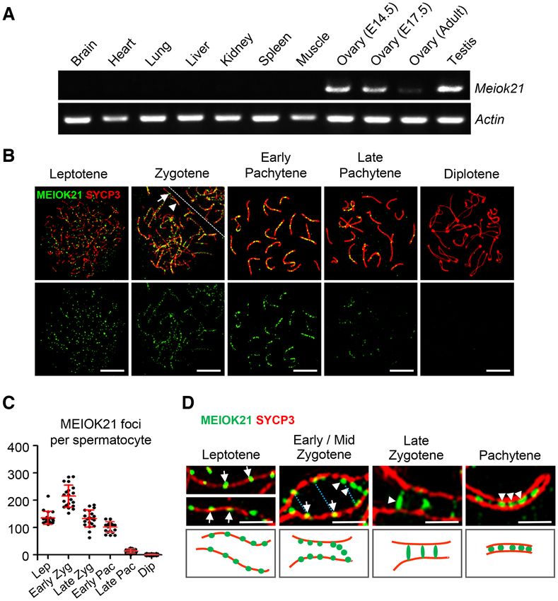
4 Nucleic Acids Research, 2020

at 95◦ C for 30 s, annealing at 60◦ C for 30 s, extension at 72◦ C   complexes (SCs) gradually disassemble and the nucleus en-
for 30 s and a final extension at 72◦ C for 5 min. The PCR           ters the diplotene stage. Finally, SCs disappear from chro-
products were analyzed on a 2% agarose gel and sequenced.            mosomes except at a few sites where chiasmata hold ho-
                                                                     mologs together (41). The last stage of meiotic prophase I
Statistical analysis                                                 is diakinesis, when SYCP3 axes break down except at cen-
                                                                     tromeres (41).
Statistical analyses were conducted using Graph-                        MEIOK21 was first observed on chromosome axes with
Pad PRISM version 5.01 (GraphPad Software, Inc.                      ∼135 foci at leptotene in spread spermatocytes immunos-
RRID:SCR 002798). The error bars show means ± SEM                    tained with anti-MEIOK21 antibodies (Figure 1B). The
or means ± SD as indicated in figure legends. Sample sizes           number of MEIOK21 foci increased to a peak of ∼200

                                                                                                                                        Downloaded from https://academic.oup.com/nar/article-abstract/doi/10.1093/nar/gkaa406/5848248 by guest on 30 June 2020
were described in figure legends. The statistical significance       at early zygotene. After that, the number of MEIOK21
of the differences between two sets of data was measured by          foci gradually decreased. At late pachytene, only a small
Student’s t-test with an unpaired, two-tailed distribution.          number of MEIOK21 foci were observed and they were
The levels of significance are indicated as follows: P < 0.05        undetectable at diplotene when SC began to disassemble
(*), P < 0.01 (**) and P < 0.001 (***).                              (Figures 1B and C). However, MEIOK21 foci were found
                                                                     on both synapsed and unsynapsed chromosome regions
RESULTS                                                              (Figure 1B, arrow vs arrowhead). At early-mid zygotene,
                                                                     ∼77% of MEIOK21 foci were on unsynapsed regions and
MEIOK21 specifically localizes on meiotic chromosomes                ∼23% were on synapsed regions. With meiosis progres-
To understand the mechanisms of meiotic recombina-                   sion, MEIOK21 focus number decreased to ∼26% on unsy-
tion, RNA sequencing of mouse germ cells was con-                    napsed regions and increased to ∼74% on synapsed regions
ducted to identify new factors involved in meiotic DSB               at late zygotene. Therefore, MEIOK21 dynamically local-
repair and recombination (33). An uncharacterized gene               izes on meiotic chromosomes during prophase of meiosis
4930432K21RIK was selected based on its preferential ex-             I.
pression in germ cells during meiosis, which was also                   The dynamic localization pattern of MEIOK21 foci
previously identified as being highly expressed in mouse             on meiotic chromosomes is quite similar to many well-
testes (38). This gene is 44.57 kb, located on chromo-               established recombination factors such as RPA and
some 8. It contains nine exons with start codon ATG in               RAD51/DMC1 recombinase (42–44). These observations
the second exon and stop codon TAA in the last exon,                 prompted us to examine the localization of MEIOK21 foci
and the ORF is 1,800 bp, encoding 600 amino acids.                   more extensively. Using structured illumination microscopy
By searching its sequence in public databases, includ-               (SIM), we found that MEIOK21 was located on the chro-
ing NCBI, Uniprot and Protein Data Bank, we found                    mosome axis as foci at leptotene (Figure 1D left, arrows). At
that 4930432K21RIK protein only exists in vertebrates                zygotene, on chromosome regions where homologous chro-
and is rather conserved (Supplementary Figure S1A). It               mosomes were still widely separated, MEIOK21 foci local-
belongs to an unknown protein superfamily (cl25823),                 ized on the axis as at leptotene (Figure 1D middle left, ar-
which contains a conserved structural domain with un-                rows), while on chromosome regions where two homolog
known function (DUF4671; https://www.ncbi.nlm.nih.gov/               axes were aligned in parallel (800–1000 nm), MEIOK21
Structure/cdd/cddsrv.cgi?uid=318011). Its human ortholog             foci were frequently seen as ‘pairs’ at the opposing sites on
is C19ORF57, which is also preferentially expressed in               the two homologs (Figure 1D top, middle left, blue dotted
testes (39). To further study the expression patterns of             lines), and many MEIOK21 foci were seen as ‘hanging foci’,
4930432K21RIK, reverse transcription qPCR (RT-qPCR)                  attached to the sides of axes (Figure 1D middle left, arrow-
was performed. We found that mRNA was highly expressed               heads).
in mouse testes and fetal ovaries (Figure 1A and Supple-                At the more advanced chromosome regions (homolog
mentary Figure S1BC). According to its biological function           axes spaced at 300–700 nm), MEIOK21 foci were seen as
described below, this gene was named Meiok21 (meiosis-               ‘bridge’ like structures or ‘fusing foci’, which is quite com-
specific 4930432K21RIK).                                             mon in late zygotene spermatocytes (Figure 1D middle
   Given that Meiok21 is highly expressed in testes and fe-          right, arrowhead). At pachytene, when all autosomes were
tal ovaries, where meiosis occurs, the expression and local-         synapsed, a majority (∼70%, n = 1192 foci) of MEIOK21
ization of MEIOK21 in spermatocytes was further exam-                foci were located between the synapsed axes, most prob-
ined in detail by chromosome spread combined with im-                ably on SC complexes (Figure 1D right, arrowheads), al-
munostaining. Meiotic stages were classified according to            though there were still some MEIOK21 foci that remained
standard cytological criteria combined with the morphol-             on the axis or as hanging foci. This progression (Figure 1D,
ogy of SYCP3, a chromosome axis marker (40). At the lep-             lower panels), from on-axis foci to ‘hanging foci’, then to a
totene stage, developing chromosome axes are labelled by             ‘bridge’ like structure and finally to single foci between axes,
short stretches of SYCP3. At the zygotene stage, these dis-          has been proved to reflect typical dynamics of recombina-
continuous SYCP3 stretches become linear axes and ho-                tion factors, such as RPA in humans and Msh4 in Sordaria,
mologs begin to synapse where the SYCP3 labelled axes                during the meiotic recombination process (31,45,46). Taken
are much thicker than unsynapsed regions (Figure 1B).                together, the distribution patterns of MEIOK21 coincide
When all autosomes completely synapsed, the nucleus en-              with typical features of recombination factors, suggesting
ters the pachytene stage. After pachytene, synaptonemal              its potential roles in meiotic recombination.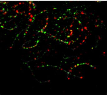
Nucleic Acids Research, 2020 5
Downloaded from https://academic.oup.com/nar/article-abstract/doi/10.1093/nar/gkaa406/5848248 by guest on 30 June 2020
Figure 1. Dynamic localization of MEIOK21 on meiotic chromosomes. (A) Meiok21 is highly expressed in testes and fetal ovaries, revealed by RT-qPCR.
(B) The dynamic localization of MEIOK21 to meiotic chromosomes in mouse spermatocytes. The meiotic stages of spermatocytes were determined by
the SYCP3 staining (red) of the chromosome axis. MEIOK21 (green) are visualized on meiotic chromosomes from leptotene to late pachytene and are
undetectable in diplotene. The arrow in zygotene indicates MEIOK21 foci on an unsynapsed axis, 77.60 ± 8.24% in early/mid zygotene and 26.08 ± 8.98%
in late zygotene. The arrowhead indicates MEIOK21 foci on a synapsed axis, 22.40 ± 8.24% in early/mid zygotene and 73.92 ± 8.98% in late zygotene.
Mean ± SD, n = 20 for each group. The dashed line separates the target cell from the adjacent unrelated cell. Scale bar, 10 m. (C) Quantification of
the numbers of MEIOK21 foci in spermatocytes from leptotene to diplotene. Spermatocyte sample sizes, n = 20 for each stage. Lep, leptotene; early
Zyg, early zygotene; late Zyg, late zygotene; early Pac, early pachytene; late Pac, late pachytene; Dip, diplotene. Error bar, mean ± SD. (D) The dynamic
localization of MEIOK21 on chromosomes investigated by structured illumination microscope (SIM). (Top panel) MEIOK21 foci first localize on the
chromosome axis (‘leptotene’, arrows). Then, on chromosome regions where two homolog axes are aligned in parallel, MEIOK21 foci were frequently
seen as ‘pairs’ at the opposing sites on the two homologs (‘early/mid zygotene’, arrows, MEIOK21 foci; blue dotted lines, focus pairs) and some foci are
released as ‘hanging foci’ (‘early/mid zygotene’, arrowheads). When homologous chromosomes aligned closely, MEIOK21 appears as ‘bridges’ or ‘fusing
foci’ (‘late zygotene’, arrowhead) and finally as fused foci (‘pachytene’, arrowheads) between synapsed axes. (Bottom panel) cartoons illustrate the dynamic
localization of MEIOK21 with meiotic progression. Scale bar, 1 m.
MEIOK21 localizes to meiotic recombination sites in a DSB- gotene, ∼40% of MEIOK21 and DMC1 foci overlapped
dependent manner with each other. At early pachytene, ∼20% of MEIOK21
foci overlapped with DMC1 foci, and ∼30% of DMC1 foci
The uniquely dynamic localization of MEIOK21 during
overlapped with MEIOK21 foci (Figure 2CD). Similar sit-
meiosis strongly indicates it may function in meiotic re-
uations were found between MEIOK21 and RAD51. From
combination. To further investigate this, MEIOK21 was co-
leptotene to zygotene, ∼60% of MEIOK21 and RAD51 foci
immunostained separately with known recombination fac-
overlapped with each other; at early pachytene, ∼20% of
tors RAD51, DMC1 and RPA protein component (RPA2).
MEIOK21 foci overlapped with RAD51 foci, and ∼30% of
We found that MEIOK21 foci colocalized with RPA foci
RAD51 foci overlapped with MEIOK21 foci (Figure 2EF).
at a high frequency, ∼85-95% of the two factors over-
The proper localization of recombination factors, includ-
lapped with each other from leptotene to early pachytene
ing RPA, RAD51 and DMC1, depends on programmed
(Figure 2AB). Partial colocalizations were found between
DSBs catalyzed by the SPO11 complex (47). Thus, we
MEIOK21 and DMC1 or RAD51. From leptotene to zy-
checked whether the localization of MEIOK21 on chromo-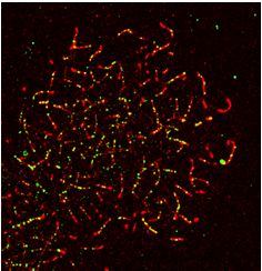
6 Nucleic Acids Research, 2020
A MEIOK21 RPA SYCP3
B MEIOK21 RPA
Overlapped foci (%)
100
80
60
40
20
0
Lep Zyg Early Pac
Downloaded from https://academic.oup.com/nar/article-abstract/doi/10.1093/nar/gkaa406/5848248 by guest on 30 June 2020
C D
MEIOK21 DMC1 SYCP3
Overlapped foci (%)
100 MEIOK21 DMC1
80
60
40
20
0
Lep Zyg Early Pac
E MEIOK21 RAD51 SYCP3
F
Overlapped foci (%)
100 MEIOK21 RAD51
80
60
40
20
0
Lep Zyg Early Pac
G WT Spo11-/- Dmc1-/- H
MEIOK21 SYCP3
WT Spo11-/- Dmc1-/-
Leptotene
500
per spermatocyte
400
MEIOK21 foci #
300
200
Zygotene
100
0
Leptotene Zygotene
Figure 2. MEIOK21 localizes to meiotic recombination sites in a DSB dependent manner. (A) MEIOK21 colocalizes with RPA. (B) Quantification shows
that ∼85-95% of the two factors overlap with each other from leptotene to early pachytene. Error bar, mean ± SD. Sample sizes (from left to right), n = 13,
13, 37, 37, 30 and 30, respectively. (C) MEIOK21 partially colocalizes with DMC1. (D) From leptotene to zygotene, ∼40% of the two factors overlap with
each other; in early pachytene, ∼20% of MEIOK21 foci overlap with DMC1 foci, and ∼30% of DMC1 foci overlap with MEIOK21 foci. Error bar, mean
± SD. Sample sizes (from left to right), n = 30, 30, 41, 41, 30 and 30, respectively. Arrowheads indicate overlapped foci. (E) MEIOK21 partially colocalizes
with RAD51. (F) From leptotene to zygotene, ∼60% of the two factors overlap with each other; in early pachytene, ∼20% of MEIOK21 foci overlap
with RAD51 foci, and ∼30% of RAD51 foci overlap with MEIOK21 foci. Error bar, mean ± SD. Sample sizes (from left to right), n = 17, 17, 42, 42,
30 and 30, respectively. Arrowheads indicate overlapped foci. (G) MEIOK21 foci are abolished in Spo11−/− spermatocytes, but more MEIOK21 foci are
observed in Dmc1−/− spermatocytes compared to WT. Note that Spo11−/− and Dmc1−/− spermatocytes can only reach the ‘zygotene’ stage. The dashed
line separates the target cell from the adjacent unrelated cell. Scale bar, 10 m (A, C, E, G). (H) The quantification of MEIOK21 foci in spermatocytes of
WT, Spo11−/− and Dmc1−/− mice. Spermatocyte sample sizes, from left to right, n = 19, 22, 17, 20, 22 and 21. Error bar, mean ± SD.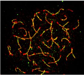
Nucleic Acids Research, 2020 7

somes was DSB dependent or not. In WT, there were abun-        dered which region of MEIOK21 mediated its nucleus lo-
dant MEIOK21 foci located on chromosomes in both lep-          calization. For this purpose, we first confirmed GFP-tagged
totene and zygotene nuclei (Figure 2G left and 2H). How-       full length MEIOK21 protein localized to the nucleus when
ever, MEIOK21 foci were rarely observed in Spo11−/− sper-      it was transiently expressed in HEK-293T cells (Figure
matocytes (Figure 2G middle and H). This supports that the     3F). Then, GFP-tagged MEIOK21 fragments (above) were
chromosome localization of MEIOK21 depends on meiotic          transfected into HEK-293T cells. The N-terminal fragment
DSBs and MEIOK21 may play important roles in DSB re-           containing the first 150 amino acids localized to the nucleus
pair. In Dmc1−/− spermatocytes, DSBs are unrepaired and        efficiently (Figure 3F), which suggests that this fragment
accumulated with extensive resection (48). The localization    contains the nuclear localization signal. However, the frag-
of MEIOK21 was further examined in Dmc1−/− sperma-             ment lacking the first 150 amino acids, MEIOK21151–600 ,

                                                                                                                               Downloaded from https://academic.oup.com/nar/article-abstract/doi/10.1093/nar/gkaa406/5848248 by guest on 30 June 2020
tocytes. Consistent with this, the number of MEIOK21 foci      localized to the cytoplasm exclusively (Figure 3F). There-
was also accumulated to a significantly higher level (Fig-     fore, MEIOK21 has at least two functional domains, the
ure 2G right versus left; H). Therefore, the localization of   N-terminal 150 amino acids for nuclear localization and the
MEIOK21 on chromosomes depends on meiotic DSBs and             C-terminal 150 amino acids for interaction with HSF2BP.
occurs prior to DMC1.                                             HSF2BP has two domains, the N-terminal coiled-coil do-
                                                               main interacting with HSF2 and the C-terminal armadillo
                                                               domain interacting with BRCA2 and BNC1 (28,51,52). Our
MEIOK21 interacts with HSF2BP
                                                               Y2H and co-IP experiments show that the N-terminal do-
To clarify the possible function of MEIOK21 during mei-        main of HSF2BP but not the C-terminal domain can suffi-
otic recombination, we searched for its potential interac-     ciently mediate its interaction with MEIOK21 (Figures 3G–
tors during this process. We focused on RPA and factors        I and Supplementary Figure S2D). However, it is still un-
that also colocalize with RPA. In mammals, the RPA com-        known whether MEIOK21 and HSF2 share the same bind-
plex is composed of RPA1, RPA2 and RPA3. Recently,             ing motif at the N-terminus of HSF2BP, and how these two
MEIOB, SPATA22 and HSF2BP have been found to colo-             factors interact with and regulate HSF2BP. These questions
calize with RPA, and they are all required for meiotic re-     remain to be further investigated.
combination (26–28,30,49,50). Thus, these proteins were
screened for their possible interactions with MEIOK21 us-
                                                               MEIOK21 is required for male fertility
ing a yeast two-hybrid (Y2H) assay. Among them, only
HSF2BP was detected to interact with MEIOK21 (Figure           MEIOK21 specifically localizes on chromosomes in a DSB
3A; Supplementary Figure S2A-C). To further confirm the        dependent manner. It colocalizes with RPA and interacts
interaction between MEIOK21 and HSF2BP, endogenous             with HSF2BP, which is involved in meiotic recombination.
co-immunoprecipitation (co-IP) assays were performed us-       All of these traits strongly suggest that MEIOK21 likely
ing WT testis extracts. Consistent with the Y2H result, en-    functions directly in meiosis. To test this, Meiok21 defi-
dogenous HSF2BP could be pulled down by MEIOK21                cient (Meiok21−/− ) mice were generated by deleting ex-
in co-IP via anti-MEIOK21 antibody. As a negative con-         ons 3–6 of Meiok21 using the CRISPR/Cas9 system (Fig-
trol, HSF2BP was not detected in co-IP using Meiok21−/−        ure 4A and Supplementary Figure S3A). The knockout ef-
testis extracts in which MEIOK21 protein is absent (Fig-       ficiency was confirmed by RT-PCR and immunostaining
ure 3B). Consistent with the interaction results, ∼75–90%      against endogenous MEIOK21 (Figures 4B and Supple-
of MEIOK21 foci colocalized with HSF2BP foci, and ∼70–         mentary Figure S3B–F). Meiok21−/− mice were viable and
90% of HSF2BP foci colocalized with MEIOK21 foci on            appeared to develop normally. Although Meiok21−/− fe-
meiotic chromosomes from leptotene to pachytene (Figure        male mice had no obvious fertility defects, Meiok21−/−
3C and Supplementary Figure S2E). Above results support        male mice were infertile (Figure 4C, Supplementary Figure
that MEIOK21 binds to HSF2BP in vivo, and functions in         S4A). Studies showed that the sizes of testes from 8-week-
meiotic recombination.                                         old Meiok21−/− male mice were much smaller than those of
   Next, we asked which region(s) of MEIOK21 inter-            WT and Meiok21+/− mice at the same age (Figure 4D, E).
act(s) with HSF2BP. Since the full- length protein is com-     However, 12-week-old Meiok21−/− ovaries were still com-
posed of an unknown structural domain (DUF4671), with-         parable to WT (Supplementary Figure S4B, C). Further
out any other known functional domains/motifs, several         analysis showed no post-meiotic round spermatids or elon-
GFP tagged truncations were constructed (Figure 3D). We        gating spermatids in the seminiferous tubules, but there
found the GFP-tagged MEIOK21301-600 fragments pulled           were numerous vacuoles in Meiok21−/− testes, which were
down Flag-tagged HSF2BP as efficiently as full length          rarely seen in WT testes (Figure 4F, upper panel). More-
MEIOK21, while MEIOK211–300 fragments could not pull           over, no spermatozoa were found in the cauda epididymis
down HSF2BP (Figure 3E, lane 9 versus lane 6). This indi-      of Meiok21−/− mice, whereas mature spermatozoa were
cates that the MEIOK21 C-terminal but not the N-terminal       present in the cauda epididymis of WT (Figure 4F, lower
fragment is responsible for its interaction with HSF2BP.       panel). TdT-mediated dUTP nick end labeling (TUNEL)
Further experiments showed that the last 150 amino acids       staining of Meiok21−/− testis sections revealed that many
(MEIOK21451–600 ) were sufficient to interact with HSF2BP      spermatocytes underwent apoptosis (Figure 4G). These re-
(Figure 3E, lane 12).                                          sults suggest that in the absence of MEIOK21, a large
   Given that MEIOK21 localized in the nucleus (specifi-       number of spermatocytes are eliminated before the com-
cally on meiotic chromosomes), but bioinformatic analysis      pletion of spermatogenesis, which results in smaller testes
did not identify a nuclear localization sequence, we won-      as often seen in synapsis- and recombination-defective mu-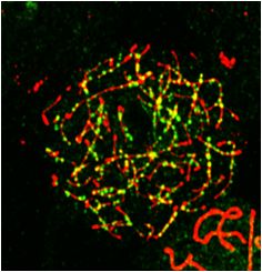
8 Nucleic Acids Research, 2020
Downloaded from https://academic.oup.com/nar/article-abstract/doi/10.1093/nar/gkaa406/5848248 by guest on 30 June 2020
Figure 3. MEIOK21 interacts with recombination factor HSF2BP. (A) MEIOK21 interacts with HSF2BP in Y2H screening, with MEIOK21 as prey and
HSF2BP as bait. About 105 yeast cells were seeded in each dot. The experiment was repeated three times. (B) HSF2BP is pulled-down in Meiok21+/+
testis lysate (+/+) (200 mg) but not Meiok21−/− testis lysate (−/−) (200 mg) by antibody against MEIOK21.The input amount is ∼5% of the lysate. IgG
acts as a negative control. The experiment was repeated three times. Nonspecific bands are labeled with *. (C) MEIOK21 colocalizes with HSF2BP on
meiotic chromosomes (see Supplementary Figure 2E for quantifications). Scale bar, 10 m. (D) Diagram of MEIOK21 truncations. (E) The C terminus of
MEIOK21 (a.a. 451–600) is required for interaction with HSF2BP. FLAG-tagged HSF2BP and GFP-tagged MEIOK21 truncations were co-transfected to
HEK-293T cells. After 48 hours, cells were lysed, and co-IP experiments were performed using GFP antibody. The input amount is ∼5% of the lysate. The
experiment was repeated twice. (F) The N terminus of MEIOK21 (a.a. 1–150) is required for its nuclear localization. GFP-tagged MEIOK21 truncations
were transfected to HEK-293T cells. After 24 hours, GFP signal was examined under a fluorescence microscope. Scale bar, 5 m. (G) The diagram of
HSF2BP truncations. (H) The coiled-coil domain of HSF2BP (a.a. 1–92) is required for MEIOK21 binding. GFP-tagged MEIOK21 and FLAG-tagged
HSF2BP truncations were co-transfected to HEK-293T cells. After 48 hours, cells were lysed, and co-IP experiments were performed with antibodies
against GFP. The input amount is ∼5% of the lysate. The experiment was repeated twice. The non-specific band (*). (I) Diagrams to illustrate interaction
domains of MEIOK21 and HSF2BP.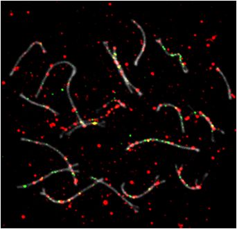
Nucleic Acids Research, 2020 9
Downloaded from https://academic.oup.com/nar/article-abstract/doi/10.1093/nar/gkaa406/5848248 by guest on 30 June 2020
Figure 4. MEIOK21 is required for male fertility. (A) The schematic of Meiok21 knockout allele. Exons 3–6 were deleted in the mutant. (B) Meiok21
knockout was confirmed by RT-PCR (Materials and Methods). RT-PCR was performed using cDNA obtained from testes as templates. PCR products
with proper size were detected from WT but not mutant samples. (C) Meiok21−/− male mice were sterile. n = 5 for each genotype. Error bar, mean ± SEM.
(D, E) Both the testis size and testis/body weight ratio of 8-week-old Meiok21−/− mice are significantly reduced compared to Meiok21+/+ and Meiok21+/−
mice. Error bar, mean ± SEM. n = 3 for each genotype. (F) No spermatids or mature spermatozoa were observed in testis or epididymis by HE staining
in Meiok21−/− mice. Scale bar, 50 m. (G) A large number of apoptotic spermatocytes (green) were observed in Meiok21−/− testis by TUNEL staining.
Scale bar, 50 m. (H) Quantitative analysis of spermatocytes at different meiotic stages from Meiok21+/+ and Meiok21−/− mice testes at postnatal day
20. There are more zygotene but less pachytene spermatocytes in Meiok21−/− testis compared to WT. n = 3 for each genotype. Error bar, mean ± SEM.
tants, such as Dmc1−/− and Msh5−/− (44,53,54). Therefore, relatively synchronized (55). The stage(s) in which the de-
MEIOK21 is essential for spermatogenesis and male fertil- fects occur can be easily inferred from meiosis progression.
ity. Spermatocytes from leptotene to diplotene can be distin-
guished based on chromosome immunostaining of SYCP3
and SYCP1, the components of meiotic chromosome axis
MEIOK21 is important for normal meiosis progression
and SC. Zygotene spermatocytes were dominant in postna-
Given that Meiok21−/− spermatocytes are eliminated via tal day 15 (PD15) testis of both WT and Meiok21−/− mice,
apoptosis, we speculate that they may undergo severe de- and there were slightly more zygotene (∼70% vs ∼65%)
fects in meiosis. Spermatogenesis in mice is a continuous and significantly less pachytene (∼7% versus ∼16%) sper-
process and occurs by waves in seminiferous tubules. Thus, matocytes in Meiok21−/− compared to WT. With meio-
spermatocytes at different stages can be seen in a single sis progression, more zygotene cells entered pachytene,
seminiferous tubule. The first wave of spermatogenesis is thus the frequency of zygotene cells decreased, and con-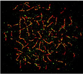
10 Nucleic Acids Research, 2020

comitantly, the frequency of pachytene cells increased. As      WT mice. (i) Synapsis partner switch, i.e. different regions
expected, at PD17, zygotene spermatocyte frequency de-          of one chromosome synapses with more than one partner
creased to ∼25% and pachytene spermatocyte frequency in-        (Figure 5A, left panel). In this case, at least one partner
creased to ∼61% in WT. However, ∼64% of spermatocytes           is a non-homolog, meaning there is also non-homologous
stayed in zygotene and only ∼22% of spermatocytes entered       synapsis, which has been reported in several recombina-
pachytene in Meiok21−/− mice. At PD20, zygotene sper-           tion mutants in mice (27,30,44). (ii) Condensed ‘zygotene’.
matocytes further decreased to ∼15% and pachytene sper-         There is a fraction of ‘zygotene’ nuclei with highly con-
matocytes increased to ∼72% in WT. But in Meiok21−/− ,          densed chromosomes as pachytene nuclei, in which there is
zygotene spermatocytes were still at a high level (∼40%)        no or only few short SYCP1 stretches. Thus, in these nu-
and pachytene spermatocytes only increased to ∼45%, (Fig-       clei homologs are unsynapsed and remained as ∼40 ‘univa-

                                                                                                                                 Downloaded from https://academic.oup.com/nar/article-abstract/doi/10.1093/nar/gkaa406/5848248 by guest on 30 June 2020
ure 4H). Therefore, there were always significantly more zy-    lents’ but not 20 bivalents as seen in WT pachytene (Figure
gotene spermatocytes and fewer pachytene spermatocytes          5A, right panel). This type of synapsis defect is often seen
in Meiok21−/− mice, compared to WT at the same ages             in recombination defective mutants (7,40,44,53). In total,
(Figure 4H). These observations suggest that in the absence     these two types of abnormal nuclei account for ∼50% of the
of MEIOK21, meiosis might be arrested or delayed at the         zygotene spermatocytes in Meiok21−/− mice (Figure 5B).
zygotene/pachytene transition, and only a subset of sper-       Such abnormalities are not found in Meiok21−/− oocytes
matocytes that have fewer defects could enter pachytene.        (Supplementary Figure S4D). We infer that these nuclei are
   Since no spermatids were found in Meiok21−/− testis, we      unable to enter the pachytene stage. This supports the idea
focused on seminiferous epithelium to examine the patho-        that the high level of zygotene nuclei in the mutants is not
logical status of spermatogenesis in Meiok21−/− testes.         due to a delay in entering pachytene, but actually due to a
Mouse spermatogenesis is a highly ordered biological pro-       fraction of nuclei that permanently arrest in the zygotene
cess, and the seminiferous epithelium can be divided into       stage because of severe synapsis failures. However, we can-
twelve stages (stages I–XII) according to the development       not exclude the possibility that these aberrant nuclei are ac-
of spermatocytes and spermatids, which can be visualized        tually at a pachytene-like stage but with pairing/synapsis
by the periodic acid-Schiff’s reaction (PAS) (55). In WT        defects.
testis, seminiferous tubules from stage I–XII can be ob-           As described above, a considerable proportion of
served, and spermatocytes from leptotene to metaphase I,        Meiok21−/− spermatocytes can still enter pachytene. In or-
together with round spermatids and elongating spermatids,       der to characterize whether these pachytene nuclei are nor-
are organized in corresponding positions (Supplementary         mal, meiotic recombination was examined. The final and
Figure S5A; upper panel). In Meiok21−/− testis, spermatids      major purpose of meiotic recombination is the formation
were never found, and stages of seminiferous epithelium         of homologous DNA crossovers (COs). COs cause the ex-
could only be identified based on the types of spermatocytes    change of homologous DNA and establish the physical con-
(Supplementary Figure S5A; lower panel) (55). Moreover,         nections between homologs required for proper chromo-
in Meiok21−/− tubules, apoptotic spermatocytes were ob-         some segregation (12). Around 90% of mouse COs can be
served as early as stage IV and throughout stages after that.   marked by MLH1 foci at pachytene (58–60). So, the number
At stage IV, pachytene spermatocytes are dominant in WT         and distribution of MLH1 foci were checked by immunos-
tubules, however, many apoptotic pachytene spermatocytes        taining in pachytene spermatocytes. Consistent with previ-
with aberrantly condensed nuclei appeared in Meiok21−/−         ous studies, in WT pachytene spermatocytes, ∼22 MLH1
tubules (55) (Supplementary Figure S5A; lower panel, ‘aP’).     foci were observed per cell and almost every autosome
These results suggest that defective Meiok21−/− pachytene       has at least one MLH1 focus, thus autosomes without an
spermatocytes may undergo apoptosis. In addition, apop-         MLH1 focus are rare (∼1%). Only ∼17 MLH1 foci were
totic diplotene (aDi) and apoptotic metaphase I (aMI) sper-     observed per Meiok21−/− pachytene spermatocyte, with
matocytes were also found in Meiok21−/− tubules (Supple-        ∼25% autosomes without even one MLH1 focus (Figures
mentary Figure S5A; lower panel, ‘aDi’ and ‘aMI’). Very         5CD, Supplementary Figure S6A).
similar results were observed in both younger and older
mice (Supplementary Figure S5BC). We speculated that a
                                                                MEIOK21 is required for normal DMC1 and RAD51 re-
small subset of Meiok21−/− spermatocytes with fewer de-
                                                                cruitment in spermatocytes
fects could bypass the pachytene checkpoint, but finally un-
dergo apoptosis. Therefore, defective Meiok21−/− sperma-        Meiotic crossover recombination originates from pro-
tocytes probably are cleared via apoptosis during prophase      grammed DSBs. Upon DSB formation, ␥ H2AX, the phos-
of meiosis I, which results in male infertility.                phorylated form of the histone variant H2AX, appears
                                                                and its level is correlated with the level of DSBs (61,62).
                                                                In WT spermatocytes, abundant ␥ H2AX signals could be
Meiok21−/− spermatocytes show defects in synapsis and
                                                                detected in the leptotene stage immediately after DSBs
crossover recombination
                                                                formed (Supplementary Figure S6B, upper two panels).
The phenotype of Meiok21−/− mice is similar to many             In early pachytene nuclei, these signals decrease on auto-
synapsis or recombination defective mutants (56,57). Thus,      somes, and bright signals appear on the XY body (63).
homologous synapsis and meiotic recombination were ex-          From mid/late pachytene to diplotene, ␥ H2AX signals are
amined in Meiok21−/− spermatocytes. By double staining          barely detectable on autosomes, however, strong signals are
of SYCP3 and SYCP1, two types of synapsis defects were          still maintained on the XY body. In Meiok21−/− sperma-
found in Meiok21−/− spermatocytes that were never seen in       tocytes, ␥ H2AX signals were comparable to WT at lep-
Nucleic Acids Research, 2020 11
Downloaded from https://academic.oup.com/nar/article-abstract/doi/10.1093/nar/gkaa406/5848248 by guest on 30 June 2020
Figure 5. MEIOK21 is required for efficient meiotic recombination. (A) Two types of aberrant synapsis are frequently observed in Meiok21−/− sperma-
tocytes, as seen in other recombination defective mutants. Partner switch (left panel), i.e. one chromosome (arrow) synapses with more than one partner
(arrowheads). (B) Quantification of the two types of aberrant ‘zygotene’ nuclei described in (A). For each genotype, 3 mice were examined. The numbers of
cells examined in each mouse are 84, 60 and 60 for WT, and 93, 48 and 64 for Meiok21−/− . (C, D) The number of crossovers, marked by MLH1 foci (red),
is significantly reduced in Meiok21−/− pachytene spermatocytes compared to WT. n = 20 and 23 for WT and Meiok21−/− , respectively. Chromosomes
without even one MLH1 focus (arrows). (E, F) In Meiok21−/− spermatocytes, the level of RPA foci is similar to that of WT at zygotene, but there is
aberrant retention of RPA on chromosomes at a late time. From left to right, n = 20, 20, 24, 13, 23, 19, 14 and 20, respectively. (G–L) The numbers of
DMC1, RAD51 and HSF2BP foci are greatly decreased in Meiok21−/− spermatocytes compared to WT. Early/Mid Zyg, early or middle zygotene; Late
Zyg, late zygotene; Pac, pachytene, Late Pac: late pachytene, Dip: diplotene. For each panel from left to right, n = 26, 36, 24, 30, 22 and 27 (H), 29, 36, 20,
27, 29 and 31 (J), 20, 21, 22, 21, 21 and 21 (L). Scale bar, 10 m (A, C, E, G, I, K). Error bar, mean ± SD (D, F, H, J, L) or mean ± SEM (B).12 Nucleic Acids Research, 2020
totene and zygotene, suggesting that a WT level of DSBs tions (CO precursors) as indicated by DMC1/RAD51 foci
are formed. Similar to WT, ␥ H2AX signals also decreased (20-40% of WT). This result suggests that CO homeostasis
on autosomes in early pachytene nuclei. However, in con- may still exist in Meiok21−/− spermatocytes. CO homeosta-
trast to WT, a number of ␥ H2AX foci were still maintained sis reflects the fact that when the number of CO precursors is
on autosomes at mid/late pachytene (45.66 ± 12.41 foci, n significantly altered, the number of COs can be maintained
= 38) and even at diplotene (33.15 ± 14.73 foci, n = 47) constantly or only affected less proportionally (66–68).
(Supplementary Figure S6B, lower two panels). This im- Two other recombination factors, MSH4 and RNF212,
plies that a considerable number of meiotic DSBs remained which act in the later stages of meiotic recombination, were
unrepaired in Meiok21−/− spermatocytes, though it seems also examined. The data showed that, without MEIOK21,
chromosomes synapsed appropriately in these nuclei. both MSH4 and RNF212 focus numbers were reduced to
Downloaded from https://academic.oup.com/nar/article-abstract/doi/10.1093/nar/gkaa406/5848248 by guest on 30 June 2020
Meiotic DSBs are processed to generate long single ∼70% of WT (Supplementary Figure S7A–D), which is
strand 3 overhangs, to which RPA binds (64). RPA foci ap- comparable to the level of MLH1 foci (∼77% of WT), but
peared from leptotene and the number peaked at zygotene much higher than the level of DMC1/RAD51 foci (20–
with a similar level in both WT and Meiok21−/− sperma- 40% of WT). This may explain why a considerable num-
tocytes (179.60 ± 12.55 versus 177.50 ± 7.98) (Figure 5E, ber of MLH1 foci can be formed in Meiok21−/− spermato-
F). This further confirms that MEIOK21 is not required for cytes, and may also suggest that CO homeostasis (and thus
DSB formation, and it is also consistent with the fact that CO/NCO differentiation) occurs at or before MSH4 foci
localization of MEIOK21 depends on DSBs but not the occurrence as previously proposed (15,61,65,68).
other way around (above). In WT spermatocytes, the num-
ber of RPA foci decreased to a very low level at early/mid MEIOK21 is essential for sufficient HSF2BP recruitment
pachytene and RPA signals are barely detectable in late
pachytene. In Meiok21−/− spermatocytes, the number of A recent study has reported that HSF2BP interacts with
RPA foci also decreased at pachytene, but were still main- BRCA2 to mediate its localization to recombination sites.
tained at a relatively high level (73.52 ± 19.11 foci, n = 20), As with Meiok21−/− spermatocytes, Hsf2bp−/− spermato-
and even at diplotene a considerable number of RPA foci cytes also have a normal level of DSBs and a low num-
could still be observed in many nuclei (13.6 ± 6.51 foci, n ber of DMC1 and RAD51 foci (30). We further found
= 20) (Figure 5EF). The aberrant retention of RPA in later that MEIOK21 interacted and colocalized with HSF2BP
prophase I is consistent with the persistence of ␥ H2AX sig- at recombination sites during meiosis. These results sug-
nals at these stages, indicating insufficient DSB repair in gest that MEIOK21 may regulate DMC1/RAD51 foci
Meiok21−/− spermatocytes. through interacting with and modulating HSF2BP. There-
In meiosis, most DSBs are repaired with homologous fore, the chromosome localization of HSF2BP was exam-
templates. Both DMC1 and RAD51 form a nucleoprotein ined in WT and Meiok21−/− spermatocytes. As reported,
filament on ssDNA of DSBs to help search and invade HSF2BP foci appeared at leptotene, reached the peak at zy-
its homologous partner for successful recombination and gotene, then gradually decreased and finally disappeared at
synapsis (13,19,65). At zygotene, abundant DMC1 foci can late pachytene in WT (Figure 5K, L). However, only a low
be observed on chromosomes in WT spermatocytes, and number of HSF2BP foci were detected in Meiok21−/− sper-
along with DSB repair, the number of DMC1 foci gradually matocytes compared to WT at corresponding stages (e.g.
decreases. However, in Meiok21−/− spermatocytes, only a 63.25 ± 3.229 versus 167.8 ± 6.786 at zygotene; Figure 5K,
very low number of DMC1 foci were observed at zygotene L). Thus, knockout of Meiok21 leads to a significantly de-
(44.14 ± 19.35 in Meiok21−/− versus 189.81 ± 54.23 in WT; creased number of HSF2BP foci on meiotic chromosomes.
Figure 5GH). Similarly, the number of RAD51 foci was
also greatly reduced in Meiok21−/− spermatocytes com- DISCUSSION
pared to WT at zygotene (60.06 ± 19.62 versus 159.48 ± We identified a new protein, MEIOK21, that is prefer-
37.33; Figure 5I, J). Western blot showed that the expres- entially expressed in testes and fetal ovaries. MEIOK21
sion levels of RAD51 and DMC1 proteins were compara- specifically localizes on meiotic recombination sites in a
ble in Meiok21−/− mutant and WT (Supplementary Figure DSB dependent manner, and colocalizes with recombina-
S6C). These results suggest that MEIOK21 does not affect tion factors RPA and HSF2BP. The dynamic and unique
the DMC1/RAD51 protein expression but is required for localization patterns of MEIOK21 show that it is a mei-
DMC1 and RAD51 loading to meiotic DSB ends or their otic recombination bridge. The C-terminal domain of
stability on chromosomes. This finding is consistent with MEIOK21 physically interacts with the N-terminal domain
two facts described above: (1) MEIOK21 does not affect of HSF2BP. In Meiok21−/− spermatocytes, HSF2BP and
DSB formation or its level, and (2) DMC1 is not required the recombinases DMC1 and RAD51 cannot efficiently lo-
for MEIOK21 localization to recombination sites. calize to recombination sites, which finally results in severe
Greatly reduced DMC1 focus number also suggest defects in synapsis and crossover recombination.
greatly reduced levels of inter-homolog recombination in-
teractions in Meiok21−/− mice (∼20–40% of WT level
MEIOK21 regulates DMC1/RAD51 foci through interac-
based on DMC1/RAD51 focus number), which will result
tion with HSF2BP
in recombination and synapsis defects (13,44). However, as
described above, in Meiok21−/− pachytene nuclei, the num- The formation of MEIOK21 foci on meiotic chromosomes
ber of MLH1 foci is still at ∼77% of the WT level, which required SPO11 but was independent of DMC1. This obser-
is less affected than inter-homolog recombination interac- vation suggests that MEIOK21 works after DSB formationNucleic Acids Research, 2020 13
but before DMC1. Further studies showed that Meiok21 female meiosis. However, we cannot completely exclude
knockout does not affect RPA recruitment but the num- the possibility that the residual low level of HSF2BP in
ber of RAD51/DMC1 foci is significantly reduced. Thus, Meiok21 knockout spermatocytes is sufficient to recruit
MEIOK21 probably works after DSB formation and RPA BRCA2 to recombination sites. Thus, further studies are re-
recruitment to regulate RAD51/DMC1 nucleoprotein fila- quired to elucidate the physical and functional interactions
ment assembly or their stability on chromosomes. between HSF2BP and BRCA2.
Efficient loading and stabilization of DMC1/RAD51
is required for proper homolog recombination. In Dmc1
Meiotic homologous recombination bridges
knockout mice, spermatocytes arrest at zygotene with se-
vere defects in meiotic recombination and synapsis (44,54). Meiotic chromosomes are proposed to be organized as
Downloaded from https://academic.oup.com/nar/article-abstract/doi/10.1093/nar/gkaa406/5848248 by guest on 30 June 2020
The proper function of DMC1 and RAD51 requires loop-axis structures (32,75). In this model, each chromatid
many accessory proteins, which regulate the assembly of is assembled in arrays of loops. The base of these loops
RAD51/DMC1 on ssDNA and/or the stability of the is the structural ‘axis’, which is decorated with a set of
nucleoprotein filaments (20,22,25–27,30,69,70). Compared proteins. Meiotic recombination occurs in the context of
with core recombinase DMC1/RAD51, the absence of dif- and is tightly regulated by this chromosome architecture
ferent accessory factors differentially impairs meiotic re- (15,32,75). Pre-DSB complexes are loaded onto chromo-
combination and synapsis. The absence of some accessory some loops and then are tethered to the axis where DSBs
factors, such as HOP2-MND1 and RPA, leads to rarely de- are formed (32,76). Based on studies mainly from yeast, it
tectable RAD51/DMC1 or MLH1 foci and severe synapsis has been proposed that after DSB ends are processed, the
defect (29,71). However, the absence of other accessory fac- first ssDNA end is released from the chromosome axis to
tors causes only moderate meiotic defects. For example, in search and invade the homologous chromosome to form a
SWS1 or SWSAP1 knockout mice, although significantly D-loop (15,31,32,77). Thus, this process brings the homol-
decreased, the numbers of RAD51/DMC1 and MLH1 foci ogous chromosomes close together. Subsequently, a small
are still observed at a considerable level in both spermato- subset of these inter-homolog recombination intermediates
cytes and oocytes (14,24). It is unclear how these accessory is selected to become COs, while the rest become NCOs
proteins work and collaborate to finely regulate meiotic re- (4,15). Proteins involved in different recombination steps
combination, and why so many factors are required. can be visualized by their different association status with
MEIOK21 specifically interacts with HSF2BP (Figure 3 chromosomes (78).
and Supplementary Figure S2). In Meiok21−/− spermato- MEIOK21 first appears as foci on the chromosome axis
cytes, RAD51/DMC1 foci are detected at ∼1/3 of the WT at leptotene. During early zygotene, MEIOK21 foci are then
level, and MLH1 foci are detected at ∼2/3 of WT level released but still associated with the axis as ‘hanging foci’.
(Figure 5). These results suggest that MEIOK21 probably Along with meiosis progression, homolog axes align closely
regulates RAD51/DMC1 foci formation and/or their sta- and MEIOK21 ‘bridges’ appear to tightly link the two axes.
bility likely through modulating HSF2BP. This proposal Finally, when homologs synapsed, bigger and brighter sin-
is supported by the fact that the number of HSF2BP foci gle MEIOK21 foci, assumed to be fused foci, can be vi-
dropped significantly in Meiok21−/− spermatocytes (Fig- sualized between the two axes. These dynamic localization
ure 5K, L). However, we cannot exclude the following pos- patterns are unique to meiotic recombination factors, such
sibilities: (i) MEIOK21 and HSF2BP regulate each other or as RPA, RAD51, DMC1 and MSH4, in diverse species
cooperate for efficient formation of RAD51/DMC1 foci; (31,32,45,46,77,79). This process has been well documented
(ii) MEIOK21 may directly regulate RAD51/DMC1 foci in Sordaria meiosis, where chromosomes in a single nu-
formation. However, the latter is less likely since no direct cleus undergo meiosis synchronously and fully aligned ho-
interaction is detected between MEIOK21 and RAD51 or molog axes can be easily observed shortly before synapsis
DMC1 in our study (Supplementary Figure S2A). (31,45). However, in other organisms including mouse, dif-
Early studies showed that BRCA2 interacts with DMC1 ferent chromosomes progress asynchronously in the nucleus
and RAD51 (72–74). A recent study found that HSF2BP during meiosis (80). Moreover, different regions of a pair of
can directly interact with RPA and BRCA2, and the local- homologs can be at significantly different stages, e.g. some
ization of GFP tagged BRCA2 is altered when it is electro- regions of chromosome are synapsed, some regions are un-
porated into Hsf2bp−/− testes (30). Based on these results, synapsed but homolog axes stay close, and some other re-
the authors proposed that RPA recruits HSF2BP, which gions of homolog axes stay far apart (Figure 1BD) (81). Re-
further recruits BRCA2-DMC1/RAD51 to DSB sites by combination bridges only can be seen in the right homolog
direct physical interactions (30). However, this electropo- regions at the right time. Recombination bridges are com-
ration result may not reflect the in vivo functional interac- posed of DNA, axis proteins, recombination complex, and
tion between BRCA2 and HSF2BP. Since BRCA2 is essen- mediator complex with intrinsic axis affinity independent of
tial for both male and female meiosis (22), if HSF2BP is re- recombination, e.g. ZIP2–ZIP4 complex (31,32).
quired to recruit BRCA2, Hsf2bp deficiency should severely The DSB-dependent localization dynamics of MEIOK21
affect both male and female meiosis. However, knockout of suggests that MEIOK21 is involved in recombination
Hsf2bp only mildly affects female meiosis (22,28,30). Our bridges as a member of the recombination complex. In the
results show that Meiok21 knockout significantly decreases absence of MEIOK21, the numbers of DMC1 and RAD51
HSF2BP focus number but has little or no effect on BRCA2 foci are greatly decreased. Therefore, MEIOK21 is required
(Supplementary Figure S7EF), and as with Hsf2bp−/− mu- for efficient formation of meiotic recombination bridges.
tants, Meiok21−/− mutants severely impair male but not This hypothesis is consistent with the fact that CO recom-You can also read