Unpicking the Roles of DNA Damage Protein Kinases in Trypanosomatids
←
→
Page content transcription
If your browser does not render page correctly, please read the page content below
MINI REVIEW
                                                                                                                                              published: 06 August 2021
                                                                                                                                         doi: 10.3389/fcell.2021.636615

                                            Unpicking the Roles of DNA Damage
                                            Protein Kinases in Trypanosomatids
                                            Gabriel L. A. Silva 1,2* , Luiz R. O. Tosi 2 , Richard McCulloch 1 and Jennifer Ann Black 1,2*
                                            1
                                             The Wellcome Centre for Integrative Parasitology, Institute of Infection, Immunity and Inflammation, University of Glasgow,
                                            Glasgow, United Kingdom, 2 Department of Cell and Molecular Biology, Ribeirão Preto Medical School, University of São
                                            Paulo, Ribeirão Preto, Brazil

                                            To preserve genome integrity when faced with DNA lesions, cells activate and
                                            coordinate a multitude of DNA repair pathways to ensure timely error correction or
                                            tolerance, collectively called the DNA damage response (DDR). These interconnecting
                                            damage response pathways are molecular signal relays, with protein kinases (PKs)
                                            at the pinnacle. Focused efforts in model eukaryotes have revealed intricate aspects
                            Edited by:
                                            of DNA repair PK function, including how they direct DDR pathways and how
                          Eesin Chen,
     National University of Singapore,      repair reactions connect to wider cellular processes, including DNA replication and
                             Singapore      transcription. The Kinetoplastidae, including many parasites like Trypanosoma spp.
                        Reviewed by:        and Leishmania spp. (causative agents of debilitating, neglected tropical infections),
                   Valentyn Oksenych,
     Norwegian University of Science
                                            exhibit peculiarities in several core biological processes, including the predominance of
              and Technology, Norway        multigenic transcription and the streamlining or repurposing of DNA repair pathways,
               Vyacheslav Yurchenko,
                                            such as the loss of non-homologous end joining and novel operation of nucleotide
        University of Ostrava, Czechia
                        H. Diego Folco,     excision repair (NER). Very recent studies have implicated ATR and ATM kinases in
    National Cancer Institute, National     the DDR of kinetoplastid parasites, whereas DNA-dependent protein kinase (DNA-
             Institutes of Health (NIH),
                          United States
                                            PKcs) displays uncertain conservation, questioning what functions it fulfills. The wide
                  *Correspondence:
                                            range of genetic manipulation approaches in these organisms presents an opportunity
                    Gabriel L. A. Silva     to investigate DNA repair kinase roles in kinetoplastids and to ask if further kinases
              gabriellamak93@usp.br
                                            are involved. Furthermore, the availability of kinase inhibitory compounds, targeting
                   Jennifer Ann Black
       Jennifer.Stortz@glasgow.ac.uk        numerous eukaryotic PKs, could allow us to test the suitability of DNA repair PKs as
                                            novel chemotherapeutic targets. Here, we will review recent advances in the study of
                    Specialty section:
                                            trypanosomatid DNA repair kinases.
         This article was submitted to
             Cell Growth and Division,      Keywords: protein kinases, PIKK, DNA damage, DNA repair, kinetoplastids, trypanosomatids
                a section of the journal
  Frontiers in Cell and Developmental
                                Biology
                                           INTRODUCTION
       Received: 01 December 2020
            Accepted: 13 July 2021
                                           Numerous DNA lesions can form within a eukaryotic cell per day, each a potential threat to genome
         Published: 06 August 2021
                                           stability (Tubbs and Nussenzweig, 2017). Genome damage can arise from a myriad of sources,
                               Citation:   including exposure to mutagenic agents, such as radiation, and endogenous cellular processes such
   Silva GLA, Tosi LRO, McCulloch R
                                           as DNA replication and metabolism. Lesions can form primarily on a single DNA strand, such as by
       and Black JA (2021) Unpicking
   the Roles of DNA Damage Protein
                                           the accumulation of unbase-paired single-stranded DNA (ssDNA), base adducts, oxidative damage,
         Kinases in Trypanosomatids.       and mismatched bases, or can affect both stands, such as through double-stranded breaks (DSBs)
       Front. Cell Dev. Biol. 9:636615.    and inter-strand cross-links. Ultimately, the persistence of all such damage can compromise high-
      doi: 10.3389/fcell.2021.636615       fidelity genome transmission to future offspring, resulting in genetic diseases, decreased fitness, or

Frontiers in Cell and Developmental Biology | www.frontiersin.org                  1                                            August 2021 | Volume 9 | Article 636615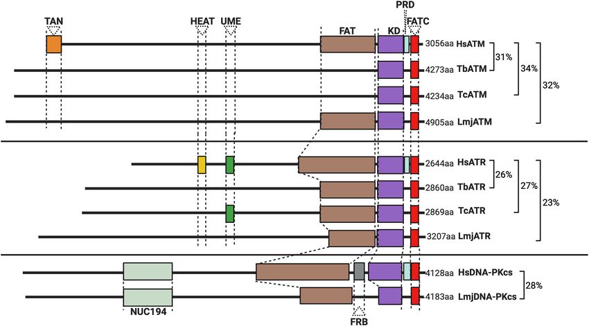
Silva et al.                                                                                                   DNA Repair Kinases in Trypanosomatids

lethality (O’Driscoll, 2012; Ribezzo et al., 2016; Chatterjee and           small-molecule inhibitors approved for clinical use (Carles et al.,
Walker, 2017). Conserved across the Eukarya, a sophisticated                2018; MRC Protein Phosphorylation and Ubiquitylation Unit,
network of pathways, collectively known as the DNA damage                   U. of D, 2020; Roskoski, 2020). Thus, rather than developing
response (DDR), operate to safeguard the genome, acting                     novel compounds that target DDR PKs, an opportunity exists
hierarchically from lesion detection to resolution. At the heart            for the repurposing of small-molecule inhibitors as novel anti-
of the DDR are evolutionarily conserved protein kinases (PKs)               parasitic treatments, particularly as the development of drugs
that act to orchestrate the repair of genome damage by signaling            targeting NTDs is routinely limited due to safety, efficacy, and
its presence and enacting the appropriate repair pathway via                funding, leaving many archaic and dangerous drugs at the
post-translational phosphorylation modifications to the hydroxyl            forefront of treatments for the foreseeable future (Field et al.,
groups of serine (S), threonine (T), or tyrosine (Y) residues on            2017; Bhattacharya et al., 2020). Additionally, using such small-
downstream factors. Additionally, DDR PKs also perform a range              molecule PK inhibitors could provide opportunities to investigate
of non-catalytic functions, such as by the allosteric regulation of         both the function and evolution of PKs, including those that
other kinases (Kung and Jura, 2016).                                        act in the trypanosomatid DDR. Such an approach may be
    The DDR and its associated PK compliment are well-                      especially attractive for less genetically tractable trypanosomatids,
characterized in “model” eukaryotes, but in trypanosomatids,                like T. cruzi and T. vivax. Here, we will focus on trypanosomatid
less is known. Trypanosomatids are parasitic members of                     DNA damage-associated PKs and their reported functions, first
the widespread and diverse Kinetoplastea class (Lukeš et al.,               discussing the known roles of canonical DDR PKs and then
2018; Butenko et al., 2020) and cause neglected tropical                    focusing on wider putative DDR PKs.
diseases (NTDs) that disproportionally affect impoverished
populations in the tropics and subtropics of the world. Human               PKs at the DDR Apex
African Trypanosomiasis (Trypanosoma brucei), Leishmaniasis                 PKs are specialized enzymes accounting for up to 3% of the
(Leishmania spp.), and Chagas disease (Trypanosoma cruzi) are               encoded genes in a typical eukaryote (Hunter and Plowman,
three of 20 NTDs targeted by the World Health Organization                  1997; Manning et al., 2002; Zulawski et al., 2014). Two
(WHO) for eradication by 2030 (World Health Organization,                   superfamilies of PKs exist including eukaryotic PKs (ePKs) and
2020). These dixenous parasites transmit from arthropod vectors             atypical PKs (aPKs), with nine subfamilies of ePKs described in
to mammalian hosts (for life cycles of each parasite, refer to Stuart       most eukaryotes. The ePK structure is largely conserved among
et al., 2008), where they cause debilitating but distinct diseases of       subfamilies, where an N-terminal lobe (composed primarily of
medical importance, which significantly impact the life quality of          β-sheets) is joined by a hinge-like region to a predominantly
the infected individual and at-risk populations, and, combined,             α-helical C-terminus, with the site of γ-phosphate transfer
are responsible for ∼80,000 deaths each year (Torres-Guerrero               (the active site) located between these extremities (Hanks and
et al., 2017; Büscher et al., 2017; Pérez-Molina and Molina, 2018).         Hunter, 1995) (for an extensive review on PK structure, refer
    Trypanosomatids are early branching eukaryotes, having                  to Taylor and Kornev, 2011). aPKs typically lack the catalytic
emerged ∼500 million years ago, close to the time mammals                   region or domains characteristic of ePKs, yet among the aPKs,
emerged from other eukaryotes (Lukeš et al., 2014). As such,                members of the phosphatidyl inositol 30 kinase-related kinase
unusual aspects of the DDR, including during DNA repair,                    (PIKK) family perform vital functions at the apex of the
have been reported. For instance, classical non-homologous end-             DDR. DNA-dependent Protein Kinase catalytic subunit (DNA-
joining (c-NHEJ) activity required for DSB repair is lacking                PKcs; absent from yeasts), ATR (Mec1 in budding yeast),
in these organisms (Burton et al., 2007; Nenarokova et al.,                 and ATM (Tel1 in budding yeast) are large enzymes (up
2019), with the result that DNA end-joining using regions of                to 500 kDa in size) sharing structural similarities with lipid
micro-homology (MMEJ) (Glover et al., 2011; Laffitte et al.,                kinases within their C-terminal kinase domains (Figure 1).
2016) or by single-strand annealing (SSA) (Glover and Horn,                 Flanking their kinase domains, they share several further
2014; Zhang and Matlashewski, 2019) appears to assume greater               conserved domains, including the FAT domain (FRAP, ATM,
prominence than in many organisms. In addition, nucleotide                  and TTRAP domain), a protein regulatory domain (PRD),
excision repair (NER) appears to have become functionally                   a LST8-binding element (LBE) domain, and FAT-C domain
streamlined (Machado et al., 2014), most likely due to the                  downstream, all of which are required for kinase function
ubiquity of multigenic transcription in kinetoplastids. Other               and activity regulation (as reviewed by Imseng et al., 2018).
peculiarities have recently emerged, with components of the 9-              The N-terminal regions of the PIKKs comprise much of their
1-1 complex playing non-canonical roles in Leishmania genome                sequence and are arranged as “superhelices” (or α-solenoids),
replication, as facilitators of genomic plasticity (Damasceno et al.,       consisting of coiled Huntingtin, elongation factor 3, protein
2016, 2018). Moreover, DDR PK activity has been implicated in               phosphatase 2A, and TOR1 (HEAT) repeats, which can modulate
developmental transitions between host and vector (Baker et al.,            kinase activity (Mori et al., 2013; Luzwick et al., 2014; Chen
2021) and as a driver of host immune evasion (Black et al.,                 et al., 2021). PIKKs typically phosphorylate substrates carrying
2020). Thus, the trypanosomatid DDR and its associated PKs                  an S/T motif followed downstream by a glutamate (Q) residue,
have potential for the discovery of novel biology and the prospect          with canonical activation of each PK occurring in a substrate-
of parasite-specific drug targets.                                          dependent manner. In addition, and as discussed below, each
    Dysregulation of PK activity is commonly reported in                    PK interacts with a number of non-kinase proteins to effect and
human disease (Cell Signaling Technology, 2020), with over 80               modulate its activity.

Frontiers in Cell and Developmental Biology | www.frontiersin.org       2                                      August 2021 | Volume 9 | Article 636615
Silva et al. DNA Repair Kinases in Trypanosomatids
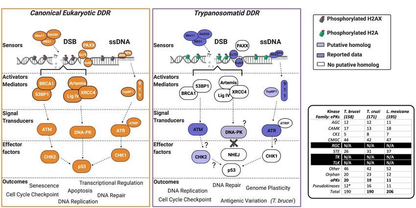
FIGURE 1 | Schematic illustration of the predicted domain locations in Trypanosomatid PIKKs compared with their human homologs. Putative domains were
identified using Pfam (http://pfam.xfam.org), Prosite (https://prosite.expasy.org), and Interpro (https://www.ebi.ac.uk/interpro/). Sequence similarity was determined
using BLAST (Altschul et al., 1997), and all sequences from trypanosomatids are compared to the corresponding human kinase sequence. Gene IDs, the
percentage identity, and the E value for each sequence are as follows: HsATM (AAB65827.1), TbATM (TbATM427_020008900; 31.47%, 2e-99), TcATM
(TcCLB.509395.20; 33.95%, 7e-108), and LmjATM (LmjF.02.0120; 31.73%, 9e-94). HsATR (NP_001175.2), TbATR (Tb427_110165100; 26.14%, 7e-119), TcATR
(TcBrA4_0103840; 27.17%, 2e-199), and LmjATR (LmjF.32.1460; 23.45%, 2e-97). HsDNA-PKcs (NP_008835.5), LmjDNA-PKcs (LmjF.36.2940; 27.58%, 4e-30).
At least two of these three PIKKs are encoded in the genomes Once active, ATM auto-phosphorylates and phosphorylates
of T. brucei, T. cruzi, and Leishmania. In the following sections, downstream substrates, including the variant histone H2AX
we will describe ATM, DNA-PKcs, and ATR, and discuss their (on serine-139) in higher eukaryotes to generate the genotoxic
reported roles in trypanosomatids (Figure 2A shows a summary stress marker yH2AX (Burma et al., 2001). However, for many
of the pathways these kinases act within). single-celled eukaryotes, for example, yeast (Downs et al.,
2000), trypanosomatids (Glover and Horn, 2012), and the
The ATM Kinase apicomplexan parasite Plasmodium falciparum (the etiological
In humans, low expression or inactivation of ATM causes agent of malaria) (Manish et al., 2021), the equivalent ATM
ataxia-telangiectasia (A-T), a neurodegenerative syndrome phosphorylation occurs on the core histone H2A. ATM can
associated with growth retardation, cancer predisposition, also activate p53 (a tumor suppressor protein) and other PKs,
immune response deficiency, and genomic instability (Savitsky including the checkpoint kinase checkpoint 2 (CHK2), halting
et al., 1995; Rothblum-Oviatt et al., 2016). Surprisingly, whereas cell cycle progression at G1 /S and G2 /M and promoting DSB
murine ATM null mutants are viable (Barlow et al., 1996; Elson repair via NHEJ (an error-prone pathway) or homologous
et al., 1996; Xu et al., 1996), kinase-dead mutants fail to survive recombination (HR; a high fidelity pathway) (Awasthi et al.,
past embryogenesis and show increased chromatid damage 2015). ATM also plays a role in telomere maintenance (Hande
associated with replication stress (Daniel et al., 2012; Yamamoto et al., 2001; Lee et al., 2015; Tong et al., 2015). ATM-deficient
et al., 2012, 2016). Thus, the inactive kinase likely inhibits other cells exhibit shortened telomeres linked to defective telomerase
repair factors from carrying out their repair functions. ATM is recruitment (an enzyme that extends telomeric sequences)
activated by DSBs detected by the Mre11-Rad50-Nbs1 (MRN) (Ritchie et al., 1999; Lee and Paull, 2005; Tong et al., 2015). ATM
complex (Figure 2A). MRN unwinds the helix and performs also acts upon dysfunctional telomeres, which are a source of
end-resection, exposing regions of ssDNA, which is pivotal for genomic instability, by eliciting a cell cycle checkpoint and cell
ATM recruitment and optimal activation (Lee and Paull, 2005). senescence (D’Adda Di Fagagna et al., 2003).
Full activation of ATM requires dissociation of the inactive The N-terminal region of the trypanosomatid ATM kinase
dimeric form of the PK, with subsequent phosphorylation is predicted to form an α-solenoid structure, accounting for
events triggering conformational changes that release one ∼57% of the enzyme (Figure 1). A FATC regulatory domain
dimer and activate the other (Bakkenist and Kastan, 2003). and a C-terminal kinase domain typical of the PIKK family can
Frontiers in Cell and Developmental Biology | www.frontiersin.org 3 August 2021 | Volume 9 | Article 636615Silva et al. DNA Repair Kinases in Trypanosomatids
FIGURE 2 | The PIKK-driven DDR pathways in Trypanosomatid parasites and the canonical Eukaryotic pathways. (A) A schematic illustration of a simplified
eukaryotic DDR pathway (left) compared to known or predicted components of the trypanosomatid DDR pathway (right). Dark shaded factors indicate that functional
characterization has been performed in one or more organisms. Light shading indicates limited data availability. White indicates no data are available or the factor is
not present in the genome, as further illustrated by question marks. For more intricate details on eukaryotic DDR factors and pathways, we encourage the reader to
refer to recent reviews (Alexander and Orr-Weaver, 2016; Blackford and Jackson, 2017; Wright et al., 2018; Sun et al., 2020; Zhao et al., 2020; Ghosh and
Raghavan, 2021). DSB, double-stranded break; DDR, DNA damage response; ssDNA, single-stranded DNA. (B) Summary table of PKs and their associated
families in T. brucei, T. cruzi, and L. mexicana. Data collated from Parsons et al. (2005), Jones et al. (2014), and Baker et al. (2021). (*) = the pseudokinases in
T. brucei are included among the counts for the other families and their respective numbers have not been adjusted to remove pseudokinase family members.
N/A = no kinases have been identified as members of these kinase families.
also be detected. However, several domains are either absent or exposed to a range of KU-55933 concentrations, a moderate
diverged in several trypanosomatids: a discernable FAT domain is slowing of parasite proliferation with little perturbation of the
absent in both T. cruzi and T. brucei, but present in Leishmania; cell cycle progression was observed, even at high concentrations
TAN domains (required for telomeric maintenance and DSB of the compound. Treatment with KU-55933 sensitized parasites
repair activities in other eukaryotes; Seidel et al., 2008) and LBE to H2 O2 , implicating ATM kinase activity in tackling oxidative
domains also appear to be lacking in all trypanosomatid ATMs. stress-derived lesions. Whether KU-55933 treatment induces
When combined with the lack of identifiable phosphorylation a more generalized sensitivity to genotoxins requires further
sites in phosphoproteomic studies in T. brucei (Urbaniak et al., investigation, as we lack information about how selective this
2013), these domain variations suggest that the regulation of inhibitor is for ATM in trypanosomatids. In a recent study, an
trypanosomatid ATM by phosphorylation is unclear and may unexpected role for the ATM gene in L. mexicana was uncovered
even differ between related trypanosomatids. (Baker et al., 2021). Deletion of ATM in promastigotes prevented
RNA interference (RNAi)-mediated depletion of ATM in the establishment of infections in the sandfly vector, implicating
mammal-infective T. brucei initially revealed a lethal phenotype ATM (and perhaps the wider DDR directed by the PK) in a
in vitro (Forsythe, 2012). However, more recent genetic screens previously unappreciated role in parasite transmission, though
(Jones et al., 2014; Stortz et al., 2017) suggest that T. brucei ATM the basis for this defect is unexplained. In fact, in both these
may be non-essential in mammal-infective cells, though effects aspects of infectivity, the L. major ATM mutants are worthy
of ATM loss in tsetse stage T. brucei are unknown. Moreover, of further study, given the inhibition data. Leishmania are
whereas in other eukaryotes ATM functions during DSB repair, intracellular parasites of mammals, developing within immune
this functionality has not been directly tested in T. brucei. Thus, cells such as neutrophils and macrophages, which generate
how ATM operates in the context of the DDR across the T. brucei reactive oxygen species (ROS). One could speculate that loss
life cycle is unclear. of ATM may increase sensitivity to ROSs generated during
In L. major, ATM function has been investigated in development in the host cell, compromising parasite viability
promastigote (sandfly-infective) cells using the small molecule and thus transmission potential. If so, ATM may be a candidate
KU-55933 (da Silva R. B. et al., 2018), which inhibits ATM activity target to block parasite transmission. To date, nothing has been
in human cells (Hickson et al., 2004). When promastigotes were reported about ATM function in T. cruzi.
Frontiers in Cell and Developmental Biology | www.frontiersin.org 4 August 2021 | Volume 9 | Article 636615Silva et al. DNA Repair Kinases in Trypanosomatids
As mentioned above, ATM phosphorylates histone H2A or ATM substrates, including H2AX, in cells lacking ATM (Stiff
H2AX in response to DNA damage. In trypanosomatids, damage- et al., 2004). DNA-PK can also orchestrate metabolic pathways
dependent phosphorylation occurs on the core histone H2A at like fatty acid synthesis (Chung, 2018). When a DSB forms, the
residue Thr130 (Glover and Horn, 2012). Following genotoxin Ku heterodimer recognizes the lesion and can recruit DNA-PKcs,
exposure, the yH2A signal can be detected either as a diffuse which, in turn, is activated by autophosphorylation, forming
nuclear signal or as foci depending on the damaging agent, the holoenzyme complex. DNA-PK phosphorylates and recruits
consistent with PK activity during the DDR. However, no work downstream substrates to effect repair. First, mismatched ends of
has shown that yH2A contributes to DNA damage repair, and the DSB are resected by nucleases, followed by gap filling by DNA
it is unknown what PK is responsible for the phosphorylation, polymerases (mainly Pol µ and Pol ε), which act in a template-
although, mutation of MRE11 abrogates the reaction (Dattani independent manner. Lastly, DNA ligase IV, in conjunction with
and Wilkinson, 2019). In addition, depletion of another DDR the x-ray repair cross-complementing protein 4 (XRCC4) and the
PK (ATR, discussed in a later section) increases yH2A levels XRCC4-like factor (XLF), seals the break (reviewed by Chung,
(Black et al., 2020). 2018; Mohiuddin and Kang, 2019; Menolfi and Zha, 2020). In
The principal downstream substrate of ATM is checkpoint recent years, a plethora of additional accessory NHEJ factors,
kinase 2 (CHK2), which can induce a G1 /S-phase cell cycle such as the Paralog of XRCC4 and XLF (PAXX; previously
stall upon activation (Matsuoka et al., 2000). A CHK2-like known as C9orf142), have been discovered, though we are yet
protein has been identified in trypanosomatids, but no work to comprehend the range of activities relating to NHEJ they
has confirmed this PK as a bonafide CHK2 homolog (Genois perform (as reviewed by Ghosh and Raghavan, 2021). Insertions
et al., 2014). Another key substrate of ATM is p53, which and deletions of the DNA template are frequent consequences
is present in metazoans (Dos Santos et al., 2016) and some of cNHEJ-directed repair. In some cases, such mutagenic repair
unicellular organisms (Lu et al., 2009; Bartas et al., 2020), though is beneficial, such as when DNA-PK acts to generate antigen
trypanosomatids appear to lack a p53 homolog. Thus, putative receptor diversity by coordinating Variable, Diverse, and Joining
events downstream of trypanosomatid ATM are unknown. V(D)J recombination (Kienker et al., 2000). Thus, mutations in
Loss of MRE11 or RAD50, the upstream recruiters of the PK the DNA-PKcs gene in mice result in severe combined immune-
(Figure 2A), affect trypanosomatid proliferation and genomic deficiency (SCID) syndrome, manifesting as profound defects
stability. In Leishmania, deletion of RAD50 can only be in T- and B-cell development. In humans, aberrant DNA-PK
achieved in an MRE11 null mutant, suggesting an unanticipated, activity correlates with the development of a range of cancers
stoichiometric balance in activities provided by these two factors (Mohiuddin and Kang, 2019).
(Laffitte et al., 2016). Both factors operate during Leishmania HR, Most kinetoplastids, including T. brucei and T. cruzi, appear
with MMEJ predominating in their absence, where chromosomal to lack DNA-PKcs, whereas across Leishmania spp., a potential
translocations are seen (Laffitte et al., 2016). In T. brucei, null DNA-PKcs homolog has been identified (Figure 1). Putative
mutants of either MRE11 or RAD50 are tolerated, with loss of the DNA-PKcs homologs have also been found in the genomes
former leading to instability in the large, transcriptionally silent of other Leishmaniiae, such as Endotrypanun monterogeii (a
Variant Surface Glycoprotein (VSG) gene-rich subtelomeres parasite of two-toed sloths) and Crithidia spp. (a monoxenous
(Robinson et al., 2002; Mehnert et al., 2021). Loss of either insect pathogen), but little is known about DNA repair in
RAD50 or MRE11 results in increased levels of VSG activation these organisms. The putative Leishmania DNA-PKcs shows
after induction of a DSB within the specialized site for VSG most sequence conservation relative to other eukaryotic DNA-
transcription (termed the bloodstream expression site), whereas PKcs proteins within its C-terminal kinase domain. Additionally,
MRE11 mutants do not display such elevation in the rate a conserved NUC194 domain, whose function is unknown,
of immune evasion without DSB induction (Robinson et al., has been identified in Leishmania DNA-PKcs, supporting this
2002). Taken together, these data raise questions about how putative PK as a homolog of human DNA-PKcs (Lees-Miller
VSG-directed HR initiates during immune evasion (da Silva et al., 2020). Functional analysis of this putative repair enzyme
M. S. et al., 2018), and analysis of ATM could be key to awaits and it is unknown if its loss alters the parasite’s
understanding this reaction. Indeed, addressing ATM function response to genotoxic stress. The putative presence of DNA-
may be informative in understanding signaling of gene family PKcs in Leishmania, and other Leishmaniiae, unlike in related
rearrangements (Weatherly et al., 2016) and gamma irradiation trypanosomatids, is especially intriguing because it is unlikely
resistance (Regis-da-Silva et al., 2006) in T. cruzi and, perhaps, to direct cNHEJ since repair of CRISPR-Cas9-generated DSBs
other trypanosomatids. in Leishmania has never been shown to occur by this repair
pathway, but instead only by MMEJ (Zhang and Matlashewski,
2015) or SSA (Zhang and Matlashewski, 2019).
DNA-PKcs: A Leishmania-Specific DDR Why Leishmania potentially possess DNA-PKcs poses another
PK? intriguing question since the Ku complex is present in T. brucei
Active DNA-PK is a holoenzyme complex consisting of DNA- and T. cruzi, which have no ortholog of the putative DNA-PKcs
PKcs and the Ku heterodimer (subunits Ku70 and Ku80 Gottlieb gene. Addressing this complex pattern of presence or absence of
and Jackson, 1993). Together, this complex initiates DSB repair components of the DNA-PK holoenzyme is further complicated
via cNHEJ. DNA-PK also shares partial functional redundancy by lack of clarity regarding what role Ku performs in the absence
with ATM; DNA-PK is capable of phosphorylating downstream of cNHEJ, with the best evidence being a role in T. brucei telomere
Frontiers in Cell and Developmental Biology | www.frontiersin.org 5 August 2021 | Volume 9 | Article 636615Silva et al. DNA Repair Kinases in Trypanosomatids
maintenance (Conway et al., 2002; Janzen et al., 2004), suggesting the activity of ATR). Once activated, ATR phosphorylates the
that this part of DNA-PK operates outside DSB repair in these effector kinase checkpoint kinase 1 (CHK1), which initiates
parasites. The nature of this critical role remains unclear, given checkpoint activation and cell cycle arrest, suppressing global
that the natural absence of both Ku proteins in Blastocrithidia origin firing, promoting dormant origin firing, and initiating
spp. does not appear to have a noticeable impact on telomere DNA repair pathways. Outside these DDR functions, ATR acts
length (Poláková et al., 2021). One possible explanation could be on centromeric R-loops to promote chromosome segregation
linked to the extensive genome plasticity observed in Leishmania, during mitosis (Kabeche et al., 2018), on genome-wide R-loops
with aneuploidy (Sterkers et al., 2011) and copy number to prevent instability (Matos et al., 2020), aids the replication
variations (CNVs) readily detected during growth (Ubeda et al., of repetitive and fragile genomic regions (Casper et al., 2002),
2008; Leprohon et al., 2009; Rogers et al., 2011; Restrepo et al., responds to mechanical stresses including nuclear and nucleolar
2019). Furthermore, the use of repair machinery for DNA deformation (Kidiyoor et al., 2016), and acts in telomere
replication (Damasceno et al., 2016, 2018, 2020) suggests that maintenance (McNees et al., 2010).
DNA repair processes are required for genome duplication. Thus, Like ATM, trypanosomatid ATR shares most sequence
the presence of a putative complete DNA-PK in Leishmania homology within the C-terminal kinase-containing region
but not in T. brucei or T. cruzi could play roles in genome (Figure 1), and ∼70% of the enzymes are composed of an
maintenance and transmission that aid plasticity. For instance, α-solenoid-like domain, which is typical of the PIKK family.
the interaction between DNA-PKcs and Ku occurring at DSBs Across all three trypanosomatids, FAT and FATC domains are
within unstable regions could activate a divergent DNA-PK present, in addition to an UME domain (NUC010; Pfam), which
pathway, perhaps amplifying repair by MMEJ or other more is characteristic of FAT and FATC domain-harboring proteins
mutagenic pathways. Though T. brucei and T. cruzi also exhibit (the function of the UME domain is unknown). Intriguingly,
genomic instability, unstable regions in T. brucei appear limited trypanosomatid ATR appears to lack a PRD domain typical of
to multicopy VSG gene families with functions in host immune PIKK kinases. This absence may be mechanistically important
evasion (Glover et al., 2013; Horn, 2014; Black et al., 2020). since the PRD domain is required for ATR activation by TOPBP1
More widespread aneuploidy and CNVs have been reported in (Mordes et al., 2008). Though a putative trypanosomatid
T. cruzi (Minning et al., 2011; Reis-Cunha et al., 2015; Callejas- homolog of TOPBP1 has been identified, its function remains
Hernández et al., 2018), though the underlying mechanics uninvestigated (Genois et al., 2014) and no interactions between
are largely uncharacterized. Thus, Leishmania DNA-PKcs may parasitic ATR and this putative TOPBP1 homolog have been
perform genus-specific functions pertaining to plasticity, though reported. In mammalian-infective T. brucei, depletion of ATR
further work is needed to demonstrate the presence and activity produces an accumulation of cells in the S-phase accompanied
of the DNA-PK holoenzyme. by growth arrest, indicating that PK is essential even in vitro
(Jones et al., 2014; Black et al., 2020). Depletion of ATR also
The ATR Kinase resulted in widespread accumulation of genotoxic stress markers,
In most eukaryotes ATR is essential for cellular proliferation. including increased levels of yH2A and formation of RAD51 and
For instance, during embryogenesis in mammals, loss of ATR RPA foci, and increased sensitivity to a range of DNA mutagens
results in mitotic catastrophe in the developing blastocyst (Brown (Black et al., 2020) implicating ATR in the trypanosomatid DDR.
and Baltimore, 2000). In adult mice, ATR depletion causes a Nonetheless, what aspect of ATR function results in T. brucei
premature aging-like syndrome that has been attributed to stem death after the loss of the PK is unknown. In this regard,
cell loss (Ruzankina et al., 2007) and appears akin to Seckel recent work in insect stage T. brucei revealed that depletion
syndrome, a complex form of microcephalic primordial dwarfism of ATR only moderately affects parasite proliferation and cell
that occurs in humans with ATR gene mutations (O’Driscoll cycle progression, despite playing an important role during HR
et al., 2003). Interestingly, loss of ATR does not predispose and damage signaling in this life cycle stage in response to
such individuals to cancer, like loss of ATM (Chanan-Khan ionizing radiation (IR) (Marin et al., 2020). This dichotomy likely
et al., 2003; Qvist et al., 2011). ATR is activated in response reflects alternative demands on repair and replication in distinct
to ssDNA accumulation at stalled DNA replication forks, at life cycle stages.
resected DSBs, or following deoxyribonucleotide triphosphate A parasite-specific and life cycle stage-specific role of ATR
(dNTP) depletion. Transcription-derived RNA-DNA hybrids (R- has been uncovered in mammalian-infective T. brucei. To evade
loops) and shortened telomeres are also prominent activators of immune clearance, stochastic switching of the VSG surface
ATR (reviewed by Saldivar et al., 2017). Briefly, ssDNA, coated antigen occurs. On any cell, at any given time, a single
with the heterotrimeric replication protein A (RPA) complex, VSG variant is expressed out of the predicted 2,000 VSGs
acts as a recruitment platform for the obligatory interaction available in the genome, the majority of which comprise a
partner of ATR, ATR Interacting Protein (ATRIP; Figure 2A). subtelomeric library (Müller et al., 2018). VSGs are transcribed
ATRIP recruits and activates ATR, resulting in a hetero- by polymerase I (Navarro and Gull, 2001; Hertz-Fowler et al.,
tetrameric complex composed of two molecules each of ATR 2008) from a specialized subtelomeric expression site known
and ATRIP. Additionally, ATR activation requires the activities as the Bloodstream Expression Site (BES), of which ∼15
of the Rad9-Rad1-Hus1 (9-1-1) complex, topoisomerase II have been reported in the laboratory-adapted Lister 427 strain
binding protein 1 (TOPBP1), and, in vertebrates, the Ewing (Müller et al., 2018). Upon ATR depletion, downregulation of
tumor-associated antigen 1 (ETAA1; the latter two regulate the actively transcribed BES occurs, correlating with increased
Frontiers in Cell and Developmental Biology | www.frontiersin.org 6 August 2021 | Volume 9 | Article 636615Silva et al. DNA Repair Kinases in Trypanosomatids
transcription from previously silent BESs, indicating that loss also raise questions about the activation of trypanosomatid ATR.
of ATR undermines BES transcriptional control. Furthermore, Trypanosomatid RPA1 can bind to the ends of telomeres and
transcripts from VSGs located in the subtelomeric library became may regulate telomere homeostasis (Pavani et al., 2016, 2018;
upregulated, suggesting increased levels of recombination events Fernandes et al., 2020). In other eukaryotes, ATR functions to
moving these VSGs into BESs. Perhaps explaining both these stabilize telomeres (McNees et al., 2010): ATR loss associated
effects on VSG expression, increased damage was detected across with R-loop and G4 structure accumulation destabilizes these
the majority of BESs and in close proximity to the VSG-associated structures, resulting in telomere dysfunction (Rhodes and Lipps,
70-bp repeats, implying that ATR may play a role in the resolution 2015; Graf et al., 2017). Given RPA activates ATR, it is possible
of lesions that accumulate within the BES. One possible form of that the kinase acts directly at the telomeres of trypanosomatids.
BES lesion is an R-loop since these structures have been shown to In support of this, loss of ATR is linked to damage accumulation
accumulate in BESs after the loss of RNase H enzymes, leading to within subtelomeric regions in T. brucei correlating with regions
the same changes in VSG expression (Briggs et al., 2018a, 2019). of R-loop formation (Briggs et al., 2018a,b; Black et al., 2020). The
Nonetheless, how ATR (and potentially R-loops) acts in VSG functions of ATR in T. cruzi are unknown.
transcriptional control and VSG recombination remains unclear
(Black et al., 2020).
In Leishmania, ATR function has been investigated in DDR Effector Kinases: What Goes on
promastigote cells using the small-molecule inhibitor VE-821 (da Downstream?
Silva R. B. et al., 2018), a selective inhibitor of the ATR kinase ATR, ATM, and DNA-PKcs, and their direct downstream
in humans (Charrier et al., 2011; Reaper et al., 2011). VE-821 substrates (discussed above), are key DDR players, coordinating
treatment was associated with a modest decrease in proliferation, much of the initial response to a DNA lesion. However, a plethora
though no cell cycle alteration was reported, as seen following of other PKs also act in a wider response to restore cellular
ATR depletion in T. brucei. However, as for ATM, VE-821-treated homeostasis after damage. In humans, up to 160 PKs (out of
cells were significantly more sensitive to H2 O2 , suggesting that ∼550 PKs encoded in the genome; Eid et al., 2017; Kanev et al.,
ATR may act during the response to oxidative stress, similar to 2019) have been linked to neoplastic cellular transformation
ATM, though work is needed to validate ATR as the target of or disease development due to mutations causing a loss or
VE-821 and to assess whether inhibiting ATR also sensitizes cells gain of function (Cell Signaling Technology, 2020). Assessing
to other genotoxins. Unlike in T. brucei, and perhaps consistent wider damage response functionality has been made possible
with the VE-821 inhibition data, ATR has been reported to be through the use of systematic high-throughput screening using
dispensable for L. mexicana survival in vitro, though effects of siRNAs, small-molecule inhibitors, or CRISPR/Cas9 technology
ATR loss were not investigated further (Baker et al., 2021). to identify novel DDR factors and map damage response
A major deficit in our understanding of the trypanosomatid pathways across several eukaryotic organisms. Indeed, a recent
ATR pathway is the initial activation of ATR itself. Other factors genome-wide screen performed in the presence or absence of a
operating within the ATR pathway include the 9-1-1 complex, panel of genotoxins has revealed ∼890 genes that may function
which has been functionally characterized in L. major, revealing during DNA repair in human cells, including ∼40 PKs (based
connections between DNA signaling pathways with genome on GO term analysis of hits on protein serine/threonine kinase
plasticity (Damasceno et al., 2018). How ATR interacts with 9-1-1 activity; Olivieri et al., 2020).
in these organisms is unknown. Given that Rad9 likely operates T. brucei, T. cruzi, and Leishmania encode for 190 (Jones et al.,
as part of an alternative complex to 9-1-1, and Hus1 is capable of 2014), 190 (Parsons et al., 2005), and 206 (Baker et al., 2021)
persisting in a monomeric form (Damasceno et al., 2016), such PKs, respectively, with several aPKs identified and members
interactions may be divergent and parasite-specific. Does ATR of all ePK groups represented, except for tyrosine-like and
interact with both complexes? Does ATR modulate their behavior tyrosine kinases (Figure 2B). Over the last decade, with the
or do they modulate the behavior of ATR? implementation of genome-wide and kinome-focused screens
The genomes of all trypanosomatids also appear to lack any in T. brucei, the roles of PKs have been investigated during
putative homologs of the obligatory ATR interaction partner drug resistance (Alsford et al., 2012), cell cycle control (Jones
ATRIP (or ETAA1), which is required for kinase activation in et al., 2014), and in vivo survival (Fernandez-Cortes et al., 2017).
other eukaryotes. We also lack information on the roles of However, only one screen to date has been performed to examine
the putative TopBP1 homolog (Genois et al., 2014), which is the parasite’s response to DNA damage. Both genome-wide and
a critical ATR activation factor (Kumagai et al., 2006) and a kinome-focused RNAi screening identified a cohort of 30 PKs
recruiter of the 9-1-1 complex, via Rad9 (Yan and Michael, (∼15% of the kinome), whose downregulation was associated
2009). TopBP1 in other eukaryotes interacts with ATR via a with increased sensitivity to MMS. Among these 30 PKs, and
small domain, the PIKK regulatory domain (PRD), upstream in addition to ATR, ATM, and the related kinase TOR4, eight
of the FATC domain; PRD deletion prevents ATR activation novel putative DDR PKs were validated. Within this cohort was
by TopBP1. The PRD domain of trypanosomatid ATR is less AUK2 (Stortz et al., 2017), a member of the aurora kinase family,
well defined and, when combined with poor conservation of the and homologous to AURKA in human cells (the function of
ATR activation domain (AAD) in the trypanosomatid TopBP1 aurora kinases is reviewed here; Tang et al., 2017; Willems et al.,
homologs, this raises questions as to whether TopBP1 plays a 2018). Deletion of AUK2 in T. brucei resulted in increased DNA
role in ATR kinase activation. Functions of the RPA complex damage sensitivity, cell cycle defects, spindle formation defects,
Frontiers in Cell and Developmental Biology | www.frontiersin.org 7 August 2021 | Volume 9 | Article 636615Silva et al. DNA Repair Kinases in Trypanosomatids
yH2A phosphorylation, and RAD51 foci formation, indicating transcriptomics may be a key strategy to examine the timing
the accumulation of DNA lesions and highlighting AUK2 as a of PK expression during parasite growth, as well as to map the
DDR kinase. AUK2 is also required for the survival of in vivo interacting PK signaling activities. In T. cruzi, no genome-wide
murine infections (Fernandez-Cortes et al., 2017). Dysregulation libraries are currently available but the recent introduction of the
of aurora kinase family members is associated with the formation CRISPR/Cas9 system in this parasite (Lander et al., 2015; Peng
of cancer, and the PKs play prominent roles during mitosis (Tang et al., 2015) could mean such screens are on the horizon.
et al., 2017). The function of AUK2 is unknown in Leishmania,
but null mutants could not be recovered in promastigote cells, CONCLUSION AND FUTURE
suggesting that it is essential (Baker et al., 2021). In T. cruzi, only
AUK1 (homologous to AURKB) function has been assessed, with
PERSPECTIVES
evidence suggesting it acts canonically during mitosis and nuclear Genome integrity must be preserved to prevent loss of
division, alongside being required during kinetoplast duplication information across generations. PKs are key facilitators of
(Fassolari and Alonso, 2018). Though the role of AUK2 was this process and their integral roles across a multitude of
not directly investigated in T. cruzi, the authors reported two DDR pathways make them opportune candidates for drug
independent forms of the protein, suggesting that this kinase development pipelines. For trypanosomatids, where many
may functionally diverge from AURKA, and indeed may display aspects of core biology are diverged, focused and broad
variation in AUK2 functions in T. brucei. approaches to study DDR PK functions have revealed novelty,
From the genome-wide screen (Stortz et al., 2017), the tousled- such as the participation of ATR in host immune evasion
like kinases 1 and 2 (TLK1/2) were identified as causing increased in T. brucei, and the proposed role for ATM in Leishmania
MMS sensitivity following their simultaneous depletion by RNAi. development in the insect vector. In contrast, we lack information
RNAi depletion resulted in a loss of proliferation, an S-phase about the DDR PK function in T. cruzi. Continued forays
stall, increasing numbers of cells lacking nuclear DNA (indicating into trypanosomatid PK function provide the prospect of new
nuclear segregation defects), and increased phosphorylation of drug targets, by re-purposing available small-molecule inhibitors,
yH2A (Stortz et al., 2017). Indeed, earlier RNAi implicated and could offer tantalizing glimpses into the evolution of core
TLK1 as the perpetrator of these defects, with TLK1 localizing biological processes in these peculiar eukaryotes.
to the nucleus of the parasites (Li et al., 2007). In metazoans,
TLKs can act during genome maintenance, in keeping with
the role of TLK1 reported for T. brucei. TLK is an essential AUTHOR CONTRIBUTIONS
gene in Leishmania, likely controlling aspects of the cell cycle,
though DDR-related roles have not been described (Baker et al., All authors listed intellectually contributed to the review,
2021). Across both screens, further investigation of candidate preparation, and submission of this article. All figures in this
DDR PKs, in addition to AUK2, revealed a further four whose review were generated using BioRender.com.
loss causes increased sensitivity to MMS, but no proliferative
defects were detected upon RNAi in vitro, suggesting that these FUNDING
kinases are required for parasite survival specifically following
genotoxic stress exposure. These four PKs belong to diverse This work was supported by funding from the Biotechnology and
PK families, including calmodulin-dependent protein kinases Biological Science Research Council (BBSRC) (BB/K006495/1,
(CAMK), which act to regulate intracellular calcium stores BB/M028909/1 and BB/N016165/1) FAPESP (16/16454-9),
including during apoptosis, and the CMGC family, which include FAPESP (20/01883-7), and FAPESP (18/14398-0) and the
regulators of cell cycle progression. In Leishmania, these four PKs Wellcome Center for Integrative Parasitology, supported by core
are non-essential, as CRISPR/Cas9-mediated null mutants are funding from the Wellcome Trust (104111).
viable in vitro (Baker et al., 2021). Further work will be needed
to investigate the role of these enzymes in the DDR, including ACKNOWLEDGMENTS
asking how they map onto the pathways elicited by ATM, ATR,
and, perhaps, DNA-PKcs. In L. mexicana, the recently developed We thank all members of the Tosi and McCulloch labs for critical
CRISPR/Cas9 bar-seq library could be used to perform the type reading of this manuscript. We apologize to any author whose
of DDR screen performed in T. brucei. In addition, single-cell work we were unable to cite due to space constraints.
REFERENCES Altschul, S. F., Madden, T. L., Schäffer, A. A., Zhang, J., Zhang, Z., Miller, W.,
et al. (1997). Gapped BLAST and PSI-BLAST: a new generation of protein
Alexander, J. L., and Orr-Weaver, T. L. (2016). Replication fork instability and the database search programs. Nucleic Acids Res. 25, 3389–3402. doi: 10.1093/nar/
consequences of fork collisions from rereplication. Genes Dev. 30, 2241–2252. 25.17.3389
doi: 10.1101/gad.288142.116 Awasthi, P., Foiani, M., and Kumar, A. (2015). ATM and ATR signaling at a glance.
Alsford, S., Eckert, S., Baker, N., Glover, L., Sanchez-Flores, A., Leung, K. F., J. Cell Sci. 28, 4255–4262. doi: 10.1242/jcs.169730
et al. (2012). High-throughput decoding of antitrypanosomal drug efficacy and Baker, N., Catta-Preta, C. M. C., Neish, R., Sadlova, J., Powell, B., Alves-Ferreira,
resistance. Nature 482, 232–236. doi: 10.1038/nature10771 E. V. C., et al. (2021). Systematic functional analysis of Leishmania protein
Frontiers in Cell and Developmental Biology | www.frontiersin.org 8 August 2021 | Volume 9 | Article 636615Silva et al. DNA Repair Kinases in Trypanosomatids kinases identifies regulators of differentiation or survival. Nat. Commun. ataxia telangiectasia mutated and Rad3 related (ATR) protein kinase as potential 12:1244. doi: 10.1038/s41467-021-21360-8 anticancer agents. J. Med. Chem. 54, 2320–2330. doi: 10.1021/jm101488z Bakkenist, C. J., and Kastan, M. B. (2003). DNA damage activates ATM through Chatterjee, N., and Walker, G. C. (2017). Mechanisms of DNA damage, repair, and intermolecular autophosphorylation and dimer dissociation. Nature 421, 499– mutagenesis. Environ. Mol. Mutagen. 58, 235–263. doi: 10.1002/em.22087 506. doi: 10.1038/nature01368 Chen, X., Xu, X., Chen, Y., Cheung, J. C., Wang, H., Jiang, J., et al. (2021). Barlow, C., Hirotsune, S., Paylor, R., Liyanage, M., Eckhaus, M., Collins, F., Structure of an activated DNA-PK and its implications for NHEJ. Mol. Cell 81, et al. (1996). Atm-deficient mice: a paradigm of ataxia telangiectasia. Cell 86, 801–810.e3. doi: 10.1016/j.molcel.2020.12.015 159–171. doi: 10.1016/S0092-8674(00)80086-0 Chung, J. H. (2018). The role of DNA-PK in aging and energy metabolism. FEBS J. Bartas, M., Brázda, V., Červeň, J., and Pečinka, P. (2020). Characterization of p53 285, 1959–1972. doi: 10.1111/febs.14410 family homologs in evolutionary remote branches of holozoa. Int. J. Mol. Sci. Conway, C., McCulloch, R., Ginger, M. L., Robinson, N. P., Browitt, A., and 21:6. doi: 10.3390/ijms21010006 David Barry, J. (2002). Ku is important for telomere maintenance, but not for Bhattacharya, A., Corbeil, A., Do Monte-Neto, R. L., and Fernandez-Prada, C. differential expression of telomeric VSG genes, in African trypanosomes. J. Biol. (2020). Of drugs and trypanosomatids: new tools and knowledge to reduce Chem. 277, 21269–21277. doi: 10.1074/jbc.M200550200 bottlenecks in drug discovery. Genes 11:722. doi: 10.3390/genes11070722 D’Adda Di Fagagna, F., Reaper, P. M., Clay-Farrace, L., Fiegler, H., Carr, P., Von Black, J. A., Crouch, K., Lemgruber, L., Lapsley, C., Dickens, N., Tosi, L. R. O., et al. Zglinicki, T., et al. (2003). A DNA damage checkpoint response in telomere- (2020). Trypanosoma brucei ATR links DNA damage signaling during antigenic initiated senescence. Nature 426, 194–198. doi: 10.1038/nature02118 variation with regulation of RNA polymerase I-transcribed surface antigens. da Silva, M. S., Hovel-Miner, G. A., Briggs, E. M., Elias, M. C., and McCulloch, Cell Rep. 30, 836–851.e5. doi: 10.1016/j.celrep.2019.12.049 R. (2018). Evaluation of mechanisms that may generate DNA lesions triggering Blackford, A. N., and Jackson, S. P. (2017). ATM, ATR, and DNA-PK: the trinity at antigenic variation in African trypanosomes. PLoS Pathog. 14:e1007321. doi: the heart of the DNA damage response. Mol. Cell 66, 801–817. doi: 10.1016/j. 10.1371/journal.ppat.1007321 molcel.2017.05.015 da Silva, R. B., Machado, C. R., Aquiles Rodrigues, A. R., and Pedrosa, A. L. Briggs, E., Crouch, K., Lemgruber, L., Hamilton, G., Lapsley, C., and McCulloch, R. (2018). Selective human inhibitors of ATR and ATM render Leishmania major (2019). Trypanosoma brucei ribonuclease H2A is an essential R-loop processing promastigotes sensitive to oxidative damage. PLoS One 13:e0205033. doi: 10. enzyme whose loss causes DNA damage during transcription initiation and 1371/journal.pone.0205033 antigenic variation. Nucleic Acids Res. 47, 9180–9197. doi: 10.1093/nar/gkz644 Damasceno, J. D., Marques, C. A., Beraldi, D., Crouch, K., Lapsley, C., Obonaga, Briggs, E., Crouch, K., Lemgruber, L., Lapsley, C., and McCulloch, R. (2018a). R., et al. (2020). Genome duplication in Leishmania major relies on persistent Ribonuclease H1-targeted R-loops in surface antigen gene expression sites can subtelomeric DNA replication. Elife 9:e58030. doi: 10.7554/ELIFE.58030 direct trypanosome immune evasion. PLoS Genet. 14:e1007729. doi: 10.1371/ Damasceno, J. D., Obonaga, R., Santos, E. V., Scott, A., McCulloch, R., and Tosi, journal.pgen.1007729 L. R. O. (2016). Functional compartmentalization of Rad9 and Hus1 reveals Briggs, E., Hamilton, G., Crouch, K., Lapsley, C., and McCulloch, R. (2018b). diverse assembly of the 9-1-1 complex components during the DNA damage Genome-wide mapping reveals conserved and diverged R-loop activities in the response in Leishmania. Mol. Microbiol. 101, 1054–1068. doi: 10.1111/mmi. unusual genetic landscape of the African trypanosome genome. Nucleic Acids 13441 Res. 46, 11789–11805. doi: 10.1093/nar/gky928 Damasceno, J. D., Obonaga, R., Silva, G. L. A., Reis-Cunha, J. L., Duncan, S. M., Brown, E. J., and Baltimore, D. (2000). ATR disruption leads to chromosomal Bartholomeu, D. C., et al. (2018). Conditional genome engineering reveals fragmentation and early embryonic lethality. Genes Dev. 14, 397–402. doi: 10. canonical and divergent roles for the Hus1 component of the 9–1–1 complex 1101/gad.14.4.397 in the maintenance of the plastic genome of Leishmania. Nucleic Acids Res. 46, Burma, S., Chen, B. P., Murphy, M., Kurimasa, A., and Chen, D. J. (2001). ATM 11835–11846. doi: 10.1093/nar/gky1017 phosphorylates histone H2AX in response to DNA double-strand breaks. J. Biol. Daniel, J. A., Pellegrini, M., Lee, B. S., Guo, Z., Filsuf, D., Belkina, N. V., et al. (2012). Chem. 276, 42462–42467. doi: 10.1074/jbc.C100466200 Loss of ATM kinase activity leads to embryonic lethality in mice. J. Cell Biol. 198, Burton, P., McBride, D. J., Wilkes, J. M., Barry, J. D., and McCulloch, R. (2007). Ku 295–304. doi: 10.1083/jcb.201204035 heterodimer-independent end joining in Trypanosoma brucei cell extracts relies Dattani, A., and Wilkinson, S. R. (2019). Deciphering the interstrand crosslink upon sequence microhomology. Eukaryot. Cell 6, 1773–1781. doi: 10.1128/EC. DNA repair network expressed by Trypanosoma brucei. DNA Repair 78, 154– 00212-07 166. doi: 10.1016/j.dnarep.2019.04.009 Büscher, P., Cecchi, G., Jamonneau, V., and Priotto, G. (2017). Human African Dos Santos, H. G., Nunez-Castilla, J., and Siltberg-Liberles, J. (2016). Functional trypanosomiasis. Lancet 390, 2397–2409. doi: 10.1016/S0140-6736(17)31510-6 diversification after gene duplication: paralog specific regions of structural Butenko, A., Opperdoes, F. R., Flegontova, O., Horák, A., Hampl, V., Keeling, disorder and phosphorylation in p53, p63, and p73. PLoS One 11:e0151961. P., et al. (2020). Evolution of metabolic capabilities and molecular features doi: 10.1371/journal.pone.0151961 of diplonemids, kinetoplastids, and euglenids. BMC Biol. 18:23. doi: 10.1186/ Downs, J. A., Lowndes, N. F., and Jackson, S. P. (2000). A role for Saccharomyces s12915-020-0754-1 cerevisiae histone H2A in DNA repair. Nature 408, 1001–1004. doi: 10.1038/ Callejas-Hernández, F., Rastrojo, A., Poveda, C., Gironès, N., and Fresno, M. 35050000 (2018). Genomic assemblies of newly sequenced Trypanosoma cruzi strains Eid, S., Turk, S., Volkamer, A., Rippmann, F., and Fulle, S. (2017). Kinmap: a reveal new genomic expansion and greater complexity. Sci. Rep. 8:14631. doi: web-based tool for interactive navigation through human kinome data. BMC 10.1038/s41598-018-32877-2 Bioinformatics 18:16. doi: 10.1186/s12859-016-1433-7 Carles, F., Bourg, S., Meyer, C., and Bonnet, P. (2018). PKIDB: a curated, annotated Elson, A., Wang, Y., Daugherty, C. J., Morton, C. C., Zhou, F., Campos-Torres, J., and updated database of protein kinase inhibitors in clinical trials. Molecules et al. (1996). Pleiotropic defects in ataxia-telangiectasia protein-deficient mice. 23:908. doi: 10.3390/molecules23040908 Proc. Natl. Acad. Sci. U.S.A. 93, 13084–13089. doi: 10.1073/pnas.93.23.13084 Casper, A. M., Nghiem, P., Arlt, M. F., and Glover, T. W. (2002). ATR regulates Fassolari, M., and Alonso, G. D. (2018). Aurora kinase protein family in fragile site stability. Cell 111, 779–789. doi: 10.1016/S0092-8674(02)01113-3 Trypanosoma cruzi: novel role of an AUK-B homologue in kinetoplast Cell Signaling Technology (2020). Kinase-Disease Associations. Available online replication. PLoS Negl. Trop. Dis. 13:e0007256. doi: 10.1371/journal.pntd. at: https://www.cellsignal.com/contents/resources-reference-tables/kinase- 0007256 disease-associations/science-tables-kinase-disease (accessed November 29, Fernandes, C. A. H., Morea, E. G. O., dos Santos, G. A., da Silva, V. L., Vieira, 2020). M. R., Viviescas, M. A., et al. (2020). A multi-approach analysis highlights the Chanan-Khan, A., Holkova, B., Perle, M. A., Reich, E., Wu, C. D., Inghirami, G., relevance of RPA-1 as a telomere end-binding protein (TEBP) in Leishmania et al. (2003). T-cell clonality and myelodysplasia without chromosomal fragility amazonensis. Biochim. Biophys. Acta Gen. Subj. 1864:129607. doi: 10.1016/j. in a patient with features of Seckel syndrome. Haematologica 88:ECR14. bbagen.2020.129607 Charrier, J. D., Durrant, S. J., Golec, J. M. C., Kay, D. P., Knegtel, R. M. A., Fernandez-Cortes, F., Serafim, T. D., Wilkes, J. M., Jones, N. G., Ritchie, R., MacCormick, S., et al. (2011). Discovery of potent and selective inhibitors of McCulloch, R., et al. (2017). RNAi screening identifies Trypanosoma brucei Frontiers in Cell and Developmental Biology | www.frontiersin.org 9 August 2021 | Volume 9 | Article 636615
Silva et al.                                                                                                                                DNA Repair Kinases in Trypanosomatids

   stress response protein kinases required for survival in the mouse. Sci. Rep.                 Kanev, G. K., de Graaf, C., de Esch, I. J. P., Leurs, R., Würdinger, T., Westerman,
   7:6156. doi: 10.1038/s41598-017-06501-8                                                           B. A., et al. (2019). The landscape of atypical and eukaryotic protein kinases.
Field, M. C., Horn, D., Fairlamb, A. H., Ferguson, M. A. J., Gray, D. W., Read, K. D.,               Trends Pharmacol. Sci. 40, 818–832. doi: 10.1016/j.tips.2019.09.002
   et al. (2017). Anti-trypanosomatid drug discovery: an ongoing challenge and a                 Kidiyoor, G. R., Kumar, A., and Foiani, M. (2016). ATR-mediated regulation of
   continuing need. Nat. Rev. Microbiol. 15, 217–231. doi: 10.1038/nrmicro.2016.                     nuclear and cellular plasticity. DNA Repair 44, 143–150. doi: 10.1016/j.dnarep.
   193                                                                                               2016.05.020
Forsythe, G. R. (2012). DNA Damage and the Trypanosoma brucei Cell Cycle.                        Kienker, L. J., Shin, E. K., and Meek, K. (2000). Both V(D)J recombination and
   Available online at: http://encore.lib.gla.ac.uk/iii/encore/record/C__Rb2926197                   radioresistance require DNA-PK kinase activity, though minimal levels suffice
   (accessed November 30, 2020).                                                                     for V(D)J recombination. Nucleic Acids Res. 28, 2752–2761. doi: 10.1093/nar/
Genois, M.-M., Paquet, E. R., Laffitte, M.-C. N., Maity, R., Rodrigue, A., Ouellette,                28.14.2752
   M., et al. (2014). DNA repair pathways in trypanosomatids: from DNA                           Kumagai, A., Lee, J., Yoo, H. Y., and Dunphy, W. G. (2006). TopBP1 activates the
   repair to drug resistance. Microbiol. Mol. Biol. Rev. 78, 40–73. doi: 10.1128/                    ATR-ATRIP complex. Cell 124, 943–955. doi: 10.1016/j.cell.2005.12.041
   mmbr.00045-13                                                                                 Kung, J. E., and Jura, N. (2016). Structural basis for the non-catalytic functions of
Ghosh, D., and Raghavan, S. C. (2021). Nonhomologous end joining: new accessory                      protein kinases. Structure 24, 7–24. doi: 10.1016/j.str.2015.10.020
   factors fine tune the machinery. Trends Genet. 37, 582–599. doi: 10.1016/j.tig.               Laffitte, M. C. N., Leprohon, P., Hainse, M., Légaré, D., Masson, J. Y., and
   2021.03.001                                                                                       Ouellette, M. (2016). Chromosomal translocations in the parasite Leishmania
Glover, L., Alsford, S., and Horn, D. (2013). DNA break site at fragile                              by a MRE11/RAD50-independent microhomology-mediated end joining
   subtelomeres determines probability and mechanism of antigenic variation                          mechanism. PLoS Genet. 12:e1006117. doi: 10.1371/journal.pgen.1006117
   in African trypanosomes. PLoS Pathog. 9:e1003260. doi: 10.1371/journal.ppat.                  Lander, N., Li, Z. H., Niyogi, S., and Docampo, R. (2015). CRISPR/Cas9-induced
   1003260                                                                                           disruption of paraflagellar rod protein 1 and 2 genes in Trypanosoma cruzi
Glover, L., and Horn, D. (2012). Trypanosomal histone γh2A and the DNA damage                        reveals their role in flagellar attachment. mBio 5:e01114-01114. doi: 10.1128/
   response. Mol. Biochem. Parasitol. 183, 78–83. doi: 10.1016/j.molbiopara.2012.                    mBio.01012-15
   01.008                                                                                        Lee, J. H., and Paull, T. T. (2005). ATM activation by DNA double-strand breaks
Glover, L., and Horn, D. (2014). Locus-specific control of DNA resection and                         through the Mre11-Rad50-Nbs1 complex. Science 308, 551–554. doi: 10.1126/
   suppression of subtelomeric VSG recombination by HAT3 in the African                              science.1108297
   trypanosome. Nucleic Acids Res. 42, 12600–12613. doi: 10.1093/nar/gku900                      Lee, S. S., Bohrson, C., Pike, A. M., Wheelan, S. J., and Greider, C. W. (2015). ATM
Glover, L., Jun, J., and Horn, D. (2011). Microhomology-mediated deletion and                        kinase is required for telomere elongation in mouse and human cells. Cell Rep.
   gene conversion in African trypanosomes. Nucleic Acids Res. 39, 1372–1380.                        13, 1623–1632. doi: 10.1016/j.celrep.2015.10.035
   doi: 10.1093/nar/gkq981                                                                       Lees-Miller, J. P., Cobban, A., Katsonis, P., Bacolla, A., Tsutakawa, S. E., Hammel,
Gottlieb, T. M., and Jackson, S. P. (1993). The DNA-dependent protein kinase:                        M., et al. (2020). Uncovering DNA-PKcs ancient phylogeny, unique sequence
   requirement for DNA ends and association with Ku antigen. Cell 72, 131–142.                       motifs and insights for human disease. Prog. Biophys. Mol. Biol. 163, 87–108.
   doi: 10.1016/0092-8674(93)90057-W                                                                 doi: 10.1016/j.pbiomolbio.2020.09.010
Graf, M., Bonetti, D., Lockhart, A., Serhal, K., Kellner, V., Maicher, A., et al. (2017).        Leprohon, P., Légaré, D., Raymond, F., Madore, É, Hardiman, G., Corbeil, J.,
   Telomere length determines TERRA and R-loop regulation through the cell                           et al. (2009). Gene expression modulation is associated with gene amplification,
   cycle. Cell 170, 72–85.e14. doi: 10.1016/j.cell.2017.06.006                                       supernumerary chromosomes and chromosome loss in antimony-resistant
Hande, M. P., Balajee, A. S., Tchirkov, A., Wynshaw-Boris, A., and Lansdorp,                         Leishmania infantum. Nucleic Acids Res. 37, 1387–1399. doi: 10.1093/nar/
   P. M. (2001). Extra-chromosomal telomeric DNA in cells from Atm-/- mice                           gkn1069
   and patients with ataxia-telangiectasia. Hum. Mol. Genet. 10, 519–528. doi:                   Li, Z., Gourguechon, S., and Wang, C. C. (2007). Tousled-like kinase in a microbial
   10.1093/hmg/10.5.519                                                                              eukaryote regulates spindle assembly and S-phase progression by interacting
Hanks, S. K., and Hunter, T. (1995). The eukaryotic protein kinase superfamily:                      with Aurora kinase and chromatin assembly factors. J. Cell Sci. 120(Pt 21),
   kinase (catalytic) domain structure and classification 1. FASEB J. 9, 576–596.                    3883–3894. doi: 10.1242/jcs.007955
   doi: 10.1096/fasebj.9.8.7768349                                                               Lu, W. J., Amatruda, J. F., and Abrams, J. M. (2009). P53 ancestry: gazing through
Hertz-Fowler, C., Figueiredo, L. M., Quail, M. A., Becker, M., Jackson, A., Bason, N.,               an evolutionary lens. Nat. Rev. Cancer 9, 758–762. doi: 10.1038/nrc2732
   et al. (2008). Telomeric expression sites are highly conserved in Trypanosoma                 Lukeš, J., Butenko, A., Hashimi, H., Maslov, D. A., Votýpka, J., and Yurchenko, V.
   brucei. PLoS One 3:e3527. doi: 10.1371/journal.pone.0003527                                       (2018). Trypanosomatids are much more than just trypanosomes: clues from
Hickson, I., Zhao, Y., Richardson, C. J., Green, S. J., Martin, N. M. B., Orr, A. I.,                the expanded family tree. Trends Parasitol. 34, 466–480. doi: 10.1016/j.pt.2018.
   et al. (2004). Identification and characterization of a novel and specific inhibitor              03.002
   of the ataxia-telangiectasia mutated kinase ATM. Cancer Res. 64, 9152–9159.                   Lukeš, J., Skalický, T., Týč, J., Votýpka, J., and Yurchenko, V. (2014). Evolution
   doi: 10.1158/0008-5472.CAN-04-2727                                                                of parasitism in kinetoplastid flagellates. Mol. Biochem. Parasitol. 195, 115–122.
Horn, D. (2014). Antigenic variation in African trypanosomes. Mol. Biochem.                          doi: 10.1016/j.molbiopara.2014.05.007
   Parasitol. 195, 123–129. doi: 10.1016/j.molbiopara.2014.05.001                                Luzwick, J. W., Nam, E. A., Zhao, R., and Cortez, D. (2014). Mutation of serine
Hunter, T., and Plowman, G. D. (1997). The protein kinases of budding yeast: six                     1333 in the ATR HEAT repeats creates a hyperactive kinase. PLoS One 9:e99397.
   score and more. Trends Biochem. Sci. 22, 18, IN1–IN4, 19–22. doi: 10.1016/                        doi: 10.1371/journal.pone.0099397
   S0968-0004(96)10068-2                                                                         Machado, C. R., Vieira-da-Rocha, J. P., Mendes, I. C., Rajão, M. A., Marcello,
Imseng, S., Aylett, C. H., and Maier, T. (2018). Architecture and activation of                      L., Bitar, M., et al. (2014). Nucleotide excision repair in Trypanosoma brucei:
   phosphatidylinositol 3-kinase related kinases. Curr. Opin. Struct. Biol. 49,                      specialization of transcription-coupled repair due to multigenic transcription.
   177–189. doi: 10.1016/j.sbi.2018.03.010                                                           Mol. Microbiol. 92, 756–776. doi: 10.1111/mmi.12589
Janzen, C. J., Lander, F., Dreesen, O., and Cross, G. A. M. (2004). Telomere length              Manish, G., Adina, H., Vera, M., Sofiya, K.-S., Kumar, S. B., Ron, D., et al. (2021).
   regulation and transcriptional silencing in KU80-deficient Trypanosoma brucei.                    Phosphorylation of the canonical histone H2A marks foci of damaged DNA in
   Nucleic Acids Res. 32, 6575–6584. doi: 10.1093/nar/gkh991                                         malaria parasites. mSphere 6:e01131-20. doi: 10.1128/mSphere.01131-20
Jones, N. G., Thomas, E. B., Brown, E., Dickens, N. J., Hammarton, T. C., and                    Manning, G., Whyte, D. B., Martinez, R., Hunter, T., and Sudarsanam, S. (2002).
   Mottram, J. C. (2014). Regulators of Trypanosoma brucei cell cycle progression                    The protein kinase complement of the human genome. Science 298, 1912–1934.
   and differentiation identified using a kinome-wide RNAi screen. PLoS Pathog.                      doi: 10.1126/science.1075762
   10:e1003886. doi: 10.1371/journal.ppat.1003886                                                Marin, P. A., Obonaga, R., Pavani, R. S., da Silva, M. S., de Araujo, C. B., Lima,
Kabeche, L., Nguyen, H. D., Buisson, R., and Zou, L. (2018). A mitosis-specific                      A. A., et al. (2020). ATR kinase is a crucial player mediating the DNA damage
   and R loop-driven ATR pathway promotes faithful chromosome segregation.                           response in Trypanosoma brucei. Front. Cell Dev. Biol. 8:602956. doi: 10.3389/
   Science 359, 108–114. doi: 10.1126/science.aan6490                                                fcell.2020.602956

Frontiers in Cell and Developmental Biology | www.frontiersin.org                           10                                              August 2021 | Volume 9 | Article 636615
You can also read