Inflammation Spreading: Negative Spiral Linking Systemic Inflammatory Disorders and Alzheimer's Disease - Frontiers
←
→
Page content transcription
If your browser does not render page correctly, please read the page content below
REVIEW
                                                                                                                                         published: 25 February 2021
                                                                                                                                     doi: 10.3389/fncel.2021.638686

                                           Inflammation Spreading: Negative
                                           Spiral Linking Systemic Inflammatory
                                           Disorders and Alzheimer’s Disease
                                           Junjun Ni 1 * and Zhou Wu 2,3 *
                                           1
                                            Key Laboratory of Molecular Medicine and Biotherapy, Department of Biology, School of Life Science, Beijing Institute of
                                           Technology, Beijing, China, 2 Department of Aging Science and Pharmacology, Faculty of Dental Science, Kyushu University,
                                           Fukuoka, Japan, 3 OBT Research Center, Faculty of Dental Science, Kyushu University, Fukuoka, Japan

                                           As a physiological response to injury in the internal body organs, inflammation
                                           is responsible for removing dangerous stimuli and initiating healing. However,
                                           persistent and exaggerative chronic inflammation causes undesirable negative effects
                                           in the organs. Inflammation occurring in the brain and spinal cord is known as
                            Edited by:     neuroinflammation, with microglia acting as the central cellular player. There is increasing
               Małgorzata Kujawska,        evidence suggesting that chronic neuroinflammation is the most relevant pathological
         Poznan University of Medical
                   Sciences, Poland
                                           feature of Alzheimer’s disease (AD), regulating other pathological features, such as the
                                           accumulation of amyloid-β (Aβ) and hyperphosphorylation of Tau. Systemic inflammatory
                        Reviewed by:
                     Rachel Raybould,      signals caused by systemic disorders are known to strongly influence neuroinflammation
   Cardiff University, United Kingdom      as a consequence of microglial activation, inflammatory mediator production, and the
              Vijayasree V. Giridharan,
   University of Texas Health Science      recruitment of peripheral immune cells to the brain, resulting in neuronal dysfunction.
    Center at Houston, United States       However, the neuroinflammation-accelerated neuronal dysfunction in AD also influences
                        Alireza Faridar,
                                           the functions of peripheral organs. In the present review, we highlight the link between
       Houston Methodist Research
               Institute, United States    systemic inflammatory disorders and AD, with inflammation serving as the common
               *Correspondence:            explosion. We discuss the molecular mechanisms that govern the crosstalk between
                          Zhou Wu          systemic inflammation and neuroinflammation. In our view, inflammation spreading
         zhouw@dent.kyushu-u.ac.jp
                          Junjun Ni
                                           indicates a negative spiral between systemic diseases and AD. Therefore, “dampening
                nijunjun@bit.edu.cn        inflammation” through the inhibition of cathepsin (Cat)B or CatS may be a novel
                   Specialty section:      therapeutic approach for delaying the onset of and enacting early intervention for AD.
        This article was submitted to
                                           Keywords: systemic inflammation, macrophages, neuroinflammation, Alzheimer’s disease, cathepsin, cytokines,
             Cellular Neuropathology,
                                           systemic inflammatory disorders
                a section of the journal
    Frontiers in Cellular Neuroscience

      Received: 07 December 2020
       Accepted: 03 February 2021
                                           INTRODUCTION
      Published: 25 February 2021
                                           Alzheimer’s disease (AD), the most common type of dementia, is becoming an international
                             Citation:     public health problem with the growth of the aged population worldwide. Developing effective
  Ni J and Wu Z (2021) Inflammation
                                           approaches for preventing and enacting early intervention for AD is an urgent issue, as relatively
   Spreading: Negative Spiral Linking
Systemic Inflammatory Disorders and
                                           little progress in crafting new therapies to combat AD has been made. Chronic neuroinflammation
                  Alzheimer’s Disease.     is well accepted as the most relevant pathological features of AD, regulating other pathological
    Front. Cell. Neurosci. 15:638686.      hallmarks of AD, such as the accumulation of amyloid-β (Aβ) and hyperphosphorylation of Tau,
     doi: 10.3389/fncel.2021.638686        both of which are involved in the neuronal dysfunction in AD (Carlyle et al., 2014; Chen, 2015).

Frontiers in Cellular Neuroscience | www.frontiersin.org                          1                                     February 2021 | Volume 15 | Article 638686
Ni and Wu                                                                                                    Inflammation Links Periphery and CNS

However, there is a great deal of evidence suggesting the                    Neuroinflammation, an inflammatory reaction in the CNS,
important role of systemic inflammation in the pathogenesis              is usually characterized by microglial activation and collateral
of AD, especially in neuroinflammation. Neuroinflammation,               brain damage and is caused by a strong inflammatory reaction.
and indeed, inflammation in general, is widely considered a              The excessive activation of microglial cells resulted in the
major contributor to the onset as well as the pathological               release of a large number of proinflammatory factors, which
process of AD (Holmes, 2013). Therapies aimed at reducing                aggravated brain damages (Spangenberg et al., 2016). In addition
systemic inflammation in individuals with mild cognitive                 to microglia, astrocytes, which constitute another type of glial
impairment (MCI) and AD have proven beneficial by delaying               cells in the CNS, are also involved in neuroinflammation. In
the cognitive decline in these individuals, suggesting that              a bioinformatics analysis, aging-related genes in the microglia
recognition of the cross-talk between systemic inflammation              were associated with the inflammatory response, whereas these
and neuroinflammation has important implications for AD                  genes in astrocytes included wildly recognized AD risk genes
therapeutic strategies (Ide et al., 2016; Zhu et al., 2018).             (Pan et al., 2020). In the progress of AD, microglia are activated
    It is well accepted that the pro-inflammatory mediators,             and secrete neuroinflammatory factors, including IL-1β, IL-6,
including interleukin (IL)-1β, IL-6, and tumor necrosis factor           and TNF-α, which can kill pathogens and promote tissue repair
(TNF)-α, are co-related factors involved in both systemic                by enhancing phagocytosis. However, the excessive release of IL-
inflammation and neuroinflammation and affecting their                   1β, IL-6, and TNF-α induces chronic neuroinflammation and
sustainment and convergence. In contrast, intracellular enzymes,         aggravates brain damage (Voet et al., 2019). IL-1β, a master
such as lysosomal cathepsins, mediate the production of                  regulator of neuroinflammation, is a very potent signaling
pro-inflammatory mediators from both the periphery and brain             molecule that is expressed normally at low levels but is induced
(Voet et al., 2019).                                                     rapidly in response to local or peripheral insults (Basu et al.,
    The present review highlights the available evidence that            2004). In AD, IL-1β, IL-6, and TNF-α are involved in causing
inflammation spreading indicates a negative spiral between               neurodegenerative changes (Griffin et al., 1998; Paganelli et al.,
systemic disorders and AD. We also discuss the molecular                 2002; Uslu et al., 2012). These proinflammatory mediators are
mechanisms underlying the crosstalk between systemic                     largely produced by microglia in the CNS, while exhibiting
inflammation and neuroinflammation. We believe that                      distinct activity pathways (Ni et al., 2015, 2019b).
‘‘dampening inflammation’’ may be a novel therapeutic approach
for delaying the onset of and enacting early intervention for AD.        Neuroinflammatory Mediators and
                                                                         Neuronal Dysfunction
NEUROINFLAMMATION IN AD                                                  Under conditions of neuroinflammation, proinflammatory
                                                                         cytokines, such as IL-1β, IL-6, and TNF-α, are released by
Microglia and Neuroinflammation in AD                                    activated microglia to promote a neuroinflammatory state. The
Making up 5%–10% of the cells in the brain, microglia are                proinflammatory cytokines can either directly affect neurons
the most abundant of the resident macrophage populations                 or indirectly regulate the expression of many other genes and
in the central nervous system (CNS). Microglia are involved              consequently affect neurons, resulting in neuronal dysfunction.
in surveillance and are characterized by branching with small            In the hippocampus, high concentrations of IL-1β act on
cell bodies and long protrusions (Michell-Robinson et al.,               neurons to inhibit synaptic strength and long-term potentiation
2015). Under this homeostatic condition, microglia can secrete           (LTP). The classical downstream signaling pathways are activated
growth factors to support and protect the electrophysiological           following IL-1β/IL-R binding. One of these pathways involves
functions of neurons, thereby maintaining CNS homeostasis                the phosphorylation and subsequent degradation of the IκB
and dynamically monitoring the synaptic function through                 subunit of nuclear factor κB (NF-κB), leading to the nuclear
synaptic contact. Also, microglia have been reported to maintain         translocation of NF-κB and its gene expression. Another pathway
the normal synaptic function through trimming or clearing                is the activation of mitogen-activated protein kinases (MAPKs),
damaged or redundant synapses (Wake et al., 2013). Following             extracellular signal-regulated kinase (ERK), and p38. Among
an insult or injury, microglia become activated and adopt                these, p38 and MAPK were reported to be activated in neurons
different states, and this polarization has resulted in the common       (Srinivasan et al., 2004), while alternative signaling involving Src
categorizations of M1-like microglia, which are suggested to have        kinase has also been reported to be activated by IL-1β/IL-R in
a more ‘classic’ role in the inflammatory response, and M2-like          hippocampal neurons (Viviani et al., 2003). Primary cultured
microglia, which exert an anti-inflammatory role to resolve              neurons exposed to recombinant IL-1β showed a significant
and suppress inflammatory responses (Ni et al., 2015, 2019a;             decrease in the level of the synaptic vesicle protein synaptophysin
Ransohoff, 2016). The elimination and repopulation of resident           and in the number of synaptic sites (Li et al., 2003). Also, IL-
microglia in aged mice significantly improved spatial memory             1β regulates dendritic spine morphology by upregulating the
and increased both neurogenesis and dendritic spine densities            transcriptional factor methyl CpG binding protein 2 (MeCP2) in
(Elmore et al., 2018). However, sustained microglial elimination         a mechanistic target of rapamycin (mTOR)-dependent manner
impaired the parenchymal plaque development in an AD mouse               (Tomasoni et al., 2017), leading to an excitatory/inhibitory
model (Spangenberg et al., 2019). These observations suggest that        unbalance by delaying the developmentally regulated switch of
microglia are a critical causative factor in the development and         gamma-aminobutyric acid (GABA) signaling (Corradini et al.,
progression of aging and AD.                                             2018). Unlike IL-1β, IL-6 can stimulate microglia and astrocytes

Frontiers in Cellular Neuroscience | www.frontiersin.org             2                                 February 2021 | Volume 15 | Article 638686
Ni and Wu Inflammation Links Periphery and CNS
to release a cascade of proinflammatory cytokines and acute- The other face of microglia appears in more advanced stages of
phase proteins, such as C-reactive protein (CRP; Querfurth and AD and has been shown to cause extensive inflammation and
Laferla, 2010). In the hippocampus, the distribution of IL-6 neurodegeneration. Nevertheless, the sustained overexpression
receptor (IL-6R) is modest in glia but prominent in perikaryon of IL-1β exacerbated tau pathology despite a reduced amyloid
and outlining the apical dendrites in neurons. IL-6R subunits in burden in AD mice (Ghosh et al., 2013). Although the roles of
the rat cerebral cortex showed that both IL-6R and its subunit are microglia in the initiation and progression of AD are heavily
localized in pre-and postsynaptic membranes (D’arcangelo et al., debated, other reports describe the protective contribution of
2000). Several downstream signaling partners are associated microglia to AD. Thus, the involvement of microglia in AD
with IL-6R activation, including Janus kinase (JAK), signal pathologies has generated strong interest.
transducer and activator of transcription 3 (STAT3), MAPK, The presence of neuroinflammation-accelerated neuronal
and phosphoinositide 3-kinases (PI3K), which are located dysfunction in AD may influence the functions of the peripheral
with postsynaptic density at hippocampal synapses (Nicolas organs. It is considered that neuronal dysfunction in AD
et al., 2012; Murase and Mckay, 2014). Therefore, IL-6/IL-6R negatively impacts the peripheral organs through peripheral Aβ
and associated signal transduction molecules are appropriately accumulation; high levels of Aβ has been found in the peripheral
positioned to influence the synaptic function. TNF-α interacts circulation of AD patients; this is attributed to a reduction of
with two cognate receptors, TNF receptor (TNF-R) I and TNF- peripheral Aβ clearances as well as peripheral Aβ production (Nie
RII. These receptors are expressed on various cell types, including et al., 2019; Gu et al., 2020). Bone is influenced by high peripheral
neurons, throughout the CNS. The binding of homotrimeric levels of Aβ. It is reported that Aβ induces the differentiation
TNF-α to either receptor can activate three major signaling and activation of osteoclasts, resulting in the promotion of bone
cascades: the Fas/caspase-8, NFκB, and c-Jun N-terminal kinase destruction (Cui et al., 2011; Li et al., 2016). Indeed, neuronal
(JNK) pathways (Park and Bowers, 2010). TNF-RI is a member dysfunction with the brain structure changes in AD has been
of the death receptor family, the activation of which may result in suggested to reduce the bone mass (Loskutova et al., 2010,
the death of neurons. 2019). This is supported by the high incidence of fracture in
AD patients with a low bone mineral density (Friedman et al.,
Neuroinflammatory Mediators and AD 2010; Wang et al., 2014). IL6 and IL17 are considered to be
Pathologies the factors linking bone destruction and neuronal dysfunction
In AD models, the neuroinflammatory cytokines have been in AD (Gu et al., 2020). The gut is influenced by peripheral
found to modulate the expression of amyloid precursor protein Aβ. It is suggested that peripheral Aβ promotes the intestinal
(APP), which leads to increased Aβ production. A previous study inflammatory process, resulting in a reduction in the barrier
reported that exposure to IL-1β increased the mRNA expression function of the gut (Wang et al., 2017). Recent research suggests
of APP in neuronal cells (Forloni et al., 1992). Similarly, that other peripheral organs, such as the liver and gums are
exposure to TNF-α upregulated APP in both neurons and also influenced by peripheral Aβ. Aβ increases macrophages to
astrocytes. Neuroinflammatory cytokines have also been shown produce IL-1β to enhance systemic inflammation, resulting in
to increase APP metabolic enzymes, including the β- and γ- a reduction in the phagocytosis ability of macrophages, which
secretase enzymatic activity (Yamamoto et al., 2007). In addition delays recovery from the pathology (Nie et al., 2019). The impact
to evidence concerning the effects of neuroinflammation on of other pathologies of AD in the peripheral organs needs to be
the APP and its processing in AD, neuroinflammation has also explored in further studies.
been reported to affect Tau tangle formation. A recent study
suggested that hippocampal synaptic pathology and microgliosis SYSTEMIC INFLAMMATORY DISORDERS
might be the earliest manifestations of neurodegeneration related AND AD
to tauopathies, and indeed, immunosuppression in the AD
model mice diminished the tau pathology and increased the Chronic systemic inflammatory conditions may be associated
animal’s lifespan (Yoshiyama et al., 2007). Activated microglia with increased AD risk and accelerated AD progression (Perry
may promote neurodegeneration; however, they also play and Holmes, 2014), and a positive link between systemic
neuroprotective roles. For example, local chronic upregulation inflammation and AD has been considered through the
of microgliosis in AD mice ameliorated plaque pathology, and deregulation of the inflammatory cascade.
brain microgliosis induced by the peripheral administration of
colony-stimulating factor led to the attenuation of Aβ plaque Bone-Related Inflammation and the Risk
and improved learning and memory in AD mice (Shaftel of AD
et al., 2007; Boissonneault et al., 2009). The dual effects of Rheumatoid arthritis (RA), the most common inflammatory
microglia-induced neuroinflammation may be attributed to a bone disease, has a prevalence of approximately 1% among
diverse set of ‘‘activation’’ phenotypes—what one may call the adults, and RA patients have higher levels of systemic
‘‘two faces’’ of disease-associated microglia. The first face is inflammation than those without RA (Mason et al., 2018). A
an immunosuppressive phenotype with an anti-inflammatory relationship between RA and AD has been reported since the
function and which is involved in the phagocytosis of Aβ. This early 1990s (Mcgeer et al., 1990). A meta-analysis including
type of microglia is seen as a defensive or neuroprotective factor 17 epidemiological studies demonstrated that Non-Steroidal
linked to amyloid clearance (Cherry et al., 2015; Onuska, 2020). Anti-Inflammatory Drugs (NSAIDs) protect against AD onset
Frontiers in Cellular Neuroscience | www.frontiersin.org 3 February 2021 | Volume 15 | Article 638686Ni and Wu Inflammation Links Periphery and CNS
(Mcgeer et al., 1996), and a prospective study of 7,000 healthy Clostridium mediate intestinal permeability by reducing the
subjects showed that the long-term use of non-steroidal formation of tight junctions (König et al., 2016). The influence
anti-inflammatory drugs (NSAIDs) protected against AD of gut microbiota on AD has been investigated. A recently
development (In T’ Veld et al., 2001). A systematic review of conducted study revealed that the increased abundance of
multiple prospective and non-prospective studies further showed proinflammatory Escherichia Shigella and decreased abundance
that NSAID exposure was associated with a decreased risk of of anti-inflammatory Eubacterium rectale might be associated
AD (Szekely et al., 2004). More recently, research has shown with systemic inflammation in AD patients (Cattaneo et al.,
that usage of classical disease-modifying antirheumatic drugs, 2017). Another study showed that the fecal microbiota profile
especially methotrexate, reduced the AD risk in RA patients in AD patients was characterized by reduced microbial diversity,
(Judge et al., 2017). Indeed, the incidence of AD in RA patients is a decreased abundance of Firmicutes and Bifidobacterium, and
higher than in the general population (Lin et al., 2018), which an increased abundance of Bacteroidetes (Vogt et al., 2017).
was consistent with the finding that AD was more prevalent The relative bacterial abundance correlated with the increase in
among RA patients than among those without RA in a nested CSF markers of AD pathology (Vogt et al., 2017). In addition
case-control study analyzing more than 8.5 million commercially to alterations in the gut microbiota composition, the increased
insured adults (Chou et al., 2016). A 21-year follow-up of the number of bacteria in the small intestine also influences the
association between RA or arthritis and dementia/AD in several permeability, which has been seen in AD patients (Kowalski
case-control and hospital- and register-based studies further and Mulak, 2019). Gut microbiota may play a regulatory
showed that the presence of joint disorders, especially RA, in role in the pathogenesis of AD (Szablewski, 2018), with the
midlife appears to be associated with cognitive decline in later relative bacterial abundance correlating with increases in CSF
life (Wallin et al., 2012). markers of AD pathology (Vogt et al., 2017). Increased intestinal
permeability can lead to an approximately three-fold increase
Oral Inflammation and the Risk of AD in levels of serum LPS in AD patients compared to healthy
Periodontitis, the most common chronic oral inflammatory controls (Zhang et al., 2009). Individuals with irritable bowel
disorder in adults with alveolar bone loss, has been recognized syndrome, a condition characterized by microbial dysbiosis,
as a risk factor for AD (Kamer et al., 2008) since morbidity are at a 1.8-fold greater risk for developing AD than the
of periodontitis is positively correlated with both the onset and general population (Chen et al., 2016), while ulcerative colitis
pathological progression of AD clinically (Kamer et al., 2015; Ide patients have a 2.4-fold higher rate of mortality due to
et al., 2016). Porphyromonas gingivalis (P. gingivalis), a keystone AD (Caini et al., 2016). DW2009, a Lactobacillus Plantarum
pathogen for periodontitis, is recognized as the major linking C29-fermented soybean, was found to improve cognitive
factor between periodontitis and AD since P. gingivalis DNA as performance in individuals with mild cognitive impairment
well as its virulence factors, including lipopolysaccharide (LPS) (MCI) in a double-blind, randomized clinical trial. Despite
and gingipain, have been detected in the cortex and cerebrospinal the possible large variance in gut microbiota composition
fluid (CSF) of AD patients (Poole et al., 2013; Dominy et al., between individuals, the consumption of DW2009 significantly
2019), and antibodies to P. gingivalis were shown to be elevated in increased the number of lactobacilli in the gut bacterial
the serum of AD patients (Kamer et al., 2015). Recent preclinical composition through stimulation of the proliferation of the
studies have suggested that periodontitis may contribute to the gut lactobacilli population (Hwang et al., 2019). Therefore, the
onset of AD, as exposure to P. gingivalis and its LPS induced microbial dysbiosis that causes increased barrier permeability
hallmarks of AD-like pathology, including Aβ accumulation, and systemic inflammation may act synergistically to accelerate
neuroinflammation, and memory decline, in middle-aged mice AD pathology.
(Wu et al., 2017; Nie et al., 2019; Gu et al., 2020; Zeng et al., 2020).
Therefore, bone-related inflammation may increase the risk of Infection and the Pathologies in AD
AD (Wu and Nakanishi, 2015). It has been demonstrated that brain infection with herpes
simplex virus type 1 (HSV1), Chlamydia pneumonia, spirochetes
Gut Inflammation and the Risk of AD as well as fungal in AD patients (Hammond et al., 2010;
Gut inflammation and dysbiosis are directly associated with Itzhaki, 2014; Miklossy, 2015; Pisa et al., 2015), suggesting
gut barrier dysfunction and increased intestinal permeability, that microbial infection in the etiology of AD, which is
and the interruption of the barrier causes the translocation of advocated as pathogen theory of AD (Itzhaki et al., 2016).
bacteria and the introduction of harmful substances into the More recently, research has shown that P. gingivalis infection
bloodstream, resulting in systemic inflammation (Bischoff et al., of the brain is involved in the pathology of AD (Liu et al.,
2014; König et al., 2016). An abundance of mucin-degrading 2017; Dominy et al., 2019). The routes of microbial infection
bacteria Akkermansia muciniphila is known to improve the of the brain have been discussed. The olfactory nerve, which
gut barrier function and reduce systemic inflammation (Alkasir leads to the lateral entorhinal cortex, is a route of entry
et al., 2017), and probiotic strains, such as Lactobacillus of HSV1 as well as Chlamydia pneumonia into the brain
Plantarum, Escherichia coli Nissle, and Bifidobacterium infantis, (Little et al., 2005; Mori et al., 2005), which is supported
enhance the intestinal barrier, increasing the expression of by olfactory dysfunction is an early symptom of AD. Indeed,
proteins that form tight junctions (Bischoff et al., 2014). In the olfactory bulb has been known as the initial site from
contrast, E. coli strains, Salmonella, and Escherichia/Shigella which characteristic AD pathology subsequently spreads through
Frontiers in Cellular Neuroscience | www.frontiersin.org 4 February 2021 | Volume 15 | Article 638686Ni and Wu Inflammation Links Periphery and CNS
the brain (Ball et al., 2013). The brainstem is known to prolonged upregulation of major histocompatibility complex
harbor latent HSV, and brainstem virus reactivation would (MHC) I and MHCII expression on cerebral capillaries. LPS
affect AD (Braak and Del Tredici, 2015). Brain microbial induces long-lasting changes to cerebral vasculature for BBB
infection is involved in the pathology of AD through the permeability, resulting in the induction of neuroinflammation
induction of neuroinflammation and brain Aβ accumulation. (Puntener et al., 2012).
It has been reported that brain infection by P. gingivalis
provokes gingipain-dependent neuroinflammation by increasing Brain Endothelial Cells and Their
microglial migration to produce IL-6 and TNF-α (Liu et al., Transporter
2017), inducing neurotoxic effects. This is demonstrated by the Systemic inflammation induced by bacteria and LPS alters the
finding that gingipain inhibition reduced P. gingivalis brain Aβ transport in brain endothelial cells, resulting in brain Aβ
infection and neuroinflammation, resulting in the blocking accumulation (Jaeger et al., 2009; Erickson et al., 2012; Zeng
of Aβ1–42 production (Dominy et al., 2019). Moreover, P. et al., 2020). Low-density lipoprotein receptor-related protein-1
gingivalis LPS has been found to induce the neuroinflammation- (LRP-1) and p-glycoprotein (Pgp) are the efflux transporter
dependent cellular accumulation of Aβ1–42 in neurons, and thus of Aβ, while receptor for advanced glycation endproducts
to contribute to memory decline in mice (Wu et al., 2017). These (RAGE) is the influx transporter for Aβ across the BBB (Deane
observations suggest that the prevention of infection could be et al., 2003, 2004). Researchers have found that the systemic
beneficial for delaying neurodegeneration in AD. However, Aβ administration of LPS inhibits Aβ efflux transport out of the
is also known as an antimicrobial peptide with potent activity brain by downregulating the expression of both LRP-1 and
against multiple bacteria and viruses, even in the brain (Bourgade Pgp in the brain endothelial cells (Jaeger et al., 2009; Erickson
et al., 2015; Kumar et al., 2016). Taken together, these findings et al., 2012). In contrast, we recently found that the systemic
suggest that systemic inflammatory disorders influence AD by administration of P. gingivalis promotes Aβ influx into the brain
amplifying inflammatory cascades (Figure 1). by upregulating the expression of RAGE in brain endothelial cells
(Zeng et al., 2020).
TRANSFER ROUTES OF SYSTEMIC Moreover, ligation of RAGE triggers a series of cellular
INFLAMMATORY SIGNALS TO signaling events, including the activation of NF-κB, leading
NEUROINFLAMMATION to the production of proinflammatory cytokines, and causing
inflammation. RAGE could also activate MAPK signaling
Systemic inflammation can initiate or exacerbate brain cascades, which thereby release and activate NF-κB downstream
pathology, even in the absence of the overt invasion of bacteria (Wang et al., 2020). The fact that RAGE-knockout mice appear
into the brain. Therefore, cellular transfer routes of systemic to be healthy and developmentally normal suggests RAGE
inflammatory signals into the brain have been intensively inhibition to be a safe therapeutic approach. FPS-ZM1, a
investigated. According to previous studies, these routes are high-affinity RAGE-specific inhibitor, has been reported to
as follows: (1) a direct pathway through the circumventricular effectively control neuroinflammation and the progression of
organs, which lack a blood-brain barrier (BBB); (2) activation Aβ-mediated neurodegeneration. These observations further
of the brain endothelial cells; (3) activation of a transporter support the notion that the activation of transport into brain
across the BBB; and (4) interaction with the vagus nerve endothelial cells transfers systemic inflammatory signals for
(Perry et al., 2003). In addition to the above classical routes, inducing or promoting lesions in the brain. A recent study
leptomeningeal cells, which cover the cortex of the brain, showed that systemic inflammation induced neuroinflammation,
are considered a new route between systemic inflammation in turn contributing to BBB dysfunction (Haruwaka et al., 2019).
and neuroinflammation, as leptomeningeal cells produce
proinflammatory mediators to induce neuroinflammation Vagus Nerve
(Wu et al., 2005, 2007; Liu et al., 2013). The vagus nerve (VN) transfers systemic inflammatory
signals into the brain, which is involved in modulating
The BBB neuroinflammation. Specifically, 80% of the VN is composed
The BBB is composed of specialized endothelial cells, glial cells, of sensory afferent fibers (Berthoud and Neuhuber, 2000)
pericytes, and a basement membrane that prevents immune cell and primary sensory afferents have been proposed to serve
migration and soluble molecule diffusion from the circulation as an anatomical substrate conveying systemic inflammatory
into the CNS, such as the brain (Perry et al., 2003; Perry, messages, triggered by locally inflammatory mediators, to the
2004). Brain endothelial cells are responsible for transferring brain (Watkins et al., 1995). Activation of glutamate afferents in
systemic signals into the brain, as these cells express functionally the brainstem medulla oblongata has been considered as the first
significant amounts of receptors, including Toll-like receptor step of the involvement of the VN in the communication pathway
(TLR)4, and are capable of producing inflammatory mediators from the systemic LPS and IL-1β to the brain (Mascarucci et al.,
(Danese et al., 2007). Repeated systemic LPS challenges, a 1998) because A2 neurons are located within the nucleus tractus
simple model of systemic inflammation, reportedly induce solitaries (located in the brainstem and medulla oblongata),
the rapid upregulation of the expression of cell adhesion which receive direct synaptic contacts from the vagal afferents
molecules, including vascular cell adhesion protein (VCAM)- (Herrera-Marschitz et al., 1996). As the longest nerve in the
1 and intercellular adhesion molecule (ICAM)-1, as well as the body, the VN is distributed in the oral cavity, neck, internal
Frontiers in Cellular Neuroscience | www.frontiersin.org 5 February 2021 | Volume 15 | Article 638686Ni and Wu Inflammation Links Periphery and CNS FIGURE 1 | Systemic inflammatory disorders influence Alzheimer’s disease (AD) by amplifying inflammation. Periodontitis, rheumatoid arthritis (RA), and gut inflammatory diseases induce/amplify systemic inflammation as well as amyloid β (Aβ) formation to induce/prolong pathological changes in the brain, including microglia-related neuroinflammation, Aβ formation, and tau hyperphosphorylation. These events contribute to the cognitive decline in AD. chest, and the internal abnormal organs. It transfers systemic 2000). Therefore, VN afferents transfer systemic inflammatory inflammatory signals from the organs to which it is distributed messages to the brain and may also be involved in the reduction into the brain, including the prefrontal cortex (Buijs and Van in neuroinflammation. Eden, 2000). Because the TLR4 are expressed on VN afferent fibers (Goehler et al., 1999), VN afferent fibers can sense bacterial Leptomeningeal Cells products, such as LPS, to activate the brain (Lal et al., 2001). Leptomeningeal cells transduce systemic inflammatory signals On the other hand, anti-inflammatory effects have been shown into the brain by producing IL-1β, TNF-α, and IL-6 (Wu et al., after VN activation. A recent report showed that VN stimulation 2005, 2007, 2008), as leptomeningeal cells express TLR2 and prevented systemically LPS-induced hippocampal microglial TLR4 that could bind to bacteria and their components (Liu activation-related neuroinflammation by reducing ionized et al., 2013). Leptomeningeal cell-produced IL-1β is involved calcium-binding adapter molecule 1 (Iba-1) immunoreactivity, in the reduction in the expression of tight junction proteins, resulting in the amelioration of cognitive dysfunction in including occludin, resulting in the amplification of systemic mice (Huffman et al., 2019). Also, VN stimulation attenuated inflammatory signals to enhance neuroinflammation (Wu et al., the upregulation of IL-6 and TNF-α in the brainstem of rat 2008). Using an in vitro cellular culture system, we found pups caused by the systemic administration of LPS (Johnson that the production of IL-1β and TNF-α in leptomeningeal et al., 2016). The anti-inflammatory effects of the VN may be cells was induced by treatment with the conditioned medium related to acetylcholine, the principal vagal neurotransmitter, from P. gingivalis LPS-stimulated macrophages; the levels of because acetylcholine can attenuate the release of IL-1β, these proteins were significantly higher than those induced by TNF-α, and IL-6 in LPS-stimulated cells (Borovikova et al., treatment with P. gingivalis LPS alone. Also, the production of Frontiers in Cellular Neuroscience | www.frontiersin.org 6 February 2021 | Volume 15 | Article 638686
Ni and Wu                                                                                                     Inflammation Links Periphery and CNS

IL-1β and TNF-α in microglia was upregulated by treatment                 involved in bone distribution (Navarro-Millán et al., 2012). Also,
with the conditioned medium from P. gingivalis LPS-treated                IL-6 is important due to its involvement in IBD, and anti-IL-
leptomeningeal cells, resulting in levels were significantly higher       6 agents have been used clinically for RA and IBD treatment
than those induced by treatment with P. gingivalis LPS alone (Liu         (Kim et al., 2015). A cohort study showed that the serum
et al., 2013). These previous findings indicate that leptomeningeal       IL-6 levels significantly increased from about 5 years before
cells transfer systemic inflammatory signals from macrophages             the onset of AD (Tilvis et al., 2004), and elevated serum IL-6
to induce microglia-related neuroinflammation in response to              levels in middle age increased the risk of developing AD in
systemic inflammatory signals.                                            the next 10 years by about 2-fold (Singh-Manoux et al., 2014).
    Taken together, the above evidence suggests that chronic              Furthermore, IL-6 contributes to the induction and maintenance
inflammation associated with systemic inflammatory disorders is           of the inflammatory process by promoting Th17 differentiation
involved in promoting the initiation and pathological processes           (Navarro-Millán et al., 2012; Dekita et al., 2017; Gu et al., 2020).
of AD.
                                                                          TNF-α
CO-RELATED PRO-INFLAMMATORY                                               As a key molecule involved in the induction and maintenance
MOLECULES BETWEEN SYSTEMIC                                                of inflammation, TNF-α induces local inflammation, resulting
INFLAMMATION AND                                                          in bone destruction in RA and periodontitis by enhancing the
                                                                          receptor activator of nuclear factor kappa-B ligand (RANKL)
NEUROINFLAMMATION                                                         expression (Marahleh et al., 2019). The application of therapies
Since inflammation is a common feature of both AD and                     targeting TNF-α has considerably improved the treatment of RA
systemic inflammatory disorders, the existence of co-related              (Chou et al., 2016). A clinical study showed that the serum levels
pro-inflammatory molecules could explain the link between                 of TNF-α were associated with cognitive decline in AD patients.
systemic inflammatory disorders and AD. Understanding the                 An increase in the serum levels of TNF-α resulted in a two-fold
inflammatory cascade pathways would thus help identify                    increase in the rate of cognitive decline over 6 months, and high
effective approaches for regulating AD. As mentioned above,               baseline levels of TNF-α were associated with a four-fold increase
inflammation is a common feature of both AD and systemic                  in the rate of cognitive decline (Holmes et al., 2009). Our recent
inflammatory disorders. Pro-inflammatory cytokines, such as IL-           clinical research showed that the intake of propolis, a resinous
1β, IL-6, and TNF-α, are representative co-related molecules              mixture produced by honey bees, for 2 years prevented the
involved in the induction and regulation of the inflammatory              cognitive decline in elderly individuals by decreasing the serum
cascade in systemic inflammatory disorders, including RA,                 levels of IL-1β, IL-6, and TNF-α. This suggests that systemic
periodontitis, and gut inflammatory diseases.                             inflammation may be a potential target for preventing AD and
                                                                          developing new therapies (Zhu et al., 2018).
IL-1β                                                                         Preclinical studies have shown that systemic inflammation
IL-1β is a critical inducer of pathogenesis and tissue damage. It         induces      age-dependent        differential    microglia-related
is considered to induce inflammatory bone disorders, including            neuroinflammation. Using a stable chronic inflammatory
RA and periodontitis (Zwerina et al., 2007; Kim et al., 2009) since       rat model of adjuvant arthritis (AA), we found that microglia in
its presence reduces the activation of osteoblasts (Hengartner            the proximity of the leptomeninges produce anti-inflammatory
et al., 2013) but increases the activation of osteoclasts (Kulkarni       cytokines, such as IL-10 and transforming growth factor-β1
et al., 2012). Clinically, IL-1β is used as a biomarker to                (TGF-β1), in young adult AA rats (Wu et al., 2005, 2007, 2008).
assess the therapeutic outcomes of patients with chronic                  In contrast, microglia, close to the leptomeninges in middle-aged
periodontitis (Buduneli and Kinane, 2011). Elevated IL-1β levels          AA rats produce pro-inflammatory mediators, including IL-
are associated with an increase in inflammatory bowel disease             1β and to a lesser degree IL-10 and TGF-β1, indicating that
(IBD) severity (Coccia et al., 2012). The involvement of IL-1β in         microglia are primed even in middle age by ‘‘microglia aging’’
gut inflammation has been evidenced by the fact that deletion of          (Nakanishi and Wu, 2009). Evidence of microglial aging was
IL-1β from monocytes consequently attenuated dextran sulfate              first identified in the brains of aged individuals based on
sodium (DSS)-induced colitis (Seo et al., 2015), and direct               morphological and immunohistochemical analyses, the aging
inhibition of IL-1β signaling reduced intestinal inflammation             microglia have been found to have an altered surveillance
in DSS-induced colitis with the chronic granulomatous disease             phenotype with less dendritic branching and reduced process
(De Luca et al., 2014). IL-1β is thus considered to act in                motility, and to exhibit lower migration rates and more sustained
conjunction with IL-6 and TNF-α to induce inflammation in IBD             pro-inflammatory responses to injury or infection (Damani et al.,
(Mao et al., 2018).                                                       2011). The alterations in dynamic microglial behavior reveal
                                                                          an aspect of the microglial aging phenotype wherein senescent
IL-6                                                                      microglia may drive features of disease progression in the aging
IL-6, a pro-inflammatory cytokine with pleiotropic biological             nervous system. A comparison of single-nucleus transcriptomics
activities, plays a key role in RA (Kim et al., 2015) and                 between humans and mice revealed a remarkable difference. For
periodontitis (Nibali et al., 2012). IL-6 contributes to the              example, variants of the microglial receptor TREM2 increase
induction and maintenance of the inflammatory process by                  the AD risk and activation of ‘‘disease-associated microglia’’
promoting T-helper 17 (Th17) cell differentiation, which is               (DAM) is dependent on TREM2 in AD mice. In human AD,

Frontiers in Cellular Neuroscience | www.frontiersin.org              7                                 February 2021 | Volume 15 | Article 638686
Ni and Wu Inflammation Links Periphery and CNS
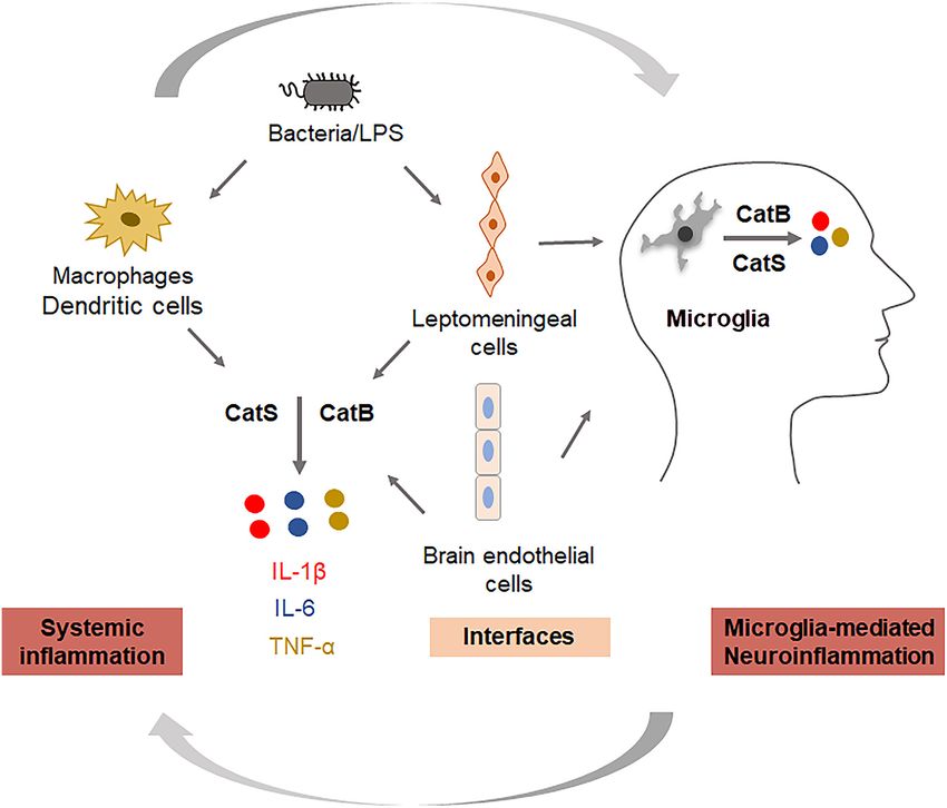
FIGURE 2 | Key roles of cathepsins in regulating systemic inflammation and neuroinflammation. Cathepsin B is involved in the production of IL-1β and TNF-α, and
cathepsin S is involved in the production of IL-6 in systemic cells (monocytes/macrophages), interfacing cells (leptomeningeal cells, brain endothelial cells), and
neural cells (microglia).
the microglia acquire a quite distinct signature characterized primarily reported to leak from lysosomes and subsequently
by the increased expression of ‘‘homeostatic’’ genes along with trigger the activation of the NLR family pyrin domain containing
genes that are absent in the DAM signature (Zhou et al., 2020). 3 (NLRP3) inflammasomes in microglia after phagocytosis of
Using single-cell RNA sequencing of live microglia purified from Aβ (Halle et al., 2008). Besides, it has also been reported
the human cerebral cortex, researchers have found at least one to be involved in the activation and processing of pro-
subset of cortical microglia that may be related to AD (Olah caspase-1 and pro-IL-1β in an NLRP3-independent manner
et al., 2020). Thus, the subset of cortical microglia related to AD (Terada et al., 2010). Since inflammasomes play either causative
should be prioritized in further validation efforts. or contributing roles, and also exaggerate the pathology in
responses to host-derived factors, the inhibition of CatB
Cellular Enzymes for Regulating may dampen neuroinflammation through the inactivation of
Co-related Pro-inflammatory Molecules NLRP3 inflammasomes. In studies using aged or middle-aged
Cathepsin (Cat) B (EC 3.4.22.1) is a lysosomal cysteine protease mice, CatB was found to activate NFκB by degrading inhibitory
that is expressed constitutively and which is linked to general κBα, an endogenous inhibitor of NFκB, via an autophagic-
protein turnover in lysosomes. The expression and activity lysosome pathway and mitochondrial oxidative stress (Wu et al.,
of CatB have been reported to be associated with several 2017; Ni et al., 2019a), and the persistent activation of NFκB
pathologies, including Alzheimer’s disease. In addition to its induced chronic neuroinflammation, which can damage neurons
role in the cleavage of amyloid precursor protein at the through highly toxic pro-inflammatory mediators and trigger the
wild-type β-secretase site (Hook et al., 2008), its regulation of breakdown of the BBB, resulting in the migration of peripheral
neuroinflammation has also received attention. CatB has been immune cells into the CNS. Also, the production of Aβ1–42
Frontiers in Cellular Neuroscience | www.frontiersin.org 8 February 2021 | Volume 15 | Article 638686Ni and Wu Inflammation Links Periphery and CNS
and Aβ3–42 was inhibited by pretreatment with a CatB inhibitor gingipain toxicity (Dominy et al., 2019). Cortexyme Inc. has
in macrophages after P. gingivalis infection. This suggests completed phase 1 clinical trials of COR388 (a gingipain
that increased levels of Aβ in the circulation may be further inhibitor) and will run a phase 2/3 study to determine
transported to the CNS, as P. gingivalis infection induced the whether it can improve cognition in patients with mild
upregulation of receptor for the advanced glycation end product to moderate AD. In addition to interventions targeting the
in cerebral endothelial cells, a process that is CatB-dependent microbiota or periodontal pathogens, some small molecules
(Nie et al., 2019; Zeng et al., 2020). CatB is also involved directly targeting inflammation are being investigated in
in the trafficking of TNF-α-containing vesicles to the plasma phase 2/3 trials, including ALZT-OP1 from AZTherapies Inc.,
membrane in macrophages (Ha et al., 2008). Taken together, Azeliragon from Pfizer, Epigallocatechin Gallate from Taiyo
CatB plays critical roles in the regulation of inflammation and International, Etanercept from Amgen Inc., and Thalidomide
the inflammatory crosstalk between the periphery and the CNS from Celgene Corporation.
via the BBB; thus, CatB appears to be a target through which the On the other hand, recently, in a 2-year double-blind
‘dampening inflammation’ may occur (Figure 2). randomized placebo-controlled trial that enrolled 195 cognitively
CatS (EC 3.4.22.27) is also a lysosomal cysteine protease intact elderly participants with a family history of AD, low-dose
with routine physiological digestion functions in antigen- naproxen did not significantly reduce the progression of the
presenting cells (APC). It was reported that cortical microglia Alzheimer Progression Score (APS), which is a composite
contain an intrinsic molecular clock and exhibit the circadian indicator of pre-symptomatic AD (Meyer et al., 2019). This
expression of CatS. The genetic ablation of CatS in mice induced work suggested the reconsideration of inflammatory disease as
hyper locomotor activity due to failure to reduce the synaptic a possible explanation for the reduced incidence of AD among
strength during sleep (Hayashi et al., 2013). Disturbed microglial NSAID users in observational studies. Even naproxen had no
circadian rhythms induced chronic inflammation in the brain benefit on any of the APS components, and to date anti-amyloid
(Ni et al., 2019b), and the increased expression of CatS was treatment strategies for AD have largely shown unimpressive
also found in the brains of AD mice, suggesting that CatS results. Thus, interest in alternative therapeutic targets, including
may indirectly induce neuroinflammation via disturbance of the anti-inflammatory strategies, is likely to continue to increase in
microglial circadian clock. Additionally, CatS-mediated cleavage the coming years.
of protease-activated receptor-2 (PAR2) results in its activation
and can cause the occurrence of inflammation in mice (Zhao CONCLUSION
et al., 2014). We previously found that CatS played a critical
role in driving splenic DC-dependent Th17 polarization through In conclusion, evidence shows that inflammation is the source
the upregulation of IL-6 by activating PAR2 after exposure of linkage between systemic diseases and AD. AD is considered
to components of periodontal bacteria (Dekita et al., 2017). a multifactorial disease affected by the negative spiral of
Therefore, the inhibition and direct targeting of CatS could be spreading inflammation. Therefore, ‘‘dampening inflammation’’
useful for the treatment of inflammation-associated pathological may be useful as a novel therapeutic approach for delaying
processes in AD (Figure 2). the onset of and enacting early intervention for AD. Inhibition
of CatB and CatS, in particular, maybe an effective target for
CLINICAL TRIALS OF dampening inflammation.
ANTI-INFLAMMATORY TREATMENT
On November 2, 2019, Green Valley announced that sodium AUTHOR CONTRIBUTIONS
oligomannate (GV971), a marine-derived oligosaccharide, had
JN and ZW conceived and drafted the manuscript. All authors
received conditional marketing approval in China to improve
contributed to the article and approved the submitted version.
the cognitive function in patients with mild to moderate
AD. GV971 was reported to restore the gut microbial profile
to normal and to lessen brain immune cell infiltration and FUNDING
inflammation in AD mice (Wang et al., 2019), and was
demonstrated to improve the cognitive function in patients with This work was supported by funding from Grants-in-Aid for
mild-to-moderate Alzheimer’s disease as early as week 4 in a Scientific Research, Japan (16K11478 to ZW), the research
phase III trial. Therefore, targeting the gut microbiota proved grant for OBT research center from Kyushu University (to
to be an effective and feasible therapy for AD. Moreover, small ZW), Beijing Institute of Technology Research Fund Program
molecule gingipain inhibitor ameliorated infection, reduced 2020CX04166 (Young Scholar to JN), and the National Natural
Aβ42 and neuroinflammation, and protected neurons from Science Foundation of China under Grant No. 32070954 to JN.
REFERENCES Ball, M. J., Lukiw, W. J., Kammerman, E. M., and Hill, J. M. (2013). Intracerebral
propagation of Alzheimer’s disease: strengthening evidence of a herpes simplex
Alkasir, R., Li, J., Li, X., Jin, M., and Zhu, B. (2017). Human gut microbiota: the virus etiology. Alzheimers Dement. 9, 169–175. doi: 10.1016/j.jalz.2012.07.005
links with dementia development. Protein Cell 8, 90–102. doi: 10.1007/s13238- Basu, A., Krady, J. K., and Levison, S. W. (2004). Interleukin-1: a master regulator
016-0338-6 of neuroinflammation. J. Neurosci. Res. 78, 151–156. doi: 10.1002/jnr.20266
Frontiers in Cellular Neuroscience | www.frontiersin.org 9 February 2021 | Volume 15 | Article 638686Ni and Wu Inflammation Links Periphery and CNS Berthoud, H. R., and Neuhuber, W. L. (2000). Functional and chemical anatomy Danese, S., Dejana, E., and Fiocchi, C. (2007). Immune regulation of the afferent vagal system. Auton. Neurosci. 85, 1–17. doi: 10.1016/S1566- by microvascular endothelial cells: directing innate and adaptive 0702(00)00215-0 immunity, coagulation and inflammation. J. Immunol. 178, 6017–6022. Bischoff, S. C., Barbara, G., Buurman, W., Ockhuizen, T., Schulzke, J. D., doi: 10.4049/jimmunol.178.10.6017 Serino, M., et al. (2014). Intestinal permeability—a new target for disease D’arcangelo, G., Tancredi, V., Onofri, F., D’antuono, M., Giovedi, S., and prevention and therapy. BMC Gastroenterol. 14:189. doi: 10.1186/s12876-014- Benfenati, F. (2000). Interleukin-6 inhibits neurotransmitter release and the 0189-7 spread of excitation in the rat cerebral cortex. Eur. J. Neurosci. 12, 1241–1252. Boissonneault, V., Filali, M., Lessard, M., Relton, J., Wong, G., and Rivest, S. doi: 10.1046/j.1460-9568.2000.00011.x (2009). Powerful beneficial effects of macrophage colony-stimulating factor on De Luca, A., Smeekens, S. P., Casagrande, A., Iannitti, R., Conway, K. L., β-amyloid deposition and cognitive impairment in Alzheimer’s disease. Brain Gresnigt, M. S., et al. (2014). IL-1 receptor blockade restores autophagy 132, 1078–1092. doi: 10.1093/brain/awn331 and reduces inflammation in chronic granulomatous disease in mice and in Borovikova, L. V., Ivanova, S., Zhang, M., Yang, H., Botchkina, G. I., humans. Proc. Natl. Acad. Sci. U S A 111, 3526–3531. doi: 10.1073/pnas. Watkins, L. R., et al. (2000). Vagus nerve stimulation attenuates the 1322831111 systemic inflammatory response to endotoxin. Nature 405, 458–462. Deane, R., Du Yan, S., Submamaryan, R. K., Larue, B., Jovanovic, S., Hogg, E., et al. doi: 10.1038/35013070 (2003). RAGE mediates amyloid-beta peptide transport across the blood-brain Bourgade, K., Garneau, H., Giroux, G., Le Page, A. Y., Bocti, C., Dupuis, G., barrier and accumulation in brain. Nat. Med. 9, 907–913. doi: 10.1038/nm890 et al. (2015). β-amyloid peptides display protective activity against the human Deane, R., Wu, Z., Sagare, A., Davis, J., Du Yan, S., Hamm, K., et al. Alzheimer’s disease-associated herpes simplex virus-1. Biogerontology 16, (2004). LRP/amyloid β-peptide interaction mediates differential brain 85–98. doi: 10.1007/s10522-014-9538-8 efflux of Abeta isoforms. Neuron 43, 333–344. doi: 10.1016/j.neuron.2004. Braak, H., and Del Tredici, K. (2015). The preclinical phase of the pathological 07.017 process underlying sporadic Alzheimer’s disease. Brain 138, 2814–2833. Dekita, M., Wu, Z., Ni, J., Zhang, X., Liu, Y., Yan, X., et al. (2017). Cathepsin S is doi: 10.1093/brain/awv236 involved in Th17 differentiation through the upregulation of IL-6 by activating Buduneli, N., and Kinane, D. F. (2011). Host-derived diagnostic markers related PAR-2 after systemic exposure to lipopolysaccharide from porphyromonas to soft tissue destruction and bone degradation in periodontitis. J. Clin. gingivalis. Front. Pharmacol. 8:470. doi: 10.3389/fphar.2017.00470 Periodontol. 38, 85–105. doi: 10.1111/j.1600-051X.2010.01670.x Dominy, S. S., Lynch, C., Ermini, F., Benedyk, M., Marczyk, A., Konradi, A., Buijs, R. M., and Van Eden, C. G. (2000). The integration of stress by the et al. (2019). Porphyromonas gingivalis in Alzheimer’s disease brains: evidence hypothalamus, amygdala and prefrontal cortex: balance between the autonomic for disease causation and treatment with small-molecule inhibitors. Sci. Adv. nervous system and the neuroendocrine system. Prog. Brain Res. 126, 117–132. 5:eaau3333. doi: 10.1126/sciadv.aau3333 doi: 10.1016/S0079-6123(00)26011-1 Elmore, M. R. P., Hohsfield, L. A., Kramar, E. A., Soreq, L., Lee, R. J., Pham, S. T., Caini, S., Bagnoli, S., Palli, D., Saieva, C., Ceroti, M., Bendinelli, B., et al. (2016). et al. (2018). Replacement of microglia in the aged brain reverses cognitive, Total and cancer mortality in a cohort of ulcerative colitis and Crohn’s disease synaptic and neuronal deficits in mice. Aging Cell 17:e12832. doi: 10.1111/acel. patients: the florence inflammatory bowel disease study, 1978-2010. Dig. Liver 12832 Dis. 48, 1162–1167. doi: 10.1016/j.dld.2016.07.008 Erickson, M. A., Hartvigson, P. E., Morofuji, Y., Owen, J. B., Butterfield, D. A., Carlyle, B. C., Nairn, A. C., Wang, M., Yang, Y., Jin, L. E., Simen, A. A., and Banks, W. A. (2012). Lipopolysaccharide impairs amyloid beta efflux et al. (2014). cAMP-PKA phosphorylation of tau confers risk for degeneration from brain: altered vascular sequestration, cerebrospinal fluid reabsorption, in aging association cortex. Proc. Natl. Acad. Sci. U S A 111, 5036–5041. peripheral clearance and transporter function at the blood-brain barrier. doi: 10.1073/pnas.1322360111 J. Neuroinflammation 9:150. doi: 10.1186/1742-2094-9-150 Cattaneo, A., Cattane, N., Galluzzi, S., Provasi, S., Lopizzo, N., Festari, C., et al. Forloni, G., Demicheli, F., Giorgi, S., Bendotti, C., and Angeretti, N. (1992). (2017). Association of brain amyloidosis with pro-inflammatory gut bacterial Expression of amyloid precursor protein mRNAs in endothelial, neuronal and taxa and peripheral inflammation markers in cognitively impaired elderly. glial cells: modulation by interleukin-1. Brain Res. Mol. Brain Res. 16, 128–134. Neurobiol. Aging 49, 60–68. doi: 10.1016/j.neurobiolaging.2016.08.019 doi: 10.1016/0169-328x(92)90202-m Chen, C.-H., Lin, C.-L., and Kao, C.-H. (2016). Irritable bowel syndrome is Friedman, S. M., Menzies, I. B., Bukata, S. V., Mendelson, D. A., and Kates, S. L. associated with an increased risk of dementia: a nationwide population-based (2010). Dementia and hip fractures: development of a pathogenic framework study. PLoS One 11:e0144589. doi: 10.1371/journal.pone.0144589 for understanding and studying risk. Geriatr. Orthop. Surg. Rehabil. 1, 52–62. Chen, M. (2015). The maze of APP processing in Alzheimer’s disease: where did doi: 10.1177/2151458510389463 we go wrong in reasoning? Front. Cell. Neurosci. 9:186. doi: 10.3389/fncel.2015. Ghosh, S., Wu, M. D., Shaftel, S. S., Kyrkanides, S., Laferla, F. M., Olschowka, J. A., 00186 et al. (2013). Sustained interleukin-1β overexpression exacerbates tau pathology Cherry, J. D., Olschowka, J. A., and O’banion, M. K. (2015). Arginase 1+ microglia despite reduced amyloid burden in an Alzheimer’s mouse model. J. Neurosci. reduce Aβ plaque deposition during IL-1beta-dependent neuroinflammation. 33, 5053–5064. doi: 10.1523/JNEUROSCI.4361-12.2013 J. Neuroinflammation 12:203. doi: 10.1186/s12974-015-0411-8 Goehler, L. E., Gaykema, R. P., Nguyen, K. T., Lee, J. E., Tilders, F. J., Maier, S. F., Chou, R. C., Kane, M., Ghimire, S., Gautam, S., and Gui, J. (2016). Treatment et al. (1999). Interleukin-1β in immune cells of the abdominal vagus nerve: a for rheumatoid arthritis and risk of Alzheimer’s disease: a nested case-control link between the immune and nervous systems? J. Neurosci. 19, 2799–2806. analysis. CNS Drugs 30, 1111–1120. doi: 10.1007/s40263-016-0374-z doi: 10.1523/JNEUROSCI.19-07-02799.1999 Coccia, M., Harrison, O. J., Schiering, C., Asquith, M. J., Becher, B., Powrie, F., Griffin, W. S., Sheng, J. G., Royston, M. C., Gentleman, S. M., Mckenzie, J. E., et al. (2012). IL-1β mediates chronic intestinal inflammation by promoting the Graham, D. I., et al. (1998). Glial-neuronal interactions in Alzheimer’s disease: accumulation of IL-17A secreting innate lymphoid cells and CD4(+) Th17 cells. the potential role of a ‘‘cytokine cycle’’ in disease progression. Brain Pathol. 8, J. Exp. Med. 209, 1595–1609. doi: 10.1084/jem.20111453 65–72. doi: 10.1111/j.1750-3639.1998.tb00136.x Corradini, I., Focchi, E., Rasile, M., Morini, R., Desiato, G., Tomasoni, R., Gu, Y., Wu, Z., Zeng, F., Jiang, M., Teeling, J. L., Ni, J., et al. (2020). Systemic et al. (2018). Maternal immune activation delays excitatory-to-inhibitory exposure to lipopolysaccharide from porphyromonas gingivalis induces bone gamma-aminobutyric acid switch in offspring. Biol. Psychiatry 83, 680–691. loss-correlated Alzheimer’s disease-like pathologies in middle-aged mice. doi: 10.1016/j.biopsych.2017.09.030 J. Alzheimers Dis. 78, 61–74. doi: 10.3233/JAD-200689 Cui, S., Xiong, F., Hong, Y., Jung, J. U., Li, X. S., Liu, J.-Z., et al. (2011). APPswe/Aβ Ha, S. D., Martins, A., Khazaie, K., Han, J., Chan, B. M., and Kim, S. O. regulation of osteoclast activation and RAGE expression in an age-dependent (2008). Cathepsin B is involved in the trafficking of TNF-α-containing manner. J. Bone Miner. Res. 26, 1084–1098. doi: 10.1002/jbmr.299 vesicles to the plasma membrane in macrophages. J. Immunol. 181, 690–697. Damani, M. R., Zhao, L., Fontainhas, A. M., Amaral, J., Fariss, R. N., and doi: 10.4049/jimmunol.181.1.690 Wong, W. T. (2011). Age-related alterations in the dynamic behavior Halle, A., Hornung, V., Petzold, G. C., Stewart, C. R., Monks, B. G., Reinheckel, T., of microglia. Aging Cell 10, 263–276. doi: 10.1111/j.1474-9726.2010. et al. (2008). The NALP3 inflammasome is involved in the innate immune 00660.x response to amyloid-beta. Nat. Immunol. 9, 857–865. doi: 10.1038/ni.1636 Frontiers in Cellular Neuroscience | www.frontiersin.org 10 February 2021 | Volume 15 | Article 638686
You can also read