Interstrand Crosslink Repair: New Horizons of DNA Damage Repair
←
→
Page content transcription
If your browser does not render page correctly, please read the page content below
Chapter

Interstrand Crosslink Repair: New
Horizons of DNA Damage Repair
Amna Aqeel, Javaria Zafar, Naureen Ehsan, Qurat-Ul-Ain,
Mahnoor Tariq and Abdul Hannan

Abstract

    Since the dawn of civilization, living organisms are unceasingly exposed to
myriads of DNA damaging agents that can temper the ailments and negatively
influence the well-being. DNA interstrand crosslinks (ICLs) are spawned by various
endogenous and chemotherapeutic agents, thus posing a somber menace to genome
solidity and cell endurance. However, the robust techniques of damage repair
including Fanconi anemia pathway, translesion synthesis, nucleotide excision and
homologous recombination repair faithfully protect the DNA by removing or toler-
ating damage to ensure the overall survival. Aberrations in such repair mechanisms
adverse the pathophysiological states of several hereditary disorders i.e. Fanconi
Anemia, xeroderma pigmentosum, cerebro-oculo-facio-skeletal syndrome and
cockayne syndrome etc. Although, the recognition of ICL lesions during interphase
have opened the new horizons of research in the field of genetics but still the
detailed analysis of conditions in which repair should occur is largely elusive.

Keywords: DNA damage repair, Interstrand cross links (ICLs), Homologous
Recombination Repair, Translesion synthesis, Non-homologous end-joining repair,
FA pathway

1. Introduction

    There is an amalgam of various environmental, endogenous as well as chemo-
therapeutic agents that are continuously having a contact with the genetic material in
living beings and making it a point of real concern throughout the globe. The attack
of reactive oxygen as well as nitrogen species on DNA have contributed towards a
large amount of defects and complex chemical structures that take place in DNA
[1]. These damages give rise to a series of simple and bulky base modifications that
distort the helical structure, abasic sites, the breaks in phosphodiester linkages along
with the interstrand crosslinks (ICLs). These lead to various mutagenic changes in
the genetic blueprint and become a reason of inhibition of the transcriptional or
replicative machinery that induce activate apoptotic divisions or necrosis [2].
    Interstrand cross-links (ICLs) are the anomaly that link the complementary
strands of DNA by the covalent linkage between the bases. These are formed
by the chemicals along with the two reactive electrophilic groups. It is a highly
sequence-dependent reaction in which the two nucleophilic groups on the opposite
strands are aligned geometrically and enable the dual reaction of the bifunctional
cross-linking agent with it. This complex chemical reaction give rise to ICLs,

1
DNA - Damages and Repair Mechanisms
mono-adducts, intrastrand cross links as well as DNA-protein cross-links [3]. The
ICLs are made with the help of reactive endogenous chemicals such as lipid per-
oxidation product known as malondialdehyde or aided with the reactive aldehyde
group of an unpromptedly formed or the enzyme-derived abasic site in the DNA
molecule with a normal base on the complementary strand [4].
A large amount of anticancer and chemotherapeutic agents such as mitomycin C
(MMC), cisplatin, nitrosoureas and nitrogen mustards are notorious for introducing
formidable blocks in the normal metabolic processes of DNA with ICLs and need
repair for cell sustenance. ICLs are also caused by various antitumor agents that
defects DNA through radical processes like C-1027, neocarzinostatin [5]. With the pas-
sage of time, the organisms have developed various complex mechanisms to alleviate
these deleterious defects from the genome. The failure to remediate the defect can con-
tribute towards cell death that can occur either through a mitotic catastrophe or the
p53-dependent apoptotic pathway. In the mammalian cells, the repair mechanisms for
ICLs repair are still ambiguous [6]. According to an estimation, about 40ICls that form
in a mammalian genome can destroy a defective cell that lacks ability to be repaired.
The in vivo study gives an overview of the elimination of the ICLs in cellular
DNA of both prokaryotes and eukaryotes. The model organisms are used for the
clear understanding of the repair mechanisms. These include E. coli and yeast.
The ICLs repair mechanisms in bacteria and yeast are replication dependent and
independent while in vertebrates, they follow repairment pathway during replica-
tion of DNA [7]. Moreover, the recent study suggests the operation of replication
independent ICL repair pathway in vertebrates.
The ICL repair pathway have been deduced from the relative sensitivity of the
DNA repair defective cell lines to the cross linking agents. Pathways of ICL repair
have mostly been inferred from the sensitivities of DNA repair defective cell lines to
crosslinking agents. During the S phase of the cell division in vertebrates, the ICL
repair is induced by the help of impeded replication forks. The process of ICL repair
needs a nexus of multiple factors along with the structure specific endonucleases,
for example TLS and HR. If a disturbance occurs during the repair, the genomic
instability results that bring forth the birth of Fanconi anemia, a cancer prone
ailment [8]. There is another ICL repair pathway that takes place in the G0/1 phase
during the cell cycle which is a replication and recombination independent pathway
[9]. In addition, the tolerance of ICLs in G1 as compared to S phase makes it an
underappreciated pathway because there, the stalled replication fork possesses high
toxicity. Contrarily, the toxicity of ICL in G1 can be depicted when it terminates the
transcription of a gene playing a vital role.
The latest studies have proposed the role of NER proteins (as they cut one side
of ICL) [7], Homologous recombination along translesion synthesis polymerases
(Polζ, Rev1) that are involved in filling the gap for both type of cells undergoing
replication as well as non-replicating ones [10]. The proteins involve in the ICL
repair have a vital role in the pathophysiology of several hereditary diseases Proteins
implicated in the repair of ICLs have a critical role in the pathophysiology of several
hereditary disorders. In addition, cells deficient in the Fanconi Anemia (FA) path-
way are highly sensitive to ICLs [11] and this pathway has been suggested to play an
important role in mammalian ICL repair at replication forks promoting homologous
recombination. There has been a series of continuous research on ICL lesions in the
past decade and it covered the various aspects of ICLs be it as their identification,
detection methods or their development along with the repair mechanisms and the
exploitation of cross linkers in the laboratory. These have paved the way towards the
better and more reliable understanding of ICLs in the complex biological samples.
This chapter foregrounds the multiple aspects of the interstrand cross-link repairs
with a reference to their pathophysiology and lesion repair mechanisms.
2Interstrand Crosslink Repair: New Horizons of DNA Damage Repair
DOI: http://dx.doi.org/10.5772/intechopen.97551
2. Basic biochemistry of ICL-generating agents
A large variety of natural and synthetic chemicals are notorious for bringing
ICLs on the front and are regarded as the ICL inducers or inducing agents. In the
same way, the metabolic byproducts formed in the cell also contribute towards ICLs
formation. Their structure and function vary greatly but ICLs inducers are known
for their bifunctional reactivity with both of the strands of DNA. The endogenous
as well as exogenous sources of ICLs are summarized as follows:
2.1 Endogenous sources of interstrand cross links
The endogenous sources of ICLs comprises of the reactive aldehydes that
are generated as a result of lipid peroxidation along with base excision repair
(BER) [12]. There are other endogenous by products of lipid peroxidation, the
α, β-unsaturated aldehydes or enals namely crotonaldehyde, acrolein, along with
the 4-hydroxynonenal (4-HNE). These are formed as a result of oxidative stress
[13]. Moreover, there are exogenous contributors as well namely cigarette smoke
and automobile exhaust to expose with acrolein and croton-aldehyde. The DNA
nucleobases interact with enals to give rise to exocyclic adducts. These adducts
then interact with proteins. The incorporation of enals to dG is done with the
help of Michael addition in which addition of N2 -amine occurs to generate N2-
(3-oxopropyl)-dG adducts. The next stage is cyclization of N1 with the aldehyde,
giving rise to N2 -γ-hydroxypropano-dG adducts [14]. These products are also
genotoxic to human beings. Shapiro and Leonard are famous for their earlier study
of nucleosides reactions with glyoxal, chloroacetaldehyde, malondialdehyd along
with related bis-electrophiles [14, 15]. The in vitro formation of ICL is attributed to
the opening of the exocyclic 1, N2-dG product that minimizes the steric hindrance
and forms ICL on exposure towards an aldehyde [16].
Moreover, there are DNA lesions that are formed as a result of accumulated
acetaldehyde in the cells. The acetaldehyde is produced as a result of alcohol
metabolism with aldehyde dehydrogenase 2 (ALDH2) as a biocatalyst. The drug
disulfiram if used, blocks the enzyme ALDH2 and accumulates the acetaldehyde in
the cells. The lesions produced are DNA adducts, breaks in single or double-strands
of DNA (DSBs), sister chromatid exchanges (SCEs), point mutations, along with
crosslinks in DNA [17]. The DNA adducts like N2-ethylidene-2′-deoxyguanosine,
N2-propano-2′-deoxyguanosine, N2-ethyl-2′-deoxyguanosine, along with N2-
etheno-2′-deoxyguanosine are vital DNA damage agents that follow the accumula-
tion of acetaldehyde in the cells. The acetaldehyde reacts with guanine and forms
a crosslink precursor known as N2 -propanoguanine (PdG) which in turn reacts
with N2 amine of guanine in 5′-CpG sequence consequently forming acetaldehyde
interstrand crosslinks (AA-ICL). In Asian continent, the irreparable detoxification
of acetaldehyde is found more often and is linked with alcohol mediated cancers
[18]. Moreover, cells in Saccharomyces cerevisiae don’t have ability to repair ICLs and
are acetaldehyde sensitive thus gives validation of acetaldehyde mediated ICLs [19].
The intestinal pathogens in human beings known as Enterobacteriaceae and
other bacteria play a vital role in the progression of colorectal cancer. They produce
colibactins that are genotoxic in nature and bring harm to human beings. With
their structural chemistry still unknown, colibactins produce ICL dependent DNA
double-strand breaks (DSBs) and activates the ICL repair pathways [20]. Cellulo
also depicts another picture of the DNA damaging mechanism in which colibac-
tin producing bacterial exposure towards the genomic DNA of cultured human
cells made it susceptible to interstrand cross links. There are different changes
observed in the intoxicated cells including the replication stress, the activation of
34
ICL AGENTS Source Adducts formed/ Clinical benefits Elimination half- Metabolism References
Target DNA sequences life of drug
Platinums
Carboplatin Synthetic Adducts: G-Pt-G and Treatment of ovarian cancer 1–2 hours Kidney [28]
Oxaliplatin (made from Pt-GG Treatment of colorectal 26 hours or Kidney [29, 30]
cisplatin) DNA sequence: 5’-GC cancer 20 hours
Mitomycin
Mitomycin C Streptomyces 5′-CG-3’ Treatment of Esophagal and Alpha-half-life of Hepatic [10, 31]
caespitosis bladder carcinoma 8.2 mins, beta-half-
life of 51.8 mins
Azinomycin
DNA - Damages and Repair Mechanisms
Azinomycin B Streptomyces 5′-GNC or 5′-GNT having antitumor activity N.A N.A [32]
sahachiroi sequences against P388 leukemia in
mice
Chloroethylnitrosoureas
Carmustine Synthetic G-C base pair Treatment of multiple 70 minutes Liver [33]
(nitrogen myelomas
mustard)
Nitrogen mustards
Cyclophosphamide Synthetic 5′ -GNC Treatment of lymphoma, 3 to 12 hours. Liver [34]
multiple myeloma and
ovarian cancer
Ifosfamide Treatment of sarcomas and 60–80% in Kidney [35]
organ cancers 72 hours
Chlorambucil Treatment of Chronic 1.5 hours Liver [36]
lymphocytic leukemia,
Hodgkins lymphoma and
Non-hodgkin lymphoma
Table 1.
Exogenous agents of Interstrand cross-link lesions.Interstrand Crosslink Repair: New Horizons of DNA Damage Repair
DOI: http://dx.doi.org/10.5772/intechopen.97551
ataxia-telangiectasia along with Rad3-related kinase (ATR), as well as the retrieval
of Fanconi anemia protein D2 (FANCD2). Contrarily, FANCD2 knockdown or ATR
inhibition decreases the survival capability of cells having an exposure towards
colibactins. The evidence ensures that collectins mediated DNA defects in infected
cells favors DNA ICLs [21].
2.2 Exogenous sources of ICLs
The other sources of ICLs are exogenous in nature. They have the same mecha-
nism of bifunctional alkylating agents but differ in their preferences for sequences,
topologically restrict the DNA and need certain processing within the cell to form
functioning ICL inducers [9]. In spite of the fact that they have a history of damag-
ing DNA, their innovative uses also aid in understanding the mechanisms they
follow to contribute in various therapeutic applications.
These include psoralens that belong to the family of furocoumarins, being
mutagenic are still a matter of contention with their photochemotherapeutic
applications in inflammatory skin diseases like psoriasis, vitiligo and eczema [22].
The Psoralens generates adducts on interaction with pyrimidines, most often with
thymine and give rise to ICLs at the sequences made up of d(TpA):d(TpA) residues
[23]. The several derivatives of psoralen form multiple changes in the DNA helical
structural framework and exhibit their toxic nature. The DNA duplex adducted
with 4′- (aminomethyl)-4,5′,8-trimethylpsoralen (AMT) exhibited 561 unwinding
and 531 bending into its major groove [24].
Another chemotherapeutic agent known as cis-platinum diamminedichloride
i-e CDDP, cisplatin also induces ICLs. It makes an adduct with purines, most often
at the N7 position of the guanines, hence ICL forms at d(GpC): d(GpC) sequences.
This is employed in various head and neck cancers, esophageal, epithelial lung,
colon, gastric, bladder along with ovarian and testicular tumors. About 90% of the
total defects are formed by 1,2-IaCL and 1,3-IaCL along ICL making only 5% of the
total DNA lesions [23].
Apart from these anticancer agents, one of prime importance is Adriamycin
which is also termed as doxorubicin. It generates a great response against a range
of tumors be it as breast tumors, acute leukemia, lymphomas, stomach, sarcomas,
multiple myelomas or bone tumors. It is employed as a singly or in combined form
[25]. The interaction of Adriamycin is clearly understood with the help of the in
vitro transcription assays that demonstrates the drug-induced DNA adducts at the
GpC sites [26]. The electrospray mass spectral analysis revealed details of GpC
drug binding regions and gives the information that the cross links are favored by
formaldehyde under the certain conditions [27]. Table 1 illustrates the exogenous
agents of Interstrand crosslink lesions.
3. ICL Repair genes and human disorders
The proteins involved in the repair of ICLs have vital role in pathophysiology of
various hereditary disorders for example xeroderma pigmentosum (XP), cerebro-
oculo-facio-skeletal syndrome (COFS), Fanconi Anaemia (FA), trichothyodis-
trophy as well as Cockayne syndrome (CS) [37]. FA is associated with aplastic
anemia, cancers (often acute myelogenous leukemia) and bone marrow failure.
The mutational changes in any FANC genes contribute towards genomic instability
and the sensitivity against the ICL agents [38]. According to an estimate 18 genes
are involved in FA and the products of genes collaborate for ICL repair during the S
phase [39]. Apart from these, the defective NER pathways also result in several rare
5DNA - Damages and Repair Mechanisms
autosomal-recessive diseases like XP, CS, TTD and COFS syndrome [40]. Moreover,
there are 11 genes that are associated with NER pathways and the defect in these
occur due to the mutations in these genes. XP is associated with pigmentation, pho-
tosensitivity as well as cancerous skin diseases. Another inherited syndrome known
as CS is present in which there are several problems arises namely ocular defects,
mental deficiency, extensive demyelination, short stature, photosensitivity, large
hands, feet, as well as ears [37]. There are wide ranging clinical spectrum of CS and
the patients acutely affected are categorized under COFS syndrome patients. TTD is
associated with neuro-ectodermal symptoms and clear sulfur-deficient brittle hair
[41]. These NER diseases are different from each other with respect to their physical
characteristics involving cutaneous ailments.
Keeping in view the various DNA repair factors, ICL genes has found to be
having a strong link with cancer. There are several genes that are revealed by
next-generation sequencing and play a part in hereditary breast cancer as well as
ovarian cancer syndrome (HBOC). These genes are BRCA1, BRCA2, PALB2, BRIP1
and RAD51C exhibiting a close link with HBOC in the ICL repair pathways [42].
The preventive medication strategy requires the early detection of the mutations
happening in BRCA1 and BRCA2 genes to help in process of recovery.
4. Recognition of ICL lesions in mammalian cells
During the course of ICL damage, the UHRF1 protein comes to rescue at the
site within a fraction of seconds [43]. These proteins identify ICLs with the help of
its SET and RING finger associated (SRA) domain, the same domain notable for
its recognition ability for the hemi-methylated DNA and employment of DNMT1
to ensure the maintenance of methylation signature in the cells of mammals [44].
The relative affinity of UHRF1 protein in response to hemi-methylated DNA as well
as ICLs are somewhat similar and proposed that UHRF1 interacted with both of
them through related mechanisms. The UHRF1 proteins are employed preceding
the incorporation of FANCD2 to ICLs [43]. About 10 minutes are lagged between
the assembling of UHRF1 and FANCD2 to ICLs. This strengthens the assumption
of other proteins being employed or the other PTM events that might occur during
this time interval. The proper mechanism of UHRF1 mediated FANCD2 repair is
not clear but implicate a direct protein–protein interaction. There has also been a
proposed role of UHRF1 in a nuclease scaffold [45]. It is also proposed that the rapid
incorporation of UHRF1 to the ICLs paves the way for FA mediated repair of lesion
later on. As ICLs vary in their structural framework, there is a probability that in
addition to UHRF1, other ICL sensor proteins do exist in the same way.
5. Factors involved in ICL repair pathway
There are several proteins that take part in the ICL repair. Along with these,
included 15 proteins that are not only specific to FA genes (A, B, C, D1, D2, E, F,
G, I, J, L, M, N, O, and P) but also to other repair pathways [46]. The important
recombination factors like RAD51, the structure-specific endonucleases like
MUS81/EME1 and XPF/ERCC1, translesion DNA polymerases and Holliday junc-
tion processing factors all contribute towards the repair of ICLs.
A rare human genetic disease known as FA, which is associated with pancyto-
penia, various developmental abnormalities and a high cancer risk [47]. The cells
procured from FA patients depict the large amount of chromosomal breakage as
well as the formation of radial chromosomes [48] that bring strength to the idea of
6Interstrand Crosslink Repair: New Horizons of DNA Damage Repair
DOI: http://dx.doi.org/10.5772/intechopen.97551
high genomic stability in the ICL repair-deficient cells. The classical FA pathway
has FA core complex (consisting of A, G, FAAP20, C, E, F, B, L, and FAAP100), an
E3 ubiquitin ligase activity and the catalytic activity dedicated to the RING domain
comprising FANCL protein. The core complex also acts on monoubiquitination of
FANC1/D2 complex and is stimulated by damaged DNA [49]. The next step is the
utilization of other downstream effectors that are attracted by the activated com-
plex. These comprises nucleases, homologous recombination factors and translesion
polymerases to remediate the lesions [50]. Whereas the exact function of monou-
biquinated FANCD2 is still ambiguous.
An ATP dependent DEAH domain helicase namely FANCM exhibit a DNA trans-
locase activity. It combines with FAAP24 and forms a complex structure comprising
a histone-fold complex i-e MHF1/MHF2. It is a significant part of activated FA path-
way [51]. The biochemical analysis also proposed that FANCM/FAAP24 complex
is responsible for stabilizing and remodeling the stopped replication forks of DNA
[52]. The complex of FAAP24 plays a vital part in the checkpoint activation that also
need ATR to begin its function [53]. However, FANCM takes part in recombination
independent ICL remediation by stimulating ubiquitination of PCNA thus promotes
the incorporation of other NER incision factors to the sites with ICLs [51].
The group of genes associated with FA comprises of FANCD1 (BRCA2), FANCJ,
FANCN, as well as FANCO are the recombination factors that forms a connection
with susceptibility for breast or ovarian cancer. The downstream processing of ICL
require the employment of recombination factors, mostly when there are the double
strand breaks in the DNA. The paralogous gene of FANCO (RAD51C) is RAD51 [54].
FANCO forms complex structures on interaction with RAD51B, RAD51D, XRCC2, as
well as XRCC3. Another significance of these paralogs is the utilization of the recom-
binase RAD51 while managing a single stranded DNA [55]. RAD51 and its paralogs
are vital to cells tolerant against ICLs and vice versa because they provide the homolo-
gous recombination in response to ICLs as well as the double strand breaks [56].
The endonucleases also pay a part in ICLs repair. Three important heterodimeric
structure-specific endonucleases are MUS81/EME1, SLX1/SLX4 and XPF/ERCC1.
SLX4 is often mutated in the complementation group consisting of FANCP [57]. The
combination of SLX4 and SLX1 make up a heterodimeric nuclease. Its function is
to resolve the Holliday junction formed during the remediation of ICls [58]. During
the process, SLX4 act as a scaffold protein that combines the multi-activity nuclease
complex comprising MUS81/EME1 as well as XPF/ERCC1. The latter acts in either
of the NER pathway as well as ICL repair. The studies proposed that NER works
independent of SLX4 with XPF/ERCC1 complex and the analysis of FANCP patients
further strengthens the idea as they were resistant against the UV radiations [59].
Further studies suggest that XPF/ERCC1 activity requiring SLX4 involves the com-
plete detaching in ICL repair. It is a replication dependent remediation of ICLs [60].
Digesting nuclease (SNM1A) then follows and digest the detached oligonucleotides
[61]. This step is a better alternative as compared to the bypass step used for synthesis.
Moreover, the lately discovered nuclease FAN1 also has a significant part in
remediation of ICL. The ubiquinated FANCD2 aids in employing FAN to ICL regions.
This step is mediated with the ubiquitin-binding zinc finger domain that is present in
FAN1 [62]. Another important domain of FAN1 exhibit 5′-3′ exonuclease activity as
well as structure-specific endonuclease activity at 5′ [63]. FAN1 thus cuts the exposed
ends of DNA along with DNA replication structures that hinders the process.
Other important participants in ICL repair are the translesion DNA polymer-
ases. The blockage of normal replicative DNA polymerases is done before reaching
the ICL regions. Other translesion polymerases in Xenopus laevis include Y-family
polymerase Rev1 as well as B-family polymerase Pol ζ (Rev3/Rev7) have a signifi-
cant part in complete removal of ICLs. These models also use replisome remodeling
7DNA - Damages and Repair Mechanisms
machinery so that the extension of stalled DNA strand occur on one base before the
ICL region [64]. On unwinding, Rev1’s deoxycytidyl transferase of Rev1 incorpo-
rates cytosine on the complementary strand across the ICL region [65]. This is then
succeeded by Pol ζ that extends the unpaired strand.
6. ICL lesion removal in quiescent G0/G1 phase
The comprehension of ICL repair is a difficult task because it has an implica-
tion on both strands of DNA. The cells in G0/G1 phase do not require homologous
recombination for ICL repair [66]. Moreover, all eukaryotic organisms ranging
from Saccharomyces cerevisiae to the human beings, require NER for the incisions
of ICL. The single stranded gap is produced at the first step of NER by the oligo-
nucleotide on ICL lesion. This can be bypassed with the help of translesion DNA
polymerases REV1 just like the DNA polymerases (η, ι, κ, and ζ,). Both the DNA
polymerases κ, and ζ, as well as REV1 are vital for this stage of NER [67].
7. ICL recognition and repair in proliferating S-phase
The repair of ICL faces several complications during the S phase. The data
exhibits the formation of double stranded breaks by interaction with ICL caus-
ing agents [59]. The ICL induced Double stranded breaks can be repaired by HR
rather than non-homologous end joining (NHEJ) method [68]. This brings to the
conclusion that ICL-induced DSBs are linked with DNA replication forks. NER
indicates ICLs in S. cerevisiae and NER function is important for ICL repair. So, all
NER-mutants exhibit hyper sensitivity to the ICL causative agents. Contrarily, the
cells deficient in XPF- as well as ERCC1- show immense hypersensitivity to the ICL
agents (mitomycin C & nitrogen mustard) in mammals. The product of XPF as well
as ERCC1 make up an endonuclease which is hetero-dimeric in nature identifies and
incise the single stranded branched structures [69]. Moreover, MUS81-EME1 along
with XPF-ERCC1, the homologous structure specific endonucleases are also keen in
repairing the ICL lesions [70]. MUS81-EME1 is notable for its binding with the dou-
ble-stranded branched structures, flaps at 3′ end, as well as Holliday junctions [71].
Either of the two XPF-ERCC1 and MUS81-EME1 are responsible for ICL-induced
double strand formation. Since, a multitude of nucleases are recognized recently
being the key players in ICLs incision, the mechanism underlying the process need
to be explored. We abridge the current knowledge about the ICL repair mechanism
in S phase. HR repairs the ICLs induced DSBs. An experiment conducted in S.
cerevisiae, gives an outline of hypersensitivity against ICL causative agents in rad51,
rad52, rad54, rad59, as well as mre11 mutants but not in case of yku70 mutants. The
hypersensitivity of rad52 yku70 double mutants to ICLs is at par with that of rad52
mutants [72]. The HR deficient strains show the increase in accumulated DSBs
successively on treating with ICL inducers as there lacks an ability to cure DSB
which means that NHEJ is not a pre-requisite to remediate DSBs stimulated by ICLs.
The mammals follow the same process in their cells. The HR deficient cells depict
hypersensitivity against ICLs like cells having mutated paralogs of RAD51, RAD54,
RAD54B, along with BRCA2, while it is not observed in cells deficient in NHEJ [73].
It significantly highlights the role of HR in repairing DSBs and re-initiating the
halted replication forks of DNA. Fanconi anemia (FA) genes are key players in the
remediation of ICL in eukaryotes. The proper role of FA gene products in biochemi-
cal reactions are still not identified properly, but are notable for their control of HR
at the replication forks of DNA [74].
8Interstrand Crosslink Repair: New Horizons of DNA Damage Repair
DOI: http://dx.doi.org/10.5772/intechopen.97551
8. Interstrand crosslinks lesion repair mechanisms
Lesions in interstrand crosslinks epitomize an arduous challenge in genome main-
tenance pathways due to the compromise of genomic information present on both
strands. Therefore, an application of non-damaged strand as a template for accurate
repair in straightforward cut and patch mechanism is not feasible. In this regard, ICL
repair employs the concerted and synchronized interaction of dynamics from numer-
ous mechanisms of DNA damage repair, including NER, homologous recombination,
mismatch repair, translesion synthesis, ataxia telangiectasia, Rad3 related and Fanconi
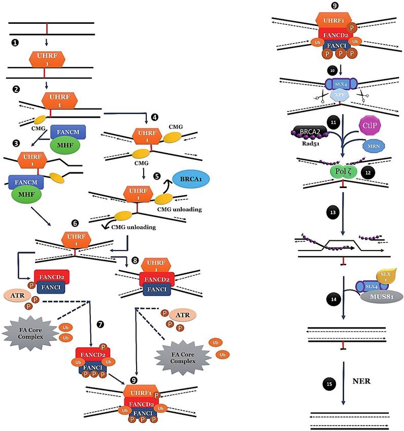
anemia pathway. Figure 1 illustrates the schematic mechanism of ICL repair [75].
Figure 1.
Schematic of ICL repair mechanism. (1) After the formation of ICLsin the cells, UHRF1 is recruited through
its SRA domain immediately. (2) Single replication fork reaches at ICL. (3) Then Replication machinery
is transversed through ICL by the help of FANCM/MHF complex and allowes the ICL for later repair. (4),
(5) On an alternate basis FANCS or BRCA1 allows the unloading of CMG helicase complex, when second
replication fork arrives at ICL. (6) Then replicative polymerase reaches at −1 position of ICL, leaving X shaped
similar to the transverse mechanism. (7) Then ATR allows the phosphorylation of FANCD2/FANCI complex
at multiple sites and meanwhile FA core complex mono-ubiquitinate at FANCD2/FANCI complex at K561 and
K523 respectively. (8) The complex is then recruited to ICL at the replication fork. (9), (10) This ubiquitinated
complex recruits SLX4/XPF on ICL in order to unhook the ICL. (11) Afterwards, CtlP an MRN complex
resect the double strand breaks and BRCA2 facilitates the formation of RAD51 filament on single stranded
DNA generated by resection. (12) Then Polζ carry out the polymerization step through the unhooked ICL. (13)
Rad51 then facilitates the invasion of strand with subsequent extension of the other strand. (14) Lastly SLX4
and nucleases resolve the Holliday junction (15) and NER repair proteins remove the damaged nucleotides.
9DNA - Damages and Repair Mechanisms
8.1 Role of homologous recombination in ICL repair
The phenomenon of homologous recombination repair (HRR) employs homolo-
gous DNA sequences as template for repair and tolerance of DNA lesions that
obstruct DNA replication in S-phase. Homologous recombination usually encom-
passes four step (i) double strand break recognition tailed by nucleolytic processing
to produce 3′ single stranded ends of DNA, (ii) protein-mediated strand invasion
of single-stranded DNA with homologous chromosome (iii) synthesis of DNA
which regenerates degraded DNA using undamaged homologous chromosome as a
template and (iv) resolution of Holliday junction intermediates. Usually the plati-
num drugs drive fruitful results in the treatment of BRCA1- and BRCA2- associated
ovarian cancers [76]. However, the protein products of these two genes give rise to
HR-mediated repair of DNA damage. A dynamic combination of BRCA1 and associ-
ated RING domain protein 1 (BARD1) exhibits ubiquitin ligase activity that is essen-
tial for the proper localization of RAD51, which is a central player in Homologous
Recombination repair. Through BRCA2 mediated interaction with RAD51, it is
specifically targeted to sites where recombination is initiated [77]. However, RAD51-
deficient cells represent hypersensitivity towards ICL-inducing agents.
In this regard, the model organism, Escherichia coli has provided deep insights in
the mechanisms involved in HRR of bacteria. Usually, RecA of bacteria has proven
to be an effective protein in all major aspects of HRR due to its ability of form-
ing nucleoprotein filament with both single and double stranded DNA. In E. coli,
RecBCD complex- combination of nuclease/helicase, initiates the phenomenon of
recombination by creating 3′-terminal single-stranded DNA substrate for the activity
of RecA protein. RecBCD complex usually binds to the end of linear double stranded
DNA and RecA in combination with single-stranded binding proteins (SSBP) allows
an incessant formation of presynaptic filament on DNA. This nucleoprotein complex
allows a rapid and efficient search for homology within the double-stranded DNA
recipient, with subsequent formation of a joint molecule. After the formation of joint
molecule, DNA PolI regenerates the sequence and the resultant Holliday junction is
resolved by the action of RuvC protein that acts in concert with RuvAB proteins to
coordinate the steps of branch migration and Holliday junction resolution [78].
In Saccharomyces cerevisiae, the incision of DNA is carried out by an anonymous
nuclease. A yeast homologue of RecA, Rad51 works in conjunction with Rad52 dis-
locates the single-stranded DNA that is ostensibly covered by RPA. The subsequent
nucleofilament works with Rad54 and Rad55/57 in DNA unwinding and strand
annealing between donor DNA and incoming Rad51 nucleoprotein. The resolution
of subsequent recombination intermediates is frequently carried out by assorted set
of mechanisms including mus81-mms4 nuclease and Resolvase A [79].
8.2 Translesion DNA synthesis in DNA interstrand crosslinks
Translesion DNA Synthesis polymerases are considered essential for ICL repair
in both S/G2 and G1 to bypass an ICL unhooked from one of the two cross-linked
strands. The phenomenon of Translesion synthesis encompasses multiple poly-
merases with a dynamic ability to carry out an insertion of nucleotide across the
lesion and others carrying out further extension. Based on genetic and biochemical
studies, an assortment of polymerases has been implied in repair of ICLs. Usually
translesion synthesis is a threefold step: (i) release of replicative polymerase after
an interruption of normal bidirectional DNA with lesion, (ii) release of specialized
translesion polymerase onto a site and starts the replication at a short distance past
the lesion, (iii) the replacement of translesion polymerase with replicative DNA
polymerase which continues the normal process of replication [80].
10Interstrand Crosslink Repair: New Horizons of DNA Damage Repair
DOI: http://dx.doi.org/10.5772/intechopen.97551
For HR-mediated repair of replication-dependent DSB and excision of ICL from
the genome, this is vital to generate an intact template. In this regard, an assortment
of polymerases allows the bypass of unhooked ICLs in vitro by using cross-linked
DNA substrate model. In Escherichia coli, PolIV can easily bypass the unhooked ICLs
of N2-N2-guanine in a non-mutagenic manner [81]. A set of human TLS polymer-
ases entail Pol η, Pol ι, Pol κ, REV1, and Pol ν that tend to insert the complementary
bases or evade anatomically varied ICLs. Competencies of such polymerase-cata-
lyzed reactions is contingent upon the structure of ICL and the amount of double-
stranded DNA around ICL.
The role of TLS polymerases in ICL repair is strongly supported by the study
of genetics. In yeast, mutations in genes encoding subunits of Polζ i.e. Rev3, Rev7
or REV1 render cells hypersensitive to cross-linking agents [72]. Polζ is majorly
important for the cross-linking resistance of non-replicating cells. However, to date
in vitro studies have not been able to show bypass of ICL damage by Pol ζ-REV1,
thus suggesting the other factors involved in lesion bypass. However, Pol η mutants
are not sensitive for cross-linking agents [82].
In mammals, Pol ζ (comprising of REV3 and REV7 subunits) and REV1 are
significant factors in ICL repair. However, the cells deficient in any of the afore-
mentioned genes are highly sensitive to various cross linking agents [83]. REV1
act as TLS polymerase scaffold and thus facilitates the polymerase exchange with
additional deoxycytidyl transferase activity that is involved in insertion of dCMP
residues opposite to ICLs.
8.3 FA proteins and ICL repair
All Fanconi Anemia patients usually indicate hypersensitivity to cross-linking
agents, signifying that FA pathway plays an indispensable role in distinguishing,
beckoning or repair of lesions generated by agents. However, the precise role of FA
proteins in response to ICLs is still in its infancy. FA pathway tends to participate in
both replication-dependent and independent pathways of ICL repair. After an expo-
sure of FA cells withy cross-linking agents, they accumulate chromosomal breaks and
radial chromosomes [84] which is an outcome of defects in cellular responses to ICLs.
After recognition of ICL and signaling cell cycle arrest, FA pathways function
to coordinate the repair of ICL. Approximately, thirteen Fanconi anemia proteins
are essential for resistance against ICLs and the clampdown of chromosomal stabil-
ity. Eight FA proteins tend to form a nuclear protein complex in order to mono-
ubiquitylate FancD2 and FancI. This event is crucial for the cellular resistance to ICL
agents. Disruption in FA core complex and ID complex tend to decrease ICL repair
efficiency [85]. The depletion of FANCD2 prevents identification of post-incision
product i.e. double-strand breaks (DSB). The programmed DSB that is promoted by
FANCI-FANCD2 complex majorly leads to the formation of Rad51 filaments and thus
allows subsequent repair via Homologous recombination. Notably, FA pathway has
been associated with proteins involved in HDR, TLS and Nucleotide excision repair.
However, the exact role of FA proteins in HDR provides a vague notion. Though,
there exists an interaction between the conduits of FA-BRCA, as FANCD1 exhibits
homology with BRCA2 and for this reason, numerous proteins of FA pathway
unswervingly interact with BRCA1 and BRCA2. In this way, it is believed that FA
pathway donot play a significant role in all Homology Directed repair mechanisms
(HDR), because of having a role in the recruitment of repair proteins in ICL damage.
Certainly, in vitro analysis recommend that FANCD1/BRCA2 play a momentous role
in ICL repair [86]. FANCD2 allies with the Mre11-Rad50-Nbs1 (MRN) complex,
that is considerably crucial for incision of DNA strands during double-strand breaks
(DSBs), a preliminary step of all homology dependent processes [87].
11DNA - Damages and Repair Mechanisms
In response to cross-linking agents, FANCD2 has been exposed to co-localize
with Nucleotide Excision Repair component, XPF that affects the solidity of
ubiquitylated FANCD2. After replication arrest, FANCD2 has also been shown to
co-localize with Rev1 [88]and core complex components of FA i.e. FANCA and
FANCG have been shown to be required for Rev1 foci formation [89]. Because of a
dynamic ability to play an indecisive role in HDR and upstream process of TLS and
NER, FA pathway orchestrates and regulate such repair mechanisms for a suitable
removal of ICL damage. In this way, inactivation of FANCD2 affect both nucleolytic
incision and translesion synthesis [90]. Recent investigations have examined the
role of FA pathway in ICL repair by means of DNA substrates carrying site-specific
ICLs in the supernatants of Xenopus.
Having a DNA substrate containing MMC-like ICL adducts significantly distorts
DNA helix. The other study has stated that ICL repair can proceed through replica-
tion dependent and independent mechanisms [85]. In nutshell, ICL repair could
take place in an absence of DNA replication in Xenopus extracts and upon transfec-
tion of an ICL- containing plasmid in G1-arrested mammalian cells is consistent
with accumulating evidence for ICL repair in G1.
8.3.1 R
UNX poly(ADP-ribosyl)ation and BLM interaction facilitate the Fanconi
anemia pathway of DNA repair
Fanconi anemia is considered as a universal genome maintenance network that
orchestrates the repair of DNA interstrand crosslinks (ICL). The tumor suppressors
RUNX1 and RUNX3 have been shown to regulate the FA pathway independent of
their canonical transcription activities, by controlling the DNA damage dependent
chromatin association of FANCD2. RUNX3 usually modifies by PARP-dependent
poly(ADP-ribosyl) ation which in turn allows RUNX binding to DNA repair
structures lacking transcription-related RUNX consensus motifs. After DNA gets
damage, the increased interaction between RUNX3 and BLM facilitates the efficient
FANCD2 chromatin localization. The mutations of RUNX-Walker motif in breast
cancers have been impaired for DNA damage-inducible PARylation, thus unveiling
an impending mechanism for FA pathway inactivation in cancers [91].
8.4 Suppression of NHEJ reduces ICL sensitivity
Even though Homologous Recombination promotes repair of double strand
break in S-phase, an alternative mechanism, Non-homologous end joining (NHEJ)
also exist to repair damaged DNA in all phases of the cell cycle. The phenomenon
of NHEJ employs a simplest mechanism of splicing to rejoin the free end of DNA.
The process involves the binding of KU70-KU80 heterodimers to the free double-
stranded ends of DNA, thus allows the binding of DNA-dependent kinase subunit
(DNA-PKcs) and initiates the activation of downstream steps [92]. DNA is pro-
cessed to remove 5′-or 3’-ssDNA tails and the subsequent ends are directly rejoined
by the activity of DNA ligase IV-XRCC4. Unlike HRR, in which homologous
sequences proofread the repair process, NHEJ generates deletions, insertions and
translocations in case of joining of incorrect ends.
In past, researches on mice and yeast has stated the notion that human cell
lines defective in factors of Non-homologous end joining i.e. KU70, KU80, Ligase,
DNA-PKcs or XRCC4, donot exhibit hypersensitivity towards ICL-inducing agents
[93]. However, recent analysis has indicated that inhibition of NHEJ pathway in cell
lines of FA patients can reduce the toxicity of ICL-inducing agents. For instance, in
a knockout model of chicken or nematode, specific FA-like defects can be salvaged
by the co-deletion of ligase IV or KU70. Moreover, through simultaneous inhibition
12Interstrand Crosslink Repair: New Horizons of DNA Damage Repair
DOI: http://dx.doi.org/10.5772/intechopen.97551
of NHEJ by PKcs inhibitor, NU7036 in FANCA- and FANCD2- deficient human cell
lines, the high sensitivity to MMC can be rescued easily. Through analysis of mitotic
spreads in these cell lines, a rare sight of uncharacteristic radial chromosomes was
observed. These annotations direct that a key purpose of the FA conduit in repair of
Interstrand crosslink lesions, is to subdue the forged ligation of ICL-induced Double
Strand breaks amid non-homologous chromosomes.
HR and NHEJ pathway provides the complementary functions in the repair of
de novo double strand breaks and the co-inhibition of these repair pathways leads
to increased cell death [94]. However, Fanconi Anemia cells are not defective in
HR per se, so the inhibition of NHEJ in FA cells still allows them to proliferate and
repair double strand breaks. This is mainly due to the reason that FA pathway mainly
endorses HR at stalled replication forks through stabilization of intermediate that is a
prerequisite for unhooking and TLS. If still the replication fork is not stabilized, HR
can befall but the generated free end of DNA likes to bound by KU70-KU80, as it has
a very high affinity for the structures [95]. By inhibition of NHEJ pathway, the less
active and less toxic FA-independent HR pathway can re-establish the replication fork.
9. Conclusion
The development of interstrand cross-links play a chief role in the mechanism
of significant chemotherapeutic agents. Emerging evidences suggest that these ICL
lesions may also be formed by environmental agents and unwanted byproducts
of metabolic processes. A better understanding of these lesions could lead to the
improvement of supplementary therapeutic agents and strategies. However, despite
the efforts of considerable investigations, the mechanism of ICL repair is still an
enigma. At the transcriptomic level, proteins involved in a number of repair path-
ways have been identified. However, the detailed analysis of conditions in which
repair should occur is largely elusive. What’s clear is that a repair of interstrand-
cross links in eukaryotes involves multiple factors from NER and HRR pathways.
Given the state of activities, it is ostensible that diverse experiments need to be done
before we get a vivid picture of this important repair mechanism.
Author details
Amna Aqeel1, Javaria Zafar1*, Naureen Ehsan1, Qurat-Ul-Ain2, Mahnoor Tariq3
and Abdul Hannan2
1 Institute of Industrial Biotechnology, GCU, Lahore, Pakistan
2 Business School, University of Central Punjab, Lahore, Pakistan
3 Institute of Business Administration, University of Punjab, Lahore, Pakistan
*Address all correspondence to: javariazafar614@gmail.com
© 2021 The Author(s). Licensee IntechOpen. This chapter is distributed under the terms
of the Creative Commons Attribution License (http://creativecommons.org/licenses/
by/3.0), which permits unrestricted use, distribution, and reproduction in any medium,
provided the original work is properly cited.
13DNA - Damages and Repair Mechanisms
References
[1] Cooke MS, Evans MD, Dizdaroglu M, [11] Auerbach AD, Wolman SR.
Lunec J. Oxidative DNA damage: Susceptibility of Fanconi’s anaemia
mechanisms, mutation, and disease. The fibroblasts to chromosome damage by
FASEB Journal. 2003;17(10):1195-214. carcinogens. Nature. 1976;261(5560):
494-6.
[2] Ou HL, Schumacher B. DNA damage
responses and p53 in the aging process. [12] Burcham PC. Genotoxic lipid
Blood. 2018;131(5):488-95. peroxidation products: their DNA
damaging properties and role in
[3] Clauson C, Schärer OD, formation of endogenous DNA adducts.
Niedernhofer L. Advances in Mutagenesis. 1998;13(3):287-305.
understanding the complex mechanisms
of DNA interstrand cross-link repair. [13] Nair U, Bartsch H, Nair J. Lipid
Cold Spring Harb Perspect Biol. 2013; peroxidation-induced DNA damage in
5(10):a012732. cancer-prone inflammatory diseases: a
review of published adduct types and
[4] Niedernhofer LJ, Daniels JS, levels in humans. Free Radic Biol Med.
Rouzer CA, Greene RE, Marnett LJ. 2007;43(8):1109-20.
Malondialdehyde, a product of lipid
peroxidation, is mutagenic in human [14] Leonard NJ. Etheno-substituted
cells. J Biol Chem. 2003;278(33):31426-33. nucleotides and coenzymes: fluorescence
and biological activity. CRC Crit Rev
[5] Hong IS, Greenberg MM. Efficient Biochem. 1984;15(2):125-99.
DNA interstrand cross-link formation
from a nucleotide radical. J Am Chem [15] Shapiro R, Cohen BI, Shiuey SJ,
Soc. 2005;127(11):3692-3. Maurer H. On the reaction of guanine
with glyoxal, pyruvaldehyde, and
[6] Deans AJ, West SC. DNA interstrand kethoxal, and the structure of the
crosslink repair and cancer. Nat Rev acylguanines. A new synthesis of
Cancer. 2011;11(7):467-80. N2-alkylguanines. Biochemistry. 1969;
8(1):238-45.
[7] Wood RD. Mammalian nucleotide
excision repair proteins and interstrand [16] Stone MP, Cho YJ, Huang H,
crosslink repair. Environ Mol Mutagen. Kim HY, Kozekov ID, Kozekova A, et al.
2010;51(6):520-6. Interstrand DNA cross-links induced by
alpha,beta-unsaturated aldehydes
[8] Hashimoto S, Anai H, Hanada K. derived from lipid peroxidation and
Mechanisms of interstrand DNA environmental sources. Acc Chem Res.
crosslink repair and human disorders. 2008;41(7):793-804.
Genes Environ. 2016;38:9.
[17] Mizumoto A, Ohashi S, Hirohashi K,
[9] Guainazzi A, Schärer OD. Using Amanuma Y, Matsuda T, Muto M.
synthetic DNA interstrand crosslinks to Molecular Mechanisms of Acetaldehyde-
elucidate repair pathways and identify Mediated Carcinogenesis in Squamous
new therapeutic targets for cancer Epithelium. Int J Mol Sci. 2017;18(9).
chemotherapy. Cell Mol Life Sci. 2010;
67(21):3683-97. [18] Hodskinson MR, Bolner A, Sato K,
Kamimae-Lanning AN, Rooijers K,
[10] Noll DM, Mason TM, Miller PS. Witte M, et al. Alcohol-derived DNA
Formation and repair of interstrand crosslinks are repaired by two distinct
cross-links in DNA. Chem Rev. mechanisms. Nature. 2020;579(7800):
2006;106(2):277-301. 603-8.
14Interstrand Crosslink Repair: New Horizons of DNA Damage Repair
DOI: http://dx.doi.org/10.5772/intechopen.97551
[19] Brendel M, Marisco G, Ganda I, [27] Taatjes DJ, Gaudiano G, Resing K,
Wolter R, Pungartnik C. DNA repair Koch TH. Alkylation of DNA by the
mutant pso2 of Saccharomyces cerevisiae anthracycline, antitumor drugs
is sensitive to intracellular acetaldehyde adriamycin and daunomycin. J Med
accumulated by disulfiram-mediated Chem. 1996;39(21):4135-8.
inhibition of acetaldehyde dehydro
genase. Genet Mol Res. 2010;9(1):48-57. [28] Cuello-Nuñez S, Larios R,
Deitrich C, Lekishvili T, Nischwitz V,
[20] Xue M, Wernke KM, Herzon SB. Sharp BL, et al. A species-specific double
Depurination of Colibactin-Derived isotope dilution strategy for the accurate
Interstrand Cross-Links. Biochemistry. quantification of platinum–GG adducts
2020;59(7):892-900. in lung cells exposed to carboplatin.
Journal of Analytical Atomic Spectro
[21] Bossuet-Greif N, Vignard J, Taieb F, metry. 2017;32(7):1320-30.
Mirey G, Dubois D, Petit C, et al. The
Colibactin Genotoxin Generates DNA [29] Lévi F, Metzger G, Massari C,
Interstrand Cross-Links in Infected Milano G. Oxaliplatin: pharmacokinetics
Cells. mBio. 2018;9(2). and chronopharmacological aspects.
Clin Pharmacokinet. 2000;38(1):1-21.
[22] Bernd A, Simon S, Ramirez Bosca A,
[30] Alcindor T, Beauger N. Oxaliplatin: a
Kippenberger S, Diaz Alperi J, Miquel J,
review in the era of molecularly targeted
et al. Phototoxic effects of Hypericum
therapy. Curr Oncol. 2011;18(1):18-25.
extract in cultures of human keratino
cytes compared with those of psoralen.
[31] van Hazel GA, Scott M, Rubin J,
Photochem Photobiol. 1999;69(2):
Moertel CG, Eagan RT, O’Connell MJ, et
218-21.
al. Pharmacokinetics of mitomycin C in
patients receiving the drug alone or in
[23] Beth A Montelone Rb. DNA Repair
combination. Cancer Treat Rep. 1983;
and Mutagenesis. Second Edition. By
67(9):805-10.
Errol C Friedberg, Graham C Walker,
Wolfram Siede, Richard D Wood, Roger
[32] Alcaro S, Coleman RS. A molecular
A Schultz, and Tom Ellenberger. The
model for DNA cross-linking by the
Quarterly Review of Biology. 2006;
antitumor agent azinomycin B. J Med
81(3):273-.
Chem. 2000;43(15):2783-8.
[24] Tomic MT, Wemmer DE, Kim SH. [33] Gerson S, Caimi P, William B,
Structure of a psoralen cross-linked Creger R, editors. Chapter 57@
DNA in solution by nuclear magnetic Pharmacology and Molecular
resonance. Science. 1987;238(4834): Mechanisms of Antineoplastic Agents
1722-5. for Hematologic Malignancies2017.
[25] DeVita VT, Lawrence TS, [34] McDonald GB, Slattery JT,
Rosenberg SA. DeVita, Hellman, and Bouvier ME, Ren S, Batchelder AL,
Rosenberg’s cancer : principles & Kalhorn TF, et al. Cyclophosphamide
practice of oncology. Philadelphia: metabolism, liver toxicity, and mortality
Wolters Kluwer/Lippincott Williams & following hematopoietic stem cell
Wilkins; 2008. transplantation. Blood. 2003;101(5):
2043-8.
[26] Phillips DR, White RJ, Cullinane C.
DNA sequence-specific adducts of [35] Lowenberg D, Thorn CF, Desta Z,
adriamycin and mitomycin C. FEBS Flockhart DA, Altman RB, Klein TE.
Lett. 1989;246(1-2):233-40. PharmGKB summary: ifosfamide
15DNA - Damages and Repair Mechanisms
pathways, pharmacokinetics and lesion recognition factor and nuclease
pharmacodynamics. Pharmacogenet scaffold. Cell Rep. 2015;10(12):1957-66.
Genomics. 2014;24(2):133-8.
[46] Wang W. Emergence of a DNA-
[36] Chlorambucil. IARC Monogr Eval damage response network consisting of
Carcinog Risk Chem Man. 1975;9:125-34. Fanconi anaemia and BRCA proteins.
Nat Rev Genet. 2007;8(10):735-48.
[37] Vermeij WP, Hoeijmakers JH,
Pothof J. Aging: not all DNA damage is [47] Lobitz S, Velleuer E. Guido Fanconi
equal. Curr Opin Genet Dev. 2014;26: (1892-1979): a jack of all trades. Nat Rev
124-30. Cancer. 2006;6(11):893-8.
[38] Taniguchi T, D’Andrea AD. [48] Auerbach AD. A test for Fanconi’s
Molecular pathogenesis of Fanconi anemia. Blood. 1988;72(1):366-7.
anemia: recent progress. Blood.
2006;107(11):4223-33. [49] Meetei AR, de Winter JP,
Medhurst AL, Wallisch M, Waisfisz Q ,
[39] Hira A, Yoshida K, Sato K, Okuno Y, van de Vrugt HJ, et al. A novel ubiquitin
Shiraishi Y, Chiba K, et al. Mutations in ligase is deficient in Fanconi anemia. Nat
the gene encoding the E2 conjugating Genet. 2003;35(2):165-70.
enzyme UBE2T cause Fanconi anemia.
Am J Hum Genet. 2015;96(6):1001-7. [50] Yamamoto KN, Kobayashi S,
Tsuda M, Kurumizaka H, Takata M,
[40] Hoeijmakers JH. DNA damage, Kono K, et al. Involvement of SLX4 in
aging, and cancer. N Engl J Med. 2009; interstrand cross-link repair is regulated
361(15):1475-85. by the Fanconi anemia pathway. Proc Natl
Acad Sci U S A. 2011;108(16):6492-6.
[41] Laugel V. Cockayne syndrome: the
expanding clinical and mutational [51] Wang Y, Leung JW, Jiang Y,
spectrum. Mech Ageing Dev. 2013; Lowery MG, Do H, Vasquez KM, et al.
134(5-6):161-70. FANCM and FAAP24 maintain genome
stability via cooperative as well as unique
[42] Chatterjee N, Walker GC. functions. Mol Cell. 2013;49(5):997-1009.
Mechanisms of DNA damage, repair,
and mutagenesis. Environ Mol Mutagen. [52] Yan Z, Delannoy M, Ling C, Daee D,
2017;58(5):235-63. Osman F, Muniandy PA, et al. A
histone-fold complex and FANCM form
[43] Liang CC, Zhan B, Yoshikawa Y, a conserved DNA-remodeling complex
Haas W, Gygi SP, Cohn MA. UHRF1 is a to maintain genome stability. Mol Cell.
sensor for DNA interstrand crosslinks 2010;37(6):865-78.
and recruits FANCD2 to initiate the
Fanconi anemia pathway. Cell Rep. [53] Schwab RA, Blackford AN,
2015;10(12):1947-56. Niedzwiedz W. ATR activation and
replication fork restart are defective in
[44] Qian C, Li S, Jakoncic J, Zeng L, FANCM-deficient cells. Embo j. 2010;
Walsh MJ, Zhou MM. Structure and 29(4):806-18.
hemimethylated CpG binding of the
SRA domain from human UHRF1. J Biol [54] Shen X, Do H, Li Y, Chung WH,
Chem. 2008;283(50):34490-4. Tomasz M, de Winter JP, et al.
Recruitment of fanconi anemia and
[45] Tian Y, Paramasivam M, Ghosal G, breast cancer proteins to DNA damage
Chen D, Shen X, Huang Y, et al. UHRF1 sites is differentially governed by
contributes to DNA damage repair as a replication. Mol Cell. 2009;35(5):716-23.
16Interstrand Crosslink Repair: New Horizons of DNA Damage Repair
DOI: http://dx.doi.org/10.5772/intechopen.97551
[55] Masson JY, Tarsounas MC, to promote DNA interstrand cross-link
Stasiak AZ, Stasiak A, Shah R, repair. Science. 2010;329(5992):693-6.
McIlwraith MJ, et al. Identification and
purification of two distinct complexes [63] Smogorzewska A, Desetty R,
containing the five RAD51 paralogs. Saito TT, Schlabach M, Lach FP,
Genes Dev. 2001;15(24):3296-307. Sowa ME, et al. A genetic screen
identifies FAN1, a Fanconi anemia-
[56] Takata M, Sasaki MS, Tachiiri S, associated nuclease necessary for DNA
Fukushima T, Sonoda E, Schild D, et al. interstrand crosslink repair. Mol Cell.
Chromosome instability and defective 2010;39(1):36-47.
recombinational repair in knockout
mutants of the five Rad51 paralogs. Mol [64] Räschle M, Knipscheer P, Enoiu M,
Cell Biol. 2001;21(8):2858-66. Angelov T, Sun J, Griffith JD, et al.
Mechanism of replication-coupled DNA
[57] Stoepker C, Hain K, Schuster B, interstrand crosslink repair. Cell.
Hilhorst-Hofstee Y, Rooimans MA, 2008;134(6):969-80.
Steltenpool J, et al. SLX4, a coordinator
of structure-specific endonucleases, is [65] Hlavin EM, Smeaton MB,
mutated in a new Fanconi anemia Noronha AM, Wilds CJ, Miller PS.
subtype. Nat Genet. 2011;43(2):138-41. Cross-link structure affects replication-
independent DNA interstrand cross-link
[58] Andersen SL, Bergstralh DT, Kohl KP, repair in mammalian cells. Biochemistry.
LaRocque JR, Moore CB, Sekelsky J. 2010;49(18):3977-88.
Drosophila MUS312 and the vertebrate
ortholog BTBD12 interact with DNA [66] Sarkar S, Davies AA, Ulrich HD,
structure-specific endonucleases in DNA McHugh PJ. DNA interstrand crosslink
repair and recombination. Mol Cell. repair during G1 involves nucleotide
2009;35(1):128-35. excision repair and DNA polymerase
zeta. Embo j. 2006;25(6):1285-94.
[59] Niedernhofer LJ, Odijk H,
[67] McHugh PJ, Sarkar S. DNA
Budzowska M, van Drunen E, Maas A,
interstrand cross-link repair in the cell
Theil AF, et al. The structure-specific
cycle: a critical role for polymerase zeta in
endonuclease Ercc1-Xpf is required to
G1 phase. Cell Cycle. 2006;5(10):1044-7.
resolve DNA interstrand cross-link-
induced double-strand breaks. Mol Cell
[68] De Silva IU, McHugh PJ, Clingen PH,
Biol. 2004;24(13):5776-87.
Hartley JA. Defining the roles of
nucleotide excision repair and
[60] Kim Y, Spitz GS, Veturi U, Lach FP, recombination in the repair of DNA
Auerbach AD, Smogorzewska A. interstrand cross-links in mammalian
Regulation of multiple DNA repair cells. Mol Cell Biol. 2000;20(21):7980-90.
pathways by the Fanconi anemia protein
SLX4. Blood. 2013;121(1):54-63. [69] Enzlin JH, Schärer OD. The active site
of the DNA repair endonuclease XPF-
[61] Wang AT, Sengerová B, Cattell E, ERCC1 forms a highly conserved nuclease
Inagawa T, Hartley JM, Kiakos K, et al. motif. Embo j. 2002;21(8):2045-53.
Human SNM1A and XPF-ERCC1
collaborate to initiate DNA interstrand [70] Abraham J, Lemmers B, Hande MP,
cross-link repair. Genes Dev. 2011; Moynahan ME, Chahwan C, Ciccia A, et
25(17):1859-70. al. Eme1 is involved in DNA damage
processing and maintenance of genomic
[62] Liu T, Ghosal G, Yuan J, Chen J, stability in mammalian cells. Embo j.
Huang J. FAN1 acts with FANCI-FANCD2 2003;22(22):6137-47.
17You can also read