Elongation and shape changes in organisms with cell walls: a dialogue between experiments and models
←
→
Page content transcription
If your browser does not render page correctly, please read the page content below
Elongation and shape changes in organisms with cell walls: a
                                                           dialogue between experiments and models

                                                                           Jean-Daniel Juliena,b , Arezki Boudaoudb,∗
                                              a Laboratoire Reproduction et Développement des Plantes, Université de Lyon, ENS de Lyon, UCB Lyon 1, CNRS,
                                                                            INRA, 46 allée d’Italie, 69364 Lyon Cedex 07, France
                                              b Laboratoire de Physique, Univ. Lyon, ENS de Lyon, UCB Lyon 1, CNRS, 46 allée d’Italie, 69364 Lyon Cedex 07,

                                                                                                     France
arXiv:1804.06456v1 [q-bio.CB] 17 Apr 2018

                                            Abstract
                                            The generation of anisotropic shapes occurs during morphogenesis of almost all organisms. With
                                            the recent renewal of the interest in mechanical aspects of morphogenesis, it has become clear
                                            that mechanics contributes to anisotropic forms in a subtle interaction with various molecular
                                            actors. Here, we consider plants, fungi, oomycetes, and bacteria, and we review the mechanisms
                                            by which elongated shapes are generated and maintained. We focus on theoretical models of the
                                            interplay between growth and mechanics, in relation with experimental data, and discuss how
                                            models may help us improve our understanding of the underlying biological mechanisms.
                                            Keywords: morphogenesis, cell wall, bacteria, fungi, yeasts, oomycetes, plants, symmetry
                                            breaking, cell polarity

                                            1. Introduction

                                                Symmetry breaking is a fascinating feature of morphogenesis: How does a sphere-like or-
                                            ganism become rod-like? How does a rod-like organism maintain its shape? Symmetry break-
                                            ing occurs within cells, when cell polarity is established and proteins or organelles are asy-
                                            metrically distributed in the cytoplasm or at the plasma membrane (Bornens, 2008; Goehring
                                            and Grill, 2013). Cell polarity has been extensively investigated, notably using modelling ap-
                                            proaches (Mogilner et al., 2012). In this review, we consider symmetry breaking when it is
                                            associated with shape changes, as exemplified by branching during hydra development (Mercker
                                            et al., 2015). More generally, symmetry breaking occurs when an initially symmetric shape —
                                            e.g. a sphere that is unchanged by rotations around its centre, or a cylinder that is unchanged
                                            by rotations around its axis — or an initially symmetric distribution of molecules (e.g. homo-
                                            geneously distributed on a sphere) becomes asymmetric — a bump appears on the sphere or on
                                            the cylinder, or the molecules become more concentrated around a point of the sphere. Here we
                                            focus on walled cells and particularly consider plants, fungi, oomycetes, and bacteria. The cells
                                            of these organisms are characterised by a relatively high internal hydrostatic pressure known as
                                            turgor pressure, which results from the concentration of solutes in the cytosol, and drives growth

                                              ∗

                                                Email address: arezki.boudaoud@ens-lyon.fr (Arezki Boudaoud)
                                            Preprint submitted to The Cell Surface                                                              April 19, 2018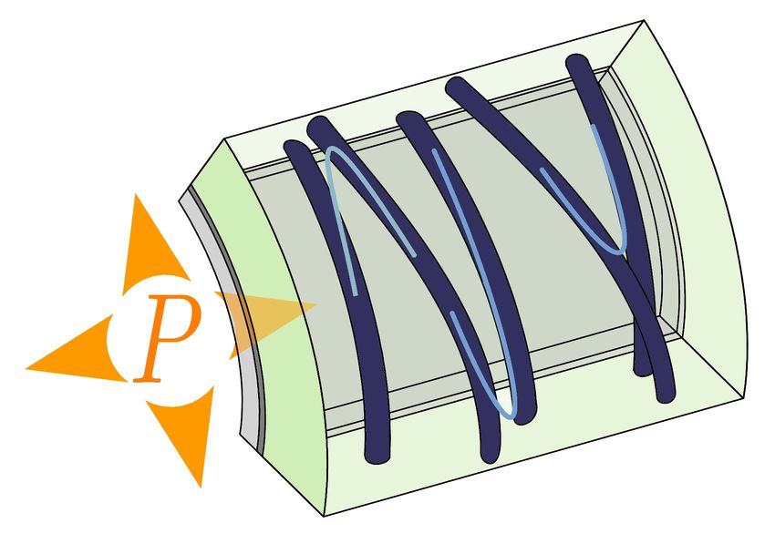
(though this is debated, see e.g. Harold et al., 1996; Rojas et al., 2014). Turgor pressure is coun-
terbalanced by a rigid shell, the extra-cellular matrix known as the cell wall, that prevents cells
from bursting (Harold, 2002). As such, the cell wall is fundamental in determining cell shape. Al-
though the cell walls of these organisms differ in their chemical composition (Lipke and Ovalle,
1998; Cosgrove, 2005; Silhavy et al., 2010), they have similar mechanical properties that are reg-
ulated so as to shape cells. In particular, pressure being a global and isotropic (non-directional)
force, inhomogeneous or anisotropic distributions of mechanical or biochemical properties seem
needed to establish and maintain asymmetric shapes.
    Previous reviews addressed such questions for specific systems, see for instance (Davı̀ and
Minc, 2015, for fission yeast), (Chang and Huang, 2014, for bacteria and fission yeast), (Kroeger
and Geitmann, 2012, for pollen tubes), or (Uyttewaal et al., 2010, for plants). In this review, we
discuss the links between mechanics and growth of elongated cells across kingdoms. We pay
specific attention to computational modelling, because it appears as a powerful tool to validate
hypotheses by setting aside all but the fundamental actors of the phenomena investigated and to
guide future experimental effort. We apologise to those whose work could not be included.

2. Systems of interest and their cell walls

    We first present a few model systems; we give a brief introduction to their cell walls in
terms of composition, structure, and mechanical properties, along with a presentation of available
mechanical models of cell walls.

2.1. Rod-shaped bacteria
     All kinds of shapes are found amongst bacteria. Actually, shape has long been an impor-
tant criterion for classification (Cabeen and Jacobs-Wagner, 2005) and is fundamental for many
bacterial functions (Young, 2006; Singh and Montgomery, 2011; Chang and Huang, 2014). The
rod is a very common shape, with Escherichia coli as a representative Gram-negative species,
making bacteria good systems to study the establishment and maintenance of elongated shapes.
Many rod-shaped bacteria expand diffusively, with new cell wall incorporated all along the rod,
while other expand at a pole or within a restricted region Cava et al. (2013).
     The typical bacterial cell wall is mostly made of peptides and glycans (Cabeen and Jacobs-
Wagner, 2007; Silhavy et al., 2010). The glycan strands are oriented circumferentially and give
the cell wall anisotropic mechanical properties (Chang and Huang, 2014), the cell wall being
stiffer in the circumferential direction than in the longitudinal direction. In Gram-positive bac-
teria, the structure of the cell wall is not as well characterised as in Gram-negative bacteria, al-
though recent studies also support a circumferential arrangement of glycan strands (Beeby et al.,
2013). A detailed mechanical model of the cell wall of Gram-negative bacteria was proposed
by Huang et al. (2008), with the peptidoglycan network simulated like a network of springs; by
adding defects in the network and modulating the type of defects and their density, this model
was able to reproduce several bacterial cell morphologies: curved, helical, snake-like, and lemon
shapes. Whereas this model did not take growth into account, it was the starting point for many
models of wall expansion, which are discussed in the next section.

2.2. Fungi and oomycetes
    Many fungi have elongated cells that grow from their tips: hyphae and yeasts. Hyphae
are very long filamentous cells, that can be collectively organised into a mycelium. Yeasts are
                                                 2
A. Rod-shaped bacterium Outer membrane
Inner membrane
Peptidoglycan network
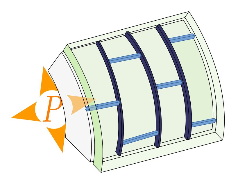
B. Fungi: hypha and fission yeast
(Toward
mycelium)
Chitin Mannan
Plasma membrane
Glucan
C. Root hair, pollen tube and SAM
Plasma membrane
(Toward
root or
pollen grain)
Callose (pollen tube)
(Shoot)
Cellulose/hemi-cellulose
network + pectin matrix
Figure 1: Systems of interest: growth mode and composition of the cell wall. (A) In many rod-shaped bacteria,
such as E. coli, growth is diffuse, localised on the whole cylindrical part on the cell. The cell wall of E. coli and other
Gram-negative bacteria is composed of a stiff layer of peptides and glycans surrounded by two lipid membranes (Silhavy
et al., 2010; Chang and Huang, 2014). (B) Tip growth is observed in fungal hyphae and fission yeast. According to
models, the cell wall would be composed of three layers, made respectively of chitin, glucan and mannan (Lipke and
Ovalle, 1998; Bowman and Free, 2006). (C) In plants, pollen tubes and root hairs are tip-growing cells. On a larger
scale, growth is focused on the tip of emerging organs around the shoot apical meristem. The plant cell wall is made of
a network of cellulose embedded in a matrix of hemicellulose and pectin (Cosgrove, 2005). Callose can also be found,
notably in pollen tubes (Chebli and Geitmann, 2011). In the three systems, the relative amounts of components may vary,
for instance between species or even spatially within a single cell.
3unicellular fungi. In particular, fission yeast (Schizosaccharomyces pombe) is a good system
to study cell polarity and the maintenance of rod shapes (Chang and Martin, 2009). Indeed,
it grows as a capped cylinder, maintaining a constant diameter (except for spores, which are
roughly spherical).
The cell wall of fungi is mostly made of glucans (excluding cellulose), mannoproteins, and
chitin (Lipke and Ovalle, 1998). Although many of these components are fibrous, it is believed
that the fungal cell wall does not have anisotropic mechanical properties because of the lack of
preferential orientation of the fibres (Chang and Huang, 2014). (Strictly speaking, the fibres are
mostly tangential, and the cell wall is transversely isotropic, being softer across the thickness
than in the directions tangential to the wall). Despite a rather well-known composition, fungal
cell walls have not been modelled in detail.
Although oomycetes grow in mycelial forms like fungi, they belong to a different taxonomic
group, Stramenopiles. Their cell walls are mostly made of glucans and, unlike fungi, they contain
some cellulose and tiny amounts of chitin (Mélida et al., 2013).
2.3. Plants: pollen tubes and root hairs
Plants provide two other systems of interest, pollen tubes and root hairs, that elongate through
tip growth. The pollen tube is a long protuberance that grows out from the pollen grain until it
reaches the ovule for fertilisation (Geitmann, 2010; Kroeger and Geitmann, 2012). Accordingly,
its growth is fast and highly directional. Its growing tip is formed of a single cell. Root hairs
are long tubular outgrowths from specialised epidermal cells of the root. They are important for
absorption of nutrients and anchorage to the soil (Carol and Dolan, 2002; Grierson et al., 2014).
The plant cell wall is mostly made of polysaccharides: cellulose microfibrils embedded in
a matrix of hemicelluloses and pectins (Varner and Lin, 1989). Cellulose fibrils can be much
longer than the cell diameter and their organisation differs according to cell type and to develop-
mental stage, ranging from highly directional circumferential alignment to random orientations
(Cosgrove, 2005). As cellulose is the stiffest component of the wall, a preferential orientation
of microfibrils can give anisotropic properties to the plant cell wall: It is stiffer in the direction
of the fibres (Kerstens et al., 2001), which may lead to less expansion in this direction, and so
drive anisotropic cell growth. In pollen tubes and root hairs, cellulose usually displays a helical
arrangement that could help resist bending forces and penetrate external medium (Aouar et al.,
2010); this arrangement could also reinforce the transition region between the tip and the cylin-
drical part, which bears the highest tension (Geitmann, 2010). An early model of the cell wall
focused on the self-organisation of cellulose due to cell geometry (Emons and Mulder, 1998;
Mulder and Emons, 2001); the condition of optimal packing of cellulose microfibrils restrains
their direction and the movement of their synthesising complexes along the axis of the cell can
generate various types of organisations with locally aligned fibres. More recently, models at-
tempted to define realistic geometries for the arrangement of polysaccharides in the cell wall and
to predict the corresponding elastic properties for small deformations (Qian et al., 2010; Kha
et al., 2010; Yi and Puri, 2012).
2.4. Plants: A multicellular case, the shoot apical meristem
Some of the relevant results that we will discuss were obtained in a multicellular context.
The shoot apical meristem (SAM) is the tissue located at the tip of any above-ground branch in
a plant; it contains a stem cell pool and is the site of organogenesis (Ha et al., 2010; Murray
et al., 2012; Gaillochet et al., 2014). Organs are initiated around the tip and emerge as protuber-
ances from the apical dome, breaking the symmetry around the axis of the dome. As discussed
4above, the deposition of cellulose microfibrils in a preferential direction can provide anisotropic
mechanical properties to the SAM.
3. Generating elongated shapes
3.1. Tip growth
We first consider geometrical models built in the context of hyphal growth. In hyphae of
many fungi, a intriguing structure localised close to the tip and known as the spitzenkörper (SPK)
concentrates vesicles and is thought to be the organising centre of tip growth. It has been pro-
posed that vesicles, containing notably multiple cell-wall regulating enzymes, are transported to
the region of the SPK by cytoplasmic microtubules. Actin microfilaments, found in the SPK,
then take over in regulating the supply of enzymes and material to the membrane. The com-
plete composition of the SPK is still unclear (see Steinberg et al., 2017, for a review on hyphal
growth). Its description as a cluster of cell wall-building enzymes has led to the Vesicle Supply
Centre (VSC) model (Bartnicki-Garcia et al., 1989) of fungal growth, first implemented in two
dimensions. In this framework, the VSC is a point in space that constantly emits vesicles in
random directions. Those vesicles move at constant velocity, and locally increase the length of
the cell wall after they have reached it. Finally, the VSC moves at a constant, prescribed veloc-
ity. This yields a steady shape that compares well with experimental observations of growing
hyphae. The three-dimensional generalisation of the VSC model (Gierz and Bartnicki-Garcia,
2001) raised the question of how the material brought by the vesicle is distributed between the
longitudinal and the circumferential directions, which led to propose additional rules for expan-
sion. A second improvement of the VSC model was to replace the ballistic motion of the vesicles
by diffusion (Tindemans et al., 2006), which only slightly modifies the shapes generated by the
model. VSC models were successful in demonstrating that self-similar tip growth can emerge
from patterns of exocytosis. However, they assumed that wall expansion is limited by supply of
materials, and that cell wall mechanics is negligible. While there is experimental evidence that
exocytosis is required for growth, it is also clear that cell wall mechanics is important to set the
pace of expansion (Kroeger and Geitmann, 2012).
Cell wall mechanics was accounted for in several generic models. A first class of models
assumes that growth can be considered as a viscous process, whereby the cell wall expands like
a viscous material under the tension generated by turgor pressure; such a process would lead
to wall thinning, and so cell wall synthesis is assumed to maintain the cell wall thickness at an
approximately constant value. Bernal et al. (2007) got inspiration from the inflation of rubber
balloons, which were modelled as elastic shells with spatially varying stiffness: More precisely,
the compliance of the material (how easy to stretch it is) is large on a narrow annular region
around the tip and small on the cylindrical part of the shell. This model was able to reproduce
observed deformations of root hairs. This work highlighted the importance of modulating the
global pressure drive by local supply of cell wall material, or by local modifications of cell wall
properties. The spatial extent of the wall deposition, and more precisely how it depends on the
size of the cell, was theoretically found to change the shape of the growing tip (Campàs and
Mahadevan, 2009). In this latter study, the cell wall was modelled as a thin viscous shell with
infinite viscosity on the flanks (so that the tube maintains its diameter). Growth is compensated
by material addition (synthesis) at the tip. The authors investigated the dependence of cell shape
on viscosity and spatial extent of material addition; they showed that tube radius increases with
viscosity and that the tip becomes more blunt as the spatial extent is increased. These results
5prompted a broad analysis of tip-growing cell shape from various species (plants, fungi and
oomycetes; Camps et al., 2012): The tip radius (radius of curvature at the tip) scales with tube
radius in plants and fungi, while the tip radius appears constant in oomycetes.
A second class of models considers growth as an incremental process whereby, at each step,
the cell wall is elastically stretched by turgor pressure, and this stretched configuration is taken
as the starting point of the next step; again, synthesis is assumed to keep cell wall thickness
constant. Goriely and Tabor (2003) used the framework of the nonlinear elasticity theory of thin
shells, which allows for large deformations of the shells. An important ingredient of their model
is that the tip of the hypha is softer than the cylindrical part. Consistent with the incremental
framework, growth is simulated by computing the deformation of the shell due to turgor pressure
then taking the deformed shape as a new initial shape that can be deformed further. So in this
model cell wall expansion is localised due to the softer tip. More recently, this model was
used to study the effect of the friction between the growing tip and the external medium. This
friction leads to a flattening of the tip that is consistent with experimental data (Goriely and
Tabor, 2008). It can be shown that incremental models are mathematically equivalent to viscous
models in the limit where pressure is relatively small (see for instance Bonazzi et al., 2014);
nevertheless, the interpretation of parameters differs between the two types of model. In viscous
models, the viscosity (inverse of extensibility) is a proxy for the rate of cell wall remodelling
under tension, but this viscosity cannot be measured directly in experiments. In elastic models,
the elastic modulus (inverse of compliance) quantifies the stiffness of the wall material, which
can be measured experimentally, but it does not necessarily predict how fast the cell wall expands
under tension. Actually, the chemistry of the cell wall is an important ingredient that is missing
from these two types of models, a limitation that applies to most of the mechanical models
presented here.
In plants, root hairs and pollen tubes are quite similar to fungi with respect to the control
of polar growth. Microtubules have two types of localisation in plants: cortical – close to the
plasma membrane, and endoplasmic. In root hairs and pollen tubes, microtubules, as well as
actin filaments, are oriented along the tube axis and are involved in targeting the supply of new
material to the tip (Sieberer et al., 2005; Gu and Nielsen, 2013; Chebli et al., 2013). In root
hairs, this organisation of microtubules depends on the cell nucleus (Ambrose and Wasteneys,
2014). Detailed measurements in root hairs have shown that cell wall expansion occurs mainly
in an annulus just behind the tip, and is isotropic there; farther from the tip, expansion becomes
mostly radial and decays with distance to the tip (Shaw et al., 2000). Dumais et al. (2006) built
a mechanical model that qualitatively reproduced expansion profiles in several tip growing cells,
including root hairs, using the following assumptions. The cell wall is viscoplastic: expansion
occurs above a threshold in tension and then increases linearly with tension; the viscosity (inverse
of extensibility) is smaller close to the tip. The thinning of the wall due to its stretching is
compensated by deposition so as to keep its thickness constant. The main result is that the
model accounts for quantitative measurements of cell geometry (curvatures) and wall expansion
(strain rates) in root hairs only if cell walls have mechanical anisotropy. More precisely, the cell
wall needs to be transversely isotropic, meaning that its properties in the direction of thickness
are different from its properties in its tangent plane. Such anisotropy can be explained by the
deposition of cellulose tangentially to the cell wall.
A similar pattern of expansion is observed in pollen tubes (Zerzour et al., 2009; Hepler et al.,
2013). The shape of tubes was reproduced with an incremental elastic model that included a
sharp gradient of stiffness at the tip (Fayant et al., 2010). The best fitting of observed shapes
was achieved assuming the cell wall transversely isotropic. Interestingly, the gradient of stiffness
6used in the simulation is consistent with the gradients of density observed for various cell wall
components, such as pectins, cellulose, and callose, that determine its mechanical properties.
As a complement to the systems studied here, we also mention trichomes — elongated hair
cells found in the aerial part of plants — because they appear to differ from pollen tubes. Yanag-
isawa et al. (2015) used a viscoelastic thin-shell model of the cell wall, expanding due to turgor-
generated tension. They needed to combine softer tip and mechanical anisotropy on the sides to
better match experimental data.
In fungi, the local delivery of new cell wall is driven by microtubules and actin filaments.
The cytoskeleton could be directly required for growth or only define the location where wall ex-
pansion takes place. Chemical treatments and mutants have demonstrated that disruption of mi-
crotubules leads to major geometrical defects in the fission yeast (Hagan, 1998). Application of
actin inhibitors can modify the dynamics of growth or completely arrest it depending on the con-
centrations used. Consequently, the microtubules and actin filaments must efficiently target the
cell tips (Sawin and Nurse, 1998; Terenna et al., 2008) to deliver the new material to the proper
location. Drake and Vavylonis (2013) modelled the coupling between microtubule dynamics, a
remodelling signal – a protein required for cell wall remodelling, and cell wall mechanics. As in
many previous studies, they considered the cell wall as effectively viscous (Campàs and Mahade-
van, 2009) and they assumed in addition that effective viscosity is reduced by the remodelling
signal. They considered microtubules as growing and shrinking flexible rods attached to the nu-
cleus. They first assumed that the level of the remodelling signal is imposed by the likelihood
of contact between microtubules and cell wall, but they found that this did not enable the main-
tenance of cell width over many generations (as observed in living cells). Maintenance of cell
width was achieved with additional assumptions: microtubules control the deposition of land-
mark proteins that in turn attract the remodelling signal; the remodelling signal has an intrinsic
dynamics (such as reaction-diffusion) that leads to its localisation over a region of well-defined
size. To summarise, Drake and Vavylonis (2013) built one of the first successful models for cell
morphogenesis that integrates cell polarity and cell wall mechanics. It would be interesting to
further probe this model by, for instance, investigating the recovery from spheroplasts (cells that
became round following wall digestion) to rod-like shapes.
Abenza et al. (2015) combined experiments and mechanical models to explore which cellular
processes among polarity, exocytosis, or wall synthesis determine the pattern of cell wall expan-
sion in fission yeast. They used an incremental elastic model and assumed the elastic modulus
to be a function of either of the cell-end localised factors involved in the three previous cellu-
lar processes. They found that exocytosis factors better predicted the observed pattern of wall
expansion. The pattern of supply of wall materials thus appears to be essential for shape, mak-
ing the connection between the concepts behind mechanical models and those behind VSC-like
models.
Overall, the qualitative agreement with experiments of a range of mechanical models strongly
supports the notion that a softer / more compliant tip is required for tip growth. However it is
yet difficult to ascertain which models are more relevant to actual cells. Further quantitative
measurements of wall expansion and wall mechanics are required to make further progress.
It is generally believed that the epidermis of aerial plant tissues is under tension (see Peters
and Tomos, 1996, for a review), which would occur for instance if the epidermis is much stiffer
than internal tissues (as inferred in Beauzamy et al., 2015). Consequently, a tissue like the shoot
apical meristem (SAM) behaves mechanically like a pressurised shell, in which the epidermis
plays the role of the shell, while inner layers corresponds to a liquid under pressure. The SAM is
therefore comparable mechanically to the unicellular systems considered so far. Quantification
7of cell wall expansion in an Arabidopsis mutant that does not produce organs (pin-formed 1,
defective in a protein that enables efflux from cells of the phytohormone auxin) revealed higher
expansion rate in an annulus that surrounds the tip Kwiatkowska (2004), consistent with stiffer
cell walls at the tip (Milani et al., 2011, 2014) and reminiscent of expansion patterns in root
hairs. However, this analogy is only partial because the mechanical properties of cell walls are
likely anisotropic in the shoot apex (see following section). Nevertheless, spatial variations in the
mechanical properties of cell walls seem to be required to establish the patterns of growth that
underlie morphogenesis. For instance, the appearance of a new growth axis – the primordium
of a lateral organ such as a leaf or a flower – on the side of the meristem is associated with a
locally softer cell wall (Peaucelle et al., 2011; Kierzkowski et al., 2012). This outgrowth requires
an increase in pectin demethylesterification (Peaucelle et al., 2011), which occurs in internal cell
walls before in surface walls, and is dependent on the accumulation of auxin (Braybrook and
Peaucelle, 2013). Note that this constitutes an important difference with pollen tubes, where
pectin demethylesterification rigidifies the cell wall and therefore inhibits growth (see Bosch and
Hepler, 2006, for more details). The three-dimensional patterns in cell wall properties prompted
Boudon et al. (2015) to develop realistic mechanical models of tissues. Each cell wall is con-
sidered as a thin surface with elastic, plastic and viscous properties. By fine tuning the stiffness
and/or viscosity of walls, Boudon et al. (2015) were able to make one or several organs emerge.
This work shows how heterogeneity in stiffness may generate complex shapes. Interestingly,
several solutions are sometimes possible for creating a given shape. For this reason, the com-
parison of computational outputs with real tissues cannot be limited to shape and requires other
experimental observations.
3.2. Anisotropic diffuse growth
The stiffness of a material is not just a number. The material can be anisotropic, i.e. it
can have different values of stiffness in different directions, like for instance a fibre-reinforced
material, which is harder to stretch in the direction of the fibres. In many cases, elongation of
walled cells requires such anisotropy.
In many rod-shaped bacteria, growth occurs on the cylindrical region of the cell (Cava et al.,
2013). New material is inserted as small patches on the cylindrical part of the cell, a process
coordinated by MreB filaments (Chang and Huang, 2014).
Despite the growth being distributed on their cylindrical part, bacteria grow as elongated
cells. This is often thought to be caused by anisotropic reinforcement of the cell wall, either
directly through a mechanical anisotropy of their material (Yao et al., 1999) or indirectly through
the bacterial cytoskeleton itself. Jiang et al. (2011) focused on the effect of MreB and built
a model of elongation that reproduced shapes, divisions, and bulging in wild-type and mutant
strains of E. coli. The cell wall was considered as a continuous transversely isotropic material
whose growth is driven by the transformation of intracellular energy into a mechanochemical
energy, that combines the elastic energy of the cell wall and the chemical energy of new bonds.
Growth is much slower than MreB dynamics, thus the mechanical effect of helical MreB fila-
ments is averaged over time and modelled as a radial force resisting turgor pressure and yielding
a preferred radius for the cell. Without this force, cells grew spherically. Whereas MreB may
bend liposomes (Hussain et al., 2018), it is unkown whether MreB is strong enough to induce
curvature of the cell wall as it is synthesised.Banerjee et al. (2016) extended this model to also
reproduce vibrio shape, where the rod-shaped bacterium is slightly bent (e.g. Caulobacter cres-
centus), by incorporating a preferred curvature of Crescentin-like proteins (with the same caveat
8as for MreB). It would be interesting to know whether observed cell shapes would still be re-
trieved by these models if forces from the cytoskeleton were replaced by anisotropy in elastic
energy or in bond energy.
Starting from a previous static model of the cell wall in Gram-negative bacteria (Huang et al.,
2008, see above), Furchtgott et al. (2011) modelled wall expansion by considering the insertion
of new short glycan strands into existing peptide cross-links, according to one of 3 scenarii:
the first scenario — random choice of the peptides — leads to bulging and loss of straightness;
the two other scenarii — uniform insertion by choosing peptides inversely proportionally to their
density or helical insertion according to the motion of synthase — enables the maintenance of rod
shapes. In order to study the molecular details of cell wall remodelling, Nguyen et al. (2015) later
built a model on a similar, coarse-grained scale; they used more realistic mechanical parameters
for the peptidoglycan network, which they inferred from molecular dynamics simulations. They
achieved maintenance of rod shapes by accounting for the spatiotemporal dynamics of enzymes
involved in cell wall remodelling and assuming coordination between enzyme activites. Thus,
the use of different microscopic hypotheses (Furchtgott et al., 2011; Nguyen et al., 2015) yields
different macroscopic outcomes, which calls for further comparison with experimental data. In
a more abstract model based on continuum mechanics, Amir and Nelson (2012) represented the
insertion of new material as the creation and movement of defects (dislocations in this case)
along the peptidoglycan lattice. Using biological relevant values of parameters, they retrieved
exponential growth in length, with a rate that is sensitive to turgor pressure. Altogether, these
studies show that circumferential insertion of glycans is a key ingredient for maintenance of
shape.
In bacteria, it is unclear whether the mechanical anisotropy of the cell wall is required for
rod shapes, as direct experimental evidence is lacking while models with defects moving in
isotropic walls (Amir and Nelson, 2012) may produce rod-shaped cells. In contrast, in plants,
reduced cellulose content induces more isotropic growth (Baskin, 2001), supporting the idea
that mechanical anisotropy is required for anisotropic diffuse growth. In the multicellular con-
text of the shoot apex, it is likely that stiffness anisotropy combines with the local softening
discussed earlier. Indeed, oriented deposition of cellulose may lead to strongly anisotropic cell
walls (Cosgrove, 2005). This would be the case in the boundary between an emerging organ and
the meristem, around the tip of the shoot, and around the tip of an organ primordium (Hamant
et al., 2008), where oriented cellulose deposition is predicted based on the orientation of micro-
tubules. The associated mechanical anisotropy would provide an additional mechanism for the
elongation of the shoot or of the organ. Indeed, using a cell-based mechanical model of tissue
growth, Boudon et al. (2015) and Sassi et al. (2014) showed that combining changes in stiffness
levels and in stiffness anisotropy ensures optimal outgrowth and better accounts for experimental
observations.
4. Feedbacks that stabilise elongated shapes
4.1. Sensing curvature
An extreme case of curvature-sensing is when wall expansion is fully determined by its cur-
vature. In tip growing cells, expansion is maximal at the tip, where the wall curvature is the
highest. Goriely et al. (2005) and Jaffar and Davidson (2013) built geometrical models of tip-
growing cells in which local wall expansion (and accordingly material supply) is an increasing
function of local curvature (Gauss curvature in 3D models). Jaffar and Davidson (2013) used
9two fitting parameters to reproduce the geometry of cells of many organisms (plants, fungi, acti-
nobacteria), though the biological relevance of these parameters is unclear. Such models show
that curvature-based expansion is sufficient to account for the stable form of tip-growing cells.
There is evidence for curvature-sensing in bacteria. Ursell et al. (2014) and Billings et al.
(2014) found that MreB localises preferentially to regions where the wall has negative curvature;
the spatiotemporal correlation of expansion and MreB indicates that MreB precedes expansion.
This causal link between curvature and MreB was confirmed by perturbation experiments. Al-
together, experimental data suggest that MreB relocalises to regions of negative curvature, in-
ducing more expansion and reverting the local geometry to cylindrical. Ursell et al. (2014) used
the model for glycan strand insertion (Furchtgott et al., 2011, see above) to test this mechanism
in E. coli. They assumed that insertion occurs preferentially at regions of low curvature and
found that this stabilised growth of a rod-shaped cell and enabled the recovery from an initially
bent shape. Hussain et al. (2018) gave further experimental support to these conclusions by
manipulating shapes of Bacillus subtilis by chemical treatments or by confinement in channels.
However, Wong et al. (2017) combined experimental and theoretical approaches to show that the
observed local enrichment in MreB in deformed bacteria is not sufficient to explain the recovery
of a straight shape.
4.2. Sensing forces
Bacteria are known to sense their mechanical state through channels that are sensitive to
membrane tension. In E. coli, Wong et al. (2017) proposed that strain-activated growth could
qualitatively explain the response and recovery of experimentally bent bacteria. Interestingly,
although the biochemical implementation of this mechanism is unclear, it was suggested to work
jointly with MreB-mediated regulation (as discussed above).
In fission yeast, spores grow roughly spherically, until an outgrowth initiates the rod-like
shape of vegetative cells. The associated transition from unstable to stable polarity is triggered
mechanically by the rupture of the outer wall of the spore, which is a very stiff thin layer that
surrounds the vegetative-like wall (Bonazzi et al., 2014). Interestingly, the ratio between the
volume at the transition and the initial volume of the spore is constant, despite a large variability
in initial volume. Bonazzi et al. (2014) modelled the outer wall as an elastic stiff shell (that may
rupture) and considered the vegetative wall either as elastic or as viscoplastic — the viscoplastic
region is where growth occurs and it corresponds to the location of the polarisome (the ensem-
ble of polarly localised proteins). The polarisome was assumed to move randomly, mimicking
experimental observations. When it is intact, the outer shell prevents outgrowth and keeps the
spore roughly spherical. The tension in the shell increases, up to a threshold over which its local
rupture initiates the outgrowth. The theoretical results thus show that stress-sensing via mechan-
ical rupture enables the outgrowth to occur when the ratio of spore volume to initial volume has
reached a well-defined threshold.
As mentioned above, morphogenesis at the shoot apex relies both on local softening and
mechanical anisotropy. The two mechanisms are tuned by a feedback from mechanics. The
local softening that initiates organ emergence is triggered by the local accumulation of the phy-
tohormone auxin (Sassi and Vernoux, 2013). Auxin patterns are determined by the polarity
of PIN FORMED1 (PIN1), a membrane-addressed protein that facilitates auxin efflux. Three
hypotheses have been proposed for the determination of auxin polarity based on experimental
observations (Abley et al., 2013; Sassi and Vernoux, 2013): flux of auxin, gradient of auxin,
and intrinsic property of the cell that may be oriented by external cues. We here focus on the
10A. Curvature-sensing in bacteria
B. Growth- and force-sensing
in fungi and pollen tubes
C. Force-sensing in the shoot apex
Figure 2: Feedbacks that stabilise elongation. (A) In E. coli, the insertion of new cell wall is increased in the region of
negative curvature. This feedback may stabilise rod shape and enable recovery from initially curved shapes (Ursell et al.,
2014; Billings et al., 2014; Hussain et al., 2018). (B) In pollen tubes and fission yeast, surface expansion feeds back on
material supply. In pollen tubes this feedback may lead to oscillatory tip growth (Rojas et al., 2011). In fission yeast,
this feedback occurs through the position of the polar cap (where polarity proteins are localised). It leads to the random
shuffling of polar cap in spores. After the rupture of the outer spore wall, the feedback promotes tip-growth (Bonazzi
et al., 2014). (C) In the plant shoot apex, two loops involving mechanosensation are coupled. By enhancing transport of
the phytohormone auxin, mechanical strain and stress focus growth at the tip of the organ. Mechanical stress may also
increase mechanical anisotropy of the cell wall via deposition of cellulose oriented by the response of microtubules to
mechanical stress (Hamant et al., 2008; Heisler et al., 2010).
11second hypothesis, because it has been related to mechanical signals. According to this hypoth-
esis, PIN1 polarity in a cell is determined by auxin concentration in neighbouring cells so that
PIN1 is polarised towards the cell with higher concentration. This so-called “up the gradient”
model can reproduce observed auxin patterns (Jönsson et al., 2006; Smith et al., 2006), though
it raises questions about how cells might sense auxin concentration in neighbouring cells. More
recent experimental evidence suggests that PIN1 proteins indirectly sense the mechanical status
of cell walls (Heisler et al., 2010; Nakayama et al., 2012; Braybrook and Peaucelle, 2013). More
precisely, PIN1 in a cell would be polarised towards the cell wall with the highest mechanical
stress/strain (which of strain or stress is sensed is still unclear), which is shared with the cell with
highest auxin concentration due to the induced softening of its walls. This chemomechanical
model has been implemented using the finite element method for the mechanics coupled with a
system of differential equation for the auxin dynamics (Heisler et al., 2010). It is able to generate
patterns of auxin accumulation and to reproduce the radial PIN1 reorientation observed around a
cell ablation.
In many plant tissues, mechanical signals also regulate the orientation of cellulose microfib-
rils (Castle, 1937; Green and King, 1966; Preston, 1988; Wasteneys and Williamson, 1987, 1989;
Williamson, 1990; Fischer and Schopfer, 1997; Hejnowicz et al., 2000; Hamant et al., 2008;
Jacques et al., 2013; Sampathkumar et al., 2014). Indeed, cellulose is synthesised following the
orientation of cortical microtubules (Baskin, 2001; Bringmann et al., 2012). Additionally, micro-
tubules orient along the direction of maximal mechanical tension (Wasteneys and Williamson,
1987, 1989; Williamson, 1990; Fischer and Schopfer, 1997; Hejnowicz et al., 2000; Hamant
et al., 2008; Jacques et al., 2013; Sampathkumar et al., 2014). Consequently, the preferential
orientation of cellulose microfibrils reinforces the cell wall in the direction of mechanical stress
(Landrein and Hamant, 2013). Several theoretical studies investigated the consequences of this
feedback loop between mechanical stress and cell wall anisotropy. Hamant et al. (2008) mod-
elled the shoot apical meristem as an elastic surface in 3D, thus only accounting for the epidermal
cell layer. They used a vertex model, meaning that they only considered cell walls orthogonal to
the surface of epidermis, represented as 1-dimensional springs. The stiffnesses of these springs
increases as they are more parallel to the local mechanical tension, mimicking the orientation of
the microtubules and their feedback on the mechanical properties of cell walls. Growth is driven
by the turgor pressure of internal tissues. Above a stress threshold, the cell walls yield and thus
deform plastically. By initiating the emergence of an organ via the local softening of a group of
cells, the cellulose reorientation leads to a circumferential pattern around the organ, reinforcing
the boundary with the apical dome and thus making the symmetry breaking more effective. Bo-
zorg et al. (2014) obtained similar results using a continuous model for the epidermal layer. They
further showed that using mechanical strain instead of stress as a directional cue is not sufficient
to account for experimental observations.
4.3. Sensing growth rate
In fission yeast, an additional result from the mechanical model discussed above (Bonazzi
et al., 2014) is that a positive feedback between growth and polarity can explain the stabilisation
during spore outgrowth. Bonazzi et al. (2014) considered three possible cues that bias the random
motion of the polarisome (polarly localised proteins): curvature of the cell surface, mechanical
stress in the surface, and expansion rate. Only the last cue led to stable cylindrical shapes, which
was supported by further experiments in which wall expansion was manipulated.
Rojas et al. (2011) developed a model coupling the deposition of new material and the me-
chanics of the cell wall. They reproduced the morphologies of pollen tubes and were able to
12explain growth oscillations that are observed in rapidly growing tubes. In this model, the rate of
deposition of wall material decreases with the velocity of cell tip, making a link between exocy-
tosis and growth rate. Consequently, the cell may either grow at constant rate or oscillate between
phases of high deposition and slow elongation and phases of low deposition and fast elongation.
This results in either cylindrical or pearled pollen tubes, respectively. At least two hypotheses
could account for such negative feedback. A ‘passive’ hypothesis is that when growth velocity
increases, exocytosis becomes relatively too slow to provide materials to the growing tip. An ‘ac-
tive’ hypothesis is that a high rate cell wall of expansion may lead to higher membrane tension
(due to limited membrane supply), leading to the opening of mechanosensitive channels and the
entry of cytosolic calcium that would downregulate the polymerisation of the actin cytoskeleton
and thus the delivery of cell wall material (Kroeger et al., 2008; Yan et al., 2009).
Rojas et al. (2017) combined experiments in Bacillus subtilis and a non-spatialised dy-
namical model to also propose that enhanced expansion would induce high membrane tension.
Mechanosensing would prevent over-expansion of the wall by reducing the supply of wall pre-
cursors when membrane tension is too large. It would be interesting to know whether this mech-
anisms is involved in regulating cell shape. Overall, the three studies discussed use models to
suggest that the rate of cell wall expansion is sensed, though the mechanisms behind are still to
be identified.
5. Conclusions
The generation of anisotropic shapes in walled cells relies mainly on two strategies. Many
cells, such as hyphae, yeasts, root hairs or pollen tubes grow directionally via the supply of
new material to the cell wall at a precise and restricted location (Tindemans et al., 2006; Drake
and Vavylonis, 2013). Several models for tip growth have been implemented and are able to
reproduce most of observed shapes. These computational approaches may give some informa-
tion about the mechanics of the cell wall (Dumais et al., 2006; Fayant et al., 2010) or about
its behaviour with respect to perturbations (Goriely and Tabor, 2008). However, the range of
hypotheses used in these models makes it difficult to know which ones are more relevant to ex-
periments. Progress should stem from more quantitative experiments and models, and from the
study of perturbations in experiments and in models. Rod-shaped bacteria grow diffusely, re-
quiring the mechanical reinforcement of their sides. This reinforcement is likely achieved thanks
to the circumferential insertion of glycan strands (Huang et al., 2008; Furchtgott et al., 2011).
On a multicellular scale, plants combine those tip growth and anisotropic diffuse growth to ini-
tiate organs morphogenesis. Theoretical models indicate that the two mechanisms are necessary
to induce the massive shape changes required for organogenesis. The major actors of this me-
chanical control of morphogenesis, each corresponding to one of the strategies discussed here,
are the phytohormone auxin, which is involved in the softening of the plant cell wall (Rein-
hardt et al., 2000), and cellulose, a stiff polymer whose oriented deposition leads to stiffness
anisotropy (Baskin, 2001).
Several feedbacks have been identified that may contribute to the maintenance of rod-shapes.
Bacteria, thanks to a simple mechanism of curvature-sensing based on the MreB protein, are
able to maintain and even to generate de novo cylindrical shapes (Billings et al., 2014; Ursell
et al., 2014; Wong et al., 2017). In fission yeast, a precise volume doubling between the germi-
nation and the outgrowth is granted by the mechanical rupture of its protective shell (Bonazzi
et al., 2014). Force-sensing is also involved in organogenesis in the plant shoot apex. Mechan-
ical stress, auxin transport, auxin-induced softening and cellulose anisotropy feed back on each
13other in complex loops that may be required for the robustness of organogenesis (Hamant et al.,
2008; Heisler et al., 2010). Finally, growth-sensing explains both the random movement and the
stabilisation of polarity before and after the triggering of the outgrowth in fission yeast (Bonazzi
et al., 2014). Growth-sensing might also be relevant for the oscillatory growth in pollen tubes
(Rojas et al., 2011). All these studies show how theoretical approaches may help unravelling the
complex feedbacks that underly organismal growth and robustness of morphogenesis. Simula-
tions of these models enable testing alternative hypotheses that can be difficult to differentiate
experimentally or may lead to the identification of key experiments.
The molecular actors behind many of these feedbacks are unknown. Curvature-sensing in
bacteria could be due to a membrane curvature-dependent binding energy of the protein complex
that includes MreB. Negative curvature and MreB localisation could also be driven by a com-
mon signal such as proteins involved in cell wall synthesis (Billings et al., 2014). In fission yeast,
mechanisms similar to the oscillatory growth of pollen tubes could explain the stabilisation of po-
larity by surface expansion (Yan et al., 2009; Rojas et al., 2011). Alternatively, polarity could be
diluted and destabilised in the absence of sufficient growth (Layton et al., 2011). Finally, growth
could be involved in the monitoring of cellular dimensions by intracellular gradients (Howard,
2012). In the context of the shoot apex, feedback mechanisms are still poorly understood and
we may only speculate. Stretching of the cell membrane or of the cell wall could activate ion
channels or modify the conformation of wall-bound proteins, triggering pathways that impact
on microtubules or on PIN1 (Landrein and Hamant, 2013). In the case of auxin transport, an
alternative hypothesis is the activation of exocytosis and inhibition of endocytosis by the ten-
sion in the cell membrane (Hamill and Martinac, 2001). All these hypotheses lack evidence, but
new insights can be expected with progress in cellular and developmental biology, together with
physically-based models of the associated processes.
We tried here to highlight concepts and generic mechanisms that hold across kingdoms. Fu-
ture directions might stem from enhanced cross-fertilisation between approaches and concepts
developed in the context of specific systems. For instance, detailed mechanical models of the
growing bacterial cell wall could provide inspiration for the more complex and less organised
cell walls of fungi, oomycetes, and plants. Conversely, continuous models developed for plants,
fungi, and oomycetes could be used to test coarse hypotheses in bacterial morphogenesis, before
dealing with more involved molecular details. More generally, modelling morphogenesis at mul-
tiple scales, with models assembled as a Russian doll to make links between successive scales
or levels, would allow to deal with a suite of simple models than can be falsified separately and
assembled to address more elaborate questions.
Acknowledgements
AB is supported by Institut Universitaire de France. AB would like to thank the Isaac Newton
Institute for Mathematical Sciences for support and hospitality during the programme Growth
form and self-organisation when this paper was finalised. This work was partially supported by
EPSRC grant number EP/K032208/1 and by a grant from the Simons Foundation.
References
Abenza, J. F., Couturier, E., Dodgson, J., Dickmann, J., Chessel, A., Dumais, J., Carazo Salas, R. E., 2015. Wall
mechanics and exocytosis define the shape of growth domains in fission yeast. Nature Communications 6, 8400.
14Abley, K., de Reuille, P. B., Strutt, D., Bangham, A., Prusinkiewicz, P., Mare, A. F. M., Grieneisen, V. A., Coen, E.,
2013. An intracellular partitioning-based framework for tissue cell polarity in plants and animals. Development 140,
2061–2074.
Ambrose, C., Wasteneys, G. O., Sep. 2014. Microtubule initiation from the nuclear surface controls cortical microtubule
growth polarity and orientation in Arabidopsis thaliana. Plant and Cell Physiology 55 (9), 1636–1645.
URL http://pcp.oxfordjournals.org/cgi/doi/10.1093/pcp/pcu094
Amir, A., Nelson, D. R., 2012. Dislocation-mediated growth of bacterial cell walls. Proceedings of the National Academy
of Sciences 109 (25), 9833–9838.
URL http://www.pnas.org/content/109/25/9833
Aouar, L., Chebli, Y., Geitmann, A., 2010. Morphogenesis of complex plant cell shapes: the mechanical role of crys-
talline cellulose in growing pollen tubes. Sexual Plant Reproduction 23 (1), 15–27.
URL https://doi.org/10.1007/s00497-009-0110-7
Banerjee, S., Scherer, N. F., Dinner, A. R., 2016. Shape dynamics of growing cell walls. Soft matter 12, 3442–3450.
Bartnicki-Garcia, S., Hergert, F., Gierz, G., 1989. Computer simulation of fungal morphogenesis and the mathematical
basis for hyphal (tip) growth. Protoplasma 153 (1-2), 46–57.
URL http://link.springer.com/article/10.1007/BF01322464
Baskin, T. I., 2001. On the alignment of cellulose microfibrils by cortical microtubules: a review and a model. Proto-
plasma 215 (1-4), 150–171.
URL http://link.springer.com/article/10.1007/BF01280311
Beauzamy, L., Louveaux, M., Hamant, O., Boudaoud, A., Nov. 2015. Mechanically, the shoot apical meristem of ara-
bidopsis behaves like a shell inflated by a pressure of about 1 mpa. Frontiers in Plant Science 6, 1038.
Beeby, M., Gumbart, J. C., Roux, B., Jensen, G. J., May 2013. Architecture and assembly of the Gram-positive cell wall.
Molecular Microbiology 88 (4), 664–672.
URL http://onlinelibrary.wiley.com/doi/10.1111/mmi.12203/abstract
Bernal, R., Rojas, E., Dumais, J., 2007. The mechanics of tip growth morphogenesis: what we have learned from rubber
balloons. Journal of Mechanics of Materials and Structures 2 (6), 1157–1168.
URL http://msp.org/jomms/2007/2-6/p10.xhtml
Billings, G., Ouzounov, N., Ursell, T., Desmarais, S. M., Shaevitz, J., Gitai, Z., Huang, K. C., Sep. 2014. De novo
morphogenesis in L-forms via geometric control of cell growth. Molecular Microbiology 93 (5), 883–896.
URL http://doi.wiley.com/10.1111/mmi.12703
Bonazzi, D., Julien, J.-D., Romao, M., Seddiki, R., Piel, M., Boudaoud, A., Minc, N., Mar. 2014. Symmetry Breaking
in Spore Germination Relies on an Interplay between Polar Cap Stability and Spore Wall Mechanics. Developmental
Cell 28 (5), 534–546.
URL http://linkinghub.elsevier.com/retrieve/pii/S1534580714000641
Bornens, M., 2008. Organelle positioning and cell polarity. Nature Reviews Molecular Cell Biology 9, 874–886.
Bosch, M., Hepler, P., 01 2006. Pectin methylesterases and pectin dynamics in pollen tubes. The Plant cell 17, 3219–26.
Boudon, F., Chopard, J., Ali, O., Gilles, B., Hamant, O., Boudaoud, A., Traas, J., Godin, C., Jan. 2015. A Computational
Framework for 3d Mechanical Modeling of Plant Morphogenesis with Cellular Resolution. PLoS Computational
Biology 11 (1), e1003950.
URL http://dx.plos.org/10.1371/journal.pcbi.1003950
Bowman, S. M., Free, S. J., Aug. 2006. The structure and synthesis of the fungal cell wall. BioEssays 28 (8), 799–808.
URL http://doi.wiley.com/10.1002/bies.20441
Bozorg, B., Krupinski, P., Jönsson, H., Jan. 2014. Stress and Strain Provide Positional and Directional Cues in Develop-
ment. PLoS Computational Biology 10 (1), e1003410.
URL http://dx.plos.org/10.1371/journal.pcbi.1003410
Braybrook, S. A., Peaucelle, A., Mar. 2013. Mechano-Chemical Aspects of Organ Formation in Arabidopsis thaliana:
The Relationship between Auxin and Pectin. PLoS ONE 8 (3), e57813.
URL http://dx.plos.org/10.1371/journal.pone.0057813
Bringmann, M., Landrein, B., Schudoma, C., Hamant, O., Hauser, M.-T., Persson, S., nov 2012. Cracking the elusive
alignment hypothesis: the microtubule-cellulose synthase nexus unraveled. Trends in Plant Science 17 (11), 666–74.
URL http://dx.doi.org/10.1016/j.tplants.2012.06.003 http://www.ncbi.nlm.nih.gov/pubmed/22784824
http://www.pubmedcentral.nih.gov/articlerender.fcgi?artid=PMC3492759
Cabeen, M. T., Jacobs-Wagner, C., Aug. 2005. Bacterial cell shape. Nature Reviews Microbiology 3 (8), 601–610.
URL http://www.nature.com/doifinder/10.1038/nrmicro1205
Cabeen, M. T., Jacobs-Wagner, C., Oct. 2007. Skin and bones: the bacterial cytoskeleton, cell wall, and cell morphogen-
esis. The Journal of Cell Biology 179 (3), 381–387.
URL http://www.jcb.org/cgi/doi/10.1083/jcb.200708001
Campàs, O., Mahadevan, L., Dec. 2009. Shape and Dynamics of Tip-Growing Cells. Current Biology 19 (24), 2102–
2107.
15URL http://linkinghub.elsevier.com/retrieve/pii/S0960982209019836
Camps, O., Cmpas, O., Rojas, E., Dumais, J., Mahadevan, L., 2012. Strategies for cell shape control in tip-growing cells.
American Journal of Botany 99 (9), 1577–1582.
URL http://www.jstor.org/stable/23251587
Carol, R. J., Dolan, L., Jun. 2002. Building a hair: tip growth in Arabidopsis thaliana root hairs. Philosophical Transac-
tions of the Royal Society B: Biological Sciences 357 (1422), 815–821.
URL http://rstb.royalsocietypublishing.org/cgi/doi/10.1098/rstb.2002.1092
Castle, E., 1937. Membrane tension and orientation of structure in the plant cell wall. Journal Of Cellular And Compar-
ative Physiology 10 (1), 113–121.
Cava, F., Kuru, E., Brun, Y. V., de Pedro, M. A., 2013. Modes of cell wall growth differentiation in rod-shaped bacteria.
Current Opinion in Microbiology 16, 731–737.
Chang, F., Huang, K. C., 2014. How and why cells grow as rods. BMC biology 12 (1), 54.
URL http://www.biomedcentral.com/1741-7007/12/54/
Chang, F., Martin, S. G., Jul. 2009. Shaping fission yeast with microtubules. Cold Spring Harbor Perspectives in Biology
1 (1), a001347.
Chebli, Y., Geitmann, A., 2011. Gravity Research on Plants: Use of Single-Cell Experimental Models. Frontiers in Plant
Science 2.
URL http://journal.frontiersin.org/article/10.3389/fpls.2011.00056/abstract
Chebli, Y., Kroeger, J., Geitmann, A., Jul. 2013. Transport Logistics in Pollen Tubes. Molecular Plant 6 (4), 1037–1052.
URL http://linkinghub.elsevier.com/retrieve/pii/S167420521460900X
Cosgrove, D. J., Nov. 2005. Growth of the plant cell wall. Nature Reviews Molecular Cell Biology 6 (11), 850–861.
URL http://www.nature.com/doifinder/10.1038/nrm1746
Davı̀, V., Minc, N., Dec. 2015. Mechanics and morphogenesis of fission yeast cells. Current Opinion in Microbiology
28, 36–45.
URL http://linkinghub.elsevier.com/retrieve/pii/S1369527415001010
Drake, T., Vavylonis, D., Oct. 2013. Model of Fission Yeast Cell Shape Driven by Membrane-Bound Growth Factors and
the Cytoskeleton. PLoS Computational Biology 9 (10), e1003287.
URL http://dx.plos.org/10.1371/journal.pcbi.1003287
Dumais, J., Shaw, S. L., Steele, C. R., Long, S. R., Ray, P. M., 2006. An anisotropic-viscoplastic model of plant cell
morphogenesis by tip growth. The International Journal of Developmental Biology 50 (2-3), 209–222.
URL http://www.intjdevbiol.com/paper.php?doi=052066jd
Emons, A. M. C., Mulder, B. M., 1998. The making of the architecture of the plant cell wall: How cells exploit geometry.
Proceedings of the National Academy of Sciences 95 (12), 7215–7219.
URL http://www.pnas.org/content/95/12/7215.short
Fayant, P., Girlanda, O., Chebli, Y., Aubin, C.-E., Villemure, I., Geitmann, A., Aug. 2010. Finite Element Model of Polar
Growth in Pollen Tubes. The Plant Cell 22 (8), 2579–2593.
URL http://www.plantcell.org/cgi/doi/10.1105/tpc.110.075754
Fischer, K., Schopfer, P., 1997. Interaction of auxin, light, and mechanical stress in orienting microtubules in relation to
tropic curvature in the epidermis of maize coleoptiles. Protoplasma 196 (1-2), 108–116.
Furchtgott, L., Wingreen, N. S., Huang, K. C., Jul. 2011. Mechanisms for maintaining cell shape in rod-shaped Gram-
negative bacteria: Rod-shape maintenance in Gram-negative bacteria. Molecular Microbiology 81 (2), 340–353.
URL http://doi.wiley.com/10.1111/j.1365-2958.2011.07616.x
Gaillochet, C., Daum, G., Lohmann, J. U., Nov. 2014. O cell, where art thou? the mechanisms of shoot meristem
patterning. Current Opinion In Plant Biology 23C, 91–97.
Geitmann, A., Mar. 2010. How to shape a cylinder: pollen tube as a model system for the generation of complex cellular
geometry. Sexual Plant Reproduction 23 (1), 63–71.
URL http://link.springer.com/10.1007/s00497-009-0121-4
Gierz, G., Bartnicki-Garcia, S., Jan. 2001. A three-dimensional model of fungal morphogenesis based on the vesicle
supply center concept. Journal of Theoretical Biology 208 (2), 151–164.
URL http://linkinghub.elsevier.com/retrieve/pii/S0022519300922094
Goehring, N. W., Grill, S. W., Feb. 2013. Cell polarity: mechanochemical patterning. Trends in Cell Biology 23 (2),
72–80.
URL http://linkinghub.elsevier.com/retrieve/pii/S0962892412002012
Goriely, A., Károlyi, G., Tabor, M., 2005. Growth induced curve dynamics for filamentary micro-organisms. Journal of
Mathematical Biology 51, 355–366.
Goriely, A., Tabor, M., May 2003. Biomechanical models of hyphal growth in actinomycetes. Journal of Theoretical
Biology 222 (2), 211–218.
URL http://linkinghub.elsevier.com/retrieve/pii/S0022519303000298
Goriely, A., Tabor, M., May 2008. Mathematical modeling of hyphal tip growth. Fungal Biology Reviews 22 (2), 77–83.
16You can also read