Biopolymer Coatings for Biomedical Applications - MDPI
←
→
Page content transcription
If your browser does not render page correctly, please read the page content below
polymers
Review
Biopolymer Coatings for Biomedical Applications
A. Joseph Nathanael *         and Tae Hwan Oh *
 School of Chemical Engineering, Yeungnam University, Gyeongsan 38541, Korea
 * Correspondence: ajosephnc@yahoo.com (A.J.N.); taehwanoh@ynu.ac.kr (T.H.O.)

 Received: 30 November 2020; Accepted: 18 December 2020; Published: 21 December 2020

 Abstract: Biopolymer coatings exhibit outstanding potential in various biomedical applications,
 due to their flexible functionalization. In this review, we have discussed the latest developments in
 biopolymer coatings on various substrates and nanoparticles for improved tissue engineering and
 drug delivery applications, and summarized the latest research advancements. Polymer coatings are
 used to modify surface properties to satisfy certain requirements or include additional functionalities
 for different biomedical applications. Additionally, polymer coatings with different inorganic ions
 may facilitate different functionalities, such as cell proliferation, tissue growth, repair, and delivery of
 biomolecules, such as growth factors, active molecules, antimicrobial agents, and drugs. This review
 primarily focuses on specific polymers for coating applications and different polymer coatings for
 increased functionalization. We aim to provide broad overview of latest developments in the various
 kind of biopolymer coatings for biomedical applications, in order to highlight the most important
 results in the literatures, and to offer a potential outline for impending progress and perspective.
 Some key polymer coatings were discussed in detail. Further, the use of polymer coatings on
 nanomaterials for biomedical applications has also been discussed, and the latest research results
 have been reported.

 Keywords: biopolymers; bioactivity; coatings; biomedical applications; tissue engineering; nanoparticles

1. Introduction
     Tissue engineering research is the combination of materials science, engineering, chemistry, biology,
and medicine; hence, it can be considered an interdisciplinary scientific research field. The regeneration
of adult tissue following an injury or degeneration is quiet a limited process. Although promising results
have been observed in certain laboratory studies, clinical trials are limited, due to certain hindrances
posed by cell integration, migration, and survival. Hence, in tissue engineering, the optimization of
biointerfaces, their integration with the corresponding tissues, and dominance over their properties
are the key guidelines for the development of novel materials. A wide range of polymers can be used in
biomedical applications, and there are numerous approaches for utilizing polymers in medical implants
and devices. In the biomedical field, synthetic biodegradable polymers find applications in various fields.
Biopolymers exhibit good bioactivity, bioresorbability, and nontoxicity. A detailed understanding of
biopolymer structure and properties can increase the applicability of biopolymers in various medical
processes and improve their use as promising and versatile candidates for coatings in such applications.
     In tissue engineering, it is important to provide appropriate biointerfaces, understand the process
of their integration with tissues, and control their properties. This control is especially important in
the construction of drug or biomolecule delivery systems. Control over the delivery system can be
achieved using suitable combinations of polymers and bioactive molecules. Over the past decades,
the importance of polymer matrices in hybrid biomaterials has been established based on the wide
variety of choices available, ranging from synthetic to natural biopolymers with a good combination of
flexibility, biodegradability, adjustable mechanical properties, bioactivity, and low toxicity. Further,

Polymers 2020, 12, 3061; doi:10.3390/polym12123061                             www.mdpi.com/journal/polymers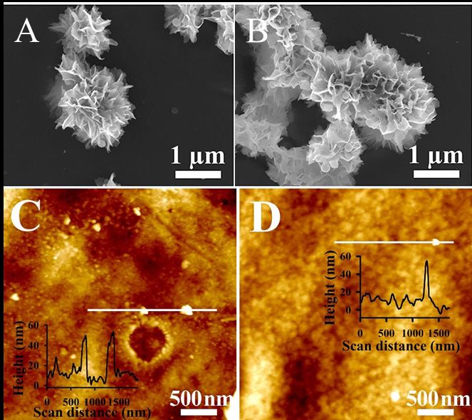
Polymers 2020, 12, 3061                                                                              2 of 26

polymeric hybrid materials can be prepared as “smart” materials, using which functionality can be
achieved through physical, chemical, or biological stimuli.
      Polymer coatings are increasingly popular in various diverse applications and segments.
From simple coatings to nanoparticle incorporated functionalized composite coatings, these polymer
coatings provide a strong functionalities to their host materials. It can be applied in various materials
of choice such as metals, ceramics, polymers and nanoparticles. In biomedical field, polymeric coatings
can play a vital role for the development of next generation biomaterials and instruments. They can be
applied as corrosion resistance, functionalize the surface, wear resistance, improve bioactivity and
even can be used as a switchable smart materials. Smart polymer coatings are the recent advancement
in the polymer coatings. Various reports indicated that, polymer are smart and have the capability
to respond to numerous stimuli, such as temperature, light, magnetic field, electric field and pH [1].
In the medical field, these smart polymers are mainly used in drug delivery applications where the
drugs can be loaded in these polymer or polymer coatings and can be delivered at the chosen location
with the aid of a stimuli. Further, shape memory polymers are gaining interest and are promising
candidates for bone tissue engineering applications.
      Another interesting aspect of the polymer coatings is the formation of the composite with other
polymers or inorganic compounds. The development of polymer-based composites are one of the main
approaches in resolving the glitches associated with the polymers in biomedical applications. Balanced
physical, chemical and biological properties can be achieved through blending the polymers with
other materials. After the emergence of nanotechnology, nanoparticle incorporation in with polymer
base materials provide improved functional characteristics. In drug delivery field, polymer based
nanocarriers are very promising owing to their ability for the encapsulation of drugs, controlled delivery,
sustained release and bioactivity. By incorporating suitable nanofillers, polymer nanocomposites can
be designed for versatile applications.
      This review aims to offer an overview of the latest progress in the biopolymer coatings and their
applications in various biomedical field, to highlight the most important results in the literature, and to
offer a potential outline for impending progress and perspective. Initially, brief overview of the polymer
coatings and their methods are described. Then, the short survey on the polymer coatings on metal
surfaces are described. Further, various biopolymer coatings and their application in the biomedical
fields are described separately. Finally, the application of biopolymer coatings on nanoparticles are
discussed. There are numerous reports available for biopolymer coatings and various polymers are
used for biomedical coatings. In this review, very recent research reports are taken into account and
some specific biopolymers are given emphasis based on their wide application and coating ability in
biomedical field along with some standout reports are provided.

2. Polymer Coatings and Films
     Polymer coating is an approach for surface property modification undertaken to satisfy the
requirements of multiple practical applications. It is a coating or paint produced using polymers with
better properties than existing ones. Polymer coatings have been used in various applications, such as
adhesion, scratch and abrasion resistance, corrosion resistance, wettability, and bioactivity. Polymer
coatings are considered highly useful in biomedical applications because they provide flexibility with
respect to the chemical groups that can be attached to surfaces, which are beneficial for biomaterial
and tissue interactions. Furthermore, their mechanical and elastic properties are comparable to those
of biological tissues. Various methods have been established and implemented for the fabrication
of polymer coatings for different applications. Highly efficient coatings with advanced properties
can be produced through a prudent choice of material, coating methods, and production parameters.
The inherent surface properties of polymers, such as poor wettability and low surface area, lead to
substandard bioactivity and make it challenging to use these in implants. Conversely, polymer-coated
implants can serve as biomimetic surfaces in the body [2]. Polymers can also be used to coat the surface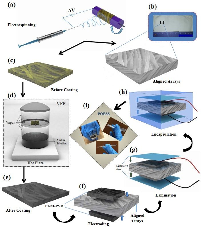
Polymers 2020, 12, 3061                                                                              3 of 26

of nanoparticles to improve their performance in the delivery of biomolecules and drugs. Polymer
coatings can be used to improve both hardness adjustment and component delivery.

3. Biopolymer Coating Methods
      There are various biopolymer coating materials available, which are well-documented in the
literature. Since this review specifically focuses on polymer coatings, this section briefly discusses
some of the main coating methods used for biopolymer coatings. The assembly of polymer into
coatings and films can be done various methods such as layer by layer (LBL) [3,4], polymer brushes,
dip coating, Langmuir-Blodgett (LB) [4–6], plasma based coating methods, spin coatings and hydrogels.
In LBL method, positively and negatively charged polyelectrolytes are coated successively. There are
number of charged polyelectrolytes are available to produce LBL film and coatings. The biggest
advantage of the LBL process is its flexibility to produce the polymer coatings. Recently Landry et al.
reviewed self-assembled layers and multilayer polymer coatings for tissue engineering applications [3].
LBL polymer films as drug delivery carriers was reviewed by Park et al. [4]. LB films are often
considered as alternate for LBL process in which the substrates are drawing at a steadily at a constant
speed from the polymer solutions. In LBL, molecules from the bulk solution are coated on the
substrate, whereas in LB, molecules from the solution surface are coated on the substrates. Leontidis
described about LB films and compared it with LBL method [5]. Polymer brush is another popular
and interesting surface modification technique in which soft material is covalently tethered on the
surface of the substrate [7] and has potential applications in various fields. Various researchers have
reviewed the applications of polymer brushes in biomedical field [7–9]. Plasma based polymer coatings
(especially non-thermal plasmas) are enable us to have controlled polymer coatings on any substrate
surfaces for various applications. This coating method provides strong adhesion to extensive range of
substrates, such as metals and ceramics. Further, this plasma coatings allows to coat even complex
shapes. Plasma based polymer coatings were reviewed and reported by various researchers [10–12].
Similarly, various other methods such as dip coating and spin coatings are used for polymer coatings.
These methods are simple, cost effective and the coating parameters can be changed easily. In a dip
coating process, the substrate is dipped into a polymer coating solution and kept in this solution
for some times, which allow substrate to absorb the polymer molecules. After that, the substrate
is withdraw from the solution and dried. Evan a large substrates can be uniformly coated by this
dip coating process. Various parameters such as solution viscosity, dipping time, drawing speed
and drying atmosphere. In spin coating, polymer solution is added dropwise in the center of the
substrate, which may be either still or set in to rotate in low speed. After that, the rotation of the
substrate is increased to high velocity to facilitate homogenous spreading of the polymer solutions on
the substrates with the combined effect of centrifugal force and surface tension. Here, the thickness of
the coating was achieved by rotation speed, viscosity and surface tension. Very recently, Song et al.
reviewed various biopolymer based coating methods [13].

4. Biopolymer Coatings on Metal Implants
     In multiple industrial applications, metal surfaces coated with organic layers provide numerous
advanced features with high potential for applicability. This technique permits the tailoring of
various characteristics, such as elasticity, wettability, bioactivity, and adhesiveness [14]. Biodegradable
polymer coatings can be applied as corrosion resistance coatings in implants to prevent corrosion post
implantation [2].
     Improved bioactivity, corrosion resistance, and antifouling properties were achieved for 316L
stainless steel (SS) by applying a pseudopeptide polymer coating. Liu et al. [15] used poly(2-
methyl-2-oxazoline) (PMOXA) as a pseudopeptide polymer to produce a non-brush bionic polymer
coating by electrochemical assembly on a 316L SS surface. bioactivity and anti-fouling properties were
observed for PMOXA with a modest degree of hydrolysis and molecular weight. Further, they found
that cell migration and proliferation were enhanced by these coatings. It was claimed that the coating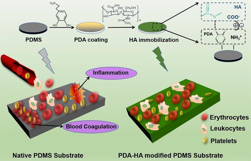
Polymers 2020, 12, 3061                                                                            4 of 26

could successfully modify the surface of the complex 3D vascular stent, which could have potential
applicability in the prevention of late stent thrombosis and in-stent restenosis [15].
     Gnedenkov et al. suggested a unique method for producing composite polymer coatings on the
surface of the magnesium alloy MA8 [16]. Significant improvements were reported in the protective
and antifriction properties of the magnesium alloy surfaces owing to the special treatment of plasma
electrolytic oxidation (PEO) coatings by super-dispersed polytetrafluoroethylene (SPTFE). Further,
the authors inferred that the thickness of the composite coating increased due to SPTFE treatment of
the PEO coating. The same group reported a similar PEO-based method for preparing hydroxyapatite-
polytetrafluoroethylene (PTFE) composite coatings on Mg–Mn–Ce alloys for use in resorbable
implants [17]. In this study, the elemental composition, phase, morphology, and multifunctional
corrosion resistance of the composite coatings were reported, and these were confirmed to impart
bioactivity to magnesium implants.
     Oosterbeek et al. attempted to produce polymer-bioceramic composite coatings on magnesium to
improve the corrosion resistance of magnesium to increase the degradation time [18]. The composite
coating was produced through a two-step process: Initial bioceramic coating by immersion in a
supersaturated calcium phosphate solution, followed by polylactic acid coating using a dip coating
process. Mechanical testing of this composite coating revealed that the polymer-only coating showed
greater adhesion strength compared to the polymer-bioceramic composite coating owing to the poor
bonding between the bioceramic layer and the substrate. The presence of corrosion resistance also
indicated that the polymer-only coating had a longer period than the other composite coatings.
     Recently, similar studies on conferring corrosion resistance and inducing biological properties in
magnesium alloys for implant applications were successfully implemented using calcium phosphate/
collagen (CaP/Col) composite coatings [19]. Chemical conversion and dip-coating methods were adapted to
fabricate composite coatings. The CaP/Col coating effectively reduced the degradation rate of magnesium
alloys and increased osteoblast adhesion in the optimal microenvironment and interface created.
     In the following section, we have reviewed specific biopolymer coatings as well as the coatings that
can be used to modify these polymer surfaces for better utilization in specific biomedical applications.

5. Biopolymer Coatings for Surface Modification

5.1. Polyvinylidene Fluoride (PVDF)
     PVDF is a polymer used widely in medical applications and has been studied extensively. It possesses
enhanced biological, textile, and piezoelectric properties, and it is a highly non-reactive thermoplastic
fluoropolymer [20,21]. Surgical meshes and sutures require non-reactivity, whereas wound-healing
applications require piezoelectricity [21]. Hence, this material is considered suitable for various
biomedical applications such as tissue engineering [22–27], physiological signal detection [28–32],
and antimicrobial and antifouling material development [22,33–38]. However, it is difficult to produce
coatings for biomedical applications using PVDF because it does not form smooth films and exhibits
issues in adhesion with other substrates. There have been reports of effective coating using PVDF
and its copolymers using the spin coating and Langmuir–Blodgett (LB) methods. Yin et al. reviewed
studies reporting the application of PVDF and its copolymer films using the spin coating and LB
methods [39]. Several studies have reported that PVDF, along with other materials, form a composite
or combination of materials that offer the advantages of both materials [22–24,28,29,31,33,35–39].
     Tien et al. [28] fabricated a flexible electronic skin (e-skin) that mimics the functions of the
human finger. The device was designed on top of a flexible platform with an array of pressure and
temperature sensor pixels, which could be used as a channel using an organic semiconductor (pentacene).
The gate dielectric material was produced from a mixture of poly(vinylidenefluoride-trifluoroethylene)
(P(VDF-TrFE)) and BaTiO3 nanoparticles. This device showed high sensitivity and could be used
successfully as e-skin. Hybrid ZnO nanoneedles and PVDF films were fabricated for applications
in wireless real-time pressure sensors to monitor the heart rate. These were highly sensitive and
Polymers 2020, 12, 3061                                                                                                                      5 of 26

wearable, and could detect pressure as low as 4 Pa [31]. A similar type of temperature-sensing material
with high thermal responsivity, stability, and reproducibility was constructed by Trung et al. [40].
This temperature sensor used reduced graphene oxide (R-GO) instead of BaTiO3 and formed a
nanocomposite with P(VDF-TrFE, (R-GO/P(VDF-TrFE)), which acted as a sensing layer. This sensor
was reported to be mechanically flexible, optically transparent, and highly responsive to temperature
changes (could detect temperature changes as low as 0.1 ◦ C).
     Apart from the PVDF coating, electrospinning of PVDF, and its copolymers and composites with
different nanomaterials can be performed to utilize advantages of both materials. These composites
are mostly used in energy harvesting and environmental remediation applications [41–44]. In recent
times, only a limited number of reports have been published on their applications in the biomedical
field. In a recent report, a self-powered piezo-organic-e-skin sensor was constructed using highly
aligned PVDF nanofibers as piezoelectric active components and polyaniline-coated PVDF NF mats as
flexible electrodes (Figure 1) [45]. This sensor was used for human health monitoring, and exhibited
remarkable sensitivity to human finger touch (10 V under 10 kPa) by converting mechanical energy into
electric energy. It was also designed to monitor various human gestures such as bending, stretching,
compression, movement, coughing, and swallowing.
                        Polymers 2020, 12, x FOR PEER REVIEW                                                                       6 of 28

                             Figure 1. (a) electrospinning process, (b) photograph of the large-scale prepared mat of highly aligned
      Figure 1. (a) electrospinning           process,
                         PVDF NFs arrays; enlarged   view (b)    photograph
                                                           exhibits                 of respective
                                                                    the structure from  the large-scale          prepared
                                                                                                  section, (c) structure   of mat of highly
                         oxidant-contained yellowish PVDF NFs mat before PANI coating, (d) VPP process, (e) structure of
      aligned PVDF NFs      arrays;    enlarged      view    exhibits     the  structure     from    respective        section,
                         deepbluish PVDF NFs mat after PANI coating, (f) electrode assembling, (g) lamination process, (h)
                                                                                                                                (c) structure
      of oxidant-contained
                         PDMS yellowish
                                encapsulation ofPVDF     NFs (i)
                                                 POESS design,   mat   beforeofPANI
                                                                  photographs              coating,
                                                                                  POESS with            (d) VPP
                                                                                              demonstration           process, (e) structure
                                                                                                              of flexibility.
                         Schematic illustration of the piezo-organic-e-skin sensor design architecture. (Reprinted with
      of deepbluish PVDF       NFs mat after PANI coating, (f) electrode assembling, (g) lamination process,
                         permission from [45] Copyright (2020), American Chemical Society.).
      (h) PDMS encapsulation of POESS design, (i) photographs of POESS with demonstration of flexibility.
                         Electrospun PVDF-nanosilica scaffolds were prepared, and their mechanical and piezoelectric
      Schematic illustration   of the piezo-organic-e-skin sensor design architecture. (Reprinted with permission
                     properties were studied for biomedical applications by Haddadi et al. [46]. Hydrophilic and
                     hydrophobic
      from [45] Copyright         silica American
                              (2020),    nanoparticles Chemical
                                                        were used to Society).
                                                                       construct the composite fiber, and the average fiber
                        diameter was increased by nanoparticle addition. Hydrophilic silica nanoparticles showed higher
                        tensile strength compared to other fibers owing to their higher dispersion and compatibility. The
                        piezoelectric property was enhanced upon the addition of silica nanoparticles; however, the addition
                        of hydrophilic silica led to an increase in the output voltage [46].
Polymers 2020, 12, 3061                                                                                                      6 of 26

      Electrospun PVDF-nanosilica scaffolds were prepared, and their mechanical and piezoelectric
properties were studied for biomedical applications by Haddadi et al. [46]. Hydrophilic and hydrophobic
silica nanoparticles were used to construct the composite fiber, and the average fiber diameter was
increased by nanoparticle addition. Hydrophilic silica nanoparticles showed higher tensile strength
compared to other fibers owing to their higher dispersion and compatibility. The piezoelectric property
was enhanced upon the addition of silica nanoparticles; however, the addition of hydrophilic silica led
to an Polymers
       increase   in12,
               2020, the   output
                        x FOR PEER voltage
                                    REVIEW [46].                                                                7 of 28
      The blending of PVDF with a conducting polymer is a method for increasing electrical output
from PVDF.  TheSengupta
                 blending of  etPVDF
                                 al. [47]with a conducting
                                           prepared   PVDFpolymer
                                                            blendsiswith
                                                                       a method
                                                                            various for polymers
                                                                                        increasing electrical output
                                                                                                    (polypyrrole      (PPy),
      from PVDF.
polyaniline    (PANI),Sengupta
                          and a et   al. [47] prepared
                                  modified             PVDF
                                               PANI with     blends with
                                                          L-glutamic       various
                                                                        acid          polymers
                                                                              (referred    to as (polypyrrole  (PPy), to
                                                                                                 PANILGA/P-LGA))
obtainpolyaniline
         different (PANI),     and aactive
                     electrically     modified  PANI with L-glutamic
                                             membranes.    Bioactivity,acid (referredconductivity,
                                                                         electrical     to as PANILGA/P-LGA))        to
                                                                                                        β-phase content,
      obtain different electrically active membranes. Biocompatibility, electrical conductivity, β-phase
and the nanostructures formed were analyzed, and bioactivity was observed to decrease in the
      content, and the nanostructures formed were analyzed, and biocompatibility was observed to
following order: p-LGA/PVDF > PANI/PVDF > PPy/PVDF > PVDF. Furthermore, while P-LGA/PVDF
      decrease in the following order: p-LGA/PVDF > PANI/PVDF > PPy/PVDF > PVDF. Furthermore,
exhibited
      whilehigher      bioactivity
              P-LGA/PVDF              and higher
                                exhibited   electrical conductivity,
                                                   biocompatibility   it electrical
                                                                    and  also exhibited      high cytotoxicity
                                                                                     conductivity,                 toward
                                                                                                    it also exhibited
HeLahigh(cancer)   cells   (Figure   2).  Hence,  this composite    material   can   be  of interest  in
             cytotoxicity toward HeLa (cancer) cells (Figure 2). Hence, this composite material can be of certain  specific
      interestapplications.
biomedical      in certain specific biomedical applications.

      FigureFigure 2: Viability
              2. Viability      of (a)
                            of (a)     HeLa
                                    HeLa       cells
                                            cells    and(b)
                                                   and    (b)MC3T3
                                                              MC3T3 cells
                                                                      cells on
                                                                            onPVDF
                                                                                PVDFandandPVDF:CP
                                                                                             PVDF:CP  electrospun fibersfibers
                                                                                                         electrospun
            (control:
      (control: PVDF).PVDF).
                         ** and** ****
                                   and signifies
                                        **** signifiesp<
                                                   p < 0.010.01
                                                             (1d)(1d)
                                                                   andandp<   0.0001(1d),
                                                                       p < 0.0001          respectively, for
                                                                                    (1d), respectively,  for both
                                                                                                             both HeLa
                                                                                                                  HeLa and
      MC3T3 andMC3T3      culture
               culture (1d);  #### (1d);     ####
                                     signifies  p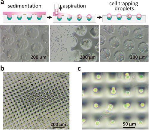
Polymers 2020, 12, 3061 7 of 26
studied. The P(VDF-TrFE) implant provided a maximum voltage and current of 6 mV and 6 nA,
respectively. Furthermore, the fibroblasts proliferated and aligned seamlessly along the electrospinning
direction of the nanofibers, and proliferation was observed to be enhanced by 1.6-fold. Based on this,
the authors claimed that the P(VDF-TrFE) scaffold could be used as a suitable tissue engineering and
wound healing material [48].
5.2. Polymethyl Methacrylate (PMMA)
PMMA is a synthetic polymer that is lightweight, cost effective, easy to manipulate, and contains
harmless subunits; these properties make it suitable for usage in biomedical applications. It is
broadly used for various medical applications, such as drug delivery, as well as in tools such as bone
cement and microsensors [49]. PMMA is also the material of choice for the production of denture
base, orthodontic retainers, and repair in dentistry [49,50]. It exhibits good mechanical properties,
slow degradation, low toxicity, and inertness. Due to these properties, it is widely used in hip-joint
transplantation. Its non-biodegradable nature makes it suitable for the construction of permanent
and mechanically stable structures, such as those used in bone tissue engineering [49,51]. The issues
associated with the coating of organic materials on metal surfaces include poor adhesion between
these two components. To address this, polymers can be covalently anchored to the substrate surface
to generate an adhesive interlayer.
A 1–2-µm PMMA layer was incorporated on a Ti substrate through alkali activation of the
surface [52]. This was achieved through the initial alkali activation of the Ti substrate followed by
surface-initiated atom transfer radical polymerization. This was performed in a heated NaOH solution.
This treatment produced a porous Ti layer rich in hydroxyl groups. Next, using phosphonic acid as the
coupling agent, the polymerization initiator was covalently grafted onto the surface [53]. The coating
was approximately of 1.9 µm and was stable in a simulated body solution. Additionally, it exhibited
good bioactivity [52]. This method can pave the way for hybrid prosthesis using personalized
medicine. In another study, the same method was used to develop a TI/PMMA/Ti sandwich structure
and study their adhesion and formability. A high bonding strength and optimal formability were
achieved. The results showed that there was no failure or delamination between the Ti and PMMA
interfaces. Hence, this type of coating and adhesion method will be advantageous in future biomedical
applications. Furthermore, the same authors reported the production of a hybrid Ti/PMMA-layered
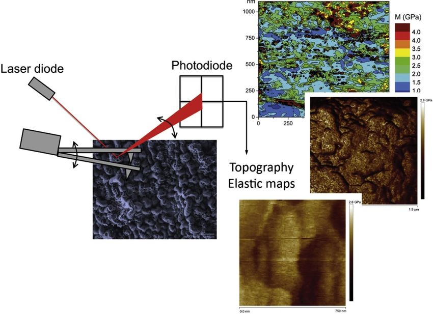
material and analyzed multiple mechanical characterizations using the same [14]. The mechanical
characterization of the thick PMMA layers on Ti substrates was performed using nanoindentation as
well as different atomic force microscopy techniques. Each of these methods indicates the mechanical
properties at different scales (Figure 3).
Biomaterial modelling for optimized methacrylate coating for Ti implant was proposed by
Sun et al. [54]. They applied cheminformatics methods to methacrylated proteins to estimate their
suitability as Ti implants coatings. They found that the bioactivity of Ti implants was higher than that
of uncoated samples when methacrylated proteins such as GelMA were used. In addition, this coating
was less susceptible to biofilm formation, which reduced the risk of osteomyelitis, which ultimately
leads to implant fixation. The development of hybrid nanoparticle coating derived from bio-polymer
was reported by Galvão et al. [55]. PMMA nanoparticles were synthesized in the presence of
poly(diallyldimethyl ammonium) chloride (PDDA) by emulsion polymerization. The antimicrobial
coating was created by spin coating or casting and drying of the nanoparticle dispersion using different
substrates such as Si, glass, or polystyrene sheets. At its highest relative content, PDD:PMMA mostly
produced homogeneous coatings. The presence of PDDA in the coatings significantly inhibited
bacterial activity, which was tested in Escherichia coli and Staphylococcus aureus. The coatings were
suggested to be suitable for different biomedical applications.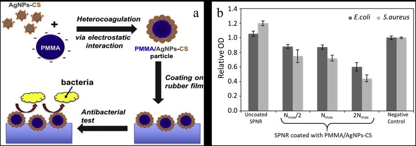
Polymers 2020, 12, 3061                                                                                              8 of 26

Polymers 2020, 12, x FOR PEER REVIEW                                                                                 9 of 28

      Figure 3.
      Figure 3. Multiscale
                Multiscale mechanical
                           mechanical analysis
                                      analysis of
                                                of polymethyl
                                                   polymethyl methacrylate
                                                               methacrylate layers
                                                                            layers grafted
                                                                                   grafted on
                                                                                           on Ti
                                                                                              Ti substrates.
                                                                                                 substrates.
      (Reprinted with
      (Reprinted with permission
                      permission from
                                  from [14]
                                        [14] Copyright
                                             Copyright (2017),
                                                        (2017),Elsevier).
                                                               Elsevier).

     Biomaterial
     The   deposition modelling     for optimized methacrylate
                          of PMMA/chitosan-silver                   coating for Tinanoparticles
                                                          (PMMA/AgNPs-CS)           implant biocompatibility
                                                                                                   on a soft rubberwas
proposed by Sun et al. [54]. They applied cheminformatics methods to methacrylated proteins to
substrate    was     achieved     by   immersion      method     which   improved     antibacterial   activity    [56].
estimate their
Positively        suitability
             charged           as Ti implants
                          AgNPs-CS      (38 nm)coatings.    They found thaton
                                                   was heterocoagulated       thethe
                                                                                  biocompatibility    of Ti implants
                                                                                      negatively charged      PMMA
was higher
cores (496 nm)thantothat   of uncoated
                        produce            samples whenwhich
                                   PMMA/AgNPs-CS,           methacrylated
                                                                   was thenproteins
                                                                             deposited such
                                                                                          on as
                                                                                              theGelMA
                                                                                                  rubberwere    used.
                                                                                                           substrate.
In addition, this coating was less susceptible to biofilm formation, which reduced the risk 4).
Antibacterial    activity   toward    E. coli and  S. aureus   was  amplified  on  the coating   surface  (Figure    of
osteomyelitis,the
Furthermore,      which    ultimately
                       cytotoxicity      leads to
                                      of L-929     implantcells
                                                fibroblast   fixation. The reduced
                                                                  was also  development     of hybrid
                                                                                     by these          nanoparticle
                                                                                                coatings.  However,
coatings derived
inhibition             fromfibroblast
            of the L-929     biocompatible     andnot
                                        cells was   antibacterial  polymers
                                                      observed. This   studywere
                                                                              showedreported   by Galvão
                                                                                        that these types ofetcoatings
                                                                                                              al. [55].
PMMA
can      nanoparticles
    be applied              were
                   to various      synthesized
                                soft  substratesininthe presence
                                                     different     of poly(diallyldimethyl
                                                                biomedical  applications. ammonium) chloride
  Polymers 2020, 12, x FOR PEER REVIEW                                                                      10 of 28
(PDDA) by emulsion polymerization. The antimicrobial coating was created by spin coating or
casting and drying of the nanoparticle dispersion using different substrates such as Si, glass, or
polystyrene sheets. At its highest relative content, PDD:PMMA mostly produced homogeneous
coatings. The presence of PDDA in the coatings significantly inhibited bacterial activity, which was
tested in Escherichia coli and Staphylococcus aureus. The coatings were suggested to be suitable for
different biomedical applications.
     The deposition of PMMA/chitosan-silver (PMMA/AgNPs-CS) nanoparticles on a soft rubber
substrate was achieved by immersion method which improved antibacterial activity [56]. Positively
charged AgNPs-CS (38 nm) was heterocoagulated on the negatively charged PMMA cores (496 nm)
to produce PMMA/AgNPs-CS, which was then deposited on the rubber substrate. Antibacterial
activity toward E. coli and S. aureus was amplified on the coating surface (Figure 4). Furthermore, the
cytotoxicity of L-929 fibroblast cells was also reduced by these coatings. However, inhibition of the
L-929 fibroblast cells was not observed. This study showed that these types of coatings can be applied
        Figure 4. (a) Schematic illustration of polymethyl methacrylate/chitosan-silver (PMMA/AgNPs-CS)
to various
     Figuresoft
             4. (a)substrates
                     Schematicin    differentof
                                 illustration  biomedical
                                                 polymethylapplications.
                                                              methacrylate/chitosan-silver (PMMA/AgNPs-CS)
        coating and its antibacterial activity; (b) optical density of suspension of Escherichia coli and
      coating and its antibacterial activity; (b) optical density of suspension of Escherichia coli and Staphylococcus
        Staphylococcus aureus. (Reprinted with permission from [56] Copyright (2019), Elsevier).
      aureus. (Reprinted with permission from [56] Copyright (2019), Elsevier).

   5.3. Polypropylene (PP)
       PP has been widely used in medical applications, especially as a surgical mesh to strengthen
   weakened tissues. PP is a thermoplastic polymer with different densities and can be classified into
   copolymer and homopolymer components. In the biomedical field, PP mesh has been extensively
   used in urogynecology [57] and hernia repair owing to specific characteristics, such as inertness,
   hydrophobicity, and strong mechanical properties, even while being lightweight [58]. It also finds
   applications in other areas of medicine such as breast reconstruction or as a supportive soft tissue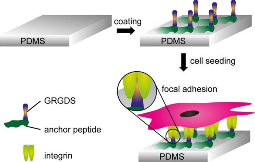
Polymers 2020, 12, 3061                                                                              9 of 26

5.3. Polypropylene (PP)
      PP has been widely used in medical applications, especially as a surgical mesh to strengthen
weakened tissues. PP is a thermoplastic polymer with different densities and can be classified into
copolymer and homopolymer components. In the biomedical field, PP mesh has been extensively
used in urogynecology [57] and hernia repair owing to specific characteristics, such as inertness,
hydrophobicity, and strong mechanical properties, even while being lightweight [58]. It also finds
applications in other areas of medicine such as breast reconstruction or as a supportive soft tissue
structure and blood oxygenator membrane. In addition, it exhibits low potential of carcinogenesis in
the human body [59]. However, certain complications are also associated with its use, including the
induction of infections and inflammatory responses within the body, which lead to a slow healing
process, insufficient drug absorption, and immune system response. Due to its high hydrophobicity,
it exerts adverse effects such as tissue damage and insertion resistance in the human body occasionally.
Hence, even though PP is a good material, its application is limited owing to its poor biological
properties. Therefore, to use PP in medical applications in the human body, surface treatment for
improving bioactivity is necessary.
      Recently, Saitaer et al. [58] reported the surface modification of a PP hernia mesh using polydopamine
(PDA) modified with cold oxygen plasma. This modification led to improved drug absorption and
longer release, and also imparted antibacterial properties. Plasma treatment is one of the best methods
conventionally used for surface modification for inducing chemical functionality and surface charge
and for increasing surface hydrophilicity [60]. Plasma-enhanced chemical vapor deposition (PECVD)
was used to modify the surface of PP implants with different chemicals to produce charged PP
substrates for layer-by-layer (LBL) coatings [60]. This PECVD method increased hydrophilicity and
the number of functional reactive groups available for molecule grafting, and was found to be suitable
for LBL deposition on PP substrates.
      To tackle the high hydrophobicity of the PP surface, Jang et al. [61] developed a matrix combining
polyvinyl pyrrolidone (PVP) and cross-linked polyethyleneglycolacrylate (PEGDA) to produce a
stable hydrogel forming layer (PVP:PEGDA) that exhibited hydrophilicity and bioactivity. This study
revealed that the hydrophilic nature of the film improved, and the mechanical as well as adhesive
strength of the PP surface could be optimized by adjusting the PVP and PEGDA ratio. Compared
to the PVP film, the combination of the PVP:PEGDA films showed 7-fold higher tensile strain at the
breaking point, and 54-fold higher adhesion strength, respectively. This type of surface modification
can be useful for the development of PP medical products.
      Cross-linked poly(styryl bisphosphonate) (poly(StBP)) thin coatings (thickness: 163 ± 8 and
175 ± 7 nm) were applied to corona-treated PP films by UV curing for bone tissue engineering
applications by Steinmetz et al. [62]. Initially, poly(StBP) nanoparticles were prepared and mixed with
poly(ethylene glycol) dimethacrylate and a photo-initiator. Next, they were spread on the PP film
and cured using UV radiation. The authors observed that the poly(StBP) nanoparticle-embedded
coating induced apatite crystal growth (Figure 5), which resulted from the strong affinity of poly(StBP)
toward calcium ions. Furthermore, the coating exhibited durability and optical properties. The authors
claimed that this coating method could be useful in various bone tissue engineering applications.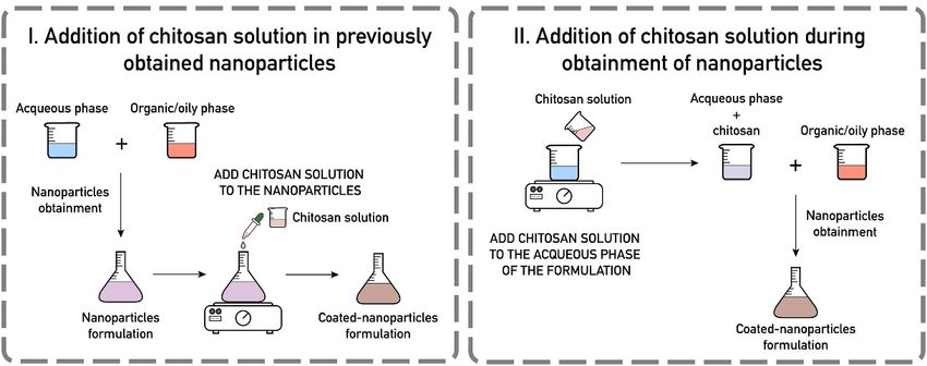
film and cured using UV radiation. The authors observed that the poly(StBP) nanoparticle-embedded
coating induced apatite crystal growth (Figure 5), which resulted from the strong affinity of poly(StBP)
toward calcium ions. Furthermore, the coating exhibited durability and optical properties. The
authors claimed
Polymers 2020, 12, 3061that this coating method could be useful in various bone tissue engineering10 of 26
applications.
Figure 5. Scanning
Figure 5. Scanningelectron
electron microscopy
microscopy (A,B)(A,B) and atomic
and atomic force microscopy
force microscopy (C,D)of images
(C,D) images of
poly(styryl
poly(styryl bisphosphonate) (poly(StBP))-6, and poly(StBP)-40 (D) films, respectively, where
bisphosphonate) (poly(StBP))-6, and poly(StBP)-40 (D) films, respectively, where “6” and “40” represent“6” and
“40” representofthe
the thickness thethickness
Mayer rod of (6
theand
Mayer rodused
40 µm) (6 and 40 µm)the
to spread used to spread
polymer the polymer
solution. solution.
(Reprinted with
(Reprinted
permissionwithfrompermission from(2020),
[62] Copyright [62] Copyright
Elsevier).(2020), Elsevier).
5.4.
5.4. Polydimethylsiloxane
Polydimethylsiloxane (PDMS)
(PDMS)
PDMS
PDMS is is aa synthetic
synthetic material
material that
that is
is used
usedextensively
extensively in invarious
variousbiomedical
biomedical tools
tools such
such asassurgical
surgical
implants, catheters,contact
implants, catheters, contactlenses,
lenses, pacemaker
pacemaker encapsulants,
encapsulants, and and biosensors,
biosensors, as wellasaswell as in
in drug drug
delivery
delivery and DNA sequencing owing to its excellent properties, such as biocompatibility, greater
and DNA sequencing owing to its excellent properties, such as bioactivity, greater flexibility, ease of
flexibility,
fabrication,ease
oxygen of fabrication,
permeability, oxygen
opticalpermeability,
transparency,optical
and low transparency, andFurthermore,
toxicity [63,64]. low toxicity it[63,64].
can be
Furthermore,
used as an ideal it organ-on-chip
can be used assubstrate
an idealtoorgan-on-chip
study stem cellsubstrate
behavior. toThe study stem cell of
characteristics behavior. The
this material
characteristics of this material make it a suitable candidate for studying cell activities, such as
make it a suitable candidate for studying cell activities, such as topography, stretching, and mechanical
topography,
and electricalstretching,
stimulations andformechanical
designingand electrical
materials for stimulations for designing
tissue engineering materials
applications [63]. for tissue
However,
engineering applications [63]. However, the interaction of PDMS with cells is limited, which
the interaction of PDMS with cells is limited, which necessitates the modification of its surface
necessitates
characteristicsthetomodification of its surface
achieve the desired characteristics
properties. Similar toto PP,achieve
plasmathe desired properties.
treatments can be usedSimilar
to modifyto
PP,
the plasma
surface treatments
of PDMS by canthebecreation
used to of modify the surface
hydroxyl groups.of PDMS by the creation of hydroxyl groups.
Gehlen
Gehlen et et al.
al. [63]
[63] reported
reported aa novel,
novel, one-step
one-step PDMS
PDMS coating
coating method
method using
using engineered
engineered anchor
anchor
peptides
peptides fused
fused to to aa cell-adhesive
cell-adhesive peptide
peptide sequence
sequence (glycine-arginine-glycine-aspartateserine).
(glycine-arginine-glycine-aspartateserine). In In this
this
method,
method, hydrophobic
hydrophobicinteractions
interactionswere wereused usedtotoattach
attach thethe
anchor
anchorpeptides
peptidesto the PDMS
to the PDMS surface by
surface
dipping the PDMS substrates in an anchor peptide solution (Figure 6). Binding performance, cell
by dipping the PDMS substrates in an anchor peptide solution (Figure 6). Binding performance,
attachment
cell attachmentof fibroblasts,
of fibroblasts,andand endothelial
endothelial cells were
cells were studied,
studied,and andthethecoating
coating conditions
conditions were were
optimized. The authors claimed that this method employed mild conditions and room temperature,
optimized. The authors claimed that this method employed mild conditions and room temperature,
and
and could
could bebeeasily
easilyusedusedto tofunctionalize
functionalizebiomedical
biomedicaldevices
deviceswithwithsensitive
sensitiveandandcomplex
complexcomponents.
components.
Ultralow friction was established in an aqueous environment for using PDMS by bonding of
a poly(acrylamide–acrylic acid) hydrogel coating on the surface of the substrate [65]. Bonding was
achieved through chemical modification of the PDMS surface and successive reaction with acrylic
acid moieties. The product reduced friction by two orders of magnitude in an aqueous environment,
which had a friction coefficient (µ) as low as 0.003 [65]. A chlorhexidine (CHX)-loaded PDMS-based
coating was applied on the surface of a 3D-printed dental polymer to induce surface wettability,Polymers
Polymers 2020, 2020, 12, x FOR PEER REVIEW
12, 3061 12 of 28
11 of 26
microstructure, and antibacterial activity [66]. CHX was encapsulated in silica nanoparticles and added
to PDMS to produce an antibacterial coating material. This was coated as a thin film on a 3D printed
specimen using oxygen plasma and by subsequent heat treatment. This coating eventually increased
the surface hydrophobicity and reduced the irregularities. Furthermore, it notably reduced bacterial
colony formation compared to that in the uncoated samples.
Polymers 2020, 12, x FOR PEER REVIEW 12 of 28
Figure 6. Schematic illustration of polydimethylsiloxane (PDMS) coating using engineered anchor
peptides fused to the cell-adhesive peptide sequence (glycine-arginine-glycine-aspartateserine,
GRGDS). (Reprinted with permission from [63] Copyright (2019), American Chemical Society).
Ultralow friction was established in an aqueous environment for using PDMS by bonding of a
poly(acrylamide–acrylic acid) hydrogel coating on the surface of the substrate [65]. Bonding was
achieved through chemical modification of the PDMS surface and successive reaction with acrylic
acid moieties. The product reduced friction by two orders of magnitude in an aqueous environment,
which had a friction coefficient (µ) as low as 0.003 [65]. A chlorhexidine (CHX)-loaded PDMS-based
coating was applied on the surface of a 3D-printed dental polymer to induce surface wettability,
Figure
Figure 6. Schematic
6. Schematic
microstructure, illustration
and illustration
antibacterialofofactivity
polydimethylsiloxane (PDMS)
[66]. CHX was (PDMS)
polydimethylsiloxane coating
encapsulated
coating using
in using engineered
silica engineered
nanoparticles anchor
and
anchor
peptides
peptides fusedto to
added tofused
PDMS to produce
the the cell-adhesive
cell-adhesive peptidepeptide
an antibacterial sequence
coating
sequence (glycine-arginine-glycine-aspartateserine,
material. This was coated as a thin filmGRGDS).
(glycine-arginine-glycine-aspartateserine, on a 3D
GRGDS). (Reprinted
printed specimen
(Reprinted withoxygen
using
with permission permission
fromplasma fromand[63]
[63] Copyright by Copyright
subsequent (2019), American
heat treatment.
(2019), American Chemical
This coating
Chemical Society). Society).
eventually
increased the surface hydrophobicity and reduced the irregularities. Furthermore, it notably reduced
Ultralow
bacterial
PDMS with friction
colony washemocompatibility
formation
enhanced established
compared to in that
an aqueous
in
was environment
thedeveloped
uncoated samples. for using
for medical implantPDMS by bonding
or device of a
application
poly(acrylamide–acrylic
by PDMSthe
modifying with acid)
enhanced
surface using hydrogel coating
hemocompatibility
a PDA and on
wasthedeveloped
hyaluronic surface of
forthe
acid (HA/PDA) substrate
medical [65].[67],
implant
composite Bonding
or device
as shownwas
in application
achieved
Figure through by modifying
7. Enhanced chemical the surface using
modification
hemocompatibility of awas
PDA
the and hyaluronic
PDMS
observedsurface aacid
for and (HA/PDA)
successive
particular composite [67], as
reactioncomposition,
HA/PDA with acrylic
usingshown
which in platelet
acid moieties. Figure 7. Enhanced
The product
adhesion hemocompatibility
reducedand friction
activationby two was
were observed
orders forcompared
a particular
of magnitude
reduced, intoHA/PDA
an aqueous
that composition,
observedenvironment,
in other
using which platelet adhesion and activation were reduced, compared to that observed in other
which had a friction
combinations of PDMS coefficient
and HA(µ)orasPDA low coatings.
as 0.003 [65]. A chlorhexidine
Furthermore, it was(CHX)-loaded
observed thatPDMS-based
along with
combinations of PDMS and HA or PDA coatings. Furthermore, it was observed that along with
coating was appliedanti-inflammatory
hemocompatibility, on the surface ofeffects a 3D-printed dental polymer
and cytotoxicity could also to induce
be alteredsurface wettability,
by adjusting the
hemocompatibility, anti-inflammatory effects and cytotoxicity could also be altered by adjusting the
microstructure,
HA and
HA PDA
and PDA and antibacterial
composition
composition ononthe activity
PDMS
the PDMSsurface.[66]. CHX
surface. Thesewas
These encapsulated
advantages
advantages cancan
be be in silica
useful
useful fornanoparticles
for the the development
development and
added
of to
medical PDMS
implants to produce
and an
devices.
of medical implants and devices. antibacterial coating material. This was coated as a thin film on a 3D
printed specimen using oxygen plasma and by subsequent heat treatment. This coating eventually
increased the surface hydrophobicity and reduced the irregularities. Furthermore, it notably reduced
bacterial colony formation compared to that in the uncoated samples.
PDMS with enhanced hemocompatibility was developed for medical implant or device
application by modifying the surface using a PDA and hyaluronic acid (HA/PDA) composite [67], as
shown in Figure 7. Enhanced hemocompatibility was observed for a particular HA/PDA composition,
using which platelet adhesion and activation were reduced, compared to that observed in other
combinations of PDMS and HA or PDA coatings. Furthermore, it was observed that along with
hemocompatibility, anti-inflammatory effects and cytotoxicity could also be altered by adjusting the
HA and PDA composition on the PDMS surface. These advantages can be useful for the development
of medical implants and devices.
Figure 7. Schematic illustration of polydimethylsiloxane (PDMS) surface modification using hyaluronic
acid and polydopamine (HA/PDA) composite coatings. (Reprinted with permission from [67].
Copyright (2017), American Chemical Society).Polymers 2020, 12, 3061 12 of 26
Another interesting and single-step surface modification for producing a long-lasting hydrophilic
surface was performed using microwell arrays, as reported by Oyama et al. [68]. In this method,
a low-energy electron beam (55 kV) was used to irradiate PDMS films in an air-produced silica-like
layer with a thickness of 40 µm. This modified surface showed prolonged hydrophilicity for more than
10 months in aqueous medium. These microwells were able to trap cells/single cells and provide stable
and promising cell adherent environments (Figure 8). Since no chemical was used in this method
for surface modification, the intrinsic bioactivity of PDMS was retained. This notable and promising
result revealed that the platform could be interesting in lab-on-chip applications, medical applications,
drug screening, and stem cell studies [68].
Polymers 2020, 12, x FOR PEER REVIEW 15 of 28
Figure 8.
Figure 8. Schematic
Schematic illustration
illustration of
of the
the cell trapping mechanism
cell trapping mechanism in in polydimethylsiloxane
polydimethylsiloxane (PDMS)
(PDMS)
microwells and the corresponding micrographs in (a) 200 µm and (b) 35 µm square microwell arrays.
microwells and the corresponding micrographs in (a) 200 µm and (b) 35 µm square microwell arrays.
(c) Single-cell
(c) Single-cell trapping
trapping demonstrated
demonstrated using
using aa combination
combination ofof bright
bright field
field microscopy
microscopy and
and fluorescence
fluorescence
imaging. (Reprinted
imaging. (Reprinted with
with permission from [68]
permission from [68] Copyright
Copyright (2018),
(2018), American
American Institute
Institute of
of Physics).
Physics).
In another study, Mahmoodi et al. [69] reported the surface modification of PDMS using PTFE
coatings to avert the absorption and adhesion of solvents onto PDMS microchannel walls, which can
be used for the encapsulation of anti-inflammatory drugs. The results showed that after the coating,
the microchannels exhibited super hydrophobicity (140.30◦ ), which effectively prevented the adhesion
and absorption of solvents by the drug-loaded nanoparticles. Furthermore, the drug release and
encapsulation efficiency were favorably altered by the coatings without any toxicity.
5.5. Polyurethane (PU)
Among synthetic polymers used in medical applications, PU is used only a small fraction
in spite of its application in various fields. PU coatings have significant uses in various fields,
including biomedical applications. In the medical field, it is primarily used to manufacture pacemaker
lead coatings, breast implant coatings, and vascular devices. In recent times, PU has garnered
Figure attention
significant 9. Different approaches
owing of chitosan coatings
to its bioactivity, for nanoparticles.
biodegradability, (Reprinted
and adaptive with permission
physical and chemical
forms.fromFurthermore,
[90] Copyrightits (2020), Elsevier).
physical and mechanical characteristics are similar to those of natural
tissues [70,71]. PUs consist of alternating hard and soft segments (HS and SS). The SS has an elastomeric
Collagen
character, is another
whereas the HSnatural polymer and
offers additional is theowing
strength richesttoconstituent
the hydrogenof ECM. As expected,urethane
bond-containing coating
a biomaterial
linkages with
[72,73]. Itscollagen can improve
biodegradability, its biocompatibility,
physicochemical its and
properties, ability to form
other an interface
properties between
can be adjusted
thealtering
by host tissue and the
the ratio implants.
between Various
the SS and HSreports provedchemical
components, that cellcomposition,
proliferation,and
differentiation
molar weightand
of
adhesion as well as new tissue formation was improved with the collagen coatings [91,92]. The cell
spreading and growth can greatly influenced by the solvent used to prepare the collagen coatings
[93]. In order to increase the mesenchymal stem cell (MSCs) adhesion, survival and proliferation
collagen coatings were tried and found to be effective [92]. A combination of collagen coatings on
chitosan shown to promote cell attachment and distribution [94].Polymers 2020, 12, 3061 13 of 26
PU in specific applications [70,74]. Joseph et al. reviewed the biomedical applications of PU and its
coatings [75].
New biodegradable freestanding PU films were produced without using any catalyst by Barrioni
et al. [70]. The authors developed an HS using hexamethylene diisocyanate and glycerol and an SS
using poly(caprolactone) triol and low-molecular-weight PEG. A highly homogeneous PU structure
with an interconnected network was formed. The deformation at break reached 425.4%, and the elastic
modulus and tensile strength were 1.6 MPs, and 3.6 MPa, respectively.
Bacterial resistance PU coatings for medical devices were developed by Roohpour et al. [76].
To inhibit microbial film formation, silver lactate and silver sulfadiazine were capped with the polymer.
The silver ions were found to be covalently bonded with PU without affecting its mechanical properties,
while an adequate bactericidal effect was exerted even when the silver content was low. This material
can be used for developing medical device coatings and associated applications. In another study,
the water resistance and bioactivity of PU were improved by the addition of isopropyl myristate,
which modified the hydrophobicity of PU [77]. The surface properties of PU changed and its surface
energy was reduced. The modified PU exhibited considerably lower water permeability than the silicon
packing materials available currently. This may be considered a suitable material for electronic implants.
Recently, PUs for medical implants and devices have been prepared by 3D printing, and the latest
developments in the 3D printing of PU in biomedical fields have been reported by Griffin et al. [78].
Similarly, a review on bio-based PU for biomedical applications was published by Wendels et al. [79].
Apart from regular coatings, PU/graphene based electrospun nanocomposite fibers were reported to
increase the electroconductivity, bioactivity and mechanical properties [80].
6. Other Biopolymer Coatings
Apart from the polymers mentioned above, various other biopolymers such as poly lactic acid
(PLA), Poly (lactide-co-glycolic) acid (PLGA), Polycaprolactone (PCL), polyethylene (PE) and some
natural polymers such as collagen and chitosan are also used for various biomedical applications.
PLA is recyclable, hydrophobic aliphatic polyester used for various biomedical applications, such as
medical devices, tissue engineering, drug delivery and 3D printed scaffolds. ZnO nanoparticles
embedded in PLA was dip coated on Mg alloy (AZ31), which helped to control the degradation and
promote antibacterial activity [81]. Incorporation of ZnO in PLA matrix provide control over surface
topography and Mg degradation rate. A free-standing PLLA micro-chamber array was developed
by dip coat PLLA solution on PDMS stamp followed by drug loading and sealing with pre-coated
polymer substrate, for drug delivery application [82]. A low frequency ultrasound trigger the release
of drug and in vitro test revealed that the full cargo of drug was completely released in 13 days under
physiological condition. It can be used as a smart polymer which can deliver the drug by stimuli.
Another biodegradable polyester is PCL which showed brilliant properties, such as bioactivity,
biodegradability and flexible mechanical properties. In biomedical field, PCL can be used for tissue
engineering applications, drug delivery and bone graft material. An -g based alloy was coated with
PCL or PCL nanocomposite coatings to improve its functionalities, such as osteogenesis, bioactivity
and adhesion strength. Kim et al. provide a uniform coating of PCL on Mg screw to improve
its osteogenesis [83]. In order to increase the adhesion between Mg and PCL, plasma electrolytic
oxidation (PEO) was performed and then PCL was coated by dip coating method. With an optimized
coating conditions, thick and dense bone formation was found around the PCL coated screw in
rat femur. In another study, PCL/fluorine doped apatite (FHA) composite duplex coatings was
performed by dip coating method, on the Mg alloy to improve its biological properties and control the
degradation rate of Mg alloy [84]. The bilayer PCL/FHA coatings provide good corrosion resistance
and biomineralization formation, which can be used for implant applications. In another study,
in order to improve the antifogging and low oxygen barrier of PCL, multilayer coatings of poly(vinyl
alcohol) (PVA) and tannic acid (TA) bilayers were used which reduced the oxygen permeability with
the presence of 20 bilayers and fogging was controlled with five bilayers [85]. This work opens up aPolymers 2020, 12, 3061 14 of 26
way to design transparent biodegradable coatings with oxygen barrier and antifogging properties for
various applications. PCL based PU electrospun microfiber with apatite nanoparticles were prepared
to enhance the biological characteristic and shape memory properties. This composite nanofibers
showed controlled drug delivery [86]. The addition of apatite with various ratio to determine the
shape memory features and 3 wt% of HA showed excellent recovery with short recovery time.
Natural polymers such as chitosan and collagen were also used as coatings for improve the
functionalities of the biomaterials. Various reports are available for the chitosan coatings for biomedical
applications. One of the coating method is electrophoretic deposition. Various recent reports provide
an overview of this coating method [87–89]. Avcu et al. recently reviewed the chitosan based composite
coatings for 8.biomedical
Figure Schematic applications
illustration of[89]. Verytrapping
the cell recently,mechanism
Frank et al.in provides a comprehensive
polydimethylsiloxane (PDMS)review
aboutmicrowells
chitosan andcoatings on nanoparticles
the corresponding [90]. Chitosan
micrographs in (a) 200 coatings
µm and (b)for35nanoparticles are carried
µm square microwell out in
arrays.
two ways: (i) Initial preparation of nanoparticle and then adding chitosan solution to the
(c) Single-cell trapping demonstrated using a combination of bright field microscopy and fluorescencenanoparticles,
(ii) addition
imaging.of(Reprinted
chitosan with
during nanoparticle
permission preparation
from [68] Copyright(Figure 9).
(2018), American Institute of Physics).
Figure 9.
Figure 9. Different
Different approaches
approaches of of chitosan
chitosan coatings
coatings for
for nanoparticles.
nanoparticles. (Reprinted
(Reprinted with
with permission
permission
from [90]
from [90] Copyright
Copyright (2020),
(2020), Elsevier).
Elsevier).
Collagen is
Collagen is another
another natural
naturalpolymer
polymerand andisisthe
therichest
richestconstituent
constituentofofECM.ECM.As Asexpected,
expected,coating
coating
a
a biomaterial
biomaterial with
with collagen
collagen cancan improve
improve itsits biocompatibility,
bioactivity, its abilityitstoability
form anto form an interface
interface between between
the host
the host
tissue andtissue and the implants.
the implants. Various
Various reports reports
proved thatproved that cell proliferation,
cell proliferation, differentiation
differentiation and adhesion and
as
adhesion
well as newas tissue
well as new tissue
formation wasformation
improvedwas improved
with withcoatings
the collagen the collagen coatings
[91,92]. The cell[91,92]. The and
spreading cell
spreading
growth canand growth
greatly can greatly
influenced influenced
by the by the
solvent used solvent used
to prepare to prepare
the collagen the collagen
coatings [93]. In coatings
order to
[93]. In order
increase to increase the
the mesenchymal stemmesenchymal
cell (MSCs) stem cell (MSCs)
adhesion, survivaladhesion, survival collagen
and proliferation and proliferation
coatings
collagen
were coatings
tried and foundweretotried and found
be effective [92].toAbe effective [92].
combination A combination
of collagen coatingsofon
collagen
chitosancoatings
shown on to
chitosan shown to promote cell attachment
promote cell attachment and distribution [94]. and distribution [94].
7. Biopolymer
7. Biopolymer Coatings
Coatings on
on Nanoparticles
Nanoparticles
In aa nanoparticle
In nanoparticle system,
system, surface
surface optimization
optimization isis needed
needed for
for the
the practical
practical use
use of
of nanoparticles
nanoparticles in
in
clinical applications.
clinical applications. In
In systematic
systematic drug
drug delivery
delivery systems,
systems, surface functionalization and
surface functionalization coatings on
and coatings on
nanoparticles can
nanoparticles canbebe highly
highly useful
useful for altering
for altering the selectivity
the selectivity of nanoparticles
of nanoparticles in theprocedure
in the delivery delivery
procedure to produce a system with better targeted drug delivery potential. The choice of the coating
to produce a system with better targeted drug delivery potential. The choice of the coating material
material
is is particularly
particularly important inimportant
biomedical inapplications,
biomedical since
applications, since surface
in some cases, in some cases, surface
modification may
alter the properties of nanoparticles, and consequently, its performance in clinical applications. This is
especially true for magnetic nanoparticles (MNPs), as some coatings may change their magnetic
properties. In medical applications, it is important to consider parameters, such as bioactivity, toxicity,
stability, and support for anchorage of other functional groups, since these coatings play a vital role
and can perform multiple functions simultaneously. One of the best tools in this respect is universal
polymer coating, which can be applied to a variety of material surfaces and has outstanding prospectsYou can also read