Stardust Interstellar Preliminary Examination VIII: Identification of crystalline material in two interstellar candidates
←
→
Page content transcription
If your browser does not render page correctly, please read the page content below
Meteoritics & Planetary Science 49, Nr 9, 1645–1665 (2014)
                    doi: 10.1111/maps.12148

   Stardust Interstellar Preliminary Examination VIII: Identification of crystalline
                         material in two interstellar candidates

  Zack GAINSFORTH1*, Frank E. BRENKER2, Alexandre S. SIMIONOVICI3, Sylvia SCHMITZ2,
Manfred BURGHAMMER4, Anna L. BUTTERWORTH1, Peter CLOETENS4, Laurence LEMELLE5,
 Juan-Angel Sans TRESSERRAS4, Tom SCHOONJANS6, Geert SILVERSMIT6, Vicente A. SOLE        4,
         Bart VEKEMANS6, Laszlo VINCZE6, Andrew J. WESTPHAL1, Carlton ALLEN7,
     David ANDERSON1, Asna ANSARI8, Sas̆a BAJT9, Ron K. BASTIEN10, Nabil BASSIM11,
        Hans A. BECHTEL12, Janet BORG13, John BRIDGES14, Donald E. BROWNLEE15,
Mark BURCHELL16, Hitesh CHANGELA17, Andrew M. DAVIS18, Ryan DOLL19, Christine FLOSS19,
George FLYNN20, Patrick FOUGERAY21, David FRANK10, Eberhard GRUN    € 22, Philipp R. HECK8,
      Jon K. HILLIER , Peter HOPPE , Bruce HUDSON , Joachim HUTH24, Brit HVIDE8,
                     31            24                25

    Anton KEARSLEY26, Ashley J. KING26, Barry LAI27, Jan LEITNER24, Hugues LEROUX28,
      Ariel LEONARD19, Robert LETTIERI1, William MARCHANT1, Larry R. NITTLER29,
Ryan OGLIORE30, Wei Ja ONG19, Frank POSTBERG23, Mark C. PRICE16, Scott A. SANDFORD31,
        Ralf SRAMA32, Thomas STEPHAN18, Veerle STERKEN20,32,33, Julien STODOLNA1,
          Rhonda M. STROUD11, Steven SUTTON27, Mario TRIELOFF23, Peter TSOU34,
      Akira TSUCHIYAMA35, Tolek TYLISZCZAK12, Joshua VON KORFF1, Daniel ZEVIN1,
                 Michael E. ZOLENSKY7, and >30,000 Stardust@home dusters36
                1
                   Space Sciences Laboratory, University of California Berkeley, Berkeley, California, USA
                              2
                                Geoscience Institute, Goethe University Frankfurt, Frankfurt, Germany
          3
           Institut des Sciences de la Terre, Observatoire des Sciences de l’Univers de Grenoble, Grenoble, France
                                     4
                                      European Synchrotron Radiation Facility, Grenoble, France
                                           5
                                            Ecole Normale Superieure de Lyon, Lyon, France
                            6
                             Department of Analytical Chemistry, University of Ghent, Ghent, Belgium
                                               7
                                                ARES, NASA JSC, Houston, Texas, USA
                                   8
                                     The Field Museum of Natural History, Chicago, Illinois, USA
                                                       9
                                                        DESY, Hamburg, Germany
                                              10
                                                 ESCG, NASA JSC, Houston, Texas, USA
                               11
                                  Naval Research Laboratory, Washington, District of Columbia, USA
                     12
                        Advanced Light Source, Lawrence Berkeley Laboratory, Berkeley, California, USA
                                                        13
                                                           IAS Orsay, Orsay, France
                                   14
                                      Space Research Centre, University of Leicester, Leicester, UK
                      15
                         Department of Astronomy, University of Washington, Seattle, Washington, USA
                              16
                                School of Physical Sciences, University of Kent, Canterbury, Kent, UK
       17
          Department of Earth and Planetary Sciences, University of New Mexico, Albuquerque, New Mexico, USA
                  18
                     Department of the Geophysical Sciences, University of Chicago, Chicago, Illinois, USA
                            19
                               Department of Physics, Washington University, St. Louis, Missouri, USA
                                          20
                                             SUNY Plattsburgh, Plattsburgh, New York, USA
                                                         21
                                                            Chigy, Burgundy, France
                                     22
                                        Max-Planck-Institut f€  ur Kernphysik, Heidelberg, Germany
                        23
                           Institut f€ur Geowissenschaften, University of Heidelberg, Heidelberg, Germany
                                          24
                                             Max-Planck-Institut f€ ur Chemie, Mainz, Germany
                                                       25
                                                          Midland, Ontario, Canada
                                                 26
                                                   Natural History Museum, London, UK
                      27
                         Advanced Photon Source, Argonne National Laboratory, Chicago, Illinois, USA
                                   28
                                      Universite des Sciences et Technologies de Lille, Lille, France
                         29
                            Carnegie Institution of Washington, Washington, District of Columbia, USA
                                      30
                                         University of Hawai’i at Manoa, Honolulu, Hawai’i, USA

                                                             1645                Published 2014. This article is a U.S. Government work
                                                                                                and is in the public domain in the USA.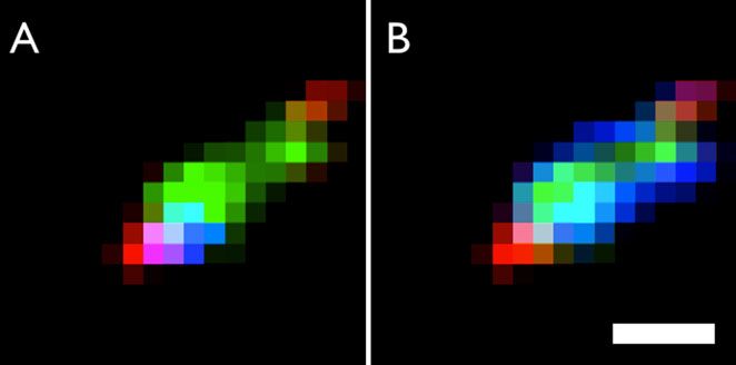
1646                                                Z. Gainsforth et al.

                             31
                                  NASA Ames Research Center, Moffett Field, California, USA
                                          32
                                             IRS, University Stuttgart, Stuttgart, Germany
                                      33
                                         IGEP, TU Braunschweig, Braunschweig, Germany
                                   34
                                      Jet Propulsion Laboratory, Pasadena, California, USA
                                                  35
                                                     Osaka University, Osaka, Japan
                                                            36
                                                               Worldwide
                                    *
                                      Corresponding author. E-mail: zackg@ssl.berkeley.edu
                                  (Received 4 December 2012; revision accepted 21 May 2013)

           Abstract–Using synchrotron-based X-ray diffraction measurements, we identified crystalline
           material in two particles of extraterrestrial origin extracted from the Stardust Interstellar
           Dust Collector. The first particle, I1047,1,34 (Hylabrook), consisted of a mosaiced olivine
           grain approximately 1 mm in size with internal strain fields up to 0.3%. The unit cell
           dimensions were a = 4.85  0.08 A,    b = 10.34  0.16 A,     c = 6.08  0.13 A    (2r).
           The second particle, I1043,1,30 (Orion), contained an olivine grain  2 mm in length and
           >500 nm in width. It was polycrystalline with both mosaiced domains varying over  20
           and additional unoriented domains, and contained internal strain fields < 1%. The unit
           cell dimensions of the olivine were a = 4.76  0.05 A, b = 10.23  0.10 A,  c = 5.99

           0.06 A (2r), which limited the olivine to a forsteritic composition [ Fo65 (2r). Orion
           also contained abundant spinel nanocrystals of unknown composition, but unit cell
           dimension a = 8.06  0.08 A    (2r). Two additional crystalline phases were present and
           remained unidentified. An amorphous component appeared to be present in both these
           particles based on STXM and XRF results reported elsewhere.

                  INTRODUCTION                                  particle size distribution has a sharp cut-off for grain
                                                                sizes above 1 lm, which is not to say that >1 lm grains
     The Stardust mission (Brownlee 2003; Tsou 2003)            are absent. With this and other information in hand, we
was a sample return mission flown as part of NASA’s             may be tempted to form some ideas of the composition
Discovery Mission program. It was effectively two               of our local interstellar medium—i.e., our immediate
missions in one spacecraft. The primary mission was to          neighborhood containing particles that would be
return the first samples from a known comet to Earth            sampled by the Stardust interstellar collector. However,
for study using instruments far too bulky to be flown in        studies of dust crystallinity, grain size, and composition
space. The primary mission resulted in an analysis of           in the interstellar medium have focused on sight lines to
cometary material captured from comet 81P/Wild 2                the galactic center or similarly bright targets easily
with unprecedented precision and detail (Brownlee et al.        accessible to astronomers. Recent studies of the local
2006). The secondary mission was to return the first            interstellar medium (LISM) have shown significant
solid samples of material from the local interstellar           chemical inhomogeneities on a parsec scale (Welsh and
medium. During  200 days of the 7 yr of spaceflight,           Lallement 2012), whereas studies of the galactic
the mission controllers exposed a second aerogel                interstellar medium (ISM) have shown multiple dust
collector to the interstellar dust stream. The discovery        populations throughout the Milky Way galaxy
and projected direction of this dust stream was based           (Sandford et al. 1991, 1995; Pendleton et al. 1994).
on observations by dust detectors on the Galileo and            These cases highlight the danger in predicting the
Ulysses missions (Gr€un et al. 1993).                           properties of the LISM dust population from the context
     Our current understanding of the interstellar              of galactic observations. In addition, the Ulysses
medium is largely derived from astronomical                     spacecraft has shown that dust reaching the inner system
observations. We expect that the dust population should         is significantly larger than the average dust size in the
comprise amorphous silicate and carbonaceous grains             interstellar medium         predicted    by astronomical
with an average size
Crystalline material in two interstellar candidates 1647
Models of dust propagation in the heliosphere show
that interstellar particles have been size sorted by
radiation pressure and Lorentz interactions within the
heliosphere. Most small particles should not penetrate to 2
AU where the Stardust collection occurred, and this
probably introduced a bias favoring collection of the
largest local interstellar medium (LISM) particles (Sterken
et al. 2014). Particles could also have been significantly
slowed, even to as low as 2 km s1 . Even at these
speeds, there is still potential for particle modification by
aerogel capture, which would complicate their study
(Frank et al. 2013; Postberg et al. 2014). The collector
itself introduces another bias. If a typical1648 Z. Gainsforth et al.
4M camera developed at the ESRF, with 2048 9 2048 powder pattern, and the observed peak characteristics
pixels/frame, a 50 9 50 mm pixel size, and a 16 bit (position, FWHM, shape) were faithful to an actual
readout. The convergence angle of the beam was 1 9 1 powder pattern.
mrad. XRD patterns were acquired in transmission To improve signal to noise for weak reflections, we
geometry. We calibrated the beamline using Al2 O3 and also employed a second algorithm that suppressed noise.
polycrystalline Al metal diffraction standards. We The standard deviation (SD) image was defined as:
recorded XRF spectra simultaneously using two Vortex-
vffiffiffiffiffiffiffiffiffiffiffiffiffiffiffiffiffiffiffiffiffiffiffiffiffiffiffiffiffiffiffiffiffiffiffiffiffiffiffiffiffiffiffiffiffiffiffiffiffiffiffiffiffi
EM 50 mm2 silicon drift detectors (SDDs). u N
u1 X
ESRF beamline ID22 (NanoImaging endstation) is Sðx; yÞ ¼ t ðDi ðx; yÞ Aðx; yÞÞ2 (2)
designed for X-ray nanodiffraction and XRF imaging N i¼1
and produces photons in the energy range of 4–37 keV
(Bleuet et al. 2008). We acquired an XRF and XRD
map of Orion by rastering with a 150 nm pixel size and so that each pixel was the standard deviation of the
using a beam spot of 225 9 235 nm. 2048 9 2048 pixel same pixel in the component images rather than the
images were acquired. We calibrated the beamline using average. In this case, pixels that were intense in only
a polysilicon standard. Orion was mapped using two one or several images were greatly amplified relative to
different beamlines as part of an agenda to characterize noise fluctuations. The centroids of the peaks were
sample alteration and to resolve oustanding science preserved as the standard deviation was a monotonic
questions arising from previous analyses. function of the variation in intensity. However, the
Keystones were mounted similarly in ID13 and shape and width of the peaks and the intensity of one
ID22. On ID22, the X-ray beam was exactly normal to peak relative to another were not preserved.
the silicon nitride membrane. On ID13, the X-ray beam The average and SD images were then azimuthally
was at a 45 angle. The XRD detector was then placed integrated to produce a 1D powder pattern that could
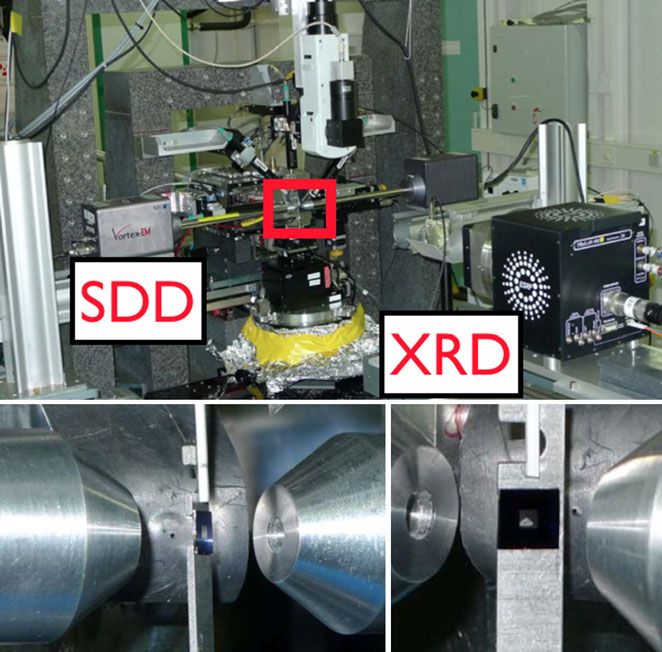
directly behind the sample. Fig. 1 shows the be used for fitting minerals (see figs. 4 and 8). As our
experimental arrangement on ID13. sample was a small particle embedded in aerogel, there
XRF analyses are discussed in companion articles was a strong background signal from amorphous SiO2
by Brenker et al. (2014) and Simionovici et al. (2014). diffraction, Rayleigh and Compton scattering. This
Scanning transmission X-ray microscopy (STXM) background was not analyzed but simply removed by
studies of these samples are discussed in Butterworth means of a spline fit using the software package Fityk
et al. (2014). (Wojdyr 2010). Similarly, we did not attempt to analyze
To identify phases from the XRD data, we employed any amorphous diffraction. As we did not sample all
two complementary approaches: A comparison of crystalline orientations as a true powder pattern would
d-spacings against a library of known minerals, and ab have, we limited ourselves to peak positions and peak
initio derivation of unit cell parameters. shapes during subsequent analysis and did not attempt
to analyze peak intensities.
Phase Identification from Virtual Powder Patterns Powder diffraction patterns can be fit using a
fingerprinting approach: observed d-spacings are
We processed the raw data into virtual powder compared against a database of minerals. We first
patterns (see Figs. 3 and 7) via two algorithms to compared the experimental data against 80 fingerprints
achieve differing objectives. In the first, we made an including presolar grain minerals that have been seen in
average image by averaging all the XRD patterns: if Di meteorites, interplanetary dust particles, astronomical
was one pattern in a rotational dataset (Hylabrook) or observations, or are theoretically expected as summarized
translational dataset (Orion), then Di ðx; yÞ was the in the review article by Lodders and Amari (2005), and
intensity of a given pixel (x, y) in that pattern. The the references therein. We also included materials used in
corresponding pixel in the average image A(x, y) was sample preparation and several additional common
meteoritic minerals. For each mineral, we simulated a
powder diffraction pattern using the energy and
1X N
Aðx; yÞ ¼ Di ðx; yÞ (1) instrumental broadening coefficients derived from our
N i¼1 beamline using the software packages CrystalDiffract
(CrystalMaker Software Ltd. 2012) and/or PDF-4+/
where i cycled through the N = 160 acquired patterns at Minerals (International Center For Diffraction Data
0.5 rotational increments for Hylabrook or N = 324 2012). We read the 80 fingerprints into MATLABâ (The
patterns for each sample translation for Orion. The MathWorks, Inc. 2011), and applied a peak finding
average image was the best approximation of an actual algorithm (Yoder 2011) to isolate a set of model peaks.Crystalline material in two interstellar candidates 1649
We then matched the experimentally measured d-spacings included and excluded the various crystal systems based
with the nearest model d-spacing, and if none could be on the observed d-spacings. This approach allowed us to
found within 2% of the measured d-spacing, we uniquely identify the unit cell to very high certainty. First,
considered that reflection unmatched. we explored which of the seven crystal systems could
We used a figure of merit defined by Smith and Snyder produce the observed pattern under any circumstances
(1979), a standard metric in the powder diffraction field, to with the assumption that only a single phase was
quantitatively assess the goodness of fit defined as diffracting. Next, we chose the highest possible symmetry
FN ¼ ð1=jD2hjÞðN=Nposs Þ, where N is the number of peaks unit cell with the smallest volume as the most likely
found experimentally and Nposs is the number of possible candidate for this crystal (Le Bail 2004). We ordered the
peaks we may expect to see in the pattern theoretically. symmetry of the seven crystal systems from highest
|D2h| is the mean difference between the matched peak symmetry to lowest symmetry: cubic, hexagonal/trigonal,
positions and their ideal values. FN thus has units of tetragonal, orthorhombic, monoclinic, triclinic. We
inverse degrees, and the inverse of FN is the average explored which of these crystal systems would produce
discrepancy between measured peak positions and model our experimental pattern using the software package
peak positions in degrees. Based on the fact that our McMaille (Le Bail 2004). The two lowest symmetry
instrumental FWHM was 0.07 at 20 , we constrained a systems (triclinic and monoclinic) were computationally
positive match to values of FN [ 14. Each fingerprint test prohibitive, and so it was impossible to fully explore the
produced a set of three numbers: (1) FN , (2) the number of space. However, we did apply McMaille’s Monte Carlo
model peaks not seen in the measured data, (3) the number approach, which was very fast and has been proven to
of measured peaks not seen in the model data. If a have a high success rate (Le Bail 2004).
measured peak was not predicted by the fingerprint, then We then placed error bars on the unit cell parameters
the fit was rejected, irrespective of the other parameters. using a Monte Carlo code we wrote in MATLABâ (The
The converse was not true: absence of a model peak in the MathWorks, Inc. 2011). This code determined what effect
measured data was expected as we had sparse powder the camera length calibration, and random errors in peak
patterns and may have missed weaker reflections. position determination had on our determination of the
However, such absences were still penalized in the FN . unit cell. Beginning with the set of experimentally
Finally, the effect of camera length uncertainty was measured d-spacings, our Monte Carlo simulation added
removed by adjusting d-spacings for the measured data by systematic (camera length) and stochastic (detector
3% in steps of 0.01%, and then choosing the best match. geometry) errors to produce 50,000 trial diffraction
The fingerprints were forsterite (two fingerprints patterns. For each pattern, it computed optimal unit cells
from separate sources), fayalite, tephroite, kirschsteinite, using an unconstrained nonlinear optimization
monticellite, laihunite 1M, laihunite 3M, liebenbergite, implemented as the function fminsearch in MATLABâ
chrysotile, talc, glaucochroite, ringwoodite, wadsleyite, (Lagarias et al. 1998). The unit cell angles a, b, and c
SiC 3C, diamond, diopside, albite, anorthite, were fixed at 90 in accordance with either a cubic or
clinoenstatite, orthoenstatite, pigeonite, gehlenite, orthorhombic structure (we did this computation only for
graphite, corundum, spinel (two from separate sources), spinel and olivine). For the systematic camera length
spinel rocksalt, u €lvospinel, defective spinel, chromite, error, the same random offset up to 1% was added to
magnetite, hercynite, trevorite, silicon, greigite, alpha- all d-spacings. For the nonsystematic errors, a random
quartz, kamacite, taenite, tetrataenite, schreibersite, offset up to 1% (Hylabrook) was computed for each
troilite, hibonite, grossite, sodalite, orthoclase, d-spacing separately. Orion had a better calibration and
perovskite, TiC, TiN, AlN, niningerite, fersilicite, the random offset was 0.2%. This produced histograms
oldhamite, halite, daubrelite, alabandite, sylvite, chlorite, allowing for the determination of error bars on the
biotite, augite, awaruite, calcite, cohenite, alpha- various lattice parameters. 2r bounds were computed by
cristobalite, grossular, hematite, maghemite, integrating the histograms.
magnesioferrite, w€ ustite, periclase, neirite, ferrosilite,
hedenbergite, suessite, pyrope, pyrite, pentlandite, Size-Strain Analysis
hexagonal pyrrhotite, monoclinic pyrrhotite 4C,
monoclinic pyrrhotite 6C. One can measure the average strain within crystals
by comparing the breadth of the reflections (FWHM) to
Ab Initio Unit Cell Determination the breadth of reflections produced by a perfect crystal
(Williamson and Hall 1953; Scherrer 1918; Stokes and
In a second approach, we analyzed the unit cell from Wilson 1944; Balzar 1999). In practice, it is necessary to
the diffraction data without assuming that the pattern remove the competing broadening caused by small
was one of our 80 chosen fingerprints. Instead, we crystallite sizes, and the instrumental optics. Broadening1650 Z. Gainsforth et al.
due to small crystallite volumes influences the breadth of intensity and 2h is the angle of the diffraction from the
all peaks equally, whereas broadening due to strain incident beam. The peak positions and shapes were
influences those reflections with small d-spacings more determined by fitting a pseudo-Voigt plus linear
dramatically. Thus, one can plot the broadening as a background with the Fityk software package (Wojdyr
function of 2h, and nanocrystallite volume will be read as 2010). The instrumental broadening measured from
inversely proportional to the y-intercept, while the slope corundum was then removed from the Hylabrook peak
will be related to the strain. This simplest approach is the breadths using the relations from Thompson et al.
Williamson-Hall method. Other approaches use similar (1987) and fed into the BREADTH software package
logic, but apply more robust mathematical models to (Balzar 1999), which implemented the double-Voigt
describe the broadening of the peaks. method to analyze size and strain.
For quantitative analysis, we used the double-Voigt The full width at half maximum (FWHM) of the
method of strain analysis (Balzar 1999) because it is pseudo-Voigt representing the instrumental broadening
more robust to noisy datasets and also utilizes the was found to be 0.07 at 2h 20 . While dedicated
distinction between two types of microstrain: local and powder synchrotron lines may be expected to achieve a
nonlocal. Strain fields due to domain boundary stresses, FWHM of approximately 0.01 , ID13 was a nanofocus
for example, permeate the entire crystal and commonly beamline so the incident beam was not perfectly parallel,
generate a broadening profile best described by a which caused additional instrumental broadening. In
Lorentzian peak shape. Strain fields due to point addition, some portion of this FWHM may have been
defects, which only strain a localized region of a few due to the small domain size of the corundum standard.
nanometers, produce a more Gaussian shaped peak
(Adler and Houska 1979; Balzar 1999). Processing Topographical Datasets
The double-Voigt method utilizes pseudo-Voigt
functions, which are a combination of Gaussian and In the case of Orion, we produced X-ray diffraction
Lorentzian curves and are the basis for separating the topographs (Black and Long 2004) using software we
two forms of microstrain. Analysis results in a volume- wrote in MATLABâ. An X-ray diffraction topograph is
weighted domain size, Dv , representing the particle similar to a darkfield image in transmission electron
diameter (analogous to a D in the Scherrer equation) and microscopy. One produces an image from X-rays
a surface-weighted domain size, Ds , also representing the diffracted into a specific direction. To accomplish this,
particle diameter. For a monodisperse spherical we generated a “SD” image as described above and
population, the actual domain diameter is related to the selected circular regions around notable reflections. Each
volume- and area-weighted sizes by D ¼ ð4=3ÞDv ¼ region acted as a mask against all images in the dataset.
ð3=2ÞDs while for lognormal distributions D\Ds \Dv For each image, we summed the pixels within the mask
(Balzar 1999). In principle, one could use the relative into a scalar value that represented the intensity of that
values of Dv and Ds to infer particle shapes or size reflection. Then we mapped each scalar value to the
distributions. spatial position of the sample. In this way, we built up
The instrumental factors must be deconvolved from an 18 9 18 pixel image (324 pixels) from the 324
the experimentally measured broadening to obtain the diffraction patterns, where the intensity of a pixel
sample’s inherent broadening and therefore measure the represented the intensity of the X-ray reflection at a
size and strain contributions to that broadening. We used spatial coordinate. As the pixels were 200 nm wide, the
a corundum standard (a-Al2 O3 ) as our calibration for topographs were 3.6 lm wide. There was a point spread
instrumental broadening, but also carried out the function (PSF) convolved with the topographical images
calculations without deconvolution of the instrumental as the beam waist was larger than the pixel size. Thus,
broadening. The latter gave a firm lower limit on the there was significant flux in the tails extending out at
domain size, and an upper limit on the strain fields least one pixel on either side of the central pixel. We
present, as the additional instrument broadening would measured the PSF using the best reflection available—
only make the particles appear smaller and more strained i.e., one in which the reflection is nearly perfectly
than they really were. As the corundum almost certainly circular showing no evidence of asterism (smearing). The
exhibits some size broadening (Thompson et al. 1987), corresponding topograph generated from this reflection
our results after subtracting the instrumental broadening is shown in Fig. 2. The point source image appeared as a
may have overestimated the particle size and single bright pixel with dimmer neighbors. Some
underestimated the strain. intensity was evident two pixels away, but was very
Ring patterns for the corundum standard were much reduced. Because of the beam waist and the
analyzed with the Area Diffraction Machine software resulting point spread function, we could not resolve
(Webb 2007) to produce I(2h) plots where I is the crystallite sizes below 400 nm, or two pixels.Crystalline material in two interstellar candidates 1651
Fig. 2. The point spread function (PSF) of the topographs
was estimated from the most pointlike topograph (left) using
an olivine 020 reflection at 5.12 A. On the right, the
corresponding diffraction spot shows no evidence of asterism
and is nearly perfectly circular. The point spread is slightly
greater than one pixel wide: significant flare occurs in
neighboring pixels and minor flare can occur two pixels away.
The scalebar is one micron.
Fig. 3. SD image for Hylabrook generated by taking the
Table 1. Simulated intensities for the 011 reflection of standard deviation of 160 images acquired at 0.5 rotational
intervals between 0 and 80 . In contrast to typical powder
kamacite nanograins in our experiemental setup. patterns, we did not see fully populated rings. Nevertheless,
Size (nm) Imax (%) FWHM ( ) the pattern was sufficiently well filled out to permit analysis of
the phase(s) present.
1000 100 0.07
50 64 0.11
20 31 0.23
10 16 0.43
5 8 0.84
2 3 2.12
Nanocrystalline and Amorphous Phases
We also investigated whether our XRD pattern may
have missed very small nanoparticulates. Table 1 shows
simulated intensities for the 011 reflection of kamacite Fig. 4. 1D powder integration patterns for Hylabrook derived
for our beam conditions and shows that particles more from the average image, the SD image, and an ideal model
in the SD pattern is a
pattern for Fo100 . The peak at 4.3 A
than about 10 nm in size should have remained easily
visible in our diffraction pattern. Even small grains such low intensity reflection, which is present in the forsterite
model, but with intensity too low to be visible in this plot. A
as 2 nm nanodiamonds should have been visible if their slight systematic shift of the SD and average images to the
modal abundance were high. right is due to an intentionally uncorrected camera length
calibration uncertainty.
RESULTS: TRACK 34 (HYLABROOK)
was used when we wished to know the FWHM and shape
Track I1047,1,34 contains one terminal particle of peaks. Figure 4 shows three 1D powder integration
named Hylabrook. We first studied it by STXM, and patterns produced from the average image, the SD image,
then by XRF/XRD on ESRF beamline ID13. We and a model pattern for pure forsterite.
acquired a set of diffraction patterns using a tomographic
mount with photons at 13,895 eV (k = 0.89229 A). We Fit Against Likely Minerals
rotated the sample through 80 in 0.5 increments, and
acquired a diffraction pattern and XRF spectrum at each Column 1 of Table 2 shows the d-spacings observed
rotation. The pattern was sufficiently complete to in our SD pattern. We fit these against our 80
determine unit cell parameters and obtain a likely mineral fingerprints (Table 3). We found that only the olivine
fit. Figure 3 shows the SD image for Hylabrook. The SD group minerals fit all the measured peaks well. Fo100
image was used when we wished to know the d-spacings produced a good match with F14 ¼ 41, and all
and azimuthal angles of reflections. The average image measured peaks were fit by the model. This meant that1652 Z. Gainsforth et al.
Table 2. Measured and model peak positions for
phases in Hylabrook.
dmeas a dmodel b hkl
5.18 5.11 020
4.39 4.32 110
3.94 3.89 021
3.80 3.73 101
3.55 3.50 111
3.48 120
3.06 3.01 121
3.00 002
2.81 2.77 130
n.d. 2.59 022
n.d. 2.56 040
2.55 2.52 131
2.50 2.46 112
2.38 2.38 200
2.36 2.35 041
2.32 210
2.30 2.27 122
2.25 140
2.20 2.16 211
2.16 220
n.d. 2.11 141
2.06 2.03 132
2.03 221 Fig. 5. Error distributions for the unit cell parameters in
a Centroids of the experimentally measured peaks for Hylabrook.
Hylabrook derived from a 50,000 trial Monte Carlo. The best
b Centroids of the expected reflections based on a Fo composition.
fit values do not exactly match the centroids of the
100 distributions since they are asymmetric.
Where the experimental pattern was unable to resolve close
reflections, two model peaks are assigned.
bars as 2r values given in Table 4: a = 4.85 0.08
we fit 14 peaks to obtain a figure of merit of 41, well b = 10.34 0.16 A,
A, c = 6.08 0.13 A (2r).
above our acceptance threshold. No other phase met The PDF-4+/Minerals database (International
the threshold requirements for a fit. Center For Diffraction Data 2012) tracks a large set of
known minerals along with their properties. The only
Ab Initio Unit Cell mineral matches in this database with unit cell
dimensions within 0.1 A of the experimentally
We used observed d-spacings to determine the unit determined values were olivine group minerals, with the
cell directly by testing all possible unit cells. We space group Pbnm. Three reflections 022, 040, and 141,
explored the cubic system (0.005 A steps, over the range were not observed in the experimental pattern, but were
of 2–20 A), the hexagonal/trigonal systems (0.01 A generally low intensity peaks and if the sample were not
the tetragonal system
steps, over the range of 2–18 A), perfectly oriented to show them, we would expect them
(0.05 A steps, over the range of 2–16 A), and the to be absent. Thus, with the exception of these weak
orthorhombic system (0.05 A steps, over the range of 022, 040, and 141 reflections, there was an exact one-to-
The orthorhombic system resulted in an
2–20 A). one correspondence between the observed pattern and a
excellent fit for a unit cell with dimensions a = 4.84, model pattern for olivine, so we concluded that
b = 10.33, c = 6.06. This unit cell produced a high FN Hylabrook probably had an olivine structure.
with a small unit cell volume, and was consistent with
the olivine mineral family. Other candidates could be Defective Structure in Hylabrook
discarded on the basis of a significantly worse FN , large
unit cell volume, and/or a lack of minerals to match Hylabrook’s diffraction pattern (Fig. 3) showed the
them. While we did explore the monoclinic and triclinic presence of asterism of the reflections on the order of 1 to
systems using Monte Carlo, we did not find any fits 10 in azimuth. Figure 6 is a magnified view of several
better than the above orthorhombic fit. The resultant peaks and shows a clockwise pattern of a single narrow
error distributions were only slightly asymmetric intense peak followed by a broader peak and then a tail.
(Fig. 5), so we assumed symmetry and quoted the error Such structures come about when there are multipleCrystalline material in two interstellar candidates 1653
Table 3. Closest fingerprint matches for Hylabrook XRD. The minerals are listed with the best fit at the top and
the worst at the bottom. The primary parameter for determining fit quality is how many measured peaks are not
predicted by the model spectrum. Only those minerals where every peak is predicted by the model are included in
this table. FN acted as a second parameter which determined goodness of fit as explained in the text. The only
minerals to fit all the experimental peaks are members of the olivine group and anorthite, and of those, the best fit
by a large margin is forsterite.
Database ID Mineral Formula Crystal system Space group FN
PDF-4+ 04-007-9021 Forsterite Mg2 SiO4 Orthorhombic Pbnm1 41
AMCSD 0000171 Forsterite Mg2 SiO4 Orthorhombic Pbnm 34
PDF-4+ 04-009-8350 Fayalite Fe2 SiO4 Orthorhombic Pbnm 19
PDF-4+ 04-007-9023 Tephroite Mn2 SiO4 Orthorhombic Pbnm 15
PDF-4+ 04-011-2883 Anorthite CaAl2 Si2 O3 Triclinic P
1 6
Table 4. Dimensions for the unit cell of the crystal in
Hylabrook with error bars.
Parameter Value 2r abs 2r rel
a 4.85 0.08 1.7%
b 10.34 0.16 1.5%
c 6.08 0.13 2.1%
a/b 0.469 0.008 1.7%
a/c 0.797 0.021 2.6%
V 304.97 10.7 3.5%
crystalline domains with a net orientation—unlike a
polycrystalline material in which the domains do not
have net orientation (Cullity and Stock 1978).
Because Hylabrook was examined on a goniometer
under microfocus conditions, slight drift of the sample
position caused small variations in the camera length
throughout the scan. These did not affect the overall
calibration as the order of these variations was smaller
than the uncertainty of the camera length calibration.
However, they did increase the FWHM when the peaks
occured in multiple frames because the centroid of the
peak varied slightly from frame to frame. Therefore, the
breadth of the Hylabrook reflections was not
determined from the average image, but from distinct
frames comprising it. Nine such peaks were examined
and chosen to represent the most diverse set of peaks in
Hylabrook. Some were polygonalized/mosaiced and
showed varying degrees of broadening. Table 5 shows
the reflections measured along with the FWHM
representing the broadening of that peak.
The reflection at 2h = [22.39,22.50] was asymmetric
and gave a poor fit with a single pseudo-Voigt, but was
well fit with two pseudo-Voigts. The centroids were 0.1
apart and could have been due to a fortuitous overlap
of the 200 and 112 reflections. From Table 5, it was Fig. 6. A number of reflections in Hylabrook had the same
evident that the reflection contained one bright well- polygonalization. In the image above, they have been rotated
focused component and one dimmer component with a about the pattern center so they can be compared. The
relative sizes of each are faithful to the original image (Fig. 3).
greater asterism. The lack of any asymmetry in the Moving clockwise, the polygonalization manifested as a single
corundum standard proved that the asymmetry was small peak, followed by a larger broader peak, and then
native to Hylabrook and not an instrumental effect. usually a tail.1654 Z. Gainsforth et al.
Table 5. Reflections measured for size-strain analysis of Hylabrook.
2ha FWHM Reflection 2h FWHM Reflection
13.24 0.119 29.38 0.179
13.56 0.087 33.66 0.155
20.62 0.123 33.98 N/A
22.39 0.122 34.48 0.155
22.50 0.115
23.46 0.180 36.63 N/A
a 2h positions and FWHM for the pseudo-Voigt used to fit the broadening.
Therefore, it was not likely to be a single domain crystallite size (Dv ) was essentially compatible with a
reflection. For this reason, we felt justified in fitting the large grain size—i.e., no resolved size broadening.
peak using two pseudo-Voigt functions with a single The strain measured by the double-Voigt analysis
linear background, and including both peaks in the size- was 0.2% 0.08% (2r). We are not aware of a
strain analysis. quantitative interpretation of this magnitude of strain in
The reflections at 2h = 33.98 and 36.63 also gave olivine. For elastic deformation, Abramson et al. (1997)
poor fits to a pseudo-Voigt and while the cause could found that bulk strains in olivine on the order of 0.2%
have been due to an overlap of neighboring reflections, were representative of stresses ≤1 GPa, depending on
there was no indication of this in the corresponding 2D the axis of the stress. However, our olivine was clearly
image shown in Table 5. Instead, the unexpected line not under external pressure such as from a diamond
shape could have been due to noise present in the anvil cell, so the strain we saw was a residual field left
experimental data, or another sample-dependent over from plastic deformation.
variation. For this reason, we chose to exclude these As noted above, corundum can cause broadening of
reflections from the size-strain analysis. its own. If we leave the instrumental broadening
The double-Voigt analysis resulted in a volume- convolved with the original peaks, we obtain a very
weighted domain size of Dv ¼ 710 420 A (2r) and a conservative lower limit on particle size and an upper
surface-weighted domain size of Ds ¼ 350 210 A limit on strain. Doing so, we found that domain sizes were
(2r). Based on our instrumental calibration, we could ≥ 27 nm (2r) and internal strain averages ≤ 0.3% (2r).
not accurately discern crystallite sizes above about In conclusion, we did not clearly resolve any size
80 nm (volume-weighted) as the crystallite broadening broadening in Hylabrook, but saw moderate strain from
becomes comparable to the instrumental broadening. crystalline defects. Furthermore, polygonalization
Therefore, the 2r upper limit on volume-weighted indicated subgrain boundaries as opposed to randomlyCrystalline material in two interstellar candidates 1655
oriented separate crystallites. With a 0.3% upper limit defects. Therefore we are uncertain whether the strain
on the strain, Hylabrook had a high crystal quality, we see is due to aerogel capture or is primary.
although we expect that a number of defects were
present and could have been imaged, for example, with EXCESS IRON CONTENT
a transmission electron microscope.
XRF data showed that Hylabrook was 25% Fe
Capture Effects by weight (Brenker et al. 2014). STXM analysis
following the ID13 analysis revealed that the Fe had
Hypervelocity capture of particles in aerogel can mobilized and did not correlate spatially with Mg
result in changes to the particle structure. Primarily, the (Butterworth et al. 2014). To verify that this
outer layers are ablated away to produce smaller, more modification did not alter our diffraction results, we
rounded grains. However, the shock and thermal examined a sequence of diffraction patterns acquired
stresses of the capture event can also modify internal throughout the rotational scan and did not find any
microstructure. Here, we discuss whether our evidence for modification of the crystalline phase. Thus,
polygonalization or microstrain measurements were the modification either occured after the ID13 XRD
capture effects, or if they were native characteristics of acquisition, or did not alter the olivine phase. While we
the extraterrestrial particle. Because the capture velocity did not see any other phases in diffraction, it is possible
of Hylabrook was 400 nm in size we could
dislocations induced in the olivine crystals from the directly measure their size using X-ray topography
capture. In the worst case, they found a strain of (Black and Long 2004) as described below. The track
approximately 1% within the dislocation loop, and was then analyzed by STXM (Butterworth et al. 2014):
computed that the pressure necessary to induce these elemental maps were acquired as well as XANES. The
defects was approximately 1.5 GPa. They postulated track was then sent back to ESRF for XRF/XRD on
that the strains were generated by transient stresses ID22 and imaged with a 350 eV bandwidth at 17000 eV
from the extreme thermal gradient that can arise during to produce XRF and XRD maps with
(k = 0.72932 A)
capture rather than impact shock itself. Models based a 150 9 150 nm pixel and 180 nm beam waist. We
on momentum balance (Anderson 1998) and acquired diffraction patterns at each pixel and
compression of a highly porous medium (Trigo- computed topographs from selected reflections.
Rodriguez et al. 2008) both suggest that peak shock At some time after the end of the ID13 acquisition
pressures at impact speeds of 6 km s1 are of order 800 but before the STXM acquisition, the particle was
MPa and only rise above 1.5–2 GPa for impact speeds modified and separated into two objects: some
in excess of 8–10 km s1 . On the other hand, we crystalline material was amorphized and displaced
measured an average strain value which is sensitive to several microns. The cause of this disruption remains
the regions devoid of defects as well as the regions with unidentified, but is discussed more thoroughly in1656 Z. Gainsforth et al.
Brenker et al. (2014), Butterworth et al. (2014), and
Simionovici et al. (2014). This provided us with an easy
method to separate phases, as we saw in total four
diffracting phases in the first ID13 scan, and fewer in
the ID22 scan. Therefore, it was easier to definitively
map specific peaks to specific phases. As the first three
scans on ID13 showed essentially identical diffraction
and XRF maps, we concluded that no modification to
the particle occured during this time. Therefore, the
initial scans from ID13 were valid, and we used the first
and only the first ID13 scan to analyze d-spacings and
FWHMs quantitatively. The second and third ID13
scans were discarded, although still useable, and the
ID22 data were used only for determining which peaks
map to the surviving phase.
Phase Determination from Topographs
The SD images of Orion taken on ID13 and ID22
are shown in Fig. 7. We saw significant broadening and
polygonalization, indicating multiple distinct diffractive
domains as well as the presence of strain. The 1D
powder integration patterns are shown in Fig. 8. The
peak positions are given in Table 6 and confirm the
presence of olivine and eight additional peaks.
Therefore, Orion was likely a multiphase object. We
compared the ID13 data with the ID22 data and found
that all the olivine peaks present in the ID13 scan were
absent in the ID22 scan with the possible exception of
one. We intepreted this to mean that the olivine was
amorphized at some point after the initial ID13 XRD
maps. Additional evidence from STXM corroborated
this (Butterworth et al. 2014). Meanwhile, all eight
peaks from the ID13 data that did not map to olivine
remained in the ID22 data. Of those eight, only five Fig. 7. Top image is the SD image of Orion from ID13.
were visible in the radial integration pattern, and of Bottom image is the SD image from ID22.
those five only four were strong enough to easily
analyze. between the two phases (Fig. 9A). Likewise, we
Using the McMaille software package (Le Bail produced a topograph from the longest two d-spacings
2004), we could not find a unit cell for the eight from spinel: 4.67 A (111) and 2.84 A (022), which were
nonolivine peaks, but fitting only the four intense unambiguously separated from olivine (Fig. 9B).
2.841 A,
reflections at 4.663 A, 2.434 A, and 2.015 A The remaining weak peaks at 8.844, 3.229, 2.215, and
Searching
resulted in a cubic unit cell with a = 8.06 A. 3.167 A could not be explained as spinel or olivine. Using
the PDF/4+ Minerals database for cubic minerals near topographs, we discovered that they were spatially
a = 8.06 A shows spinel minerals are closest. Many separated from the olivine and spinel and therefore were
chemical compositions are compatible with this unit a third phase. We attempted to find a mineral match
cell, including magnesiospinel, chromite, hercynite, using PDF 4/+ and to determine a unit cell using the
gahnite, and ringwoodite. We could not constrain the McMaille software package, but did not find any
chemistry with our data. satisfactory results. Therefore, it may be multiphase, or
We produced topographs from 30 olivine reflections have a very large, low symmetry unit cell. We call this
at d-spacings of 5.12 A, 3.72 A, and 3.50 A phase the unknown core phase as it is situated near the
corresponding to hkls 020, 101, and 111/120, olivine in the core of the particle (Fig. 9C).
respectively. These d-spacings were well separated from XRF and STXM found the bulk composition of
spinel reflections and could be used to discriminate Orion was 25% Fe by weight (Brenker et al. 2014;Crystalline material in two interstellar candidates 1657
Table 6. Measured and model peak positions for
phases in Orion.
a b
dmeas dmodel FWHM hklfo hklsp
8.844 0.207
5.116 5.113 0.092 020
4.663 4.668 0.164 111
4.309 4.317 0.051 110
3.888 3.890 0.129 021
3.721 3.729 0.129 101
3.498 3.503 0.141 111
3.485 120
3.229 *c
3.167 *
2.996 3.013 0.165 121
2.997 002
2.841 2.858 0.368 022
2.775 2.772 0.138 130
n.d. 2.586 n.d. 022
n.d. 2.556 n.d. 040
2.515 2.516 0.200 131
2.459 2.462 0.189 112
2.434 2.438 0.242 113
n.d. 2.381 n.d. 200
2.351 2.351 0.116 041
Fig. 8. The top panel compares the 1D powder integration n.d. 2.334 n.d. 222
from the ID13 SD image for Orion with model spectra for 2.317 2.319 0.182 210
olivine and spinel. The 2-D pattern was processed to remove 2.267 2.272 0.267 122
bad pixel instrumental artifacts showing as false peaks before
2.252 140
integrating. The bottom panel compares the 1D powder
integration pattern from the ID22 SD image for Orion with a 2.215 0.127
model spectrum for spinel. 2.163 2.163 0.178 211
2.158 220
2.097 2.108 0.208 141
Butterworth et al. 2014), so we considered the 2.015 2.035 0.420 132
possibility of Fe metal and sulfide phases. Kamacite, 2.031 221
the most common room temperature structure of pure 2.021 004
Fe metal, has an 011 reflection at 2.02 A. We a Centroids of the experimentally measured peaks for Orion.
produced a topograph of the 2.02 Å d-spacing and b Centroids expected from Fo
100 or spinel. Where the experimental
found a phase appearing as a shell around the olivine. pattern was unable to resolve close reflections, two model peaks are
assigned. In this case bold text indicates a peak is far more intense
Additionally, the FWHM of the 2.02 Å reflection was
than the others at the same d-spacing. Spinel peaks are also
nearly double the FWHM of nearby reflections, which italicized to distinguish them from olivine.
pointed to phase which was either highly strained or c The peak position was too weak in the radial integration to
nanoparticulate. With further investigation we found determine a FWHM.
that the 2.434 Å spinel peak had a topograph that
overlapped 2.02 Å topograph in some areas. They unknown core phase was at one end, slightly to the
were not identical, however, so it may have had a side. Orion was a heterogeneous object because the
contribution from another phase. We named the 2.02 Å olivine, spinel, and the unknown phases were segregated
topograph the unknown shell phase to reflect this spatially.
uncertainty. Regardless, if the olivine were wrapped in
a polycrystalline material, or an amorphous material Olivine Analysis
with nanocrystalline inclusions, then the topograph
should have appeared as it does in Fig. 9D, viz. a 2D All olivine reflections are shown in Table 5. We
projection of a hollow cylinder or ellipsoid. computed the Smith-Snyder figure of merit, FN (Smith
Synthesizing the topographs from Figs. 9 and 10, it and Snyder 1979) in the same fashion as for Hylabrook
is clear that the center of Orion was olivine and spinel and found that F13 ¼ 51 for the raw data when fit to
appeared on each end. The unknown shell phase was Fo100 , and F13 ¼ 110 after removing any systematic
most intense around the rim of the olivine. The error due to camera length calibration. In this case, the1658 Z. Gainsforth et al.
Table 7. Computed unit cell parameters with error
bars for Orion olivine.
Parameter Value 2r abs 2r rel
a 4.76 0.05 1%
b 10.23 0.10 1%
c 5.99 0.06 1%
a/b 0.465 0.0014 0.3%
a/c 0.795 0.0028 0.4%
V 291 8 3%
only 0.001% of the strongest line. Similarly to
Hylabrook, we fit our 80 fingerprints and found that
only olivine group minerals fit all the peaks seen and
still maintained a high FN . To measure the unit cell, we
applied the same Monte Carlo approach used for
Hylabrook with 50,000 trials: a = 4.76 0.05 Å, b =
10.23 0.10 Å, c = 5.99 0.06 A where all error
bars are 2r (Table 7). Figure 11 shows the distribution
of values computed by the Monte Carlo. The detector
Fig. 9. A) Topograph generated from 30 reflections of olivine geometry calibration for Orion was of higher quality
showing the position of the olivine phase in Orion. B)
Topograph generated from the two longest d-spacings of than it was for Hylabrook. As a result, the
spinel showing the spinel phase in Orion/Sirus. C) Topograph nonsystematic variation was only 0.2% rather than 1%
generated from the d-spacing at 8.84 A corresponding to an for Hylabrook. The systematic error (camera length
unknown phase. D) Topograph generated from the 2.02 A calibration) remained at 1% as it was for Hylabrook.
reflection, which contains olivine, spinel, and a third phase. 1 The distributions for the unit cell parameters a, b, and c
lm scalebar.
and volume had a boxcar shape. Shown in Fig. 11 the
boxcars reflect the fact that all unit cell parameters
could be systematically altered by 1%, but were well
known relative to each other. The ratios a/b and a/c,
which were essentially independent of the systematic
error, remained much closer to normal distributions.
In contrast with Hylabrook, we had enough
accuracy in the unit cell measurement to examine
chemical composition. The unit cell of olivine varies
with cation concentration and the cation radius (Fisher
and Medaris 1969; Lumpkin and Ribbe 1983; Koch
et al. 2004). Using Lumpkin and Ribbe (1983) and a
Monte Carlo code we wrote in MATLABâ, we computed
the radius of the tetrahedral cation to be 0.28 0.08 Å
Fig. 10. A) Composite RGB image of spinel (red), olivine (Fig. 12A). Si and Be are the only known cations in this
(green), and an unknown phase (blue). Olivine is the mineral range of radii within olivine structure minerals. Likewise,
at the core of Orion, while spinel appears to occupy the two Fig. 12B shows the M1 site radius to be 0.75 0.08 A.
ends, along with the unknown phase at one end. B) RGB
For comparison, Mg is 0.72, Fe is 0.78 A, and Ca is
image of spinel (red), olivine (green), and the As M1 and M2 cations in the beryllonates have
olivine+spinel+third phase (blue). Because there are pixels 1.00 A.
which are intense in the blue, but not the red or green, we we ruled this out and claimed
radii of 0.54 – 0.62 A,
conclude that despite contamination from the olivine and that Si was the tetrahedral cation. To compute the M1/
spinel reflections, there must be a third diffracting phase, M2 ratio, we took advantage of the fact that the ratios
which appears to be more intense around the periphery of the
olivine. 1 lm scalebar.
of the unit cell parameters a/b and a/c were better
known than the absolute values for a, b, and c. Figures
reflection at 2.015 Å has been excluded because it 12C and 12D show computed results using the ratioed
overlapped spinel and shell phase peaks, and the peak unit cell and assuming silicon for the tetrahedral cation.
at 2.097 was not counted in FN because its intensity was M1/M2 is tightly constrained between 0.96 and 1.00Crystalline material in two interstellar candidates 1659
Fig. 12. Error distributions for the computed atomic site radii
in Orion olivine. A shows the radius of the tetrahedral site
B shows the size of the
usually occupied by silicon (0.260 A).
M1 site (Mg = 0.72, Fe = 0.78). C shows the ratio of M1/M2
after removing errors due to camera length calibration, and
assuming the tetrahedral cation is Si. D shows the M1
computation after removing errors and assuming tetrahedral
Si.
Fig. 11. Error distributions for the unit cell parameters of
olivine in Orion.
were better elucidated by the topographical analysis, as
(Fig. 12C). The M1 site is between 0.71 and 0.75 A. many olivine domains were larger than the pixel size
Pure forsterite is entirely consistent with these results. and could be resolved in real space.
Pure fayalite is inconsistent. Because the M1/M2 ratio is
close to 1, the size of the cations in M1 and M2 must Olivine Mosaicism
be similar. In olivines with large and small cations, the
larger cations take the M2 site leading to a reduction in The topographs showed that the olivine was heavily
M1/M2 from 1. Therefore, our optimal choice for a mosaiced. Figure 13 shows a mosaic with an asterism of
composition comes from the centroids of our error about 20 . Topographs produced from eight positions
distributions: M1/M2 = 0.98 and M1 = 0.73 A, which along the asterism are shown in juxtaposition and labeled
matches Fo85 . Other olivines are consistent within by their azimuthal coordinate, v, defined as the
our error bars. For example, (Mg1:91 Ca0:09 )SiO4 has counterclockwise azimuthal angle from 3 o’clock in
M1 = 0.72, and an M1/M2 = 0.97 while Fo71 has the diffraction image. The point spread function blurred
M1 = 0.74, and M1/M2 = 1. Therefore, the 2r upper the topograph view of the mosaic domains slightly, so we
limits for binary composititions are: fayalite may consider each of the topographs to be approximately1660 Z. Gainsforth et al.
Fig. 13. A mosaiced olivine reflection at 2.16 A (lower right image) corresponds to the 211 or 220 reflection of olivine. The
numbered images are topographs where the number represents the azimuthal position (v) in the diffraction pattern showing that
this mosaic spread occurs over approximately 20 in azimuth. A fixed coordinate is marked in all topographs to clarify the fact
that different real-space domains are responsible for the reflections. The topograph pixel sizes are 190 nm. The mosaic extends
almost two microns in real space.
and c are the three radii of the ellipsoid. Because of the
/ ¼ 2 e tanð2hÞ (3) point spread function of the topographs (Fig. 2), we
should remove one pixel from the side of each topograph,
where / is the azimuthal broadening, and e is the and then we can determine two radii of the olivine as 1.1
variation in the angle of the diffractive domains and 0.3 lm. If we assume that the depth dimension was
(mosaicity). For this reflection at 2.16 A (2h = 23.8 ),
equal to the smallest lateral dimension (0.3 lm radius),
we obtained e = 23 . This variation was clearly too then the volume of the ellipsoid was 0.41 lm3 . Using the
large to be due to internal strain or size broadening and density for forsterite of 3.25 g cm-3, this gave a total
so was due to grain boundaries or disconnected grains. olivine mass of 1300 fg, and a total Mg mass of 460 fg.
Conversely, a smaller number of tiny olivine The STXM results estimated 1 pg of forsterite
domains were also visible such as the one shown in Fig. (Butterworth et al. 2014), which is in good agreement
2. These were essentially single pixel reflections, blurred considering we cannot know exactly how well the
only by the PSF of the beamline. They did not have an ellipsoid approximation represented the actual particle.
obvious orientation to other crystallites, and so it
appears that polycrystalline olivine was also present. We
have an upper limit of 400 nm from the topographs for Spinel Analysis
the size of the polycrystalline domains. Therefore, the
mosaic crystal should account for the bulk of We previously calculated an 8.06 A cubic unit cell
the olivine mass as it would require a large number of from from the four brightest nonolivine reflections in
the polycrystallites to equal the volume of the mosaic ID13. Again, we applied our Monte Carlo routines to
seen in Fig. 13. obtain error bars for the unit cell dimensions. Cubic
If we assume that the olivine was compact, then we symmetry requires a = b = c and a ¼ b ¼ c ¼ 90 .
can make an order of magnitude estimate of its size by The resulting unit cell had a dimension: a = 8.06
approximating the shape seen in the sum topograph (Fig. (2r). The figure of merit for spinel was F4 ¼ 28
0.08 A
9A) as an ellipsoid with volume V = (4p/3)abc where a, b, for the raw data and F4 ¼ 57 after removing anyCrystalline material in two interstellar candidates 1661
Fig. 14. Two reflections produced by the longest d-spacing in
(111).
spinel: 4.66 A
camera length calibration error. A size-strain analysis
was difficult for the spinel as only the two longest
(highest d-spacing) peaks were clearly separated from
the olivine peaks. The reflections were so sparsely
populated that it was not possible to get a good
quantitative understanding of the spreading, although
we observed distinct crystallites with variable spreading,
either due to crystallite size, strain, or both. Figure 14 Fig. 15. Topographs for all the 4.66 A spinel reflections. The
shows images for two reflections from the longest spinel number in each box represents the azimuthal position in the
d-spacing, 4.66 A. Whereas one appeared as a nearly diffraction pattern (v). Lower-right inset is a cluster of
perfect crystal, the other was barely discernable as the reflections with an azimuthal spread over 20 .
asterism was so great. Therefore, the characteristics of
the spinel crystals were highly inhomogenous.
The spinel topographs did not appear to bear the We might expect to have seen nanoparticulate Fe
same mosaicity relationship seen in the olivine. Figure from glass with embedded metal and sulfides (Bradley
15 shows topographs for all the peaks at the longest 2007), space weathering of olivine (Hapke 2001), or
d-spacing for spinel, 4.66 A corresponding to the 111 aerogel capture of sulfides (Leroux et al. 2009). If
reflection. There appeared to be a sequence of closely nanoparticulate Fe were present, it would have shown
spaced peaks with an azimuthal spread of 20 going as the 2.02 A reflection such as in the shell phase.
from v ¼ 34 to 54 . However, the topographs showed However, nano-Fe could not have made the 2.434 A
that the peaks mapped to crystallites on opposite sides reflection. There are no iron sulfides with both these
of the olivine grain and did not show a clear mosaicity reflections dominating others, although pyrite can make
or polygonalization. There may still have been some the 2.434 A reflection. We should note that known
relationship, for example, the topographs for v ¼ 34 , spinel-sulfide phases such as those found in IDPs (Dai
41 , and 48 appear to be close, but it is also important and Bradley 2001) and greigite probably did not
to note that each of these topographs is essentially the produce these reflections since their unit cell is too
PSF for the beamline so there is no guarantee that the large. Because sulfides readily decompose upon aerogel
grains were large enough to overlap. One reasonable capture into an Fe-metal core and Fe sulfide rim
explanation is that the grains were nanocrystals within a (Leroux et al. 2009), it follows that absence of a sulfide
volume of amorphous material. signature does not rule out the presence of sulfides prior
to the collection. However, the presence of S in the
Fitting Unknown Phases XRF could be explained if sulfides present in the
incident particle decomposed due to aerogel capture in
We know from XRF analysis (Brenker et al. 2014) the same fashion as we see in the cometary samples
that significant iron and sulfur were present in the (Leroux et al. 2009). We would expect a residual nano-
particle, and that these were among the most mobile Fe core, which would show up as a 2.02 A ring and the
elements during synchrotron irradiation. Additionally, sulfur would remain in amorphous phases invisible to
carbon was invisible to XRF and STXM due to the XRD.
experimental limitations so we also considered whether Graphite could also produce the 2.02 A reflection,
Fe, sulfide, or carbonaceous materials could fit either of
but should produce a 3.36 A reflection as well that we
the unknown phases. did not see, so we conclude that graphite wasYou can also read