Peptidoglycan Salvage Enables the Periodontal Pathogen Tannerella forsythia to Survive within the Oral Microbial Community
←
→
Page content transcription
If your browser does not render page correctly, please read the page content below
Research Article

                                                             Microb Physiol 2021;31:123–134                                                             Received: February 17, 2021
                                                                                                                                                        Accepted: April 22, 2021
                                                             DOI: 10.1159/000516751                                                                     Published online: June 9, 2021

Peptidoglycan Salvage Enables the
Periodontal Pathogen Tannerella
forsythia to Survive within the Oral
Microbial Community
Isabel Hottmann a Marina Borisova a Christina Schäffer b Christoph Mayer a
aInterfaculty Institute of Microbiology and Infection Medicine, Organismic Interactions/Glycobiology, Eberhard Karls

Universität Tübingen, Tübingen, Germany; bDepartment of NanoBiotechnology, NanoGlycobiology Unit, Universität
für Bodenkultur Wien, Vienna, Austria

Keywords                                                                                         port summarizes T. forsythia’s strategies to survive in the oral
Oral biofilm · Periodontitis · Pathogen · N-acetylmuramic                                        habitat by means of PGN salvage pathways, including recov-
acid · Peptidoglycan · Cell wall recycling                                                       ery of exogenous MurNAc and PGN-derived fragments but
                                                                                                 also polymeric PGN, which are all derived from cohabiting
                                                                                                 bacteria either via cell wall turnover or decay of cells. Salvage
Abstract                                                                                         of polymeric PGN presumably requires the removal of pep-
Tannerella forsythia is an anaerobic, fusiform Gram-negative                                     tides from PGN by an unknown amidase, concomitantly with
oral pathogen strongly associated with periodontitis, a mul-                                     the translocation of the polymer across the outer mem-
tibacterial inflammatory disease that leads to the destruc-                                      brane. Two recently identified exo-lytic N-acetylmuramidas-
tion of the teeth-supporting tissue, ultimately causing tooth                                    es (Tf_NamZ1 and Tf_NamZ2) specifically cleave the pep-
loss. To survive in the oral habitat, T. forsythia depends on                                    tide-free, exogenous (nutrition source) PGN in the periplasm
cohabiting bacteria for the provision of nutrients. For axenic                                   and release the MurNAc and disaccharide substrates for the
growth under laboratory conditions, it specifically relies on                                    transporters Tf_MurT and Tf_AmpG, respectively, whereas
the external supply of N-acetylmuramic acid (MurNAc),                                            the peptide-containing, endogenous (the self-cell wall) PGN
which is an essential constituent of the peptidoglycan (PGN)                                     stays unattached. This review also outlines how T. forsythia
of bacterial cell walls. T. forsythia comprises a typical Gram-                                  synthesises the PGN precursors UDP-MurNAc and UDP-N-
negative PGN; however, as evidenced by genome sequence                                           acetylglucosamine (UDP-GlcNAc), involving homologs of
analysis, the organism lacks common enzymes required for                                         the Pseudomonas sp. recycling enzymes AmgK/MurU and a
the de novo synthesis of precursors of PGN, which rational-                                      monofunctional uridylyl transferase (named Tf_GlmU*), re-
izes its MurNAc auxotrophy. Only recently insights were ob-                                      spectively.                             © 2021 The Author(s).
tained into how T. forsythia gains access to MurNAc in its oral                                                                                        Published by S. Karger AG, Basel

habitat, enabling synthesis of the own PGN cell wall. This re-

karger@karger.com      © 2021 The Author(s).                                                     Correspondence to:
www.karger.com/mip     Published by S. Karger AG, Basel                                          Christoph Mayer, christoph.mayer @ uni-tuebingen.de
                       This is an Open Access article licensed under the Creative Commons
                       Attribution-NonCommercial-4.0 International License (CC BY-NC)
                       (http://www.karger.com/Services/OpenAccessLicense), applicable to
                       the online version of the article only. Usage and distribution for com-
                       mercial purposes requires written permission.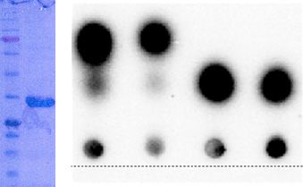
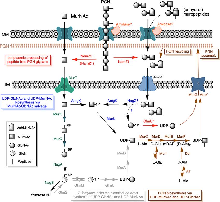
Introduction                                                        2019]. Thereby a flexible mesh-like structure is generated,
                                                                       the PGN sacculus, that allows the bacterial cell to with-
    Tannerella forsythia, originally designated Bacteroides            stand intracellular osmotic pressure (turgor), thus pro-
forsythius, is an anaerobic Gram-negative bacterium tax-               tecting from lysis [Vollmer and Seligman, 2010]. T. for-
onomically grouped to the Porphyromonadaceae within                    sythia possesses a typical Gram-negative PGN [Mayer et
the Bacteroidetes phylum [Tanner et al., 1986; Maiden et               al., 2019b]. However, genome analysis of T. forsythia re-
al., 2003]. It was first described by Anne Tanner at the               vealed that the bacterium lacks orthologs of murA/murB
Boston Forsyth Institute as a “slow growing fusiform Bac-              and glmS/glmM/glmU (shown in Fig. 1; proteins missing
teroides” and was isolated from apical plaque samples                  in T. forsythia but commonly present in bacteria are de-
from patients suffering from advanced periodontitis                    picted in gray). These genes encode generally essential
[Tanner et al., 1979; Tanner et al., 1986]. Remarkably, the            enzymes for the de novo biosynthesis of the PGN precur-
bacterium could not be obtained in pure culture, but it                sors UDP-MurNAc and UDP-GlcNAc [Chen et al., 2005;
grew in the presence of a helper bacterium, such as Fuso-              Friedrich et al., 2015]. Specifically, due to the lack of
bacterium nucleatum, even when the two bacteria were                   murA/murB, T. forsythia is unable to synthesize UDP-
separated by a semipermeable membrane [Tanner et al.,                  MurNAc from UDP-GlcNAc, rationalizing the MurNAc
1986; Dzink et al., 1987]. This indicated that survival of             auxotrophy of T. forsythia. Moreover, due to the lack of
T. forsythia depends on an essential growth factor that is             glmS/glmM/glmU, T. forsythia is also unable to synthesize
released from the helper bacterium. This mysterious                    UDP-GlcNAc from fructose 6-phosphate (fructose 6P)
compound was subsequently identified by Chris Wyss at                  (Fig. 1).
the University of Zürich as N-acetylmuramic acid (Mur-                     Since T. forsythia apparently fails to de novo synthesize
NAc), which supported axenic growth of T. forsythia                    UDP-MurNAc and UDP-GlcNAc, it depends on the sal-
[Wyss, 1989]. Together with N-acetylglucosamine                        vage of MurNAc and, presumably, also of GlcNAc from
(GlcNAc), MurNAc is an essential carbohydrate constit-                 the environment to synthesize its cell wall. It is conceiv-
uent of the peptidoglycan (PGN) of the bacterial cell wall.            able to assume that the polymicrobial flora in the oral cav-
The glycan backbone of PGN is built from these two car-                ity, harboring more than 700 bacterial species [Lamont et
bohydrates, which are connected via β-1,4 glycosidic                   al., 2018], provides to T. forsythia distinct options to ac-
bonds in an alternate arrangement and multiple of these                cess various sources of MurNAc and GlcNAc. Indeed, the
glycan chains are cross-linked via short peptides linked to            ability to establish itself in a polymicrobial biofilm con-
MurNAc [Weidel and Pelzer, 1964; Walter and Mayer,                     sortium, commonly known as dental plaque, is supposed

Fig. 1. Overview of the PGN/MurNAc salvage and biosynthetic            self-cell wall) PGN within the periplasm. Peptide-free poly-
pathways of T. forsythia crucial for its survival within polymicro-    GlcNAc-MurNAc chains are readily degraded in the periplasm by
bial biofilms. T. forsythia lacks homologs of GlmS, GlmM, a bi-        the recently identified paralogous exo-N-acetylmuramidases Tf_
functional GlmU, MurA, and MurB (light gray), which are gener-         NamZ1 and Tf_NamZ2, that act on peptide-free PGN-glycans re-
ally essential enzymes in bacteria, generating UDP-GlcNAc and          leasing GlcNAc-MurNAc disaccharides and MurNAc, respective-
UDP-MurNAc required for PGN biosynthesis. The later steps of           ly. Subsequently, these cleavage products are translocated across
PGN biosynthesis via MurCDEF, MurI, Alr, Ddl, MraY, and MurG           the IM by the transporters Tf_AmpG and Tf_MurT, respectively.
are present in T. forsythia (brown). Hence, T. forsythia depends on    PGN-derived disaccharides may be cleaved by an N-acetylglucos-
the salvage of external MurNAc-sources. Exogenous MurNAc (for          aminidase (Tf_NagZ) (cf. Table 1) within the cytoplasm. MurNAc
symbols refer to legend) and GlcNAc-MurNAc/GlcNAc-anhMur-              can either be shuttled into the PGN synthesis pathway via the Tf_
NAc disaccharides are internalized via the inner membrane (IM)         AmgK/Tf_MurU enzymes of T. forsythia (blue) thereby generat-
transporters Tf_MurT (MurT) and Tf_AmpG (AmpG), respec-                ing UDP-MurNAc, or enters the PGN catabolic pathway via Tf_
tively. Translocation across the outer membrane (OM) is suppos-        MurK/Tf_MurQ (green), leading to formation of GlcNAc 6P,
edly facilitated via SusC-like TonB-dependent uptake machineries       which is further catabolized by Tf_NagA and Tf_NagB. Tf_AmgK
that work in complex with SusD-like glycan-binding lipoproteins.       may phosphorylate GlcNAc, besides MurNAc, as shown for the
Uptake of both, (anhydro-)muropeptides and longer PGN chains,          Pseudomonas homolog [Gisin et al., 2013]. In this work, we report
presumably occurs by concomitant removal of the stem peptides          on the discovery of a monofunctional GlcN 1P uridylyl transferase
by a so far unknown PGN amidase, thereby releasing peptide-free        (GlmU*) that converts GlcNAc 1P to UDP-GlcNAc. Thus, T. for-
disaccharides and PGN-glycans (poly-GlcNAc-MurNAc chains)              sythia is able to bypass the missing PGN de novo biosynthetic en-
into the periplasm. The removal of the peptide side chains from        zymes MurA and MurB (gray) by using salvage pathways for ex-
the PGN upon entry in the cell likely allows T. forsythia to distin-   ogenous PGN and fragments thereof (red and blue).
guish between exogenous (nutrition source) and endogenous (the
                                                                                                              (For figure see next page.)

124                    Microb Physiol 2021;31:123–134                                        Hottmann/Borisova/Schäffer/Mayer
                       DOI: 10.1159/000516751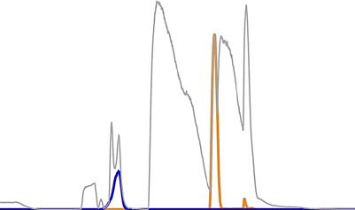
to be a prerequisite for the survival of T. forsythia [Bloch    oral surfaces for the subsequent adherence and growth of
et al., 2019]. Formation of multispecies dental plaque bio-     so called "bridge bacteria" [Socransky et al., 1998; Kolen-
films follows a clear spatiotemporal scheme and its pro-        brander, 2000]. During biofilm maturation, these “bridge
gression plays a crucial role in periodontitis. Periodontitis   bacteria,” including Fusobacterium nucleatum, create a
is a multifactorial inflammatory disease, characterized by      suitable environment for the so-called “late colonizers”
the irreversible destruction of the teeth-supporting tissue     [Diaz et al., 2000; 2002]. Among these are the periodontal
and leading to tooth loss if untreated [Holt and Ebersole,      pathogens T. forsythia, the related Bacteroidetes species
2005]. A distinction is drawn between the different colo-       Porphyromonas gingivalis, and the spirochete Treponema
nizers and stages of maturation of dental plaque biofilms,      denticola, which together build up the so-called “red
according to which the so-called “early colonizers,” which      complex” [Paster et al., 2006]. The late colonizing red
are mainly Gram-positive streptococci, precondition the         complex consortium is associated with severe forms of

                                                                                                                              1

Tannerella forsythia Peptidoglycan                              Microb Physiol 2021;31:123–134                         125
Salvage                                                         DOI: 10.1159/000516751
periodontitis [Holt and Ebersole, 2005]. Given the fas- teins, and importantly, it relies on the supply of UDP-
tidious growth requirements of T. forsythia in a labora- GlcNAc and UDP-MurNAc for efficient synthesis of its
tory monoculture, the question arises, which natural PGN.
sources the bacterium can exploit to ensure its survival in
the oral cavity. In this review, we describe the most recent
advances in the understanding of the salvage pathways PGN Biosynthesis in T. forsythia in Comparison to
used by T. forsythia within oral microbial communities to Other Bacteria and Characterization of GlmU*
acquire the essential growth factor MurNAc and how this
is metabolized to allow PGN biosynthesis. Given the presence of multiple amino sugars in its dif-
ferent cell envelope structures, T. forsythia surprisingly
lacks generally essential proteins of the amino sugar bio-
PGN Type and Cell Envelope Structure of T. forsythia synthetic pathway. The bacterium lacks a glucosamine
6-phosphate synthase (glutamine-fructose 6P amido-
As a Gram-negative bacterium, T. forsythia contains a transferase; GlmS), which converts the fructose 6-phos-
thin, moderately crosslinked PGN layer located within phate intermediate of glycolysis to glucosamine 6-phos-
the periplasm, embedded between the inner membrane phate (GlcN 6P) [Badet et al., 1987]. This enzyme is the
and the lipopolysaccharide (LPS)-containing outer mem- initial amino sugar biosynthetic enzyme of most bacteria,
brane. A recent compositional analysis of the PGN of T. indicating that T. forsythia is unable to de novo synthesize
forsythia revealed the typical carbohydrate building GlcN 6P and, hence, amino sugars derived from it. This
blocks GlcNAc and MurNAc as well as the 1,6-anhydro- enzyme is also the main regulator of amino sugar metab-
form of MurNAc (anhMurNAc), constituting the termi- olism in other bacteria. To adjust the cellular level of GlcN
nal ends of the PGN chains [Mayer et al., 2019b]. T. for- 6P to the growth requirements, glmS expression is con-
sythia PGN was assigned to the classical A1γ-type PGN trolled via a GlmS-riboswitch in the Gram-positive bac-
of Gram-negative bacteria with meso-diaminopimelic terium Bacillus subtilis and via small regulatory RNAs
acid (mDAP) involved in direct crosslinks [Schleifer and and the sensor protein RapZ in the Gram-negative bacte-
Kandler, 1972; Mayer et al., 2019b]. The LPS of T. for- rium Escherichia coli [Klein and Ferre-D'Amare, 2006;
sythia is of the rough type and contains the membrane Kalamorz et al., 2007]. Since T. forsythia lacks GlmS, the
anchored lipid A, inner and outer core – but lacks a distal regulation of amino sugar metabolism has to be regulated
O-specific antigen [Raetz and Whitfield, 2002; Posch et in a different way in this organism. The presence of the
al., 2013]. A characteristic of the cell envelope of T. for- subsequent enzyme of the general amino sugar anabolic
sythia is the additional coverage of the outer membrane pathway, which isomerizes GlcN 6P to glucosamine α-1-
by a two-dimensional crystalline surface (S)-layer, con- phosphate (GlcN 1P) [Mengin-Lecreulx and van Heijen-
sisting of self-assembling glycoproteins (Tf_TfsA and Tf_ oort, 1996], is uncertain in T. forsythia. A glucosamine-
TfsB) [Lee et al., 2006; Posch et al., 2011b; Sekot et al., phosphate isomerase (Tf_GlmM; BFO_1882; Tanf_
2012]. A glycosylated S-layer is rare among Gram-nega- 04345) annotated in the genome of T. forsythia reveals
tive bacteria and characterizes different T. forsythia higher similarity to phosphogluco-/phosphomannomu-
strains, containing either a pseudaminic acid or legion- tases (e.g., 30% amino acid identity with AlgC of Pseudo-
aminic acid derivative at the non-reducing terminal end monas putida) than to GlmM-like phosphoglucosamine
of a complex S-layer oligosaccharide. These sialic acid- mutases. In most bacteria, GlcN 1P is further converted
like nonulosonic acids (9-carbon backbone 2-keto acid to UDP-GlcNAc by a bifunctional N-acetyltransferase/
sugars) were shown to influence pathogenicity of the bac- uridylyl transferase (GlmU), which comprises an N-ter-
terium [Posch et al., 2011a; Friedrich et al., 2017; Bloch et minal GlcN 1P acetyltransferase domain and a C-termi-
al., 2019] and their synthesis was shown to depend on nal GlcNAc 1P uridylyl transferase domain [Mengin-Lec-
either UDP-GlcNAc or GDP-GlcNAc, respectively, as reulx and van Heijenoort, 1994; Gehring et al., 1996]. T.
precursors [Schoenhofen et al., 2006; Schoenhofen et al., forsythia lacks a bifunctional UDP-GlcNAc synthase
2009; Friedrich et al., 2017]. Since T. forsythia constantly [Hottmann et al., 2018; Ruscitto and Sharma, 2018]. We
builds up its complex cell envelope during growth, the found, however, that the organism possesses a mono-
organism relies on the supply of a suitable amino sugar functional GlcNAc 1P uridylyl transferase (designated
precursors for an efficient synthesis of its complex cell Tf_GlmU*; BFO_1878; Tanf_04325), which is a homolog
wall glycoconjugates, including LPS, S-layer glycopro- of a monofunctional Cc_GlmU* (43% overall amino acid
126 Microb Physiol 2021;31:123–134 Hottmann/Borisova/Schäffer/Mayer
DOI: 10.1159/000516751tion was analyzed using [32P]-GlcNAc α-1-phosphate
(GlcNAc 1P) as a substrate (shown in Fig. 2; also see ma-
Gl d
U*
d dar
terial and methods). After the addition of GlmU* and
m
rif tan
Pu in s
ie UTP, we observed the direct conversion of GlcNAc 1P to
e
ot
Pr
UDP-GlcNAc, which was not detectable in the negative
UDP- control. For an unequivocal product determination, a re-
GlcNAc
action mixture with non-labeled GlcNAc 1P was also ana-
GlcNAc 1P
lyzed by electrospray-ionization time-of-flight (ESI-TOF)
kDa
32 mass spectrometry as described earlier [Gisin et al., 2013]
25 (shown in Fig. 3). The substrate GlcNAc 1P appeared with
γ32-ATP a mass of (M-H)- = 300.063 m/z in negative ion mode,
Start
which is in accordance with the theoretical mass of GlcNAc
+ GlmU* + H2O 1P ((M-H)- = 300.049 m/z) (Fig. 3; in blue). After incuba-
0 3 0 3 tion with Tf_GlmU*, the substrate was partly converted
Time, h into a product with (M-H)- = 606.100 m/z, which is in
agreement with the expected mass of UDP-GlcNAc ((M-
H)- = 606.074 m/z) (Fig. 3b; in orange)).
Fig. 2. Purity and activity of recombinant Tf_GlmU* analyzed by
SDS-PAGE and by UDP-GlcNAc formation using a radioactive
The biosynthesis of PGN is a conserved process within
enzyme assay. Left: Coomassie stained 12% SDS-PAGE gel; lane 1, bacteria that can be divided into three major steps occur-
protein standard mixture with molecular masses as indicated; lane ring in different cell compartments. In the first step, the
2, purified, recombinant GlmU*. Right: Radioactive assay of re- soluble precursor molecules UDP-MurNAc and UDP-
combinant GlmU*. [32P]-GlcNAc α-1P (GlcNAc 1P) was gener- MurNAc-pentapeptide are synthesized from UDP-
ated from GlcNAc and [32P]-ATP using an anomeric MurNAc/
GlcNAc kinase (AmgK from P. putida KT2440) [Gisin et al., 2013;
GlcNAc in the cytoplasm (shown in Fig. 1). In the second
Renner-Schneck et al., 2015]. Furthermore, [32P]-GlcNAc α-1P step, the undecaprenyl-phosphate-anchored PGN pre-
(GlcNAc 1P) was incubated with 5 µg of Tf_GlmU* and 50 mM cursors lipid I and lipid II are synthesized at the cytoplas-
UTP (control with water instead of enzyme) and reactions were mic membrane, which, in the last step, are assembled to
spotted on a TLC plate immediately (0) and after 3 h (3) of incuba- polymeric, crosslinked PGN in the periplasm. UDP-Mur-
tion at 37°C.
NAc, the first dedicated precursor of PGN, is synthesized
from UDP-GlcNAc via the enzymes MurA and MurB,
representing the known route for MurNAc synthesis in
nature. In the first reaction, MurA catalyzes the transfer
sequence identity to Ccan_15070) that has been identi- of an enolpyruvate from phosphoenolpyruvate to the
fied in the oral pathogen Capnocytophaga canimorsus 3′-hydroxyl group of UDP-GlcNAc. The thereby gener-
[Renzi et al., 2015]. C. canimorsus is auxotrophic for ated UDP-GlcNAc-enolpyruvate is subsequently reduced
GlcNAc (not MurNAc), but is able to salvage this amino by MurB using one reduction equivalent of NADPH to
sugar from salivary mucins present in the oral cavity. As generate UDP-MurNAc [Benson et al., 1993]. In T. for-
an adaption to the availability of GlcNAc in the oral bac- sythia, these first committed steps in the biosynthesis of
terium’s settling, the dog mouth, C. canimorsus appar- PGN are not possible due to the lack of the initiating en-
ently has lost its capability to de novo synthesize UDP- zymes MurA/MurB. All the following reactions leading
GlcNAc, hence, its genome has lost the genes encoding to UDP-MurNAc-pentapeptide (MurC-F, Ddl, MurI,
GlmM and the bifunctional GlmU [Renzi et al., 2015]. Alr) and the further reactions leading to lipid II (MraY,
To determine the function of Tf_GlmU*, the 309-ami- MurG), are present in T. forsythia (shown in Fig. 1). These
no acid recombinant enzyme containing a C-terminal reactions are responsible for the sequential ATP-depen-
His6-tag was expressed in E. coli BL21 (DE3) cells. The dent ligation of single amino acids. MurC (BFO_0400;
protein was purified using nickel-affinity chromatogra- Tanf_11285) transfers L-alanine to UDP-MurNAc, and
phy, followed by size exclusion chromatography (using MurD (BFO_0403; Tanf_11300) and MurE (BFO_0405;
HiLoad 16/60 Superdex 200 column, GE Healthcare) to Tanf_11310) further add D-glutamic acid (generated by
near homogeneity (shown in Fig. 2). With a radioactive the L-/D-glutamate racemase MurI; BFO_1448;
uridylyl transferase assay as described earlier [Gisin et al., Tanf_09790) and mDAP, yielding UDP-MurNAc-tri-
2013; Renner-Schneck et al., 2015], the Tf_GlmU*-reac- peptide. Finally, MurF (BFO_1349; Tanf_06315) is re-
Tannerella forsythia Peptidoglycan Microb Physiol 2021;31:123–134 127
Salvage DOI: 10.1159/0005167512.0 2.0 UDP-GlcNAc
(M-H)– 606.100 m/z
1.5 1.5
MS intensity × 106, cps
MS intensity × 106, cps
GlcNAc 1P
(M-H)– 300.062 m/z
BPC BPC
1.0 1.0 GlcNAc 1P
(M-H)– 300.062 m/z
0.5 0.5
0 0
0 10 20 30 0 10 20 30
a Time, min b Time, min
Fig. 3. Analysis of uridylyl transferase activity of recombinant Tf_ ed ion chromatograms (EICs) for GlcNAc 1P (blue) and UDP-
GlmU* by LC-MS. The reaction mixture containing GlcNAc 1P in GlcNAc (orange). The measured mass-to-charge ratios of the
Tris-HCl buffer (pH 7.6) was analyzed prior (a) and after addition compounds, indicated in the figure, are in agreement with the the-
of UTP and purified recombinant Tf_GlmU* of T. forsythia (b). oretical m/z values of (M-H)- = 300.049 m/z for GlcNAc 1P and of
Shown are the base peak chromatograms (BPCs; grey) and extract- (M-H)- = 606.074 m/z for UDP-GlcNAc.
quired for the addition of the dipeptide D-Ala-D-Ala, (BFO_0400; Tanf_11295) and RodA (BFO_3355;
which in generated by ligation of two D-alanine molecules Tanf_08125) can be found on the T. forsythia genome, as
by the ligase Ddl (BFO_3290; Tanf_02600), yielding well as Rod-complex and divisome components and pen-
UDP-MurNAc-pentapeptide [Smith, 2006]. In the sec- icillin-binding protein PGN synthases.
ond step, the membrane-anchored precursors lipid I and
lipid II are synthesized by MraY (BFO_0404; Tanf_11305)
and MurG (BFO_0401; Tanf_11290), respectively (shown MurNAc Uptake and Metabolism in T. forsythia:
in Fig. 1). The prenyl sugar transferase MraY firstly trans- Evidence for an AmgK-MurU Salvage Route
fers phospho-MurNAc-pentapeptide to the lipid carrier
undecaprenyl phosphate, generating undecaprenyl pyro- Since T. forsythia cannot de novo synthesize UDP-
phosphoryl-MurNAc-pentapeptide, named lipid I [Al- MurNAc due to the lack of murA and murB orthologs
Dabbagh et al., 2008]. Subsequently, lipid II is generated (Fig. 1), it relies on the salvage of exogenous MurNAc or
by the action of the glycosyltransferase MurG that trans- alternative sources of MurNAc. As mentioned above, T.
fers a GlcNAc moiety from UDP-GlcNAc onto lipid I forsythia's dependency on external MurNAc was first re-
[Bouhss et al., 2004]. In the final third step, lipid II is ported by Wyss in 1989, who observed that exogeneous
translocated to the outer leaflet of the cytoplasmic mem- MurNAc is a crucial growth factor and required for the
brane, which is mediated by MurJ-like flippases belong- maintenance of a rod-shaped (fusiform) cell morphology
ing to the multidrug/oligosaccharidyl-lipid/polysaccha- [Wyss, 1989]. We confirmed the MurNAc-dependency
ride (MOP) exporter superfamily, allowing the assembly of T. forsythia and the transition from healthy rod-shaped
of PGN by PGN-synthases [Typas et al., 2012; Sham et al., to fusiform cells upon MurNAc starvation [Hottmann et
2014; Cho et al., 2016]. Surprisingly, T. forsythia lacks an al., 2018]. This observation clearly showed that T. forsyth-
obvious MurJ homolog, and it is thus unclear how lipid ia must utilize an uptake system for MurNAc (and other
II is translocated in this organism. Importantly, however, MurNAc-sources) and a biosynthesis pathway leading to
an undecaprenyl-phosphate carrier system is present, as UDP-MurNAc that substitutes for MurA and MurB.
T. forsythia contains both an undecaprenyl diphosphate The ability to recover components of their own cell
synthase (UppS; BFO_1444; Tanf_09810) and an undeca- wall is a common feature of bacteria. During growth of
prenyl diphosphatase (UppP/BacA, BFO_1764; bacteria, the cell wall undergoes a permanent turnover,
Tanf_03865). Additionally, homologs of FtsW and the released breakdown products are recovered in a
128 Microb Physiol 2021;31:123–134 Hottmann/Borisova/Schäffer/Mayer
DOI: 10.1159/000516751process known as PGN recycling [Mayer et al., 2019a]. In two metabolites, MurNAc and the PGN precursor mol-
Gram-negative bacteria, PGN recycling involves the ecule UDP-MurNAc-pentapeptide [Hottmann et al.,
cleavage of the PGN sacculus by lytic transglycosylases 2018]. The accumulation of the latter provided that Mur-
generating anhydromuropeptides (GlcNAc-anhMur- NAc can also be channeled into the anabolic PGN synthe-
NAc-peptides) which are efficiently recovered via the sis route as evidenced by a growth advantage of a T. for-
AmpG transporter [Mayer et al., 2019a]. Some bacteria sythia ΔmurK mutant in MurNAc-depleted growth me-
can grow on PGN components provided as nutrients. For dium [Hottmann et al., 2018].
instance, in E. coli, the uptake of MurNAc is mediated by Pseudomonas spp. possess an alternative route for the
a phosphotransferase system (PTS) transporter that im- synthesis of MurNAc that bypasses the de novo biosyn-
ports and simultaneously phosphorylates MuNAc, yield- thesis of UDP-MurNAc and recovers MurNAc directly as
ing MurNAc 6P [Dahl et al., 2004]. The typical PTS trans- UDP-MurNAc [Gisin et al., 2013]. This pathway in-
ports and phosphorylates sugars via the concerted action cludes, first, the phosphatase MupP, which dephosphory-
of the cytoplasmatic enzymes EI (enzyme I) and HPr (his- lates MurNAc 6P forming MurNAc [Borisova et al.,
tidine protein), and the EII complex, which is composed 2017]. Next, the anomeric kinase AmgK phosphorylates
of the sugar-specific domains EIIA and EIIB, and the the amino sugars GlcNAc and MurNAc forming GlcNAc
transmembrane domain EIIC) [Siebold et al., 2001]. Mur- α-1P an MurNAc α-1P, respectively. Subsequently, the
NAc 6P is the substrate of MurQ, a key enzyme of cell wall α-1-phosphorylated sugars can be used by GlmU and the
recycling, i.e., degradation of anhydromuropeptides, and uridylyl transferase MurU for the generation of UDP-
MurNAc metabolism, which converts MurNAc 6P to GlcNAc and UDP-MurNAc, respectively. Biochemical
GlcNAc 6P [Jaeger et al., 2005; Borisova et al., 2016]. Ge- characterization of MurU of P. putida revealed stringent
nome analysis of T. forsythia revealed the presence of an specificity for the uridylation of MurNAc α-1P and not
ortholog of murQ but not of murP [Ruscitto et al., 2016]. GlcNAc α-1P [Renner-Schneck et al., 2015]. Similar func-
In view of the genetic context, Tf_murQ is part of a three- tions might be provided by the orthologs Tf_amgK and
gene cluster encoding a putative sodium solute transport- Tf_murU found in T. forsythia (unpublished data). Most
er (Tf_MurT; BFO_0041; Tanf_08375), a putative sugar likely, the Tf_AmgK/Tf_MurU pathway is the principal
kinase (Tf_MurK; BFO_0042; Tanf_08380), and the pu- route for PGN synthesis in T. forsythia and replaces the
tative MurNAc 6P lactyl ether hydrolase MurQ (Tf_ missing MurA/MurB pathway. The deficit of genes is a
MurQ; BFO_0044; Tanf_08385). The Tf_MurT trans- phenomenon reported from other bacteria that thrive in
porter was characterized as a PTS-independent MurNAc biofilms. For so far unknown reason, this might provide
transporter, the first known MurNAc uptake system of an increased fitness [Koskiniemi et al., 2012].
this kind, and Tf_MurK and Tf_MurQ were character-
ized as a specific MurNAc kinase and a bona fide MurNAc
6P lacyl ether hydrolase, respectively [Ruscitto et al., T. forsythia Salvages PGN Turnover Products from
2016; Hottmann et al. 2018]. The clustered organization Cohabiting Bacteria
of the murTKQ genes seems quite common among or-
ganisms from the Bacteroidetes phylum [Ruscitto et al., T. forsythia grows on MurNAc, yet anhydromuropep-
2016; Ruscitto and Sharma, 2018]. tides (GlcNAc-AnhMurNAc-peptides) and muropep-
For the metabolization of sugars their phosphoryla- tides (GlcNAc-MurNAc-peptides) are more commonly
tion is mandatory. As MurNAc is not phosphorylated in found in the natural environment [Fujimoto and Fukase,
T. forsythia by the transporter MurT, the concomitance 2011]. Anhydromuropeptides constitute the general
of a sugar kinase (Tf_MurK) also encoded within the PGN turnover products of bacteria, while muropeptides
murTKQ cluster is consequential [Ruscitto et al., 2016; represent PGN degradation products resulting from the
Hottmann et al., 2018]. Biochemical investigations of ki- activity of lysozyme-like muramidases [Mayer et al.,
nase activity of MurK revealed stringent specificity for 2019a]. Thus, it is reasonable to assume that T. forsythia
MurNAc phosphorylation at the position C6, thereby salvages anhydromuropeptides and muropeptides which
generating MurNAc 6P, the substrate for the etherase both likely occur in oral biofilms. Anhydromuropeptides
MurQ and catabolism [Hottmann et al., 2018]. The phos- are generated in large amounts during cell wall turnover,
phorylation position of MurNAc represents a metabolic whereby the PGN layer is steadily cleaved by autolytic en-
branching point in T. forsythia. Intriguingly, the deletion zymes (autolysins), namely lytic transglycosylases and
of Tf_murK (ΔTf_murK) results in the accumulation of endopeptidases to enable bacterial growth [Höltje, 1995;
Tannerella forsythia Peptidoglycan Microb Physiol 2021;31:123–134 129
Salvage DOI: 10.1159/000516751Table 1. Occurrence of selected recycling/turnover enzymes in T. forsythia and cohabiting bacteria
PGN recycling & turnover Reference strain T. forsythiaa F. nucleatuma T. denticolaa P. gingivalisa
enzyme (Uniprot ID)
MurQ (P76535) E. coli K12 46%b, c nd nd 47%f
AmpG (P0AE16) E. coli K12 24%b, c nd nd nd
NagZ (P75949) E. coli K12 28%b, c nd 33%d 31%f
AnmK (P77570) E. coli K12 nd nd nd nd
AmgK (Q9I5U1) P. aeruginosa PA01 25%b, 26%c nd nd nd
NamZ (P40407) B. subtilis 168 47, 36, 38% nd 46%e 37%f
(three orthologs)b, c
a
BlastP analysis was performed using the non-redundant protein sequences database for searching of orthologs in the genome of T.
forsythia 92A2 (taxid: 203275) and ATCC 43037 (accession number JUET00000000 [Friedrich et al., 2015]), F. nucleatum (taxid: 851),
T. denticola (taxid: 158) and P. gingivalis (taxid: 837). Identity percentages relative to the indicated query protein are shown for orthologs
from b T. forsythia strain 9 A2A, c T. forsythia stain ATCC 43037, d T. denticola strain ATCC 35405, e T. denticola strain SP3, f P. gingivalis
strain W83. nd, orthologs not detected.
van Heijenoort, 2011; Mayer et al., 2019a]. In E. coli, per treatment of the (anhydro-)muropeptides with a recom-
generation, about 50% of the PGN of the cell wall is binant E. coli amidase (AmiD), yielded identical growth
cleaved by autolysins releasing anhydromuropeptides rates compared with the peptide-containing (anhydro-)
into the periplasm, from where they are efficiently inter- muropeptides. Thus, apparently, an endogenous amidase
nalized (recycled) by the inner membrane permease in T. forsythia is able to remove the peptide portions from
AmpG [Park and Uehara, 2008; Mayer et al., 2019a]. the (anhydro)-muropeptides, yielding disaccharides. In-
While in T. forsythia, a AmpG transporter homolog triguingly, the T. forsythia ΔampG mutant, in contrast to
(BFO_0039; Tanf_08365) was identified, several cohabi- the T. forsythia wild-type, did not grow in medium sup-
tating bacteria of T. forsythia (shown in Table 1), such as plemented with the peptide-free disaccharides GlcNAc-
F. nucleatum, T. denticola, and P. gingivalis, lack ortho- MurNAc and GlcNAc-anhMurNAc. This distinguishes
logs of the E. coli AmpG permease. This indicates that T. the T. forsythia AmpG-ortholog from E. coli AmpG, as
forsythia might benefit from PGN turnover products re- the specificity of the latter is restricted to anhydromuro-
leased by these bacteria within the oral biofilm. Although peptides and GlcNAc-anhMurNAc but does not accept
Tf_AmpG shows rather low (24%) sequence identity to E. GlcNAc-MurNAc [Cheng and Park, 2002].
coli AmpG [Ruscitto et al., 2017], its function as an anhy-
dromuropeptide permease was initially proposed on the
basis the ability of Tf_ampG to complement an E. coli PGN Serves as MurNAc-Source for T. forsythia
ampDampG mutant using a reporter assay [Ruscitto et al.,
2017]. Subsequently, we showed that T. forsythia grows An exciting recent finding was that T. forsythia could
on anhydromuropeptides derived from E. coli PGN also use intact PGN as a growth factor [Mayer et al., 2020].
cleaved with a lytic transglycosylase, while a T. forsythia PGN from the cohabiting bacteria P. gingivalis and F. nu-
ΔampG deletion mutant cannot [Mayer et al., 2020]. In- cleatum, from E. coli, as well as from T. forsythia, served
triguingly, we also revealed Tf_AmpG to be responsible as a growth factor for T. forsythia, replacing MurNAc or
for growth in the presence of muropeptides derived from (anhydro-)muropeptides. In contrast, the T. forsythia
muramidase-cleaved PGN of either F. nucleatum or E. ∆ampG mutant was severely impaired in growth when
coli [Mayer et al., 2020]. Surprisingly, growth on muro- cultured with intact PGN instead of MurNAc, indicating
peptides was clearly favoured over that on anhydromuro- that transport via AmpG is critical to sustain growth on
peptides, as evidenced by a final OD about twice as high PGN [Mayer et al., 2020]. This observation implies that
with muropeptides as compared to identical amounts of T. forsythia is able to degrade PGN mainly to GlcNAc-
anhydromuropeptides [Mayer et al., 2020]. Further, we MurNAc or GlcNAc-anhMurNAc disaccharides rather
could show that peptide-free disaccharides, i.e. GlcNAc- than to MurNAc. In our group, we recently identified and
anhMurNAc and GlcNAc-MurNAc generated by pre- characterized a novel exo-N-acetylmuramidase in B. sub-
130 Microb Physiol 2021;31:123–134 Hottmann/Borisova/Schäffer/Mayer
DOI: 10.1159/000516751tilis named NamZ [Müller et al., 2021]. This enzyme re- strates of the T. forsythia AmpG and MurT transporters, leases MurNAc from glycoside substrates carrying a Mur- respectively. Based on our findings, we suggest that PGN NAc at the non-reducing end, such as the chromogenic is internalized by T. forsythia through the outer mem- reporter substrate para-nitrophenyl-MurNAc (pNP- brane, possibly via a SusCD-like uptake complex (shown MurNAc), the natural PGN-derived disaccharide Mur- in Fig. 1). Sus complexes are common uptake complexes NAc-GlcNAc as well as peptide-free PGN chains termi- within the polysaccharide utilization loci (PUL) of Bacte- nating with a MurNAc residue. NamZ was found to con- roidetes bacteria, consisting of TonB-dependent outer stitute the founding member of a novel family of membrane transporter (SusC) and cell surface glycan- exo-acting PGN β-N-acetylmuramidases [Müller et al., binding proteins (SusD) [Grondin et al., 2017; Lapebie et 2021]. In the light of MurNAc-auxotrophy of T. forsythia, al., 2019]. An amidase, as mentioned earlier, is required we reasoned that the presence of enzymes exhibiting exo- to remove the peptides from PGN upon internalization. β-N-acetylmuramidase activity could be beneficial. In- Peptide-free exogeneous PGN is then processed by triguingly, the genome of T. forsythia encodes three or- NamZ1 and NamZ2 to yield GlcNAc-MurNAc/GlcNAc- thologs of B. subtilis NamZ (Tf_NamZ1: BFO_0040; anhMurNAc and MurNAc (shown in Fig. 1) and subse- Tanf_08370; Tf_NamZ2: BFO_2140; Tanf_00660, and quentyl these fragments are taken up via Tf_AmpG and Tf_NamZ3: BFO_2092; Tanf_00855; see Table 1). Fur- Tf_MurT and are further processed intracellularly by thermore, NamZ orthologs were also found in P. gingiva- shuttling to the UDP-MurNAc biosynthesis pathway, via lis as well as in T. denticola (Table 1), consistent with the Tf_AmgK and Tf_MurU, or through the catabolic path- finding that the NamZ family glycosidases (CAZy family way via Tf_MurK and Tf_MurQ (shown in Fig. 1). GH171; www.cazy.org/GH171) are prominently found within the members of the Fibrobacteres-Chlorobi-Bacte- roidetes FCB superphylum [Müller et al., 2021]. The T. Conclusion forsythia NamZ2 was shown to have a similar function to NamZ of B. subtilis. This enzyme cleaves peptide-free The MurNAc auxotrophic oral pathogen T. forsythia PGN glycans and MurNAc-glycosides with a MurNAc at compensates for its fastidious growth requirements in the reducing end, and generates MurNAc, which can be the oral cavity by salvaging of MurNAc, and MurNAc- transported into the cell via MurT (shown in Fig. 1). The containing PGN fragments such as GlcNAc-MurNAc other NamZ ortholog, Tf_NamZ1, was identified as a and GlcNAc-anhydroMurNAc disaccharides as well as GlcNAc-MurNAc disaccharide-releasing exolytic β-N- (anhydro-)muropeptides, which are common cell wall acetylmuramidase. This enzyme also releases MurNAc by turnover products, presumably released into the environ- hydrolysis of MurNAc-glycosides, albeit less efficiently ment by cohabiting bacteria. Intriguingly, we revealed than NamZ2; its main function is the cleavage of peptide- that many of the cohabiting bacteria lack an ortholog of free PGN yielding GlcNAc-MurNAc disaccharides (as AmpG, and thus cannot recover the GlcNAc-MurNAc well as GlcNAc-anhMurNAc disaccharides, which com- and GlcNAc-anhydroMurNAc disaccharides released monly resides at the reducing ends of the glycans). The from their PGN cell wall during turnover. Sometimes namZ1 gene is located directly adjacent to ampG on the they even lack further important PGN recycling enzymes T. forsythia genome, which encodes the transporter of (MurQ, AnmK, AmgK; see Table 1). Thus, these bacteria GlcNAc-MurNAc and GlcNAc-anhMurNAc [Mayer et likely provide T. forsythia with essential PGN turnover al., 2020]. Intriguingly, the subsequent gene downstream fragments. Moreover, T. forsythia is able to salvage intact of ampG, lytB, displays significant similarity to SpoIID- PGN, which is processed by two members of a newly domain containing lytic transglycosylases [Morlot et al., identified family of exo-β-N-acetylmuramidases (Tf_ 2010]. Moreover, T. forsythia possesses at least three fur- NamZ1 and Tf_NamZ2) within the periplasm. Since ther putative lytic transglycosylases belonging to glycosyl these enzymes require peptide-free PGN as substrates, we hydrolase family 23 (www.cazy.org). These enzyme may suggest that the initial removal of the peptide stems from be involved in trimming peptide-free, exogeneous PGN incoming PGN by an unknown amidase is critical to de- or be involved in PGN recycling in T. forsythia. In sum- grade exogeneous PGN within the periplasm. This might mary, the exo-N-acetylmuramidases Tf_NamZ1 and Tf_ be a strategy of T. forsythia to distinguish between exog- NamZ2 presumably degrade peptide-free PGN fragments enous PGN (nutritional source) and endogenous PGN in the periplasm to yield GlcNAc-MurNAc (besides (i.e., its own cell wall), because the exo-β-N- GlcNAc-anhMurNAc) and MurNAc, which are the sub- acetylmuramidases specifically cleave peptide-free exo Tannerella forsythia Peptidoglycan Microb Physiol 2021;31:123–134 131 Salvage DOI: 10.1159/000516751
genous PGN and leave the structural PGN unattached.                    tion mixture was analyzed before and after the addition of GlmU*
Follow-up studies will identify the proposed amidase and               and 50 mM UTP, using HPLC connected to an electrospray-ion-
                                                                       ization time-of-flight (ESI-TOF) mass spectrometer (Micro-TOF
clarify whether exogenous PGN is transported through                   II; Bruker) [Gisin et al., 2013].
the outer membrane via SusCD-like complexes. These
complexes are clustered within so-called PULs and fre-
quently used by Bacteroidetes to forage polysaccharides.                  Acknowledgement
However, a PGN-specific PUL has so far not been identi-
fied in the T. forsythia genome. Moreover, the roles of                   We thank Valentin Friedrich for very helpful discussions and
AmgK and MurU in PGN biosynthesis remains to be                        Libera lo Presti for linguistic editing.
clarified. The understanding of cell wall and amino sugar
metabolism in T. forsythia and the elucidation of the dif-
ferent MurNAc-containing sources and routes of uptake                     Statement of Ethics
and metabolism used by this bacterium to survive within
                                                                           Ethics approval was not required, as the work includes only an
the oral microbial community will help to understand its               in vitro characterization of a carbohydrate metabolic enzyme.
physiological demands and set the basis to develop new
approaches for the treatment of periodontitis.
                                                                          Conflict of Interest Statement

   Materials and Methods/Experimental Procedures                          The authors have no conflicts of interest to declare.

    Plasmid Construction, Expression, and Purification of GlmU*
    To determine the function of Tf_GlmU*, the encoding gene
                                                                          Funding Sources
was amplified from genomic DNA of T. forsythia ATCC 43037 us-
ing the primer pair GlmU*fw_ TACGAACATATGAAACCAA-
                                                                          This work was supported by the Deutsche Forschungsgemein-
CATTATTTGTGCTGGCGGC/GlmU*rev_ GCGCTCGAGAC-
                                                                       schaft (DFG; German Research Foundation, projects GRK1708/
CGAACAGCTGAGCGGGATATTC (introduced restriction
                                                                       B2 MA2436/7-1 and SFB766/A15; as well as the Germany' Excel-
sites are shown in bold) and cloned in vector pET29b for the ex-
                                                                       lence Strategy -EXC2124 - 390838134) and the Austrian Science
pression as a C-terminally His6-tagged-fusion protein. The PCR
                                                                       Fund FWF (project I 2875-B22).
product and vector pET29 were digested with NdeI and XhoI
(NEB, Frankfurt, Germany) and ligated using T4 DNA ligase
(Thermo Fisher Scientific, Waltham, MA, USA). The resulting
plasmid (pET29_GlmU*) was transformed into E. coli BL21(DE3)              Author Contributions
cells for overproduction.
                                                                          I.H. performed all experiments. I.H., M.B., C.S. and C.M. con-
    Radioactive Uridylyl Transferase Assay                             ceived and wrote the manuscript.
    The Tf_GlmU* reaction was analyzed with a radioactive uridy-
lyl transferase assay using [32P]-GlcNAc α-1-phosphate as a sub-
strate, which was prepared from GlcNAc and γ-[32P]-ATP using
AmgK from P. putida as described earlier [Gisin et al., 2013;
Renner-Schneck et al., 2015]. For the radioactive uridylyl transfer-      References          Al-Dabbagh B, Henry X, El Ghachi M, Auger G,
ase assay, 5 µL of the AmgK reaction mixture were incubated with                                 Blanot D, Parquet C, et al. Active site mapping
50 mM UTP and 1 µg of Tf_GlmU* in 100 mM Tris-HCl buffer (pH                                     of MraY, a member of the polyprenyl-phos-
7.6) and spotted immediately and after 3 h of incubation at 37°C                                 phate N-acetylhexosamine 1-phosphate
                                                                                                 transferase superfamily, catalyzing the first
on a TLC plate (silica gel 60 F254; Merck, Darmstadt, Germany).
                                                                                                 membrane step of peptidoglycan biosynthe-
After separation in h n-butyl alcohol:methanol:25% (wt/vol) am-                                  sis. Biochemistry. 2008 Aug 26; 47(34): 8919–
monium hydroxide:water (5:4:2:1), the TLC plate was analyzed us-                                 28.
ing a Typhoon Trio Biomolecular Imager (GE Healthcare).                                       Badet B, Vermoote P, Haumont PY, Lederer F,
                                                                                                 LeGoffic F. Glucosamine synthetase from
   LC-MS Uridylyl Transferase Assay                                                              Escherichia coli: purification, properties, and
   A non-radioactive, LC-MS uridylyl transferase assay was per-                                  glutamine-utilizing site location. Biochemis-
formed as described above but using unlabeled GlcNAc α-1P as the                                 try. 1987 Apr 7;26(7):1940–8.
substrate, which was prepared according to a published protocol                               Benson TE, Marquardt JL, Marquardt AC, Etz-
                                                                                                 korn FA, Walsh CT. Overexpression, purifi-
[Gisin et al., 2013]. 25 mM of GlcNAc α-1P was used in a 100-µL                                  cation, and mechanistic study of UDP-N-
reaction mixture containing 100 mM Tris-HCl (pH 7.6), 50 mM                                      acetylenolpyruvylglucosamine          reductase.
UTP, 1 U baker’s yeast inorganic pyrophosphatase and 5 µg of                                     Biochemistry. 1993 Mar 2;32(8):2024–30.
Tf_GlmU*. The reaction was incubated for 3 h at 37°C. The reac-

132                    Microb Physiol 2021;31:123–134                                         Hottmann/Borisova/Schäffer/Mayer
                       DOI: 10.1159/000516751
Bloch S, Tomek MB, Friedrich V, Messner P, Gehring AM, Lees WJ, Mindiola DJ, Walsh CT, for an opinion. Int J Syst Evol Microbiol. 2003
Schäffer C. Nonulosonic acids contribute to Brown ED. Acetyltransfer precedes uridylyl- Nov;53(Pt 6):2111–2.
the pathogenicity of the oral bacterium Tan- transfer in the formation of UDP-N-acetyl- Mayer C, Kluj RM, Mühleck M, Walter A, Unsle-
nerella forsythia. Interface Focus. 2019 Apr 6; glucosamine in separable active sites of the ber S, Hottmann I, et al. Bacteria's different
9(2):20180064. bifunctional GlmU protein of Escherichia ways to recycle their own cell wall. Int J Med
Borisova M, Gaupp R, Duckworth A, Schneider coli. Biochemistry. 1996 Jan 16;35(2):579–85. Microbiol. 2019a Nov;309(7):151326.
A, Dalügge D, Mühleck M, et al. Peptidogly- Gisin J, Schneider A, Nägele B, Borisova M, May- Mayer VMT, Hottmann I, Figl R, Altmann F,
can recycling in Gram-positive bacteria is er C. A cell wall recycling shortcut that by- Mayer C, Schäffer C. Peptidoglycan-type
crucial for survival in stationary phase. mBio. passes peptidoglycan de novo biosynthesis. analysis of the N-acetylmuramic acid auxo-
2016 Oct 11;7(5):e00923-16. Nat Chem Biol. 2013 Aug;9(8):491–3. trophic oral pathogen Tannerella forsythia
Borisova M, Gisin J, Mayer C. The N-acetylmu- Grondin JM, Tamura K, Déjean G, Abbott DW, and reclassification of the peptidoglycan-type
ramic acid 6-phosphate phosphatase MupP Brumer H. Polysaccharide Utilization Loci: of Porphyromonas gingivalis. BMC Microbi-
completes the Pseudomonas peptidoglycan Fueling Microbial Communities. J Bacteriol. ol. 2019b Sep 2;19(1):200.
recycling pathway leading to intrinsic fosfo- 2017 Aug 1;199(15). Mayer VMT, Tomek MB, Figl R, Borisova M,
mycin resistance. mBio. 2017 Mar 28;8(2). Holt SC, Ebersole JL. Porphyromonas gingivalis, Hottmann I, Blaukopf M, et al. Utilization of
Bouhss A, Crouvoisier M, Blanot D, Mengin-Le Treponema denticola, and Tannerella for- different MurNAc sources by the oral patho-
creulx D. Purification and characterization of sythia: the "red complex", a prototype poly- gen Tannerella forsythia and role of the inner
the bacterial MraY translocase catalyzing the bacterial pathogenic consortium in periodon- membrane transporter AmpG. BMC Micro-
first membrane step of peptidoglycan biosyn- titis. Periodontol 2000. 2005 Jun;38:72–122. biol. 2020 Nov 17;20(1):352.
thesis. J Biol Chem. 2004 Jul 16; 279(29): Höltje JV. From growth to autolysis: the murein Mengin-Lecreulx D, van Heijenoort J. Copurifi-
29974–80. hydrolases in Escherichia coli. Arch Micro- cation of glucosamine-1-phosphate acetyl-
Chen T, Abbey K, Deng WJ, Cheng MC. The bio- biol. 1995 Oct;164(4):243–54. transferase and N-acetylglucosamine-
informatics resource for oral pathogens. Nu- Hottmann I, Mayer VMT, Tomek MB, Friedrich V, 1-phosphate uridyltransferase activities of
cleic Acids Res. 2005 Jul 1; 33(Web Server is- Calvert MB, Titz A, et al. N-Acetylmuramic Escherichia coli: characterization of the glmU
sue):W734–40. acid (MurNAc) auxotrophy of the oral patho- gene product as a bifunctional enzyme cata-
Cheng Q, Park JT. Substrate specificity of the gen Tannerella forsythia: characterization of a lyzing two subsequent steps in the pathway
AmpG permease required for recycling of cell MurNAc kinase and analysis of Its role in cell for UDP-N-acetylglucosamine synthesis. J
wall anhydro-muropeptides. J Bacteriol. 2002 wall metabolism. Front Microbiol. 2018;9:19. Bacteriol. 1994 Sep;176(18):5788–95.
Dec;184(23):6434–6. Jaeger T, Arsic M, Mayer C. Scission of the lactyl Mengin-Lecreulx D, van Heijenoort J. Character-
Cho H, Wivagg CN, Kapoor M, Barry Z, Rohs ether bond of N-acetylmuramic acid by Esch- ization of the essential gene glmM encoding
PDA, Suh H, et al. Bacterial cell wall biogen- erichia coli "etherase". J Biol Chem. 2005 Aug phosphoglucosamine mutase in Escherichia
esis is mediated by SEDS and PBP polymerase 26;280(34):30100–6. coli. J Biol Chem. 1996;271(1):32–9.
families functioning semi-autonomously. Nat Kalamorz F, Reichenbach B, März W, Rak B, Morlot C, Uehara T, Marquis KA, Bernhardt TG,
Microbiol. 2016 Sep 19;1:16172. Görke B. Feedback control of glucosamine- Rudner DZ. A highly coordinated cell wall
Dahl U, Jaeger T, Nguyen BT, Sattler JM, Mayer 6-phosphate synthase GlmS expression de- degradation machine governs spore morpho-
C. Identification of a phosphotransferase sys- pends on the small RNA GlmZ and involves genesis in Bacillus subtilis. Genes Dev. 2010
tem of Escherichia coli required for growth on the novel protein YhbJ in Escherichia coli. Feb 15;24(4):411–22.
N-acetylmuramic acid. J Bacteriol. 2004 Apr; Mol Microbiol. 2007 Sep;65(6):1518–33. Müller M, Calvert M, Hottmann I, Kluj RM,
186(8):2385–92. Klein DJ, Ferré-D'Amaré AR. Structural basis of Teufel T, Balbuchta K, et al. The exo-β-N-
Diaz PI, Zilm PS, Rogers AH. The response to ox- glmS ribozyme activation by glucosamine- acetylmuramidase NamZ from Bacillus subti-
idative stress of Fusobacterium nucleatum 6-phosphate. Science. 2006 Sep 22;313(5794): lis is the founding member of a family of exo-
grown in continuous culture. FEMS Micro- 1752–6. lytic peptidoglycan hexosaminidases. J Biol
biol Lett. 2000 Jun 1;187(1):31–4. Kolenbrander PE. Oral microbial communities: Chem. 2021 Mar 5;100519.
Diaz PI, Zilm PS, Rogers AH. Fusobacterium nu- biofilms, interactions, and genetic systems. Park JT, Uehara T. How bacteria consume their
cleatum supports the growth of Porphyromo- Annu Rev Microbiol. 2000;54:413–37. own exoskeletons (turnover and recycling of
nas gingivalis in oxygenated and carbon-di- Koskiniemi S, Sun S, Berg OG, Andersson DI. Se- cell wall peptidoglycan). Microbiol Mol Biol
oxide-depleted environments. Microbiology lection-driven gene loss in bacteria. PLoS Rev. 2008 Jun;72(2):211–contents.
(Reading). 2002 Feb;148(Pt 2):467–72. Genet. 2012 Jun;8(6):e1002787. Paster BJ, Olsen I, Aas JA, Dewhirst FE. The
Dzink JL, Smith CM, Socransky SS. Development Lamont RJ, Koo H, Hajishengallis G. The oral mi- breadth of bacterial diversity in the human
of a broth medium for Bacteroides forsythus. crobiota: dynamic communities and host in- periodontal pocket and other oral sites. Peri-
J Clin Microbiol. 1987 May;25(5):925. teractions. Nat Rev Microbiol. 2018 Dec; odontol 2000. 2000;42:80–7.2006
Friedrich V, Janesch B, Windwarder M, Maresch 16(12):745–59. Posch G, Andrukhov O, Vinogradov E, Lindner
D, Braun ML, Megson ZA, et al. Tannerella Lapebie P, Lombard V, Drula E, Terrapon N, B, Messner P, Holst O, et al. Structure and im-
forsythia strains display different cell-surface Henrissat B. Bacteroidetes use thousands of munogenicity of the rough-type lipopolysac-
nonulosonic acids: biosynthetic pathway enzyme combinations to break down glycans. charide from the periodontal pathogen Tan-
characterization and first insight into biologi- Nat Commun. 2019 May 3;10(1):2043. nerella forsythia. Clin Vaccine Immunol.
cal implications. Glycobiology. 2017 Apr; Lee SW, Sabet M, Um HS, Yang J, Kim HC, Zhu 2013 Jun;20(6):945–53.
27(4):342–57. W. Identification and characterization of the Posch G, Pabst M, Brecker L, Altmann F, Messner
Friedrich V, Pabinger S, Chen T, Messner P, De- genes encoding a unique surface (S-) layer of P, Schäffer C. Characterization and scope of
whirst FE, Schäffer C. Draft genome sequence Tannerella forsythia. Gene. 2006 Apr 12; S-layer protein O-glycosylation in Tannerella
of Tannerella forsythia type strain ATCC 371(1):102–11. forsythia. J Biol Chem. 2011a Nov 4;286(44):
43037. Genome Announc. 2015 Jun 11;3(3). Maiden MF, Cohee P, Tanner AC. Proposal to 38714–24.
Fujimoto Y, Fukase K. Structures, synthesis, and conserve the adjectival form of the specific Posch G, Pabst M, Brecker L, Altmann F, Messner
human Nod1 stimulation of immunostimula- epithet in the reclassification of Bacteroides P, Schäffer C. Characterization and scope of
tory bacterial peptidoglycan fragments in the forsythus Tanner et al. 1986 to the genus Tan- S-layer protein O-glycosylation in Tannerella
environment. J Nat Prod. 2011 Mar 25;74(3): nerella Sakamoto et al. 2002 as Tannerella for- forsythia. J Biol Chem. 2011b Nov 4; 286(44):
518–25. sythia corrig., gen. nov., comb. nov. request 38714–24.
Tannerella forsythia Peptidoglycan Microb Physiol 2021;31:123–134 133
Salvage DOI: 10.1159/000516751Raetz CR, Whitfield C. Lipopolysaccharide endo- Schleifer KH, Kandler O. Peptidoglycan types of Tanner AC, Haffer C, Bratthall GT, Visconti RA,
toxins. Annu Rev Biochem. 2002;71:635-700. bacterial cell walls and their taxonomic impli- Socransky SS. A study of the bacteria associ-
doi: 10.1146/annurev.biochem.71.110601. cations. Bacteriol Rev. 1972;36(4):407–77. ated with advancing periodontitis in man. J
135414. Epub 2001 Nov 9. Schoenhofen IC, McNally DJ, Brisson JR, Logan Clin Periodontol. 1979 Oct;6(5):278–307.
Renner-Schneck M, Hinderberger I, Gisin J, SM. Elucidation of the CMP-pseudaminic Tanner ACR, Listgarten MA, Ebersole JL,
Exner T, Mayer C, Stehle T. Crystal structure acid pathway in Helicobacter pylori: synthesis Strezempko MN. Bacteroides forsythus sp.
of the N-acetylmuramic acid α-1-phosphate from UDP-N-acetylglucosamine by a single nov, a slow-growing, fusiform Bacteroides sp.
(MurNAc-α1-P) uridylyltransferase MurU, a enzymatic reaction. Glycobiology. 2006 Sep; from the human oral cavity. Int J Syst Bacte-
minimal sugar nucleotidyltransferase and po- 16(9):8C–14C. riol. 1986 Apr;36(2):213–21.
tential drug target enzyme in Gram-negative Schoenhofen IC, Vinogradov E, Whitfield DM, Typas A, Banzhaf M, Gross CA, Vollmer W. From
pathogens. J Biol Chem. 2015 Apr 24;290(17): Brisson JR, Logan SM. The CMP-legionamin- the regulation of peptidoglycan synthesis to
10804–13. ic acid pathway in Campylobacter: biosynthe- bacterial growth and morphology. Nat Rev
Renzi F, Manfredi P, Dol M, Fu J, Vincent S, Cor- sis involving novel GDP-linked precursors. Microbiol. 2012 Dec 28;10(2):123–36.
nelis GR. Glycan-foraging systems reveal the Glycobiology. 2009 Jul;19(7):715–25. van Heijenoort J. Peptidoglycan hydrolases of
adaptation of Capnocytophaga canimorsus to Sekot G, Posch G, Oh YJ, Zayni S, Mayer HF, Pum Escherichia coli. Microbiol Mol Biol Rev.
the dog mouth. MBio. 2015 Mar 3; 6(2): D, et al. Analysis of the cell surface layer ultra- 2011 Dec;75(4):636–63.
e02507. structure of the oral pathogen Tannerella for- Vollmer W, Seligman SJ. Architecture of peptido-
Ruscitto A, Honma K, Veeramachineni VM, Ni- sythia. Arch Microbiol. 2012 Jun;194(6):525– glycan: more data and more models. Trends
shikawa K, Stafford GP, Sharma A. Regula- 39. Microbiol. 2010 Feb;18(2):59–66.
tion and Molecular Basis of Environmental Sham LT, Butler EK, Lebar MD, Kahne D, Bern- Walter A, Mayer C. Chapter 6: Peptidoglycan
Muropeptide Uptake and Utilization in Fas- hardt TG, Ruiz N. Bacterial cell wall. MurJ is structure, biosynthesis, and dynamics during
tidious Oral Anaerobe Tannerella forsythia. the flippase of lipid-linked precursors for pep- bacterial growth. In: Cohen E, Merzendorfer
Front Microbiol. 2017;8:648. tidoglycan biogenesis. Science. 2014 Jul 11; H, editors. Extracellular Sugar-Based Bio-
Ruscitto A, Hottmann I, Stafford GP, Schäffer C, 345(6193):220–2. polymers Matrices. Berlin: Springer; 2019. p.
Mayer C, Sharma A. Identification of a novel Siebold C, Flükiger K, Beutler R, Erni B. Carbohy- 237–99.
N-acetylmuramic acid transporter in Tanner- drate transporters of the bacterial phospho- Weidel W, Pelzer H. Bagshaped Macromolecules
ella forsythia. J Bacteriol. 2016 Nov 15; enolpyruvate: sugar phosphotransferase sys- – A new outlook on bacterial cell walls. Adv
198(22):3119–25. tem (PTS). FEBS Lett. 2001 Aug 31; 504(3): Enzymol Relat Subj Biochem. 1964; 26: 193–
Ruscitto A, Sharma A. Peptidoglycan synthesis in 104–11. 232.
Tannerella forsythia: scavenging is the modus Smith CA. Structure, function and dynamics in Wyss C. Dependence of proliferation of Bacteroi-
operandi. Mol Oral Microbiol. 2018 Apr; the mur family of bacterial cell wall ligases. J des forsythus on exogenous N-acetylmuramic
33(2):125–32. Mol Biol. 2006 Sep 29;362(4):640–55. acid. Infect Immun. 1989 Jun;57(6):1757–9.
Socransky SS, Haffajee AD, Cugini MA, Smith C,
Kent RL Jr. Microbial complexes in subgingi-
val plaque. J Clin Periodontol. 1998 Feb;25(2):
134–44.
134 Microb Physiol 2021;31:123–134 Hottmann/Borisova/Schäffer/Mayer
DOI: 10.1159/000516751You can also read