Anti-Inflammatory Properties of Statin-Loaded Biodegradable Lecithin/Chitosan Nanoparticles: A Step Toward Nose-to-Brain Treatment of ...
←
→
Page content transcription
If your browser does not render page correctly, please read the page content below
ORIGINAL RESEARCH
                                                                                                                                       published: 24 September 2021
                                                                                                                                      doi: 10.3389/fphar.2021.716380

                                           Anti-Inflammatory Properties of
                                           Statin-Loaded Biodegradable
                                           Lecithin/Chitosan Nanoparticles: A
                                           Step Toward Nose-to-Brain Treatment
                                           of Neurodegenerative Diseases
                                           Adryana Rocha Clementino 1,2, Cinzia Marchi 1, Michele Pozzoli 3, Franco Bernini 1,
                                           Francesca Zimetti 1 and Fabio Sonvico 1,4*
                                           1
                                             Department of Food and Drug, University of Parma, Parma, Italy, 2Conselho Nacional do Desenvolvimento Científico e
                                           Tecnológico-CNPq, Brasilia, Brazil, 3The Woolcock Institute for Medical Research, Discipline of Pharmacology, Sydney Medical
                                           School, University of Sydney, Sydney, NSW, Australia, 4University Research Centre for the Innovation of Health Products
                                           (Biopharmanet-TEC), University of Parma, Parma, Italy

                                           Nasal delivery has been indicated as one of the most interesting alternative routes for the
                                           brain delivery of neuroprotective drugs. Nanocarriers have emerged as a promising
                           Edited by:      strategy for the delivery of neurotherapeutics across the nasal epithelia. In this work,
                     Roberto Paganelli,
  University of Studies G. d’Annunzio
                                           hybrid lecithin/chitosan nanoparticles (LCNs) were proposed as a drug delivery platform
              Chieti and Pescara, Italy    for the nasal administration of simvastatin (SVT) for the treatment of neuroinflammatory
                       Reviewed by:        diseases. The impact of SVT nanoencapsulation on its transport across the nasal
            Hedwich Fardau Kuipers,
                                           epithelium was investigated, as well as the efficacy of SVT-LCNs in suppressing
        University of Calgary, Canada
                       Luigia Trabace,     cytokines release in a cellular model of neuroinflammation. Drug release studies were
            University of Foggia, Italy    performed in simulated nasal fluids to investigate SVT release from the nanoparticles
                  *Correspondence:         under conditions mimicking the physiological environment present in the nasal cavity. It
                        Fabio Sonvico      was observed that interaction of nanoparticles with a simulated nasal mucus decreased
                fabio.sonvico@unipr.it
                                           nanoparticle drug release and/or slowed drug diffusion. On the other hand, it was
                    Specialty section:
                                           demonstrated that two antibacterial enzymes commonly present in the nasal secretions,
         This article was submitted to     lysozyme and phospholipase A2, promoted drug release from the nanocarrier. Indeed,
          Translational Pharmacology,
                                           an enzyme-triggered drug release was observed even in the presence of mucus, with a
               a section of the journal
            Frontiers in Pharmacology      5-fold increase in drug release from LCNs. Moreover, chitosan-coated nanoparticles
            Received: 28 May 2021          enhanced SVT permeation across a human cell model of the nasal epithelium (×11). The
        Accepted: 09 August 2021           nanoformulation pharmacological activity was assessed using an accepted model of
     Published: 24 September 2021
                                           microglia, obtained by activating the human macrophage cell line THP-1 with the
                             Citation:
 Clementino AR, Marchi C, Pozzoli M,
                                           Escherichia coli–derived lipopolysaccharide (LPS) as the pro-inflammatory stimulus.
    Bernini F, Zimetti F and Sonvico F     SVT-LCNs were demonstrated to suppress the pro-inflammatory signaling more
(2021) Anti-Inflammatory Properties of
                                           efficiently than the simple drug solution (−75% for IL-6 and −27% for TNF-α vs.
Statin-Loaded Biodegradable Lecithin/
       Chitosan Nanoparticles: A Step      −47% and −15% at 10 µM concentration for SVT-LCNs and SVT solution,
  Toward Nose-to-Brain Treatment of        respectively). Moreover, neither cellular toxicity nor pro-inflammatory responses were
          Neurodegenerative Diseases.
        Front. Pharmacol. 12:716380.
                                           evidenced for the treatment with the blank nanoparticles even after 36 h of incubation,
      doi: 10.3389/fphar.2021.716380       indicating a good biocompatibility of the nanomedicine components in vitro. Due to their

Frontiers in Pharmacology | www.frontiersin.org                                   1                                     September 2021 | Volume 12 | Article 716380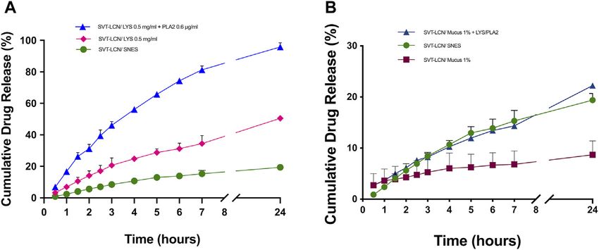
Clementino et al.                                                                                                      SVT-LCNs for Neuroinflammation

                                           biocompatibility and ability to promote drug release and absorption at the biointerface,
                                           hybrid LCNs appear to be an ideal carrier for achieving nose-to-brain delivery of poorly
                                           water-soluble drugs such as SVT.
                                           Keywords: hybrid nanoparticles, enzymatic degradation, RPMI2650 cell line, statins, neuroinflammation,
                                           microglia-like THP-1 cell line, nasal drug delivery

INTRODUCTION                                                                   antioxidant and protective properties on neurovasculature
                                                                               appear relevant for AD development (Zimetti et al., 2021).
The term “neuroinflammation” is used to describe the                                The need for effective treatments to prevent
inflammatory response originated in the central nervous                         neuroinflammation-related          damage       associated     with
system (CNS) after local damage signals, including trauma,                     neurodegenerative diseases has boosted the interest in drug
infectious agents, oxidative species release, β-amyloid                        repurposing (Athauda and Foltynie, 2018; Ballard et al., 2020).
oligomers, and τ protein neurofibrillary tangle formation                       In fact, it is believed that existing drugs with multiple
(Morales et al., 2014). Immune responses in the CNS occur                      pharmacological effects against the neuroinflammation genesis
very frequently and are mostly mediated by resident innate                     and progression could contribute to address the multifaceted
glial immune cells. Indeed, microglia represent the major                      nature of neurodegenerative pathology and/or diminish the
cellular component of the innate immune system of the brain.                   cumulative effects of inflammation in the brain. In this regard,
An acute inflammatory response in the CNS is caused by the                      in the past few years, our group has proposed, for the first time,
immediate and early activation of microglia cells in response to               the nose-to-brain delivery of encapsulated statins as an innovative
noxious stimuli and by the expression of pro-inflammatory                       approach in the treatment of neurodegenerative diseases
mediators, such as cytokines and reactive oxygen species                       (Clementino et al., 2016). Statins, as a result of HMG-CoA
(ROS) (Fakhoury, 2016). The formation and release of                           reductase inhibition, are lipid-lowering agents widely used as a
protective/resolving inflammatory mediators is a defensive                      first-line therapy in cholesterol-related cardiovascular diseases.
response contribution to tissue repair and resolution of                           However, several clinical investigations have also suggested a
inflammation. However, if the stimulus remains persistent, a                    beneficial role of statins on the pathogenesis of AD (Sodero and
pathological chronic inflammatory condition can develop,                        Barrantes, 2020) via the modulation of brain cholesterol levels as
causing cumulative damage over time. The hyperactivation of                    well as through pleiotropic effects, acting, for example, on
microglia cells can cause an excessive release of cytotoxic factors,           β-amyloid production and accumulation in plaques
with consequent deleterious effects also for healthy tissues,                  (Fassbender et al., 2001; Ehehalt et al., 2003).
especially on the neuronal cells. Indeed, recent data suggest                  Clinical–mechanistic studies evidenced that simvastatin (SVT)
that local brain immune activation and evolution of the                        is able to modulate the microglia activity and the release of
inflammation response have the capacity to facilitate and                       inflammatory markers in the injured brain (Li et al., 2009;
trigger the pathophysiological conditions typical for many                     Manickavasagam et al., 2020). Furthermore, statins can
neurodegenerative diseases (Hennessy et al., 2015). Apparently,                suppress cellular apoptosis in the CNS, stimulate the
glial cells secrete and respond to a vast array of signaling                   expression of neurotrophic factors (NGF and BDNFG) (Yang
molecules, producing exaggerated amounts of chemokines                         et al., 2012), and reduce the expression of inducible nitric oxide
(CCL2 and CXCL1) and cytokines (IL-6, TNF-α, and IL-1β)                        synthase (iNOS) (Roy and Pahan, 2011) while maintaining the
(Elain et al., 2014). Elevated levels of chemo/cytokines have been             blood–brain barrier (BBB) integrity. However, these effects have
detected in the serum and cerebrospinal fluids of patients                      proven to be quite elusive after oral administration, possibly as a
suffering from Alzheimer’s disease (AD), Parkinson’s disease                   direct consequence of high hepatic extraction and poor crossing
(PD), multiple sclerosis (MS), and Huntington’s disease (HD).                  of the BBB (McGuinness et al., 2014; Sonvico et al., 2017).
Together with other factors, these elevated chemo/cytokine levels              Importantly, an efficient delivery of statins to the cerebral
appear to play an important role in the neurodegenerative                      parenchyma could be decisive for the management of several
processes of these illnesses since these diseases share the                    neurodegenerative disorders.
ubiquitous feature of chronic inflammation (Erta et al., 2012).                     Recently, nasal drug administration has been indicated as one
In AD and depression animal models, the release of                             of the most interesting alternative routes for brain delivery. In
neuroinflammatory cytokines appears to have a role in the                       fact, nasal delivery allows the avoidance of systemic drug
pathogenesis mediated by increased levels of β-amyloid, either                 metabolism and the necessity of drug transport across the BBB
via reduced clearance or increased production from the amyloid                 (Illum, 2000). In addition, intranasal administration, being
precursor protein (Alasmari et al., 2018; Morgese et al., 2020).               noninvasive and not requiring the intervention of healthcare
Moreover, mounting evidence indicates that the oxidative and                   professionals, may offer additional advantages in terms of
inflammatory imbalance accompanying cardiovascular disease                      patient compliance and enhanced adherence to the therapy in
could be related to the pathogenesis and progression of AD. In                 the case of chronic therapy. However, the extent of the nasal drug
particular, the alteration of the proteic composition of high-                 absorption is highly dependent on the formulation and on its
density lipoproteins (HDLs) and the reduction of their                         residence time on the nasal mucosa, even more so in the case of

Frontiers in Pharmacology | www.frontiersin.org                            2                               September 2021 | Volume 12 | Article 716380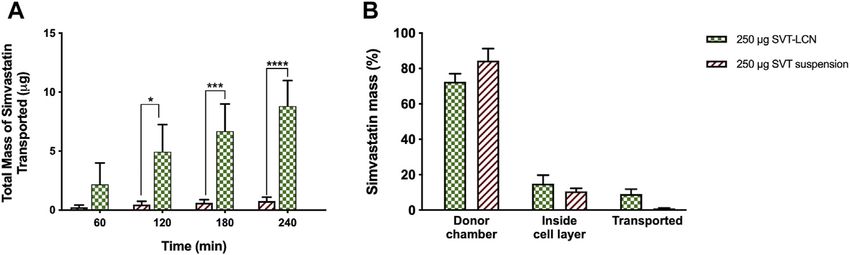
Clementino et al.                                                                                                   SVT-LCNs for Neuroinflammation

nose-to-brain delivery (Wu et al., 2008). Consequently, the ability         lipopolysaccharide from Escherichia coli, and thiazolyl blue
to deliver therapeutically relevant amounts of                              tetrazolium    bromide   (MTT)     were     obtained    from
drugs directly from the nose to brain strictly depends on the               Sigma–Aldrich (St. Louis, MO, United States). All other
availability of efficient drug delivery systems (Mistry et al., 2015).       chemicals were of analytical grade. Ultrapure and degassed
    Lecithin/chitosan nanoparticles (LCNs) have been proposed               ultrapure water (Purelab Flex, ELGA-Veolia LabWater, High
as a delivery system for hydrophobic drugs through mucosal                  Wycombe, United Kingdom) were used in all experiments.
tissues (Barbieri et al., 2015; Şenyiğit et al., 2016). The chitosan
surface layer characterizing their structure appears adapted for            Methods
nasal delivery as it promotes mucoadhesion and enhances                     Simvastatin-Loaded Lecithin/Chitosan Nanoparticle
epithelial permeability (Gerelli et al., 2010; Bruinsmann et al.,           (SVT-LCN) Preparation
2019). However, the idea that slow drug–releasing nanoparticles             SVT-LCNs were prepared through a self-emulsifying process
would provide a significant improvement of nose-to-brain                     involving lecithin, a negatively charged phospholipid, and
delivery has been questioned (Illum, 2007). Conversely, rapidly             chitosan, a derivative of the natural polysaccharide chitin,
biodegraded nanoparticles could improve the brain target and                according to a protocol previously reported (Clementino et al.,
bioavailability of lipophilic drugs, by tailoring prompt drug               2016). Briefly, 10 mg of SVT was dissolved in 0.8 ml of an ethanol
release and absorption across the nasal mucosal surface.                    solution of lecithin (2.5%), containing 20 mg of both Labrafac and
Previously, we developed simvastatin-loaded lecithin/chitosan               Maisine oil vehicles. Then, the lecithin solution containing SVT
nanoparticles (SVT-LCNs) presenting several desirable features              and oils was injected into 10 ml of a 0.01% w/v chitosan aqueous
in view of nose-to-brain delivery (Clementino et al., 2016). Small          solution, under constant agitation (magnetic stirring, 300 rpm)
particle size, narrow size distribution, high positive surface              and temperature (40°C). Freshly prepared nanoparticle
charge, and high drug-encapsulation efficiency and stability                 suspensions were stirred at constant temperature until
were obtained for nanoparticles produced with an efficient                   complete evaporation of ethanol from the system. SVT-LCNs
self-assembly process. Hence, in the present work, we carefully             were characterized for the particle size, polydispersity index, and
investigated features of the nanoformulation that are critical for          surface zeta potential using a Malvern Zetasizer Nano ZSP
SVT to enter the cerebral parenchyma while exploiting the nasal             (Malvern Instruments Ltd., United Kingdom). Nanoparticles’
route of administration. In particular, nanoparticles’                      encapsulation efficiency (EE) was determined indirectly by
composition, physicochemical properties affecting drug release,             centrifugation and ultrafiltration (Vivaspin® 2, MWCO 30,000,
interaction of nanoparticles with mucosal surfaces, and drug                Sartorius, Germany) to separate nanoparticles from a
transport across nasal epithelia were explored. Last, the                   nonencapsulated drug material, as previously reported
efficacy of SVT-loaded hybrid LCNs in attenuating cytokine                   (Bruinsmann et al., 2019). SVT content was quantified
release was evaluated for the first time using a myeloid cell                applying a validated high-performance liquid chromatography
line as a model for microglia.                                              (HPLC) analytical method with a UV/Vis detector set at 238 nm
                                                                            and published elsewhere (Clementino and Sonvico, 2018).

MATERIALS AND METHODS                                                       Nanoparticle In Vitro Drug Release
                                                                            In vitro drug release experiments were conducted using vertical
Materials                                                                   Franz type diffusion cells (0.6 cm2 diffusional area) (Disa, Milan,
Chitosan (Chitoclear FG, deacetylation degree 95%, viscosity                Italy) assembled with a dialysis cellulose membrane (MWCO
45 cP) was provided by Primex (Siglufjordur, Iceland) and                   14,000 Da, Sigma–Aldrich, St. Louis, MO, United States)
used without further purification. Lecithin (Lipoid S45) was                 separating the donor and receptor compartments. Receptor
obtained from Lipoid AG (Ludwigshafen, Germany).                            compartments were accurately filled with 4 ml of a simulated
                                                  ™
Pharmaceutical grade oils such as Maisine 35-1 (glycerol                    nasal electrolyte solution (SNES, pH 6.5), which is used as a
                                     ™
monolinoleate) and Labrafac Lipophile WL 1349 (medium
chain triglycerides, EP) were a gift from Gattefossé (Saint-
                                                                            release medium (Castile et al., 2013). Assembled Franz cells were
                                                                            maintained at constant temperature (37.0 ± 0.2°C) and
Priest, France). Simvastatin USP 99%, mucin from a porcine                  magnetically stirred (800 rpm) during 24 h of the experiment.
stomach (Type III), human lysozyme, and phospholipase A2 were               SVT-LCN drug release was performed dispersing 25 µl of
supplied by Sigma–Aldrich (Steinheim, Germany).                             formulation in 1) SNES, 2) an artificial mucus model (1% of
   Cell line RPMI 2650 (CCL-30) was purchased from American                 porcine stomach mucin type II dispersed in SNES) (Griffiths
Type Culture Collection (ATCC, Manassas, VA, United States).                et al., 2010), 3) SNES, or 4) artificial mucus containing 0.5 mg/ml
Human monocyte THP-1 cell line was purchased from the                       of lysozyme and 0.6 µg/ml of phospholipase A2 (Sigma–Aldrich,
European Collection of Authenticated Cell Culture (ECACC,                   St. Louis, MO, United States). Samples were then added to the
Porton Down, United Kingdom). Minimum essential cell                        donor compartment. At predetermined time points, aliquots of
culture medium (MEM), RPMI 1640, and fetal bovine serum                     500 µl were withdrawn from the receptor compartment and
(FBS) were acquired from Life Technologies (Thermo Fisher                   replaced with the same amount of fresh SNES medium to
Scientific, Monza, Italy). Cell culture inserts and other culture            maintain sink conditions. Collected samples were analyzed by
plastics were acquired from Corning® Life Science (Tewksbury,               HPLC-UV for SVT content. At the end of the experiment, the
MA, United States). Phorbol 12-myristate 13-acetate (PMA),                  content donor compartments from each Franz cell and the

Frontiers in Pharmacology | www.frontiersin.org                         3                               September 2021 | Volume 12 | Article 716380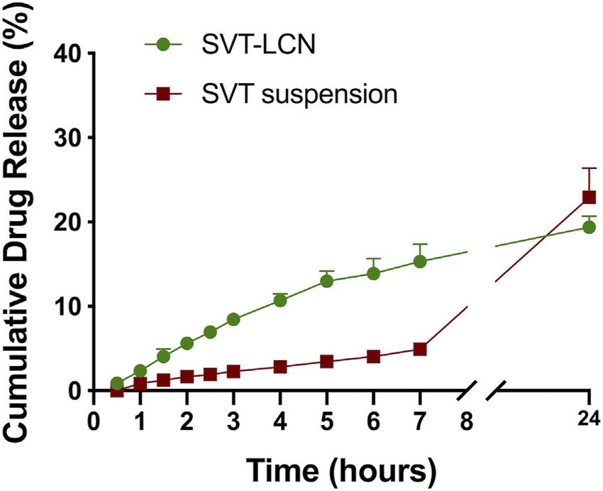
Clementino et al.                                                                                                  SVT-LCNs for Neuroinflammation

dialysis membranes were also assayed for SVT content to                    36 h of incubation, the MTT assay was performed on cell
calculate the overall mass balance. SVT release experiments                monolayers. In detail, SVT-LCN and SVT treatments were
were performed at least in triplicate.                                     stopped, and cells were treated with a solution of 1 mg/ml of
                                                                           MTT in RPMI supplemented with 5% FBS, at 37°C for 2 h in the
Study of SVT-LCN Permeation Across RPMI2650                                dark. The solution was then removed, and the resulting
Nasal Cells                                                                intracellular formazan crystals were dissolved in 200 µl of
In vitro permeation studies were performed against a cellular              DMSO under shaking for 10 min. Finally, absorbance was
model of nasal mucosa, as previously reported (Pozzoli et al.,             measured at a wavelength of 570 nm using an absorbance
2016). Briefly, RPMI 2650 human nasal epithelial cell lines                 microplate reader (Spark®, Tecan, Männedorf, Switzerland).
(ATCC, Manassas, VA, United States) were cultivated in the                 Absorbance values were directly correlated with the cellular
minimum essential medium (MEM, Life Technologies, Thermo                   viability, and the percentage of cell viability for each treatment
Fisher Scientific, Monza, Italy) enriched with 10% (v/v) of fetal           was compared to control values obtained for untreated cells.
bovine serum (FBS) and 1% (v/v) of nonessential amino acid
solution, and incubated at 37°C with 95% air humidity and 5%               Investigation of the Anti-Inflammatory Activity of
CO2 atmosphere. When confluence was 90%, cells were seeded at               SVT-LCNs
the concentration of 2.5 × 10⁶ cells/ml on collagen-coated                 In order to assess the anti-inflammatory activity of SVT-LCNs,
Snapwell® polyester membrane inserts (Costar, Corning Life                 THP-1 cells were seeded and differentiated as previously
Sciences, Tewksbury, MA, United States, 1.13 cm2, 0.4 µm pore              described, treated for 2 h with LPS, and subsequently
size, coated with 200 µl of 1 × collagen PBS solution 24 h prior to        incubated with fresh RPMI for 6 h in order to promote
seeding). Then, 24 h after seeding, the medium on the apical               cytokine secretion and accumulation in the medium. Initially,
chamber was completely removed to transfer cells to an air–liquid          an evaluation at different incubation times for both SVT-LCNs
interface (ALI) condition, allowing cells to differentiate in a            and SVT solution was performed to investigate the optimal
pseudostratified monolayer. Acceptor compartments were                      treatment conditions. In the first condition tested (indicated as
filled with 1.5 ml of the cell culture medium and replaced                  protocol A), SVT or SVT-LCNs at 10 µM were added 24 h before
3 times per week. Transepithelial electric resistance (TEER)               and during the 2 h treatment with LPS. In the second condition
measurements were recorded using an EVOM2 epithelial                       (indicated as protocol B), cells were treated with free SVT or SVT-
voltohmmeter (World Precision Instruments Inc., Sarosata, FL,              LCNs at 10 µM before and during incubation with LPS, as well as
United States) to evaluate the monolayer integrity and suitability         for further 6 h after the treatment with LPS. Once the optimal
for the transport studies. Finally, 14 days after seeding, 250 µl of       experimental condition was identified, which was set as protocol
1 mg/ml SVT-LCNs or 1 mg/ml simvastatin suspension in HBSS,                B, THP-1 cells were finally treated with increasing drug
which was used as a control, was added on the cell surface to carry        concentrations of SVT-LCNs and SVT (0.1, 1, and 10 μM), for
out permeation studies, in triplicate. Samples of 200 µl were              comparison purposes. At the end of the treatment, supernatants
collected from the acceptor compartment every hour and                     were collected, and IL-6 and TNF-α production in the medium
analyzed by HPLC-UV for SVT content. After 4 h, the apical                 was quantified by ELISA assay kits according to the
surface of cell epithelia was washed twice with HBSS to collect any        manufacturer’s instructions (Thermo Fischer Scientific,
remaining non-permeated drug. Thereafter, cells were scraped               Waltham, MA, United States).
out from SnapWell® inserts and lysed with CelLytic buffer
(Invitrogen, Thermo Fisher Scientific, Waltham, MA,
                                                          ™                Statistics
United States) to extract and quantify the amount of SVT                   The statistical analyses were performed with Prism 8 software
accumulated within the cells.                                              (GraphPad Software, San Diego, CA, United States). Each
                                                                           experiment was run at least in triplicate, and results were
Effect of SVT-LCNs on Pro-Inflammatory Glia-Like Cell                       expressed with the mean and the standard deviation. The
Viability                                                                  results were analyzed using Student’s t-test for unpaired
To evaluate the impact of SVT-LCNs on cell viability, human                samples when comparing two groups (indicated as T in Figure
monocyte-derived macrophages THP-1 were used. These cells are              legends) or ordinary one-way ANOVA to compare more than
widely accepted as the surrogate of microglial cells to evaluate           two conditions, applying the Tukey post hoc test for multiple
cytokine secretion in the context of neuroinflammation in AD                comparisons (indicated as F in Figure legends).
(McFarland et al., 2017; Wiatrak and Balon, 2021). THP-1
monocytes were cultured in RPMI 1640 medium
supplemented with 10% FCS at 37°C in 5% CO2. To perform                    RESULTS
the experiments, cells were seeded in 24-well plates at the density
of 5·105 cells/well in the presence of 100 ng/ml PMA for 72 h to           Physicochemical Characterization of
allow differentiation into macrophages. After differentiation, cells       SVT-LCNs
were treated for 2 h with lipopolysaccharide from E. coli (LPS,            SVT-LCNs were formed by the electrostatic self-assembly of
0.25 μg/ml) as pro-inflammatory stimulus (Wiatrak and Balon,                lecithin and chitosan and have a multilayered structure
2021). Cells were then treated with SVT-LCNs or with SVT                   alternating chitosan aqueous-rich layers and phospholipid
dissolved in DMSO at concentrations of 0.1, 1, and 10 µM. After            bilayers, as previously reported (Gerelli et al., 2008b; Gerelli

Frontiers in Pharmacology | www.frontiersin.org                        4                               September 2021 | Volume 12 | Article 716380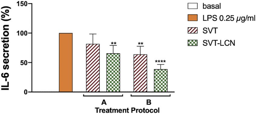
Clementino et al. SVT-LCNs for Neuroinflammation
TABLE 1 | Nanoparticles’ physicochemical properties and simvastatin encapsulation efficiency (EE).
Batch code Particle size (nm) ζ Potential (mV) PDI EE (%)
Blank LCN 147.0 ± 26.4 +57.3 ± 2.6 0.380 ± 0.025 —
SVT-LCN 217.8 ± 12.1 +44.3 ± 2.1 0.096 ± 0.032 98.0 ± 1.2
suspension (approximately 20% of the drug was released) during
the 24 h of the release experiment, nonencapsulated SVT showed
an extremely low dissolution rate at early time points while the
release from nanoparticles was prompt. In fact, over the first 7 h
of the experiment, 15.31% (±1.06%) of SVT was released from the
nanoparticles, displaying a significantly faster release in
comparison to the SVT suspension (4.93 ± 0.57%).
Subsequently, with the aim to simulate the physiological
conditions present in the nasal cavity more closely, drug
release studies were carried out using SNES containing
mucus and/or two antibacterial enzymes physiologically
present in nasal secretions, that is, lysozyme and
phospholipase A2 (PLA2) as dissolution media (Travis
et al., 2001). While, as shown previously, in the simple
SNES electrolyte solution, SVT was released from SVT-
LCNs in a controlled manner, a rather different behavior
was observed when enzymes were added to SNES
(Figure 2A). When lysozyme was added to SNES, the total
FIGURE 1 | Simvastatin release from drug suspension (red squares) and drug release from SVT-LCNs doubled, attaining 50% at 24 h.
SVT-LCNs (green dots) in simulated nasal electrolyte solution (SNES), pH 6.5 Interestingly, when both lysozyme and PLA2 were added to
(n 3, ± S.D.). Experiments were carried out in a vertical Franz cell apparatus
the dissolution medium, nearly all the encapsulated SVT was
using a cellulose dialysis membrane (MWCO 14,000 Da).
released (96.82 ± 2.62%) within 24 h. Finally, to understand
how mucus could impact nanoparticle delivery following nasal
administration, in vitro release studies were performed using a
et al., 2008a). In order to improve drug encapsulation efficiency, simulated nasal mucus. Figure 2B illustrates that when
nanoparticles were produced with the addition of pharmaceutical nanoparticles were dispersed in the artificial mucus (SNES
grade oils to help drug encapsulation in the lipophilic domain of containing 1% porcine mucin), SVT release was significantly
LCNs. The physicochemical characteristics of SVT-LCNs are hindered (19.40 ± 1.30% vs. 8.69 ± 2.63%). However, when the
presented in Table 1. The encapsulation of the statin into the enzymes were added to the simulated nasal mucus, the total
LCN structure did not affect the main physicochemical features of release of SVT was boosted back up to 22%.
the nanomedicine. SVT-LCNs showed a relatively small particle
size (218 ± 12 nm), with narrow size distribution
(PDI 0.096 ± 0.032) and positive surface charge (+44.3 ± SVT Permeation Across an In Vitro Model of
2.1 mV). High drug encapsulation efficiency was achieved with the Nasal Epithelium
nanoparticles encapsulating 98% (±1.2%) of the total amount of The ability of SVT-LCNs to promote SVT absorption through
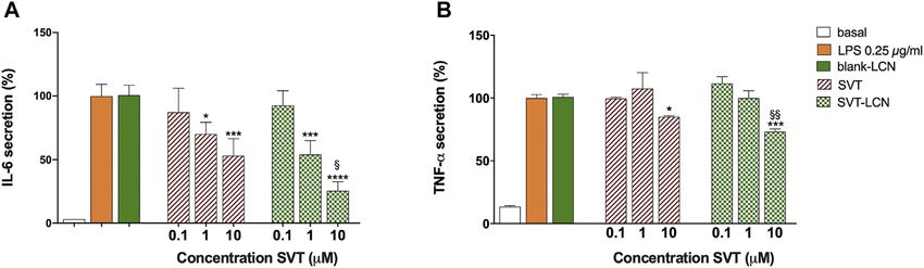
SVT used in the preparation (1 mg/ml). the nasal mucosa was investigated using a mucus-producing
the nasal epithelium model obtained using the human nasal
septum cell line RPMI 2650 (Pozzoli et al., 2017). Figure 3A
SVT Release From SVT-LCNs in Biorelevant illustrates the total amount of SVT which had permeated
Conditions across the nasal epithelial model after the deposition of
To investigate the hybrid nanoparticle drug release in vitro, a SVT suspension and SVT-LCNs on the cells’ pseudo-
simple electrolyte solution containing sodium, potassium, and monolayer surface. After the first hour, approximately 3 µg
calcium salts (SNES, pH 6.5) was initially used as the release (±1.8 µg) of SVT from SVT-LCNs was already transported,
medium to simulate nasal secretions. Release studies were while only 0.25 µg (±0.17 µg) of SVT was found in the acceptor
performed using vertical Franz diffusion cells and a compartment of the drug suspension used as a control. At the
semipermeable membrane to separate released/dissolved drug end of the experiment after 4 h, 9 µg (±2.16 µg) of SVT from
from drug-releasing nanoparticles. Drug release profiles obtained nanoparticles permeated the nasal cells vs just 0.8 µg
for SVT-LCNs and SVT suspension in SNES are presented in (±0.34 µg) for SVT suspension (11-fold increase; p < 0.001).
Figure 1. Although no significant difference was found between It was important to note that a progressive, almost linear,
the amount of SVT released from nanoparticles and from the increase in the drug permeation for simvastatin-loaded
Frontiers in Pharmacology | www.frontiersin.org 5 September 2021 | Volume 12 | Article 716380Clementino et al. SVT-LCNs for Neuroinflammation
FIGURE 2 | Panel (A)-SVT-LCN release in simulated nasal electrolyte solution (SNES) pH 6.5 (green dots) and with the addition of lysozyme 0.5 mg/ml (pink
diamonds) and lysozyme 0.5 mg/ml + phospholipase A2 (PLA2) 0.6 µg/ml (blue triangles). Panel (B)-SVT-LCN release in SNES (green dots), simulated nasal mucus
(SNES plus porcine stomach mucin type II at 1% w/v, pH 6.5; red squares) and with the addition of lysozyme 0.5 mg/ml + phospholipase A2 (PLA2) 0.6 µg/ml (blue
triangles). Experiments (n 3, ± S.D.) were carried out in a vertical Franz cell apparatus using a cellulose dialysis membrane over a 24-h period.
FIGURE 3 | Panel (A)–Amount of simvastatin transported across the RPMI2650 nasal cell pseudo-monolayer over time after deposition on the cells apical surface
of simvastatin-suspension (red bars) or simvastatin-loaded nanoparticles (SVT-LCNs, green bars). Panel (B)–Percentage distribution of total simvastatin recovered after
4 h of transport studies across the RPMI2650 nasal cell pseudo-monolayer using simvastatin suspension (red bars) or simvastatin-loaded nanoparticles (SVT-LCNs,
green bars) (n 3, ± S.D.). Permeability experiments were carried out on RPMI2650 cells grown under air–liquid interface conditions (TEER values were around
120 Ω·cm2 before and after transport studies).
nanoparticles was evidenced, while the permeation from followed by 36 h incubation and using SVT solution in
simvastatin suspension occurred at a very slow rate, DMSO as control. Results are reported in Supplementary
reaching almost a plateau. These data suggest an enhanced Materials (Supplementary Figure 1) and show that neither
drug permeability when lecithin/chitosan nanoparticle the raw material (SVT) nor the nanoparticle formulation
technology is applied. (SVT-LCNs) affected the glia-like cells’ viability at the drug
As shown in Figure 3B, after 4 h of permeation, 84.42 ± 6.79% concentrations tested (0.1, 1, and 10 µM). Indeed, cell viability
and 72.94 ± 4.52% of the drug remained on the surface of the cell remained around 100% for all treated groups compared to
layer for SVT suspension and SVT-LCNs, respectively. Around untreated control cells.
14.94 ± 4.75% of SVT from the nanoparticle formulation and
11.03 ± 1.74% from the SVT suspension were found inside the
cells, suggesting a higher internalization of nanoparticles within Investigation of the Anti-Inflammatory
the cells of the nasal epithelium. Activity of SVT-LCNs
The in vitro efficacy of SVT-LCN formulation was further
assessed using LPS-treated glia-like THP-1 as a surrogate
Investigation of SVT-LCN Cytotoxicity model of neuroinflammation (McFarland et al., 2017). Time-
Against the Glia-Like THP-1 Human Cell and dose-dependent effects of LPS stimulation on TNF-α and IL-
Line 6 release from THP-1 cells were evaluated and are presented in
The biocompatibility of the nanoparticle formulation on the the Supplementary Material. Experimental evidence showed that
THP-1 cell line was assessed by determining cell viability LPS has a significant pro-inflammatory effect in the glia-like cell
Frontiers in Pharmacology | www.frontiersin.org 6 September 2021 | Volume 12 | Article 716380Clementino et al. SVT-LCNs for Neuroinflammation
SVT-LCNs prior (24 h), during (2 h), and after (6 h) the LPS
stimulus (total of 32 h of incubation).
For both protocols and treatments, that is, SVT solution and
SVT-LCNs, a reduction in LPS-induced IL-6 cytokine release
from the THP-1 cells was observed. However, considering the
first treatment condition (Figure 4, A protocol), only SVT-LCNs
displayed a statistically significant reduction of IL-6 levels (−37%
IL-6 concentration vs. LPS, p < 0.05). Nevertheless, a significant
reduction in LPS-induced IL-6 secretion was observed, for both
SVT solution (−38% IL-6 concentration vs. LPS, p < 0.05) and
SVT-LCNs (−63% IL-6 concentration vs. LPS, p < 0.01), when the
FIGURE 4 | Effect of treatment with 10 μM SVT solution and SVT-LCNs
on the secretion of IL-6 from THP-1 glia-like cells according to two different treatments were simulating a chronic treatment (Figure 4,
treatment protocols: A-cells were pretreated with 10 µM of SVT solution or Protocol B). Then, the extent to which SVT-LCN treatment
SVT-LCNs for 24 h followed by 2 h of simultaneous treatment with LPS would impact the pro-inflammatory signaling and microglia
0.25 µg/ml; B-cells were treated with 10 µM of SVT solution or SVT-LCNs activation was assessed by measuring IL-6 and TNF-α release
prior (24 h), during (2 h), and after LPS treatment (6 h). Data are expressed as
mean percentage ± S.D. of cytokine secretion from at least three repeated
by THP-1 glia-like cells as a consequence of the LPS stimulus. The
measurements. Statistical significance of the results is expressed with respect SVT solution was used as the positive control for drug efficacy,
to the LPS control treatment (*: p < 0.05; **p < 0.01 compared to LPS, F). while blank nanoparticles were tested to exclude any effect by the
nanocarrier components. SVT-LCNs and the SVT solution
exhibited a dose-dependent reduction of the activation of glia-
like THP-1 cells in terms of release of IL-6 (Figure 5A). In the
model at the concentration of 0.25 µg/ml with a sustained release case of TNF-α, only the highest investigated drug concentration
of TNF-α and IL-6 pro-inflammatory cytokines observed beyond was able to significantly reduce the cytokine secretion for both
the 2 h of incubation with LPS (data not shown) and attained a treatments (Figure 5B). However, only in the case of treatment
peak concentration after 6 h (Supplementary Figure 2). with SVT-LCNs, the attenuation of cytokine release has been
The release of pro-inflammatory cytokines followed by LPS relevant. In fact following LPS stimulation, only the treatment
stimuli was used to evaluate the potential effect of SVT-LCNs to with SVT-LCNs (10 µM) showed at the same time a highly
counteract neuroinflammation. The experimental conditions
significant suppression of IL-6 release (−75% IL-6
were designed and optimized to simulate a preventive and a
concentration vs. LPS, p < 0.001) and of TNF-α levels (−27%
chronic neuroinflammation treatment. LPS-treated cells were
exposed to two different treatment protocols with SVT of TNF-α levels vs. LPS, p < 0.01). Moreover, the reduction
solution or SVT-LCNs (Figure 4). In the first case, protocol induced by SVT-LCNs was more pronounced than that of SVT
A, cells were pretreated with 10 µM of SVT solution or SVT- solution (p 0.05 and p 0.005 for IL-6 and TNF-α,
LCNs for 24 h, followed by 2 h of simultaneous treatment with respectively). Additionally, blank LCNs did not show
0.25 µg/ml of LPS (total of 26 h incubation). Conversely, for pharmacological activity, with cytokine levels superimposable
protocol B, cells were treated with 10 µM of SVT solution or to those obtained for the LPS stimulus alone, ruling out a
FIGURE 5 | Dose–response evaluation of the treatment with SVT solution and SVT-LCNs on the secretion of IL-6 (A) and TNF-a (B) from THP-1 glia-like cells after
LPS treatment (0.25 µg/ml) following protocol B as shown in Figure 4. Basal cytokine levels, LPS stimulus alone, and blank LCNs (the nanoparticle amount
corresponding to 10 µM of SVT) are presented as controls. Data are expressed as mean percentage ± S.D. of cytokine secretion from at least three repeated
measurements. Statistical significance of the results is expressed in comparison to that of the LPS treatment (*p < 0.05; ***p < 0.001; p****Clementino et al. SVT-LCNs for Neuroinflammation
potential anti-inflammatory activity from the nanoparticle predict the drug bioavailability from a nasal formulation.
components. Indeed, the nanoparticle formulation increased in 4-fold
SVT release within the first 3 h compared to the drug
suspension. However, considering the composition of the
DISCUSSION proposed nanocarrier, we hypothesized that enzymes
naturally present in the nasal cavity trigger drug release
The nose is an attractive alternative route for delivery of drugs to toward hybrid lecithin/chitosan nanoparticles. In a
the brain. However, the complexity of the nasal anatomy and different study, Barbieri et al. have shown the
physiology can pose a remarkable challenge in achieving the susceptibility of comparable lecithin/chitosan
delivery of intranasally administered drugs to the brain. The use nanoparticles to biodegradation by gastrointestinal
of nanomedicine is a promising approach for the nose-to-brain enzymes, promoting drug release and transport through
transport of therapeutics across the nasal mucosa (Mistry et al., the intestinal epithelium (Barbieri et al., 2015). Lysozyme
2009; Ahmad et al., 2017; Junior et al., 2020). However, the and phospholipase A2 are antibacterial proteins naturally
delivery of therapeutically relevant amounts of present in nasal secretions (Travis et al., 2001) and have the
drugs exploiting the nasal route is strongly dependent on the potential to degrade the two main components of the hybrid
availability of efficient nanoparticulate drug carriers (Mistry et al., nanosystem. Lysozyme is capable to degrade chitosan
2015). The major approaches to increase drug bioavailability via (Nordtveit et al., 1996), while PLA2 catalyzes the
intranasally administered nanoparticles are as follows: 1) the use hydrolysis of phospholipids, affecting the lipidic
of mucoadhesive polymers, increasing the local drug residence component of nanoparticles (Andresen et al., 2010). In
time; 2) incorporation of ingredients controlling the drug release drug release studies employing lysozyme, an increase in
rate; and 3) the employment of permeation enhancers (Sonvico SVT release from nanoparticles was observed, with a
et al., 2018). doubling of the cumulative amount of the drug released
Hybrid chitosan/lecithin nanoparticles display good compared to that of SNES (see Figure 2A). Interestingly,
mucoadhesion properties because of the cationic the addition of both enzymes determined a dramatic increase
polysaccharide surface coating and good encapsulation in the drug release rate with nearly all of the loaded SVT
properties for poorly soluble drugs due to the high lipid released within 7 h. SVT release from lecithin/chitosan
content (Gerelli et al., 2010; Şenyiğit et al., 2016). Together nanoparticles could be actually triggered by the enzymatic
with a relatively small and narrow particle size distribution, action on nanoparticle structure biodegradation after
these properties appear highly desirable for nanoscale-based deposition in the nasal cavity. These results corroborate
formulations intended for nasal administration. However, in the hypothesis that hybrid phospholipid/polysaccharide
order to evaluate the suitability of lecithin/chitosan nanoparticles are mucoadhesive systems prone to
nanoparticles for the nasal delivery of SVT, in vitro drug biodegradation when they are in contact with the mucosal
release of nanoparticles was evaluated since it represents an surface.
important tool in the development of nanomedicines (Tinkle As aforementioned, the major drawback affecting the
et al., 2014; Sainz et al., 2015). Conditions were selected so availability of nasally administered lipophilic drugs is the
that they could be relevant for SVT release from mucociliary clearance. Therefore, additional in vitro release
nanoparticles in the physiological environment present in studies were performed for SVT-LCNs dispersed in simulated
nasal cavities. In a relatively simple SNES, after an initial artificial nasal mucus. A decrease in the cumulated amount of
faster release rate evidenced for SVT-LCNs, the overall drug SVT released suggests an interaction between nanoparticles
release from nanoparticles after 24 h did not differ from that and mucins, resulting in the hampering of drug release and/or
of the control drug suspension. The decrease on the release drug diffusion through the mucus network. In fact, the mucus
rate of the nanoencapsulated SVT was attributed to the layer has been recognized as a barrier for the diffusion of
multilayered organization structure of nanoparticles poorly water-soluble drugs, which interact with local
hindering drug release at a later point. In fact, the glycoproteins and lipids, leading to an overall reduced
polymer coating the nanoparticles acts as a drug-release diffusion rate (Sigurdsson et al., 2013). The presence of the
barrier. Hence, the drug solubility and diffusion in/or antibacterial enzymes resulted in a boost of drug release in the
across the polymeric or lipid layers become a determining presence of mucus, confirming once again the relevance of
factor in drug release. Moreover, the fact that SVT is a enzyme-triggered release of SVT from hybrid lecithin/
lipophilic drug which has a high affinity for the lipid chitosan nanoparticles in the nasal physiological
component of the nanoparticles and its release could be environment. Thus, the proposed hybrid chitosan-coated
hindered at an oil–water interface due to the low drug nanoparticles interacted with the mucus secretion. Even
aqueous solubility has to be considered. Nevertheless, though this interaction could prevent drug availability and
considering the specific conditions present in the nasal consequent permeation through the nasal epithelium, their
cavity and the rapid turnover of the mucus layer due to biodegradation by glycoside hydrolases and phospholipase
mucociliary clearance, the initial hours of in vitro drug enzymes naturally present in the nasal cavity could represent
release from the drug delivery system are critical to a new strategy to overcome the mucus barrier.
Frontiers in Pharmacology | www.frontiersin.org 8 September 2021 | Volume 12 | Article 716380Clementino et al. SVT-LCNs for Neuroinflammation
When considering these results, it is important to emphasize 1) inhibiting signal transduction pathways activating pro-
that drug release experiments in the present work employed inflammatory transcription factors, such as nuclear factor κB;
Franz vertical diffusion cells. Even though in vitro experiments 2) by activating PPARα receptors and the anti-inflammatory
applying Franz cells have become one of the most frequent activity; and 3) affecting the expression of the MHC II
methods adopted for drug release studies of nonconventional histocompatibility complex (Paumelle et al., 2006; McFarland
drug dosage forms, they present the drawback of being a static et al., 2014), possibly through the disruption of cholesterol-
diffusion method in which the dialysis membrane separating the containing microdomains, which transport and concentrate
donor and receptor compartments poses as a significant MHC-II molecules to the cell surface (Kuipers and Elsen,
additional barrier to the diffusion of the drug (Ng et al., 2010; 2005; Kuipers et al., 2005).
Modi and Anderson, 2013). All these considerations lead to the In the present study, we have demonstrated an anti-
hypothesis that the released fractions of SVT could be even higher inflammatory activity of both SVT-LCNs and SVT, with a
in vivo. greater effect exerted by the nanoformulation. In the case of
In the transport studies across the RPMI 2650 nasal epithelium SVT, our findings were in agreement with previously published
model, the amount of SVT transported by nanoparticles was data, indicating the capacity of statins to mitigate microglia and
determined and compared with that obtained from a suspension its activation and to reduce the production of neuroinflammatory
of the free drug. Hybrid lecithin/chitosan nanoparticles increased mediators through the modulation of several signaling pathways
the permeation of SVT by 11-fold. Interestingly, the amount of (Bagheri et al., 2020). On the contrary, another previous study
SVT found within the cells and the mucosa were similar for suggested a pro-inflammatory effect of statins able to increase the
encapsulated SVT and free raw materials, suggesting that secretion of cytokines in Toll-like receptor–induced primary
nanoparticles do not increase drug internalization and monkey microglia (Putten et al., 2012). The reason for this
accumulation inside the epithelial nasal cells. The role of discrepancy among findings is not clear, but it may be very
chitosan as a permeation enhancer is well known (Smith et al., likely related to the use of different in vitro models and/or the type
2004). However, it cannot be assumed that it is the only factor of pro-inflammatory stimuli.
responsible for the hybrid nanoparticle permeation enhancement Among the possible mechanisms to explain the enhanced anti-
since in the case of SVT, one of the main limiting steps is the inflammatory activity of SVT-LCNs, we believe that there is a
drug’s poor water solubility. Considering that the release studies direct uptake and digestion of nanoparticles by cells, as suggested
supported nanoparticle biodegradation, it is likely that LCN for other nanosystems (Tulbah et al., 2019). Indeed, it has been
permeation enhancement was also supported by the previously demonstrated that LCNs could effectively enter cells
biodegradation of the delivery system in the mucus barrier or via the energy-dependent caveolae-mediated endocytosis and
by intracellularly providing a supplementary driving force to the macropinocytosis (Chu et al., 2019). Moreover, surface charge
drug diffusion across the mucosal barrier. In a recent study, has also been shown to be an important parameter determining
Barbieri et al. investigated the permeation of tamoxifen across the the degree of particle uptake. For instance, Thiele et al. found that
intestinal epithelium of rats using lecithin/chitosan nanoparticles positively charged microparticles showed much greater uptake by
(Barbieri et al., 2015). Authors demonstrated that tamoxifen- human-derived macrophages and dendritic cells (Thiele et al.,
loaded nanoparticles significantly increased the drug transport ex 2001).
vivo across rat intestinal tissue when pancreatin or the lipase In the present study, the enhanced anti-inflammatory activity
enzyme was present, suggesting that nanoparticle degradation detected for SVT-LCNs is to be attributed to the direct uptake and
increased the extent of drug intestinal permeation via an digestion of nanoparticles by the macrophages since empty
enhanced paracellular transport. Interestingly, when a nanoparticles did not present any effect in the release of IL-6
semipermeable membrane was used to prevent nanoparticles and TNF-α.
contacting with the mucosal tissue, the amount of drug Notably, both SVT and STV-LCNs were more effective in
permeated from the nanoparticle formulation was similar to suppressing IL-6 than TNF-α secretion. This differential
that obtained from the drug suspension. These results suggest effect may be very likely related to the extent of cytokine
a decisive role of the hybrid lecithin/chitosan nanoparticle production triggered by LPS. Indeed, after LPS exposure,
structure in determining interactions at the biointerface and TNF-α reached the concentration of about 15,000 pg/ml,
biodegradation processes within the mucosal tissue able to while IL-6 secretion was about 1,000× fold lower. This
increase drug absorption and bioavailability. milder inflammatory setting may better highlight the
Recent data pointed out that local brain immune activation inhibitory effect of statin formulation at lower
has the capacity to facilitate and trigger the pathological and concentrations. In addition, despite some concerns about
physiological modifications in neurodegenerative diseases. In potential cytotoxicity of cationic materials which have
particular, it has been shown that microglia cells play an been raised by some studies in the past, LCN formulation
important role in the management of inflammation in was demonstrated to be well tolerated by human THP-1 glia-
neurodegenerative pathologies. Beyond the lipid-lowering like cells. The association of nonconventional and pluripotent
effect, many pleiotropic effects have been attributed to statins anti-inflammatory group of compounds such as statins with
due to the inhibition of the mevalonic acid pathway and the nanocarriers capable to deliver drugs directly from the nose
formation of isoprenoid intermediates (Bellosta et al., 2000; Bedi to the brain constitutes an innovative and promising strategy
et al., 2016). In fact, statins modulate inflammatory responses by in the treatment of neurodegenerative diseases.
Frontiers in Pharmacology | www.frontiersin.org 9 September 2021 | Volume 12 | Article 716380Clementino et al. SVT-LCNs for Neuroinflammation
CONCLUSION on microglia cells with in vivo studies on relevant
neuroinflammation animal models. Overall, it can be
In summary, in the present work, we investigated the hybrid concluded that hybrid lecithin/chitosan nanoparticles
lecithin/chitosan nanoparticles designed as a platform for the appear as a highly attractive drug delivery platform for the
nasal administration of SVT for treating neuroinflammatory nose-to-brain delivery of drugs with the potential to increase
disease. We observed that nanoparticle interaction with a drug availability at the target organ and possibly its efficacy.
simulated nasal mucus decreased nanoparticle drug release
and/or slowed drug diffusion. We also evidenced the effect of
enzymes present in nasal secretions, such as lysozyme and DATA AVAILABILITY STATEMENT
PLA2, in promoting drug release from the nanocarrier.
Moreover, chitosan-coated nanoparticles enhanced SVT The raw data supporting the conclusions of this article will be
permeation across a human cell model of the nasal made available by the authors, without undue reservation.
epithelium. Finally, we demonstrated an effect of SVT-LCNs
in suppressing the pro-inflammatory stimulus in a myeloid cell
line model for microglia. Taking the presented data into AUTHOR CONTRIBUTIONS
consideration, the encapsulation of SVT into lecithin/
chitosan nanoparticles had been shown to be a beneficial Investigation and writing—original draft preparation, AC and
strategy for the nasal delivery of lipophilic statins. Hybrid CM; methodology, MP; resources and conceptualization, FB;
lecithin/chitosan nanoparticles not only provided a prompt supervision and writing—review and editing, FZ and FS.
drug release in biorelevant conditions, bypassing the mucus
barrier and some enzymes naturally present in nasal
secretions, but also enhanced the in vitro transport of SVT FUNDING
across a model of the nasal absorptive epithelium. The
capability of exploiting innate immunity antibacterial AC would like to acknowledge the Brazilian government as
enzymes to trigger drug release and possibly promoting recipients of CNPq grants in the program “Ciências sem
drug permeation across cellular surfaces represents an Fronteiras” (grant number: 202558/2015-0).
innovative concept of controlled drug release for
nanoparticle delivery carriers designed for nasal
administration. In addition, the encapsulation of SVT was SUPPLEMENTARY MATERIAL
also demonstrated to improve the efficacy of the drug
compound against a glial-like cell neuroinflammation model The Supplementary Material for this article can be found online at:
although we are aware that our “proof of principle” https://www.frontiersin.org/articles/10.3389/fphar.2021.716380/
observation needs to be integrated with further evaluations full#supplementary-material
Intestine from Lecithin/chitosan Nanoparticles. Int. J. Pharm. 491, 99–104.
REFERENCES doi:10.1016/j.ijpharm.2015.06.021
Bedi, O., Dhawan, V., Sharma, P. L., and Kumar, P. (2016). Pleiotropic
Ahmad, E., Feng, Y., Qi, J., Fan, W., Ma, Y., He, H., et al. (2017). Evidence of Nose- Effects of Statins: New Therapeutic Targets in Drug Design. Naunyn
To-Brain Delivery of Nanoemulsions: Cargoes but Not Vehicles. Nanoscale 9, Schmiedeberg Arch. Pharmacol. 389, 695–712. doi:10.1007/s00210-016-
1174–1183. doi:10.1039/c6nr07581a 1252-4
Alasmari, F., Alshammari, M. A., Alasmari, A. F., Alanazi, W. A., and Alhazzani, K. Bellosta, S., Ferri, N., Arnaboldi, L., Bernini, F., Paoletti, R., and Corsini, A. (2000).
(2018). Neuroinflammatory Cytokines Induce Amyloid Beta Neurotoxicity Pleiotropic Effects of Statins in Atherosclerosis and Diabetes. Diabetes Care 23
through Modulating Amyloid Precursor Protein Levels/Metabolism. Biomed. (Suppl. 2), B72–B78.
Res. Int. 25, 1–8. doi:10.1155/2018/3087475 Bruinsmann, F. A., Pigana, S., Aguirre, T., Souto, G. D., Pereira, G. G., Bianchera,
Andresen, T. L., Thompson, D. H., and Kaasgaard, T. (2010). Enzyme- A., et al. (2019). Chitosan-Coated Nanoparticles: Effect of Chitosan Molecular
triggered Nanomedicine: Drug Release Strategies in Cancer Therapy Weight on Nasal Transmucosal Delivery. Pharmacetutics 11, 86. doi:10.3390/
(Invited Review). Mol. Membr. Biol. 27, 353–363. doi:10.3109/ pharmaceutics11020086
09687688.2010.515950 Castile, J., Cheng, Y.-H., Simmons, B., Perelman, M., Smith, A., and Watts, P.
Athauda, D., and Foltynie, T. (2018). Drug Repurposing in Parkinson’s Disease. (2013). Development of In Vitro Models to Demonstrate the Ability of PecSys®,
CNS Drugs 32, 747–761. doi:10.1007/s40263-018-0548-y an In Situ Nasal Gelling Technology, to Reduce Nasal Run-Off and Drip. Drug
Bagheri, H., Ghasemi, F., Barreto, G. E., Sathyapalan, T., Jamialahmadi, T., and Dev. Ind. Pharm. 39, 816–824. doi:10.3109/03639045.2012.707210
Sahebkar, A. (2020). The Effects of Statins on Microglial Cells to Protect against Chu, X.-Y., Huang, W., Wang, Y.-L., Meng, L.-W., Chen, L.-Q., Jin, M.-J., et al.
Neurodegenerative Disorders: A Mechanistic Review. Biofactors 46, 309–325. (2019). Improving Antitumor Outcomes for Palliative Intratumoral Injection
doi:10.1002/biof.1597 Therapy through Lecithin–Chitosan Nanoparticles Loading
Ballard, C., Aarsland, D., Cummings, J., O’Brien, J., Mills, R., Molinuevo, J. L., et al. Paclitaxel–Cholesterol Complex. Int. J. Nanomed 14, 689–705. doi:10.2147/
(2020). Drug Repositioning and Repurposing for Alzheimer Disease. Nat. Rev. ijn.s188667
Neurol. 16, 661–673. doi:10.1038/s41582-020-0397-4 Clementino, A., Batger, M., Garrastazu, G., Pozzoli, M., Favero, E. D., Rondelli, V.,
Barbieri, S., Buttini, F., Rossi, A., Bettini, R., Colombo, P., Ponchel, G., et al. (2015). et al. (2016). The Nasal Delivery of Nanoencapsulated Statins – an Approach for
Ex Vivo permeation of Tamoxifen and its 4-OH Metabolite through Rat Brain Delivery. Int. J. Nanomed 11, 6575–6590. doi:10.2147/ijn.s119033
Frontiers in Pharmacology | www.frontiersin.org 10 September 2021 | Volume 12 | Article 716380Clementino et al. SVT-LCNs for Neuroinflammation
Clementino, A., and Sonvico, F. (2018). Development and Validation of a RP- McFarland, A. J., Davey, A. K., and Anoopkumar-Dukie, S. (2017). Statins Reduce
HPLC Method for the Simultaneous Detection and Quantification of Lipopolysaccharide-Induced Cytokine and Inflammatory Mediator Release in
Simvastatin’s Isoforms and Coenzyme Q10 in Lecithin/chitosan an In Vitro Model of Microglial-like Cells. Mediat Inflamm. 2017, 1–10.
Nanoparticles. J. Pharm. Biomed. Anal. 155, 33–41. doi:10.1016/ doi:10.1155/2017/2582745
j.jpba.2018.03.046 McGuinness, B., Craig, D., Bullock, R., Malouf, R., and Passmore, P. (2014). Statins
Ehehalt, R., Keller, P., Haass, C., Thiele, C., and Simons, K. (2003). Amyloidogenic for the Treatment of Dementia. Cochrane Database Syst. Rev. 7, CD007514.
Processing of the Alzheimer β-amyloid Precursor Protein Depends on Lipid doi:10.1002/14651858.cd007514.pub3
Rafts. J. Cel Biol. 160, 113–123. doi:10.1083/jcb.200207113 Mistry, A., Stolnik, S., and Illum, L. (2009). Nanoparticles for Direct Nose-To-
Elain, G., Jeanneau, K., Rutkowska, A., Mir, A. K., and Dev, K. K. (2014). The Brain Delivery of Drugs. Int. J. Pharm. 379, 146–157. doi:10.1016/
Selective anti-IL17A Monoclonal Antibody Secukinumab (AIN457) Attenuates j.ijpharm.2009.06.019
IL17A-induced Levels of IL6 in Human Astrocytes. Glia 62, 725–735. Mistry, A., Stolnik, S., and Illum, L. (2015). Nose-to-Brain Delivery: Investigation
doi:10.1002/glia.22637 of the Transport of Nanoparticles with Different Surface Characteristics and
Erta, M., Quintana, A., and Hidalgo, J. (2012). Interleukin-6, a Major Cytokine Sizes in Excised Porcine Olfactory Epithelium. Mol. Pharm. 12, 2755–2766.
in the Central Nervous System. Int. J. Biol. Sci. 8, 1254–1266. doi:10.7150/ doi:10.1021/acs.molpharmaceut.5b00088
ijbs.4679 Modi, S., and Anderson, B. D. (2013). Determination of Drug Release Kinetics from
Fakhoury, M. (2016). Immune-mediated Processes in Neurodegeneration: where Nanoparticles: Overcoming Pitfalls of the Dynamic Dialysis Method. Mol.
Do We Stand? J. Neurol. 263, 1683–1701. doi:10.1007/s00415-016-8052-0 Pharm. 10, 3076–3089. doi:10.1021/mp400154a
Fassbender, K., Simons, M., Bergmann, C., Stroick, M., Lütjohann, D., Keller, P., Morales, I., Guzmán-Martínez, L., Cerda-Troncoso, C., Farías, G. A., and
et al. (2001). Simvastatin Strongly Reduces Levels of Alzheimer’s Disease Maccioni, R. B. (2014). Neuroinflammation in the Pathogenesis of
β-amyloid Peptides Aβ42 and Aβ40 In Vitro and In Vivo. Proc. Natl. Acad Alzheimer’s Disease. A Rational Framework for the Search of Novel
Sci 98, 5856–5861. doi:10.1073/pnas.081620098 Therapeutic Approaches. Front Cel Neurosci 8, 112. doi:10.3389/
Gerelli, Y., Barbieri, S., Bari, M. T. D., Deriu, A., Cantù, L., Brocca, P., et al. (2008a). fncel.2014.00112
Structure of Self-Organized Multilayer Nanoparticles for Drug Delivery. Morgese, M. G., Schiavone, S., Maffione, A. B., Tucci, P., and Trabace, L. (2020).
Langmuir 24, 11378–11384. doi:10.1021/la801992t Depressive-like Phenotype Evoked by Lifelong Nutritional omega-3 Deficiency
Gerelli, Y., Bari, M., Barbieri, S., Sonvico, F., Colombo, P., Natali, F., et al. (2010). in Female Rats: Crosstalk Among Kynurenine, Toll-like Receptors and Amyloid
Flexibility and Drug Release Features of Lipid/saccharide Nanoparticles. Soft Beta Oligomers. Brain Behav. Immun. 87, 444–454. doi:10.1016/
Matter 6, 685–691. j.bbi.2020.01.015
Gerelli, Y., Bari, M. T. D., Deriu, A., Cantù, L., Colombo, P., Como, C., et al. Ng, S.-F., Rouse, J. J., Sanderson, F. D., Meidan, V., and Eccleston, G. M.
(2008b). Structure and Organization of Phospholipid/polysaccharide (2010). Validation of a Static Franz Diffusion Cell System for In Vitro
Nanoparticles. J. Phys. Condens Matter 20, 104211. doi:10.1088/0953-8984/ Permeation Studies. AAPS Pharmscitech 11, 1432–1441. doi:10.1208/
20/10/104211 s12249-010-9522-9
Griffiths, P. C., Occhipinti, P., Morris, C., Heenan, R. K., King, S. M., and Nordtveit, R. J., Vårum, K. M., and Smidsrød, O. (1996). Degradation of Partially
Gumbleton, M. (2010). PGSE-NMR and SANS Studies of the Interaction of N-Acetylated Chitosans with Hen Egg white and Human Lysozyme. Carbohyd
Model Polymer Therapeutics with Mucin. Biomacromolecules 11, 120–125. Polym. 29, 163–167. doi:10.1016/0144-8617(96)00003-3
doi:10.1021/bm9009667 Paumelle, R., Blanquart, C., Briand, O., Barbier, O., Duhem, C., Woerly, G., et al.
Hennessy, E., Griffin, É. W., and Cunningham, C. (2015). Astrocytes Are Primed (2006). Acute Antiinflammatory Properties of Statins Involve Peroxisome
by Chronic Neurodegeneration to Produce Exaggerated Chemokine and Cell Proliferator–Activated Receptor-α via Inhibition of the Protein Kinase C
Infiltration Responses to Acute Stimulation with the Cytokines IL-1β and TNF- Signaling Pathway. Circ. Res. 98, 361–369. doi:10.1161/
α. J. Neurosci. 35, 8411–8422. doi:10.1523/jneurosci.2745-14.2015 01.res.0000202706.70992.95
Illum, L. (2007). Nanoparticulate Systems for Nasal Delivery of Drugs: A Real Pozzoli, M., Ong, H. X., Morgan, L., Sukkar, M., Traini, D., Young, P. M., et al.
Improvement over Simple Systems? J. Pharm. Sci. 96, 473–483. doi:10.1002/ (2016). Application of RPMI 2650 Nasal Cell Model to a 3D Printed Apparatus
jps.20718 for the Testing of Drug Deposition and Permeation of Nasal Products. Eur.
Illum, L. (2000). Transport of Drugs from the Nasal Cavity to the central Nervous J. Pharm. Biopharm. 107, 223–233. doi:10.1016/j.ejpb.2016.07.010
System. Eur. J. Pharm. Sci. 11, 1–18. doi:10.1016/s0928-0987(00)00087-7 Pozzoli, M., Traini, D., Young, P. M., Sukkar, M. B., and Sonvico, F. (2017).
Junior, E. R. de. O., Santos, L. C. R., Salomão, M. A., Nascimento, T. L., Oliveira, G. Development of a Soluplus Budesonide Freeze-Dried Powder for Nasal Drug
de. A. R., Lião, L. M., et al. (2020). Nose-to-brain Drug Delivery Mediated by Delivery. Drug Dev. Ind. Pharm. 43, 1–31. doi:10.1080/03639045.2017.1321659
Polymeric Nanoparticles: Influence of PEG Surface Coating. Drug Deliv. Transl Putten, C. V. D., Kuipers, H. F., Zuiderwijk-Sick, E. A., Straalen, L. V., Kondova, I.,
Re 10, 1688–1699. doi:10.1007/s13346-020-00816-2 Elsen, P. J. V. D., et al. (2012). Statins Amplify TLR-induced Responses in
Kuipers, H. F., Biesta, P. J., Groothuis, T. A., Neefjes, J. J., Mommaas, A. M., and Microglia via Inhibition of Cholesterol Biosynthesis. Glia 60, 43–52.
Elsen, P. J. van. den. (2005). Statins Affect Cell-Surface Expression of Major doi:10.1002/glia.21245
Histocompatibility Complex Class II Molecules by Disrupting Cholesterol- Roy, A., and Pahan, K. (2011). Prospects of Statins in Parkinson Disease. Neurosci
Containing Microdomains. Hum. Immunol. 66, 653–665. doi:10.1016/ 17, 244–255. doi:10.1177/1073858410385006
j.humimm.2005.04.004 Sainz, V., Conniot, J., Matos, A. I., Peres, C., Zupanǒiǒ, E., Moura, L., et al. (2015).
Kuipers, H. F., and Elsen, P. J. van. den. (2005). Statins and Control of MHC2TA Regulatory Aspects on Nanomedicines. Biochem. Bioph Res. Co 468, 504–510.
Gene Transcription. Nat. Med. 11, 365–366. doi:10.1038/nm0405-365 doi:10.1016/j.bbrc.2015.08.023
Li, B., Mahmood, A., Lu, D., Wu, H., Xiong, Y., Qu, C., et al. (2009). Simvastatin Şenyiğit, T., Sonvico, F., Rossi, A., Tekmen, I., Santi, P., Colombo, P., et al. (2016).
Attenuates Microglial Cells and Astrocyte Activation and Decreases In Vivo Assessment of Clobetasol Propionate-Loaded Lecithin-Chitosan
Interleukin-1b Level after Traumatic Brain Injury. Neurosurgery 65, Nanoparticles for Skin Delivery. Int. J. Mol. Sci. 18, 32. doi:10.3390/
179–186. doi:10.1227/01.neu.0000346272.76537.dc ijms18010032
Manickavasagam, D., Lin, L., and Oyewumi, M. O. (2020). Nose-to-brain Co- Sigurdsson, H. H., Kirch, J., and Lehr, C.-M. (2013). Mucus as a Barrier to
delivery of Repurposed Simvastatin and BDNF Synergistically Attenuates LPS- Lipophilic Drugs. Int. J. Pharm. 453, 56–64. doi:10.1016/j.ijpharm.2013.05.040
Induced Neuroinflammation. Nanomed Nanotechnol Biol. Med. 23, 102107. Smith, J., Wood, E., and Dornish, M. (2004). Effect of Chitosan on Epithelial Cell
doi:10.1016/j.nano.2019.102107 Tight Junctions. Pharm. Res. 21, 43–49. doi:10.1023/b:
McFarland, A. J., Anoopkumar-Dukie, S., Arora, D. S., Grant, G. D., McDermott, pham.0000012150.60180.e3
C. M., Perkins, A. V., et al. (2014). Molecular Mechanisms Underlying the Sodero, A. O., and Barrantes, F. J. (2020). Pleiotropic Effects of Statins on Brain
Effects of Statins in the Central Nervous System. Int. J. Mol. Sci. 15, Cells. Biochim. Biophys. Acta Biomembr 1862, 183340. doi:10.1016/
20607–20637. doi:10.3390/ijms151120607 j.bbamem.2020.183340
Frontiers in Pharmacology | www.frontiersin.org 11 September 2021 | Volume 12 | Article 716380You can also read