On the Embryonic Development of the Nasal Turbinals and Their Homology in Bats - Frontiers
←
→
Page content transcription
If your browser does not render page correctly, please read the page content below
ORIGINAL RESEARCH
                                                                                                                                             published: 23 March 2021
                                                                                                                                       doi: 10.3389/fcell.2021.613545

                                            On the Embryonic Development of
                                            the Nasal Turbinals and Their
                                            Homology in Bats
                                            Kai Ito 1* , Vuong Tan Tu 2,3 , Thomas P. Eiting 4 , Taro Nojiri 5,6 and Daisuke Koyabu 7,8,9*
                                            1
                                              Department of Anatomy, Tissue and Cell Biology, School of Dental Medicine, Tsurumi University, Yokohama, Japan,
                                            2
                                              Institute of Ecology and Biological Resources, Vietnam Academy of Science and Technology, Hanoi, Vietnam, 3 Graduate
                                            University of Science and Technology, Vietnam Academy of Science and Technology, Hanoi, Vietnam, 4 Department
                                            of Neurobiology and Anatomy, University of Utah, Salt Lake City, UT, United States, 5 Graduate School of Agricultural
                                            and Life Sciences, The University of Tokyo, Tokyo, Japan, 6 The University Museum, The University of Tokyo, Tokyo, Japan,
                                            7
                                              Research and Development Center for Precision Medicine, University of Tsukuba, Tsukuba, Japan, 8 Jockey Club College
                                            of Veterinary Medicine and Life Sciences, City University of Hong Kong, Kowloon, Hong Kong, 9 Department of Molecular
                                            Craniofacial Embryology, Tokyo Medical and Dental University, Tokyo, Japan

                          Edited by:        Multiple corrugated cartilaginous structures are formed within the mammalian nasal
               Juan Pascual-Anaya,          capsule, eventually developing into turbinals. Due to its complex and derived
         RIKEN Cluster for Pioneering
             Research (CPR), Japan
                                            morphology, the homologies of the bat nasal turbinals have been highly disputed
                        Reviewed by:
                                            and uncertain. Tracing prenatal development has been proven to provide a means
                    Oleksandr Yaryhin,      to resolve homological problems. To elucidate bat turbinate homology, we conducted
  Max Planck Institute for Evolutionary
                                            the most comprehensive study to date on prenatal development of the nasal capsule.
                     Biology, Germany
              Sérgio Ferreira-Cardoso,      Using diffusible iodine-based contrast-enhanced computed tomography (diceCT), we
      UMR 5554 Institut des Sciences        studied in detail the 3D prenatal development of various bat species and non-bat
  de l’Evolution de Montpellier (ISEM),
                                France
                                            laurasiatherians. We found that the structure previously identified as “maxilloturbinal”
                 *Correspondence:
                                            is not the true maxilloturbinal and is only part of the ethmoturbinal I pars anterior. Our
                     Daisuke Koyabu         results also allowed us to trace the evolutionary history of the nasal turbinals in bats. The
            dsk8evoluxion@gmail.com
                                            turbinate structures are overall comparable between laurasiatherians and pteropodids,
                              Kai Ito
            ocean42.rhino@gmail.com         suggesting that pteropodids retain the ancestral laurasiatherian condition. The absence
                                            of the ethmoturbinal I pars posterior in yangochiropterans and rhinolophoids has
                     Specialty section:
                                            possibly occurred independently by convergent evolution.
          This article was submitted to
  Evolutionary Developmental Biology,       Keywords: Chiroptera, evo-devo, skull, microCT (µCT), homology
                 a section of the journal
   Frontiers in Cell and Developmental
                                 Biology
                                            INTRODUCTION
         Received: 02 October 2020
        Accepted: 08 February 2021
                                            The mammalian nasal cavity contains a series of bony and cartilaginous plate-like structures called
          Published: 23 March 2021
                                            turbinals, which together project into the nasal cavity and provide surface area for various functions
                              Citation:     (Moore, 1981; Van Valkenburgh et al., 2014; Smith et al., 2015). Generally, the roles of the nasal
   Ito K, Tu VT, Eiting TP, Nojiri T and
                                            cavity are twofold: to heat and humidify inhaled air before entering the lungs and to provide
  Koyabu D (2021) On the Embryonic
  Development of the Nasal Turbinals
                                            surface area for odorant deposition and olfactory sensation (Negus, 1958; Hillenius, 1992). The
         and Their Homology in Bats.        turbinals projecting into the nasal cavity primarily provide a surface area, offering a scaffold for
      Front. Cell Dev. Biol. 9:613545.      blood vessels, secretory cells, and olfactory cells (Negus, 1958; Moore, 1981; Smith and Rossie, 2008;
      doi: 10.3389/fcell.2021.613545        Van Valkenburgh et al., 2011; Wagner and Ruf, 2019, 2020).

Frontiers in Cell and Developmental Biology | www.frontiersin.org                 1                                            March 2021 | Volume 9 | Article 613545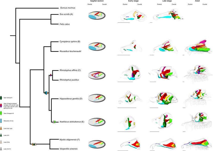
Ito et al.                                                                                                       Development of Bat Nasal Turbinals

    Several types of turbinal can be recognized in the mammalian          transversalis extends from the lateral walls of the nasal cavity
nasal cavity. The marginoturbinal and atrioturbinal are found             and attaches to the nasal septum, separating the ethmoturbinal
in the outer nasal cartilage in the rostral part of the nasal             recess from the nasopharyngeal duct (Lozanoff and Diewert,
cavity. The marginoturbinal begins at the lateral margin of               1989; Macrini, 2012; Smith et al., 2015).
the external nasal opening and continues into the atrioturbinal               As for the general developmental pattern for mammals,
(Maier, 1980, 2000). The shape of these turbinals forms the               initially, these turbinals and laminae appear as simple ridges
naris and permits effective airflow (Göbbel, 2000; Maier and              along the lateral wall of the nasal capsule (Dieulafe, 1906). The
Ruf, 2014). These turbinals remain cartilaginous in adults and            nasal capsule, which is the rostral part of the chondrocranium,
are continuous with the maxilloturbinal (Voit, 1909; Reinbach,            undergoes drastic morphological changes through ontogeny
1952a,b; Maier, 1980, 1993b; Zeller, 1987; Smith et al., 2015).           (Maier and Ruf, 2014; Van Valkenburgh et al., 2014; Smith
The maxilloturbinal is ventrally positioned in the nasal cavity           et al., 2015). Morphogenesis of the nasal capsule in mammals is
(Negus, 1958; Moore, 1981; Smith et al., 2015). This turbinal             attributed to three mesenchymal condensations: the parietotectal
projects from the medial surface of the maxilla and is covered            cartilage aside from the tectum, paranasal cartilage, and
with the respiratory epithelium (Scott, 1954; Adams, 1972; Van            orbitonasal lamina (De Beer, 1937; Reinbach, 1952b; Moore,
Valkenburgh et al., 2011) to add humidity to and increase the             1981; Zeller, 1987; Rossie, 2006; Smith and Rossie, 2006,
temperature of inhaled air (Scott, 1954). The maxilloturbinal             2008; Van Valkenburgh et al., 2014). The nasal tectum of
generally becomes the largest and most complex in adults (Maier,          the parietotectal cartilage condenses in the rostrocaudal and
1993b; Maier and Ruf, 2014; Van Valkenburgh et al., 2014; Smith           mediolateral direction (De Beer, 1937; Smith and Rossie, 2008).
et al., 2015). The nasoturbinal projects from the roof of the nasal       As the mesenchyme condenses, the rostral ridge of the paranasal
cavity (Moore, 1981). This turbinal articulates with the inferior         cartilages overlaps the parietotectal cartilage, and the caudal ridge
margin of the nasal bone and medial surface of the maxilla and            of the paranasal cartilages overlaps the orbitonasal lamina (Smith
extends caudally into the ethmoid complex (Moore, 1981; Smith             and Rossie, 2008; Van Valkenburgh et al., 2014). As a result,
et al., 2015). The ethmoturbinals project from the lateral mass of        the lamina semicircularis is formed rostrally and ethmoturbinal
the ethmoid bone (Smith et al., 2015). Several ethmoturbinals are         I is formed caudally within the nasal capsule. Subsequently,
found (Van Gilse, 1927; Maier, 1993a; Maier and Ruf, 2014) and            ethmoturbinals II to IV are formed rostrocaudally within the
are generally covered with olfactory epithelium (Adams, 1972;             orbitonasal lamina (Rossie, 2006; Smith and Rossie, 2008; Van
Gross et al., 1982; Martinez et al., 2020). Each ethmoturbinal            Valkenburgh et al., 2014). The nasal capsule then becomes
is arranged one behind the other in parallel (Voit, 1909). Voit           gradually enclosed by exocranial facial bones (Maier and Ruf,
(1909) denoted the ethmoturbinals by Roman numerals in                    2014). Through prenatal ontogeny, the structure of each turbinal
rostrocaudal sequence. Ethmoturbinal I protrudes toward the               changes in shape and becomes complicated, filling the nasal
nostrils and is usually the largest among the ethmoturbinals              cavity (Maier and Ruf, 2014). Prenatally, the nasal epithelium
(Voit, 1909). It makes the front border of the ethmoturbinal              sinks at specific sites, where the initial folds are created. Within
recess, which is the restricted space in the caudal part within           the initial folds, mesenchymal condensations constitute the
the nasal cavity (Smith and Rossie, 2008; Maier and Ruf, 2014).           primitive morphology of the chondral template of turbinals
The number of ethmoturbinals varies among species (Paulli,                (Smith et al., 2020). Later, these mesenchymal condensations
1900a,b,c; Rowe et al., 2005). To our knowledge, the minimum              chondrify. In perinatal and postnatal stages, cartilages change
number is seen in Tursiops in odontocetes with the absence of             their shape into lamellae (Smith et al., 2020). In the adult,
the ethmoturbinal (Mead and Fordyce, 2009). Orycteropus afer              the cartilaginous lamellae is fully ossified with the process of
has the maximum number of ethmoturbinal so far with “at least             endochondral ossification except for the marginoturbinal and
nine” (Stößel et al., 2010). The frontoturbinals are located within       atrioturbinal (Voit, 1909; Martineau-Doizé et al., 1992; Ruf et al.,
the frontoturbinal recess, which is the dorsocaudal space of the          2015; Smith et al., 2020). Additional turbinals branch off from
lateral recess bounded ventrally by the root of ethmoturbinal I           each turbinal, scroll, and fold and also merge with one another
(Maier, 1993a; Rossie, 2006). The accessory scrolls between the           (Parker, 1885; Maier, 1980, 2000; Deleon and Smith, 2014; Maier
frontoturbinals within the frontoturbinal recess are known as             and Ruf, 2014; Smith et al., 2020). The ossified remnant of
interturbinals (Maier, 1993b; Maier and Ruf, 2014; Ruf, 2014).            the nasal capsule becomes the ethmoid bone (Patterson, 1977).
The number of frontoturbinals and interturbinals may vary                 An emerging consensus agrees with the bauplan (body plan)
depending on the species (Smith et al., 2015).                            of cartilaginous nasal capsule having a tripartite composition:
    In addition to turbinals, the nasal cavity presents other             the anterior part (pars anterior), lateral part (pars lateralis),
sheet-like ossifications such as the lamina semicircularis, lamina        and posterior part (pars posterior) (Figure 1) (Maier, 1993a;
horizontalis, and lamina transversalis (Maier and Ruf, 2014). The         Rossie, 2006; Smith and Rossie, 2006, 2008; Maier and Ruf, 2014;
lamina semicircularis is the medial wall of the maxillary recess          Van Valkenburgh et al., 2014).
and frontoturbinal recess (Ruf, 2014). This lamina is continuous              The turbinate anatomy of various mammalian species at the
with the posterior part of the nasoturbinal (Macrini, 2012; Smith         adult stage has been described by the classic works of Paulli,
et al., 2015). The lamina horizontalis separates the lateral recess       providing a major source of current information on the diversity
into the dorsal and ventral chambers: the dorsal chamber is the           of mammalian turbinals (Paulli, 1900a,b,c). However, his studies
frontoturbinal recess and the ventral chamber is the maxillary            erroneously interpreted the lamina semicircularis as a turbinal,
recess (Smith and Rossie, 2008; Maier and Ruf, 2014). The lamina          due to the lack of observations on fetal stages of nasal structures

Frontiers in Cell and Developmental Biology | www.frontiersin.org     2                                      March 2021 | Volume 9 | Article 613545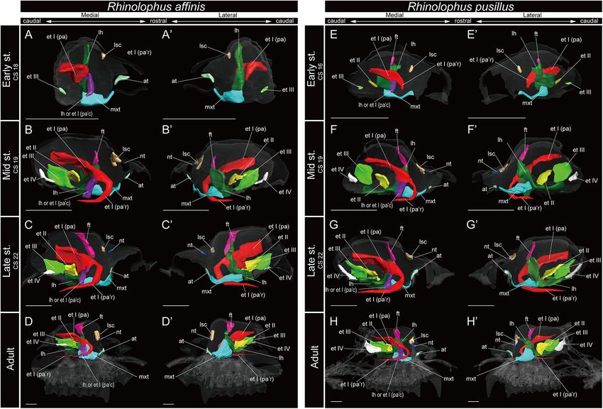
Ito et al.                                                                                                                                       Development of Bat Nasal Turbinals

  FIGURE 1 | Generalized schematic mammalian nasal capsule (modified from Maier, 1993a). (A) Medial view of parasagittal section; (B) horizontal section. These
  images show the nasal structure without facial exocranial (dermal) bones except for the maxilla and palatine bones. at, atrioturbinal; et I (pa), ethmoturbinal I pars
  anterior; et I (pp), ethmoturbinal I pars posterior; et II–IV, ethmoturbinals II–IV; etr, ethmoturbinal recess; ft, frontoturbinal; in, interturbinal; lh, lamina horizontalis; lsc,
  lamina semicircularis; mt, marginoturbinal; mx, maxillare; mxr, maxillary recess; mxt, maxilloturbinal; nac (pa), nasal capsule pars anterior; nac (pl), nasal capsule pars
  lateralis; nac I (pp), nasal capsule pars posterior; nph, nasopharyngeal duct; nt, nasoturbinal; pal, palatinum.

(Maier and Ruf, 2014). Since the nasal structure becomes highly                                (Springer et al., 2001; Teeling et al., 2002, 2003, 2005).
complicated, particularly during prenatal development, it is                                   Apart from most members of the family Pteropodidae of
virtually impossible to correctly establish turbinate homologies                               the Yinpterochiroptera, many bat species can use laryngeal
between species solely by comparisons of adult anatomy (Maier                                  echolocation. Most echolocating bat species emit their calls
and Ruf, 2014). In contrast, the turbinate structure in fetal stages                           orally, but in some families, echolocation calls are emitted
is rather simple, and observations on fetal series allow us for                                nasally (Brigham et al., 2004; Feldhamer et al., 2007). Olfactory
tracing the structural changes of the nasal capsule and turbinals                              capabilities in bats have been suggested to vary between species
(Maier, 1993a; Macrini, 2014; Maier and Ruf, 2014). Thus,                                      (Bhatnagar and Kallen, 1974a; Hutcheon et al., 2002). Bat
previous studies have emphasized the importance of comparative                                 turbinals have been studied by many authors (Grosser, 1900;
embryological approaches for understanding turbinate homology                                  Frick, 1954; Bhatnagar and Kallen, 1974a,b, 1975; Cooper and
among mammals (Novacek, 1993; Maier, 1993a; Maier and Ruf,                                     Bhatnagar, 1976; Göbbel, 2002; Giannini et al., 2006, 2012;
2014). However, few studies incorporate fetal samples (Reinbach,                               Nelson et al., 2007; Smith et al., 2012; Eiting et al., 2014a; Curtis
1952a,b; Maier, 1980; Smith and Rossie, 2008; Giannini et al.,                                 and Simmons, 2017; Curtis et al., 2020; Yohe et al., 2018), but
2012; Maier and Ruf, 2014; Ruf et al., 2015; Ruf, 2020), possibly                              the complex and diverse anatomy of bat turbinals has caused
due to the difficulty in obtaining rare fetal samples.                                         much confusion regarding their homology, possibly owing to
   Bats lack the prenatal information on turbinate anatomy                                     the variations in echolocation behavior and olfactory functions
with unresolved turbinate homology. They are the second                                        (Curtis and Simmons, 2017; Curtis et al., 2020).
most speciose order of mammals, exceeding 1,400 recognized                                        A handful of studies have attempted to discuss the
species (Wilson and Mittermeier, 2019; Simmons and Cirranello,                                 homologies of bat turbinals (Bhatnagar and Kallen, 1974a;
2020). Phylogenetically, they are presently divided into two                                   Kämper and Schmidt, 1977; Curtis and Simmons, 2017) using
suborders, i.e., Yangochiroptera and Yinpterochiroptera                                        adult specimens; however, as noted earlier, homologies of the

Frontiers in Cell and Developmental Biology | www.frontiersin.org                          3                                                March 2021 | Volume 9 | Article 613545
Ito et al.                                                                                                                 Development of Bat Nasal Turbinals

                                                                                      Our samples include Cynopterus sphinx and R. leschenaultii
                                                                                      from Pteropodidae, Rhinolophus affinis and Rhinolophus
                                                                                      pusillus from Rhinolophidae, Hipposideros gentilis and Aselliscus
                                                                                      stoliczkanus from Hipposideridae of Yinpterochiroptera, and
                                                                                      Myotis siligorensis and Vespertilio sinensis from Vespertilionidae
                                                                                      species of Yangochiroptera (Figure 2). Fetuses of three non-bat
                                                                                      species of Laurasiatheria, Suncus murinus, Sus scrofa, and
                                                                                      Felis catus, were included as outgroups (Figure 2). Samples
                                                                                      belong to the curatorial collections at the Institute of Ecology
                                                                                      and Biological Resources of Vietnam Academy of Science and
                                                                                      Technology and the University Museum of the University of
                                                                                      Tokyo (Supplementary Table 1). These samples were fixed
                                                                                      and preserved with 70% ethanol solution. Grayscale images of
                                                                                      the specimens’ crania were obtained using microCT (InspeXio
                                                                                      SMX-90CT Plus, Shimadzu Co, Japan) with 90 kV source voltage
                                                                                      and 100 mA source currents. To enhance the contrast of the
                                                                                      CT images, we followed the image enhancement techniques of a
                                                                                      previous study (Gignac and Kley, 2014; Gignac et al., 2016) and
                                                                                      dipped the specimens with iodine-based solutions (1% iodine,
                                                                                      I2 KI in 99% ethanol solution) (Sohn et al., in press). Staining
                                                                                      duration was between 6 and 24 h depending on the size of the
                                                                                      specimen. Voxel size ranged from 8 to 35 µm. Images were
                                                                                      reconstructed with dimensions of 1,024 × 1,024 pixels and
                                                                                      in 12-bit grayscale. We reconstructed the cartilage and bones
                                                                                      within turbinals by manual segmentation of grayscale images
                                                                                      for each specimen using Segmentation Editor Tool in Amira 5.3
                                                                                      (Visage Imaging, Berlin, Germany) (Supplementary Table 1).
  FIGURE 2 | Phylogenetic relationships of bats and outgroup species in this          The cartilaginous structures are stained poorly by iodine-based
  study. Phylogenetic framework is based on Teeling et al. (2005) and Li et al.
                                                                                      solutions. We identified them indirectly from the connective
  (2007).
                                                                                      tissue like perichondria, which are readily stained with iodine-
                                                                                      based solutions (Gignac et al., 2016). When interpreted from
mammalian nasal structures are hardly possible to establish                           the surrounding structure, it is possible to distinguish ossified
without studying fetal anatomy. To date, our knowledge                                and cartilaginous structures. Supplementary Figure 1 shows the
on prenatal turbinals in bats is still in its infancy and                             ossified and cartilaginous structure with enhanced contrast of
restricted to only a few studies on some bat species, including                       the CT images from iodine solution. The crown-rump length
Rousettus aegyptiacus (Jurgens, 1962; Fehse, 1990), Pipistrellus                      (CRL) of each specimen was measured using sliding calipers
pipistrellus, Rhinolophus ferrumequinum, Vespertilio murinus                          (N20, Mitutoyo, Japan). Bat specimens were staged following
(Grosser, 1900), Miniopterus schreibersii (Fawcett, 1919; De Beer,                    Cretekos et al. (2005), which has been developed based on the
1937), Myotis myotis (Frick, 1954), Pteropus lylei (Giannini                          Carnegie system for human development. Bat fetal specimens
et al., 2012), Megaderma lyra (Smith et al., 2012), and Rousettus                     of stages CS18, CS19, and CS22 or CS23 of Cretekos’ staging
leschenaultii (Smith et al., 2020).                                                   system (which respectively correspond to CS18, CS19, and CS22
    Similarly, the fetal anatomy and ontogenetic periods to                           in the human Carnegie system), in which turbinate development
adult stages in bats are still largely unexplored or poorly                           and splitting can be observed, were here compared. In this
studied. Here, using diffusible iodine-based contrast-enhanced                        study, specimens assigned as stage 18 are hereafter referred to
computed tomography (diceCT) imaging, we describe the                                 as “early stage,” stage 19 as “mid stage,” and stages 22 and 23 as
detailed embryonic development of the nasal cavity in eight                           “late stage” for simplification. For R. pusillus, a fetal specimen
species of bats, dividing into two suborders: Yangochiroptera and                     of stage 15 was additionally studied to observe the initial onset
Yinpterochiroptera. We revise turbinate homologies among bats                         of the turbinate projection. Gestation day 29 and postnatal day
and reconstruct the evolutionary history of the nasal turbinal of                     1 of S. murinus are respectively referred as “mid stage” and
bats in light of the modern phylogenetic framework.                                   “late stage” (which roughly correspond to CS22 and CS23 in
                                                                                      the human Carnegie system). Gestation day 28 and gestation
                                                                                      day 40 of S. scrofa are respectively referred as “mid stage” and
MATERIALS AND METHODS                                                                 “late stage” (which roughly correspond to CS22 and CS23 in
                                                                                      the human Carnegie system). Gestation day 38 and gestation
We observed multiple developmental stages from the                                    day 49 of F. catus are referred as “mid stage” and “late stage,”
fetus to adult of eight species of bats. Stages and basic                             respectively (which roughly correspond to CS22 and CS23 in
measurements are summarized in Supplementary Table 1.                                 the human Carnegie system). S. scrofa and F. catus were aged

Frontiers in Cell and Developmental Biology | www.frontiersin.org                 4                                     March 2021 | Volume 9 | Article 613545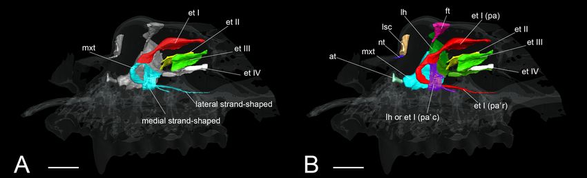
Ito et al. Development of Bat Nasal Turbinals
TABLE 1 | Terminology for turbinals and laminas.
Structure name Synonyms from other authors
Marginoturbinal –
Atrioturbinal –
Maxilloturbinal Inferior concha (Moore, 1981, p. 255)
Nasoturbinal Nasoturbinal, mucosal part (Smith and Rossie, 2008), rostral
nasoturbinal (Giannini et al., 2012)
Lamina Crista semicircularis (Voit, 1909), endoturbinal I (Paulli,
semicircularis 1900a,b,c; Moore, 1981), semicircular crest (Smith and Rossie,
2008), caudal nasoturbinal (Giannini et al., 2012), nasoturbinal
osseous part (Smith et al., 2015)
Lamina Anterior root of ethmoturbinal I (De Beer, 1937), lateral root of
horizontalis ethmoturbinal I (Rossie, 2006), frontomaxillary septum (Smith
and Rossie, 2008), lamina transversalis posterior (Macrini, 2014)
Ethmoturbinal I Endoturbinal I (Allen, 1882; Giannini et al., 2012), endoturbinal II
pars anterior (Paulli, 1900a,b,c; Moore, 1981), middle concha (Moore, 1981,
p. 255), ethmoturbinals I (Smith and Rossie, 2008),
endoturbinal I in adult (Macrini, 2014)
Ethmoturbinal I Ethmoturbinal I lobule (Allen, 1882), endoturbinal II, lower
pars posterior lamella (Paulli, 1900a,b,c; Moore, 1981), middle concha
(Moore, 1981, p. 255), ethmoturbinals II (Smith and Rossie,
2008), endoturbinal I in adult (Macrini, 2014)
Ethmoturbinal II Endoturbinal II (Allen, 1882; Giannini et al., 2012), endoturbinal
III (Allen, 1882; Paulli, 1900a,b,c; Moore, 1981), superior
concha (Moore, 1981, p. 255), ethmoturbinals III (Smith and
Rossie, 2008), endoturbinal II in adult (Macrini, 2014)
Ethmoturbinal III Endoturbinal III (Allen, 1882; Giannini et al., 2012), endoturbinal
IV (Paulli, 1900a,b,c; Moore, 1981), highest concha (Moore,
1981, p. 255), ethmoturbinal IV (Smith and Rossie, 2008)
Interturbinal Ectoturbinal (Allen, 1882; Paulli, 1900a,b,c; Moore, 1981;
Giannini et al., 2012)
Frontoturbinal Ectoturbinal (Allen, 1882; Paulli, 1900a,b,c; Moore, 1981;
Giannini et al., 2012), ectoturbinal in adult (Macrini, 2014)
based on Evans and Sack (1973). Specimen ID, CRL, stages,
and scanning parameters of all specimens are summarized in
Supplementary Table 1.
Terminology
The anatomical terminology for turbinals varies between studies
(Table 1), but here we adopted the bauplan proposed by Maier
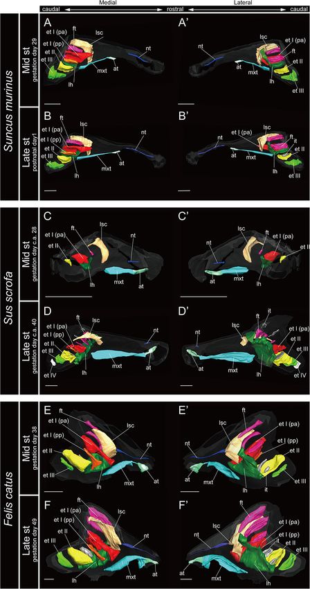
(1993a) and followed the anatomical terminology of Voit (1909) FIGURE 3 | 3D reconstructions of the developing turbinals in non-bat
laurasiatherians. (A,A0 ,C,C0 ,E,E0 ) Mid stage fetus; (B,B0 ,D,D0 ,F,F0 ) late stage
(Figure 1). We chose this terminology because it takes into fetus or postnatal; (A–F) medial view; (A0 –F0 ) lateral view. Scale bars, 1 mm.
account the topography, ontogeny, and homology of turbinal at, atrioturbinal; et I (pa), ethmoturbinal I pars anterior; et I (pp), ethmoturbinal I
bones (Maier and Ruf, 2014). pars posterior; et II–IV, ethmoturbinals II–IV; ft, frontoturbinal; mxt,
maxilloturbinal; lh, lamina horizontalis; nt, nasoturbinal; lsc, lamina
semicircularis.
RESULTS
Marginoturbinal and Atrioturbinal early stage in R. affinis and H. gentilis and from the mid stage
The marginoturbinal and the atrioturbinal were cartilaginous in R. pusillus (Figures 5A,A0 ,F,F0 , 6A,A0 ). In addition, the
structures in all species examined here. The atrioturbinal of atrioturbinal developed toward the rostrocaudal direction
all outgroup species and all bats was positioned ventrally, starting with the late stage (Figures 5, 6). While the
and it was continuous with the maxilloturbinal caudally atrioturbinal was visible, the marginoturbinal was partly
(Figures 3–7). As the nasal capsule enlarged, the atrioturbinal visible in our scans. Hence, the marginoturbinal cannot be
became more rostrocaudally elongated in the outgroup reconstructed. The contrast between the thick cartilage and
species as well as in Pteropodidae (Figures 3, 4). The surrounding soft tissue was not clear enough to identify
atrioturbinal is more rostrocaudally elongated from the the boundary.
Frontiers in Cell and Developmental Biology | www.frontiersin.org 5 March 2021 | Volume 9 | Article 613545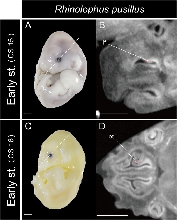
Ito et al. Development of Bat Nasal Turbinals
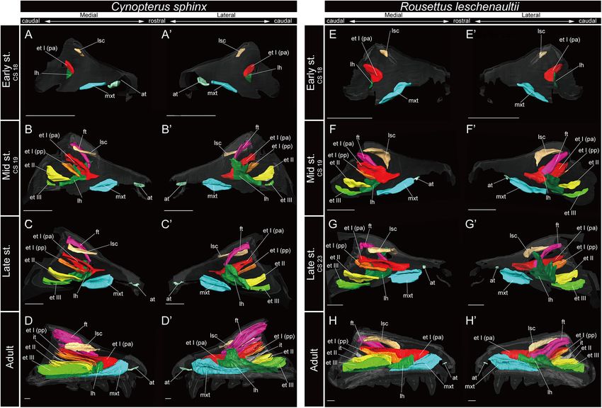
FIGURE 4 | 3D reconstructions of the developing turbinals in Cynopterus sphinx and Rousettus leschenaultii. (A–D0 ) C. sphinx; (E–H0 ) R. leschenaultii; (A,A0 ,E,E0 )
early stage fetus; (B,B0 ,F,F0 ) mid stage fetus; (C,C0 ,G,G0 ) late stage fetus; (D,D0 ,H,H0 ) adult. (A–D) Medial view and (A0 –D0 ) lateral view of C. sphinx; (E–H) medial
view and (E0 –H0 ) lateral view of R. leschenaultii. Scale bars, 1 mm. at, atrioturbinal; et I (pa), ethmoturbinal I pars anterior; et I (pp), ethmoturbinal I pars posterior; et
II–IV, ethmoturbinals II–IV; ft, frontoturbinal; mxt, maxilloturbinal; lh, lamina horizontalis; nt, nasoturbinal; lsc, lamina semicircularis.
Maxilloturbinal structure was replaced and ossified in the adult (Supplementary
In most specimens examined in this study, the maxilloturbinal Figures 3, 4).
was positioned caudally to the atrioturbinal. The maxilloturbinal Within Rhinolophoidea, all species presented similar
was a rostrally positioned structure within the nasal cavity, and maxilloturbinal morphologies. The maxilloturbinal enlarged and
its ventral side folded inward. The maxilloturbinal enlarged as it only partially ossified in the early to late stages (Figures 5, 6
developed in all outgroup species (Figure 3 and Supplementary and Supplementary Figures 5–8). The maxilloturbinal also
Figure 2). At the same time, it showed a double scroll in S. scrofa fused with the lamina horizontalis caudally such that it
(Figures 3C–D0 and Supplementary Figures 2C,D) and a single occurred in the early stage in Rhinolopus and A. stoliczkanus
scroll in S. murinus and F. catus (Figures 3A–B0 ,E–F0 and (Figures 5A,A0 ,E,E0 , 6E,E0 ) and in the mid stage in H. gentilis
Supplementary Figures 2A,B,E,F). The maxilloturbinal of the (Figures 6B,B0 ). Nonetheless, the maxilloturbinal was reduced
outgroup fetus was cartilaginous. compared with other turbinals and laminae after the late stage
Among bats, the developmental pattern of Pteropodidae in all species belonging to Rhinolophoidea (Figures 5, 6 and
resembled that in outgroup species. The maxilloturbinal Supplementary Figures 5–9). In addition, only the caudal side
of Pteropodidae was the largest among all turbinals and of the maxilloturbinal was ossified in the adult, and the rostral
laminae starting in the early stage (Figures 4A,A0 ,E,E0 ). side remained cartilaginous (Supplementary Figure 9).
Beginning at the mid stage in C. sphinx and late stage The maxilloturbinal of M. siligorensis and V. sinensis was
in R. leschenaultii, the maxilloturbinal started branching rostrocaudally elongated and lateromedially narrow (Figure 7).
(Supplementary Figures 3B, 4C). From the mid stage in It was slim and rod-shaped from the early to late stages
C. sphinx and late stage in R. leschenaultii, the maxilloturbinal (Figures 7A–C0 ,E–G0 ). Unlike the outgroup and Pteropodidae,
developed dorsal and ventral branches which were both laterally the maxilloturbinal did not branch, and it showed a single
scrolled as in the late stage of S. scrofa (Figures 3D,D0 , 4B,B0 ,G,G0 scroll ventrally as it developed from the late stage to adult
and Supplementary Figures 2D, 3B, 4C). Also, the cartilaginous (Supplementary Figures 2–4, 10, 11). It extended lateromedially,
Frontiers in Cell and Developmental Biology | www.frontiersin.org 6 March 2021 | Volume 9 | Article 613545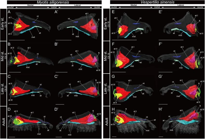
Ito et al.                                                                                                                                       Development of Bat Nasal Turbinals

  FIGURE 5 | 3D reconstructions of the developing turbinals in Rhinolophus affinis and R. pusillus. (A–D0 ) R. affinis; (E–H0 ) R. pusillus; (A,A0 ,E,E0 ) early stage fetus;
  (B,B0 ,F,F0 ) mid stage fetus; (C,C0 ,G,G0 ) late stage fetus; (D,D0 ,H,H0 ) adult. (A–D) Medial view and (A0 –D0 ) lateral view of R. affinis; (E–H) medial view and (E0 –H0 )
  lateral view of R. pusillus. Scale bars, 1 mm. at, atrioturbinal; et I (pa), ethmoturbinal I pars anterior; et I (pa’c), caudal part of ethmoturbinal I pars anterior; et I (pa’r),
  rostral part of ethmoturbinal I pars anterior; et II–IV, ethmoturbinals II–IV; ft, frontoturbinal; mxt, maxilloturbinal; lh, lamina horizontalis; nt, nasoturbinal; lsc, lamina
  semicircularis.

becoming a plate-like structure where it attached to the                                          The nasoturbinal of Rhinolophus and H. gentilis from
inner lateral nasal wall in the adult (Figures 7D,D0 ,H,H0 and                                 the mid stage and of A. stoliczkanus from the late stage
Supplementary Figures 10, 11).                                                                 consisted of a tiny cartilaginous structure, continuing with the
                                                                                               lamina semicircularis (Figures 5B–D0 ,F–H0 , 6B–D0 ,G–H0 and
Nasoturbinal                                                                                   Supplementary Figures 5–8).
We observed the nasoturbinal in mid and late stages of                                            The nasoturbinal of M. siligorensis and V. sinensis was
the outgroup species. The nasoturbinal slightly projected                                      much more well-developed than other chiropteran species
ventrally from the nasal wall (Figure 3). It did not show any                                  and projected slightly ventrally (Figure 7). It did not
scrolling, and it extended rostrocaudally and was observed                                     show any scrolling and extended rostrocaudally beyond
near the naris in both mid and late stages in all outgroups                                    the atrioturbinal–maxilloturbinal contact. While it formed
(Figure 3 and Supplementary Figure 2). The length of                                           a short rod-like structure in the early and mid stages
the nasoturbinal varied among the outgroup species such                                        (Figures 7A–B0 ,E–F0 and Supplementary Figures 10, 11),
that the nasoturbinal of S. murinus and F. catus was                                           in the late stage and adult, it formed a long rod-like structure
rostrocaudally longer than S. scrofa (Figure 3). The nasoturbinal                              rostrocaudally (Figures 7C–D0 ,G–H0 and Supplementary
of S. murinus and F. catus elongated rostrocaudally such                                       Figures 10, 11).
that its length was comparable to that of the maxilloturbinal
(Figures 3A–B0 ,E–F0 ).                                                                        Lamina Semicircularis
   In Pteropodidae, the nasoturbinal was absent during                                         The lamina semicircularis was observed in all outgroup
prenatal developmental stages (Figure 4 and Supplementary                                      species (Figure 3). This lamina extended from the inner
Figures 3, 4). In the adult, a slight projection was observed                                  wall in the central region of the nasal capsule toward
dorsally to the nasal cavity near the naris (Supplementary                                     the lateromedial, dorsoventral, and caudorostral directions. It
Figures 3, 4).                                                                                 expanded transversally on the dorsal side of the nasal cavity

Frontiers in Cell and Developmental Biology | www.frontiersin.org                          7                                                 March 2021 | Volume 9 | Article 613545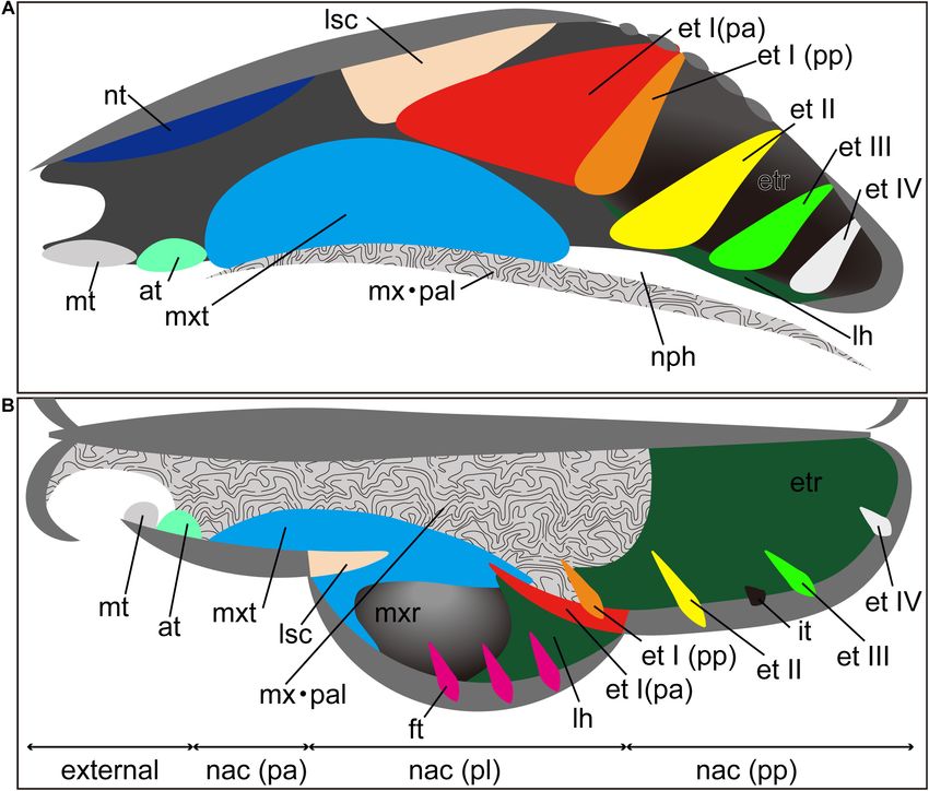
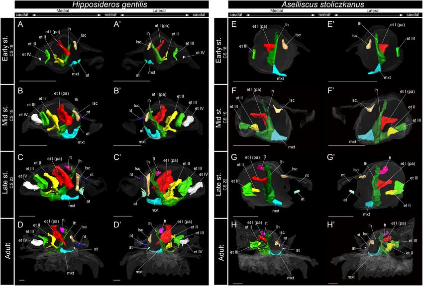
Ito et al.                                                                                                                                    Development of Bat Nasal Turbinals

  FIGURE 6 | 3D reconstructions of the developing turbinals in Hipposideros gentilis and Aselliscus stoliczkanus. (A–D0 ) H. gentilis; (E–H0 ) A. stoliczkanus; (A,A0 ,E,E0 )
  early stage fetus; (B,B0 ,F,F0 ) mid stage fetus; (C,C0 ,G,G0 ) late stage fetus; (D,D0 ,H,H0 ) adult. (A–D) Medial view and (A0 –D0 ) lateral view of H. gentilis; (E–H) medial
  view and (E0 –H0 ) lateral view of A. stoliczkanus. Scale bars, 1 mm. at, atrioturbinal; et I (pa), ethmoturbinal I pars anterior; et I (pp), ethmoturbinal I pars posterior; et
  II–IV, ethmoturbinals II–IV; ft, frontoturbinal; mxt, maxilloturbinal; lh, lamina horizontalis; nt, nasoturbinal; lsc, lamina semicircularis.

as it developed from the mid stage to the late (Figure 3 and                                 starting in the early stage (Figures 6A,A0 and Supplementary
Supplementary Figure 2).                                                                     Figure 7A). The lamina semicircularis then elongated toward
   Among bats, the lamina semicircularis of C. sphinx and                                    the lateromedial and dorsoventral directions from the inner
R. leschenaultii showed an almost identical developmental                                    wall of the nasal capsule in the mid to late stages (Figures 6B–
pattern as that of the outgroup species in terms of the transverse                           C0 and Supplementary Figures 7B,C). In the late stage, the
expansion. The lamina semicircularis was observed in the early                               lateromedially and dorsoventrally extended lamina semicircularis
stage for both species (Figures 4A,A0 ,E,E0 and Supplementary                                formed a wall that separated the anterior and the middle region of
Figures 3, 4). The lamina semicircularis extended in the                                     the nasal capsule (Figures 6C,C0 and Supplementary Figure 7C).
caudorostral direction rather than the dorsoventral direction                                In the adult, the lamina semicircularis was partly ossified, but not
which is different from the observation in the outgroup. While                               scrolled in all Rhinolophoidea (Figures 5, 6 and Supplementary
the lamina semicircularis of C. sphinx and R. leschenaultii was                              Figures 5–8).
not as large as that of the outgroup species, it was as large as                                In the adult of M. siligorensis and V. sinensis, the lamina
the frontoturbinal and the ethmoturbinal III from the mid stage                              semicircularis was not observed in the caudal region of the
(Figures 4B–D0 ,F–H0 ).                                                                      nasal cavity, which was surrounded by the maxilla and palatine
   The developmental pattern of the lamina semicircularis                                    (Figures 7D,D0 ,H,H0 and Supplementary Figures 10, 11).
was similar between Rhinolophoidea species (Figures 5, 6 and                                 The nasoturbinal was found in most of the dorsal region of
Supplementary Figures 5–8). The lamina semicircularis was                                    the external nasal cartilage (rostral side of the nasal cavity),
observed in all fetal stages and in the adult of Rhinolophoidea                              but the laminar structure was not seen in the caudal side
(Figures 5, 6 and Supplementary Figures 5–8). The lamina                                     of the nasoturbinal (Figures 7D,D0 ,H,H0 and Supplementary
semicircularis of H. gentilis was the largest among all                                      Figures 10, 11). While the nasoturbinal was observed, the
Rhinolophoidea (Figures 5, 6 and Supplementary Figures 5–8).                                 laminar structure was also not observed in the caudal side of
It projected ventrally from the inner wall of the nasal capsule                              the nasoturbinal in any fetal stages (Figures 7A–C0 ,E–G0 and

Frontiers in Cell and Developmental Biology | www.frontiersin.org                        8                                               March 2021 | Volume 9 | Article 613545
Ito et al.                                                                                                                                        Development of Bat Nasal Turbinals

  FIGURE 7 | 3D reconstructions of the developing turbinals in Myotis siligorensis and Vespertilio sinensis. (A–D0 ) M. siligorensis; (E–H0 ) V. sinensis; (A,A0 ,E,E0 ) early
  stage fetus; (B,B0 ,F,F0 ) mid stage fetus; (C,C0 ,G,G0 ) late stage fetus; (D,D0 ,H,H0 ) adult. (A–D) Medial view and (A0 –D0 ) lateral view of M. siligorensis; (E–H) medial
  view and (E0 –H0 ) lateral view of V. sinensis. Scale bars, 1 mm. at, atrioturbinal; et I (pa), ethmoturbinal I pars anterior; et I (pp), ethmoturbinal I pars posterior; et II–III,
  ethmoturbinal II–III; ft, frontoturbinal; mxt, maxilloturbinal; lh, lamina horizontalis; nt, nasoturbinal; lsc, lamina semicircularis.

Supplementary Figures 10, 11). Hence, the lamina semicircularis                                it fused with the ventral side of the ethmoturbinal I pars
was not formed in both M. siligorensis and V. sinensis.                                        anterior (Figures 3C,C0 , 4A,A0 ,E,E0 and Supplementary
                                                                                               Figures 2C, 3A, 4A). The lamina horizontalis likely extended
Lamina Horizontalis                                                                            toward the lateromedial, rostrocaudal, and caudorostral
The lamina horizontalis horizontally separates the                                             directions and fused with the ventromedial part of the
nasopharyngeal duct and the ethmoturbinal recess, which                                        ethmoturbinal I pars posterior and the ethmoturbinal II
includes several turbinals in the outgroup species (Figure 3                                   from the early to the mid stages (Figures 4A–B0 ,E–F0 and
and Supplementary Figure 2). The lamina horizontalis of                                        Supplementary Figures 3, 4). The lamina horizontalis enlarged
the outgroup species extended from the inner wall of the                                       lateromedially and elongated rostrocaudally from the mid stage
nasal capsule toward the lateromedial, rostrocaudal, and                                       to adult (Figures 4B–D0 ,F–H0 and Supplementary Figures 3, 4).
caudorostral directions. In the mid and late stages, the dorsal                                In all fetal stages, it was cartilaginous in both species, but ossified
side of the lamina horizontalis fused with the ventral side of                                 in the adult (Supplementary Figures 3, 4).
the ethmoturbinal I pars anterior and the ethmoturbinal I pars                                     The lamina horizontalis of Rhinolophoidea showed a different
posterior except for the mid stage of S. scrofa (Figure 3 and                                  developmental pattern compared with that of all outgroups and
Supplementary Figure 2).                                                                       Pteropodidae (Figures 3–6). In all members of Rhinolophoidea,
   Among bats, C. sphinx and R. leschenaultii showed a                                         the lamina horizontalis that projected from the inner wall
developmental pattern similar to the outgroup species,                                         of the nasal capsule extended toward the lateromedial and
whereas the developmental patterns of Rhinolophoidea and                                       dorsoventral directions from the early stage to adult. The lamina
Yangochiroptera were different from those of the outgroup                                      horizontalis formed a wall perpendicular to the rostrocaudal
species. Similar to the mid stage of S. scrofa, the lamina                                     plane (Figures 5, 6 and Supplementary Figures 5–8).
horizontalis projected from the inner wall of the nasal capsule                                    Among Rhinolophoidea, the presumptive developmental
in the early stage of C. sphinx and R. leschenaultii, and dorsally,                            pattern of the lamina horizontalis of Rhinolophus was somewhat

Frontiers in Cell and Developmental Biology | www.frontiersin.org                          9                                                 March 2021 | Volume 9 | Article 613545
Ito et al. Development of Bat Nasal Turbinals
dissimilar to that of H. gentilis or A. stoliczkanus, such that the
lamina horizontalis scrolled ventrolaterally beginning in the early
stage (Figures 5A,A0 ,E,E0 and Supplementary Figures 5A, 6A).
The apex of the lamina horizontalis projected inward in the
early stage and then gradually extended ventrally and medially
from the mid stage to the adult (Figure 5 and Supplementary
Figures 5, 6). In the adult, its apex extended caudally into
the nasopharyngeal duct (Supplementary Figures 5, 6). When
observed medially from the sagittal plane, it formed a hairpin-
shaped structure (Figures 5D,D0 ,H,H0 ). When seen in coronal
sections, the lamina horizontalis extended from the dorsolateral
side of the nasal wall toward the ventromedial side. At the medial
portion of the nasal capsule, the ethmoturbinal I pars anterior
is positioned dorsally from the lamina horizontalis from the
early stage of Rhinolophus (Supplementary Figures 5, 6). The
ventromedial part of the lamina horizontalis bent lateralward
from the point in which the ethmoturbinal I pars anterior
extended from the late stage to adult in R. affinis and from mid
sage to adult of R. pusillus (Supplementary Figures 5, 6). At
the caudal portion of the nasal cavity of adult Rhinolophus, the
ventral edge of the ventromedial part of the lamina horizontalis
was round and appeared inside of the nasopharyngeal duct
(Supplementary Figures 5, 6). Nonetheless, this hairpin-shaped
structure of the lamina horizontalis may be the ethmoturbinal I FIGURE 8 | The onset of the turbinate projections in Rhinolophus pusillus.
pars anterior (Figure 5). The structure which is most probably Broken lines indicate the location of section. Scale bar, 1 mm. (A) Early stage
the lamina horizontalis was ossified in the adult (Supplementary (CS 15) fetus embryo; (B) early stage fetus (CS 15) cross section; (C) early
stage fetus (CS 16) embryo; (D) early stage fetus (CS 16) cross section. et I,
Figures 4D, 5D, 9).
ethmoturbinal I; if, initial fold.
The lamina horizontalis was not observed in M. siligorensis
and V. sinensis in any fetal stages or the adult, unlike all outgroup
species as well as Pteropodidae and Rhinolophoidea (Figure 7
and Supplementary Figures 10, 11). In Rhinolophus, the developmental pattern of the
ethmoturbinal I pars anterior differed from that of all other
Ethmoturbinal I Pars Anterior species and formed a distinctive structure (Figure 5). The early
The ethmoturbinal I pars anterior of the outgroup species stage (CS 15) of R. pusillus was the smallest of all samples
projected toward the lateromedial and caudorostral directions examined in this study (Figures 8A,B). In the early stage (CS 15)
from the inner wall of the nasal capsule already in the mid stage, of R. pusillus, the cartilage of the nasal capsule was obscured in
fusing with the dorsal side of the lamina horizontalis (Figure 3 the scan, but an initial fold was observed in the inner wall of the
and Supplementary Figure 2). It was the largest turbinal among nasal capsule (Figure 8B). The cartilage which we believe as the
all ethmoturbinals from the mid to late stages in all outgroups ethmoturbinal I pars anterior of the late phase of early stage (CS
(Figure 3). The ethmoturbinal I pars anterior of S. scrofa was less 16) was likely embedding within this initial fold of the early stage
developed than that of F. catus and S. murinus in the mid stage (CS 15) in R. pusillus (Figures 8C,D).
(Figures 3A,A0 ,C,C0 ,E,E0 ). The fusion of the ethmoturbinal I pars anterior with the
The ethmoturbinal I pars anterior of Pteropodidae showed lamina horizontalis extended toward the lateromedial direction
the same developmental pattern as that of the outgroup species from the inner wall in the early stage of Rhinolophus,
(Figures 3, 4). The developmental pattern from the early to mid like the mid stage of S. scrofa, and the early stage of
stages, in particular, resembled that from the mid to the late stages Pteropodidae (Figures 3–5, and Supplementary Figures 2–6).
of S. scrofa (Figures 3C–D0 , 4A–B0 ,E–F0 ). The ethmoturbinal I The alternative interpretation is that the lamina horizontalis
pars anterior of Pteropodidae fused ventrally with the lamina did not form the hairpin-shaped structure in medial side.
horizontalis and projected toward the lateromedial direction Consequently, the ethmoturbinal I pars anterior split into rostral
from the inner wall of the nasal capsule (Figures 4A–B0 ,E–F0 ). and caudal parts from the early stage (Figures 5A,A0 ,E,E0
The ethmoturbinal I pars anterior extended rostrally from the and Supplementary Figures 5, 6). In this case, the rostral
early to mid stages, and in the adult, it reached as far as the dorsal part of the ethmoturbinal I pars anterior extended rostrally
border of the maxilloturbinal (Figures 4A–B0 ,D,D0 ,E–F0 ,H,H0 ). and curved ventrally in the mid stage of R. affinis and the
The ethmoturbinal I pars anterior of Pteropodidae was the largest early stage of R. pusillus (Figures 5B,B0 ,E,E0 ). Subsequently,
turbinal among all ethmoturbinals from the early stage to the it entered the nasopharyngeal duct, extending caudally in
adult (Figure 4). The ethmoturbinal I pars anterior was ossified the mid stage of R. affinis and the late stage of R. pusillus
in the adult (Supplementary Figures 4, 5). (Supplementary Figures 5B, 6C). When observed medially
Frontiers in Cell and Developmental Biology | www.frontiersin.org 10 March 2021 | Volume 9 | Article 613545Ito et al. Development of Bat Nasal Turbinals
from the sagittal plane, it formed a hairpin-shaped structure Ethmoturbinal I Pars Posterior
(Figures 5B,B0 ,E,E0 ). The apex of the rostral part of the The ethmoturbinal I pars posterior was observed from the ventral
ethmoturbinal I pars anterior extended caudally also from the side of the ethmoturbinal I pars anterior starting in the mid
mid stage (Figures 5B,B0 ,E,E0 ). It formed the freestanding stage of S. murinus and F. catus and in the late stage of S. scrofa
structure in the adult (Figure 5). (Figures 3A,A0 ,D,D0 ,E,E0 and Supplementary Figures 2A,D,E).
The caudal part of the ethmoturbinal I pars anterior, which Ethmoturbinal I pars posterior of Pteropodidae was located
can be identified as a part of the lamina horizontalis in at the same place as that of the outgroup species (Figures 3, 4).
the alternative interpretation, scrolled medially in the mid It was absent in the early stage (Figures 4A,A0 ,E,E0 and
stage of R. affinis and R. pusillus (Figures 5B,B0 ,F,F0 and Supplementary Figures 3, 4) but appeared starting in the mid
Supplementary Figures 5, 6). Subsequently, the apex of the stage (Figures 4B,B0 ,F,F0 and Supplementary Figures 3, 4).
caudal part of the ethmoturbinal I pars anterior that projected In contrast to the outgroup species and Pteropodidae,
inward gradually extended ventrally and medially until the late ethmoturbinal I pars posterior was absent from the early stage
stage (Figure 5). In the adult, this apex extended caudally and to adult in all members of Rhinolophoidea, M. siligorensis, and
became another freestanding structure in the nasopharyngeal V. sinensis (Figures 5–7).
duct (Figures 5D,D0 ,H,H0 and Supplementary Figures 5, 6).
From the sagittal plane, it also formed a hairpin-shaped structure
medially (Figures 5D,D0 ,H,H0 ). When seen in coronal sections, Turbinals in the Ethmoturbinal Recess
ethmoturbinal I pars anterior was present at the rostral portion Ethmoturbinals II and III projected from the inner wall of
of the nasal capsule. At the medial portion of the nasal capsule, the nasal capsule starting in the mid stage in S. murinus and
the rostral and caudal part of ethmoturbinal I pars anterior F. catus (Figures 3A,A0 ,E,E0 and Supplementary Figures 2A,E).
was branched off from the lamina horizontalis. The rostral part In S. scrofa, ethmoturbinal II was observed in the mid stage,
extended toward the dorsomedial side, and the caudal part while ethmoturbinal III was only present in the late stage
toward the ventromedial side from the early stage to adult (Figures 3C–D0 and Supplementary Figures 2C,D). In the
(Supplementary Figures 5, 6). At the caudal portion of the outgroup species, ethmoturbinal II was the second largest among
nasal cavity of adult, the ventral edge of the caudal part of all ethmoturbinals after ethmoturbinal I pars anterior except for
ethmoturbinal I pars anterior was round and appeared inside S. murinus (Figure 3).
of the nasopharyngeal duct medially to the rostral part of In Pteropodidae, ethmoturbinal II and ethmoturbinal III were
ethmoturbinal I pars anterior (Supplementary Figures 5, 6). absent in the early stage; however, they projected from the inner
In the early stage of H. gentilis and A. stoliczkanus, the ventral wall of the nasal capsule after the mid stage (Figure 4 and
side of the ethmoturbinal I pars anterior fused at the dorsal side Supplementary Figures 3, 4). In terms of size, ethmoturbinal II
of the dorsoventrally elongated lamina horizontalis and projected was larger compared with ethmoturbinal III in Pteropodidae in
from the inner wall of the nasal capsule (Figures 6A,A0 ,E,E0 the mid and late stages (Figures 4B–C0 ,F–G0 ). Ethmoturbinal III
and Supplementary Figures 7, 8). The ethmoturbinal I pars was larger than ethmoturbinal I pars posterior from the late stage
anterior was large after the early stage, and it developed dorsally to adult (Figures 4C–D0 ,G–H0 ).
and caudally to the lamina horizontalis in H. gentilis and In R. affinis, ethmoturbinal II appeared from the inner wall
A. stoliczkanus, respectively (Figures 6A,A0 ,E,E0 ). Still, it did of the nasal capsule in the mid stage, while in R. pusillus,
not elongate rostrally like that of S. murinus and Pteropodidae it was observed in a similar position in the early stage
(Figures 3A–B0 , 4, 5). The ethmoturbinal I of Rhinolophoidea (Figures 5B,B0 ,E,E0 and Supplementary Figures 5B, 6A).
was ossified in the adult (Supplementary Figures 5–8). In both species, ethmoturbinal III projected from the inner
Similar to the outgroup species and other bats, the wall of the nasal capsule in the early stage (Figures 5A,A0 ,E,E0
ethmoturbinal I of M. siligorensis and V. sinensis was the largest and Supplementary Figures 5, 6). In R. pusillus, the size
turbinal from the early stage to adult (Figure 7). On the other of ethmoturbinal II and ethmoturbinal III was mostly
hand, it differed from the outgroup species and Pteropodidae comparable in the early stage (Figures 5E,E0 ). Ethmoturbinal
in that it did not split into pars anterior and pars posterior III was larger compared with ethmoturbinal II in the mid
(Figures 3, 4, 7 and Supplementary Figures 2–4, 10, 11). stage (Figures 5B,B0 ,F,F0 ). Also, ethmoturbinal IV arose
The ventral side of the rostral end of ethmoturbinal I formed in the mid stage of Rhinolophus (Figures 5B,B0 ,F,F0 and
a horizontal plate-like process from the mid stage to adult Supplementary Figures 5, 6). Ethmoturbinal III was the
(Figures 7B–D0 ,F–H0 and Supplementary Figures 10, 11). In largest, and ethmoturbinals II and IV were of the same size
addition, since the lamina horizontalis was absent in all stages, among these three turbinals from the late stage Rhinolophus
ethmoturbinal I projected solely from the inner wall of the nasal (Figures 5C,C0 ,G,G0 ).
capsule (Figure 7 and Supplementary Figures 10, 11). While it In H. gentilis, ethmoturbinals II, III, and IV projected from the
enlarged dorsoventrally from the early to late stages (Figures 7A– inner wall of the nasal capsule in the early stage (Figures 6A,A0 ).
C0 ,E–G0 and Supplementary Figures 10, 11), ethmoturbinal I Ethmoturbinal III was large in the early stage (Figures 6A,A0 ).
extended toward the caudorostral direction from the late stage to Moreover, in H. gentilis, ethmoturbinal II was larger than
adult (Figures 7C–D0 ,G–H0 and Supplementary Figures 10, 11). ethmoturbinal III in the mid stage; however, the size became
Ethmoturbinal I of Yangochiroptera was ossified in the adult similar from the late stage (Figures 6B–D0 ). In A. stoliczkanus,
(Supplementary Figures 10, 11). ethmoturbinal III appeared in the early stage (Figures 6E,E0 ).
Frontiers in Cell and Developmental Biology | www.frontiersin.org 11 March 2021 | Volume 9 | Article 613545Ito et al. Development of Bat Nasal Turbinals
Then, in the mid stage, ethmoturbinal II appeared rostrally to echolocation, while Cynopterus does not engage in such behavior)
ethmoturbinal III (Figures 6F,F0 ). Ethmoturbinal IV was not are not reflected in the turbinate structures of R. leschenaultii and
observed from the early stage to adult (Figures 6H,H0 ). C. sphinx (Figure 4 and Supplementary Figures 3, 4).
Ethmoturbinal II appeared from the inner wall of the nasal Based on adult specimens, Allen (1882) and Paulli (1900c)
capsule in the early stage of M. siligorensis and V. sinensis; suggested that Pteropodidae [Cynopterus, Epomophorus
however, after the mid stage, ethmoturbinal II shifted laterally, gambianus, Pteropus giganteus, Pteropus sp., Rousettus
approaching ethmoturbinal I during late ontogeny as the nasal (Cyonycteris)] have “four endoturbinals.” Given Allen’s (1882)
capsule enlarged (Figures 7B–D0 ,F–H0 and Supplementary schematic for the turbinal of E. gambianus, we assume that he
Figures 10, 11). The caudal part of ethmoturbinal II curved incorrectly split the true ethmoturbinal I into “endoturbinal
and fused with the laterally positioned ethmoturbinal I from the I” and “endoturbinal II” (Supplementary Figure 12). Allen
late stage to adult (Figures 7C–D0 ,G–H0 and Supplementary (1882) drew other schematics for the turbinal of non-volant
Figures 10, 11). Both ethmoturbinal II and ethmoturbinal mammals, in which the author identified ethmoturbinal I
III extended toward the caudorostral and the ventrodorsal pars anterior and pars posterior as the “endoturbinal I and
directions, forming a triangular shape from the mid stage lobule.” This suggests that the misidentification of the turbinal
(Figures 7B–D0 ,F–H0 ). of E. gambianus reported by Allen (1882) was clearly not caused
by the difference in nomenclature. Only tentative inferences
can be made, as Paulli (1900c) provided no schematic for
DISCUSSION the turbinals of Pteropodidae, but we assume Paulli labeled
the lamina semicircularis as “endoturbinal I.” Thus, there are
Turbinate Ontogeny and Homology in “four endoturbinals” for the Pteropodidae species in Paulli’s
Laurasiatheria and Pteropodidae view. Giannini et al. (2012) identified the turbinals based on
We found that the turbinate structures are principally a histological section of one fetal stage of Pteropus sp. and a
comparable between Laurasiatheria and Pteropodidae. The CT-scanned image of the adult P. lylei. They pointed out that
fetuses of outgroup species, S. murinus, S. scrofa, and F. catus, there is one additional “endoturbinal” in both studies of Allen
together with the fetuses of Pteropodidae, appear to share a (1882) and Paulli (1900c) compared with their observation
ventrally positioned and enlarged maxilloturbinal, which is the (Figures 4C–D0 ,G–H0 and Supplementary Figure 12). Our
largest among all turbinals (Figures 3, 4 and Supplementary observation on the turbinals of C. sphinx and R. leschenaultii
Figures 2–4). Ethmoturbinal I is the largest to develop among shows that ethmoturbinal III projects from the inner nasal wall
ethmoturbinals, splitting into pars anterior and pars posterior in from the mid stage (Figures 4B–D0 ,F–H0 and Supplementary
all outgroup species (Figures 3A–B0 ,D–F0 ). In our study, adult Figures 3, 4). Following this, no more ethmoturbinals are formed
specimens of outgroup species were not examined. Previous (Figure 4 and Supplementary Figures 3, 4). Allen’s study on
studies showed the ventrally positioned, branched off, and E. gambianus (with our assumption that ethmoturbinal I splits
enlarged maxilloturbinal and ethmoturbinal I pars anterior into two parts; Supplementary Figure 12), Giannini’s study on
and pars posterior in the adult S. murinus (Kuramoto, 1980), Pteropus, and our study on C. sphinx and R. leschenaultii indicate
S. scrofa (Paulli, 1900a), and F. catus (Moore, 1981). These that the number of ethmoturbinals is three in Pteropodidae
characteristics were also seen in the adult Pteropodidae studied (Figure 4 and Supplementary Figures 3, 4).
here. Furthermore, the lamina horizontalis of the outgroup Giannini et al. (2012) compared Pteropus with non-bat
species and Pteropodidae divides the nasopharyngeal duct mammals and claimed that the turbinate element composition
and the ethmoturbinal recess that includes several turbinals in Pteropus is comparable with that of non-human primates
(Figures 3, 4 and Supplementary Figures 2–4). The enlarged (Smith and Rossie, 2006) and rodents (Paulli, 1900c) in terms
ethmoturbinal recess with a vast space formed dorsally which of the reduced number of turbinals. They also concluded that
is filled with the olfactory mucosa suggests that Pteropodidae the number of frontoturbinals in Pteropus differs from that
are equipped with high olfactory ability (Negus, 1958; Craven of the hedgehog Erinaceus (Paulli, 1900c) and the marsupial
et al., 2007, 2010; Eiting et al., 2014b). The developmental Monodelphis (Rowe et al., 2005; Macrini, 2014) such that
structure and position of the turbinal and lamina of the outgroup Pteropus has one while the hedgehog and marsupial have two
species studied here are congruent with the generality seen frontoturbinals. The turbinate structure of Pteropus is both
among previously reported non-volant mammals (Paulli, similar and different from that of carnivorans and ungulates
1900a,b,c; Voit, 1909; Moore, 1981; Smith and Rossie, 2008; which have increasing number and complexity (Stößel et al.,
Van Valkenburgh et al., 2014). 2010) in ethmoturbinals, frontoturbinals, and interturbinals
Some authors mention the possibility that the unique (Giannini et al., 2012). Carnivorans have three ethmoturbinals
turbinate morphology of Rhinolophus (which was also seen in like Pteropodidae, which are similar to our results (Paulli,
our study) is related to echolocation (Curtis and Simmons, 2017; 1900a,b,c; Wagner and Ruf, 2019, 2020) (Supplementary
Curtis et al., 2020). Given this, and the fact that Rhinolophus Table 1). However, Paulli reported that the maximum of
belongs to Yinpterochiroptera as do Pteropodidae, we expected the frontoturbinal and interturbinal combined (ectoturbinal in
to see similar morphology in echolocating Rousettus prior to our Paulli) is five to ten (only Meles) (Paulli, 1900c). This is different
experiment (Feldhamer et al., 2007). However, such behavioral to Pteropodidae with one frontoturbinal and one interturbinal.
differences (Rousettus is known for using tongue clicks for The number of ethmoturbinal of ungulates varies such that
Frontiers in Cell and Developmental Biology | www.frontiersin.org 12 March 2021 | Volume 9 | Article 613545Ito et al. Development of Bat Nasal Turbinals
Capra has three and Dicotyles labiatus has seven. Moreover, to Yangochiroptera (Teeling et al., 2002, 2003, 2005). Although
the number of frontoturbinal and interturbinal largely varies most bats of Yangochiroptera emit sonar from their oral
with seven in Tragulus javanicus and 31 in Equus caballus apparatus, Phyllostomidae and Nycteridae are characterized
(Paulli, 1900c; Moore, 1981). Capra showed the same number by emitting sonar from the naris (Jones and Teeling, 2006;
of ethmoturbinal as Pteropodidae; however, the number of Feng et al., 2012). As for the turbinals of Nycteridae, Allen
ethmoturbinal and frontoturbinal and interturbinal combined (1882) is the only study that identifies turbinals in this
in ungulates is generally larger than that of Pteropodidae. family, in which the author described the adult Nycteris
Nonetheless, Giannini et al. (2012) pointed out that the turbinate thebaica. He stated that N. thebaica has two endoturbinals
homology of elements in Pteropus can be traced without difficulty (endoturbinal I has a lobule) and one ectoturbinal. Moreover,
based on the position of turbinals in canids (Vulpes vulpes). he claimed that it has a nasoturbinal that is larger than
Even though the number varies for certain turbinals (caudal the endoturbinal. Regarding Phyllostomidae, Bhatnagar and
ethmoturbinal, interturbinal, and frontoturbinal) among non- Kallen (1974a) studied Artibeus jamaicensis; Kämper and
volant Laurasiatheria and Pteropodidae, the turbinal element Schmidt (1977) studied Artibeus lituratus, Carollia perspicillata,
composition of pteropodids is easily traceable from that of Glossophaga soricina, Phyllostomus discolor, and Sturnira lilium;
our non-volant laurasiatherians. Therefore, we agree with and Yohe et al. (2018) studied A. jamaicensis, Brachyphylla
Giannini et al. (2012) that the turbinate element composition of pumila, Erophylla bombifrons, and Phyllonycteris poeyi of
Pteropodidae is rather similar to that of other mammals (even for Phyllostomidae. Principally, the members of Phyllostomidae
species with complex turbinals). reportedly have seven turbinals. Although details of the
identification varies among studies, all these studies agree that
Yangochiroptera in Phyllostomidae the ethmoturbinal recess within the nasal
The lamina horizontalis is not formed throughout ontogeny cavity is separated with the nasopharyngeal duct rostrally by
in the studied Vespertilionidae (Figure 7 and Supplementary the ethmoturbinal and caudally by the lamina (Bhatnagar and
Figures 10, 11). Consequently, there is no clear separation Kallen, 1974a; Kämper and Schmidt, 1977; Yohe et al., 2018).
between the nasopharyngeal duct and the ethmoturbinal recess. Presenting coronal sections of the ethmoturbinal recess in
This anatomical setting suggests that these species are not three bats (Anoura geoffroyi, S. lilium, Uroderma bilobatum),
specialized to keep the inspired air within the nasal cavity, Eiting et al. (2014a) showed that the lamina that separates
allowing for better odorant sorption (Negus, 1958; Adams, 1972; the nasopharyngeal duct and the ethmoturbinal recess is well-
Craven et al., 2010; Eiting et al., 2014b). Based on the nasal cavity developed. However, these studies are all based on adult species,
structure, we suggest that the studied Vespertilionidae members and fetal information of Phyllostomidae is still largely lacking
are less capable of catching odorants compared with the outgroup (Bhatnagar and Kallen, 1974a; Kämper and Schmidt, 1977; Eiting
mammals and Pteropodidae which have the lamina horizontalis et al., 2014a; Yohe et al., 2018).
and an independent space of the ethmoturbinal recess. Compared with the patterns reported for Phyllostomidae,
Fetal turbinals of P. pipistrellus, V. murinus (Grosser, we recognize that M. siligorensis and V. sinensis do not have
1900), M. schreibersii (Fawcett, 1919; De Beer, 1937), and the lamina horizontalis, which is the lamina separating the
M. myotis (Frick, 1954) were studied previously using histological nasopharyngeal duct and ethmoturbinal recess (Figure 7). This
sections. The figures given by Fawcett (1919); De Beer (1937), was particularly obvious in the caudal region of the nasal cavity
and Frick (1954) did not show the lamina, which separates (Bhatnagar and Kallen, 1974a; Kämper and Schmidt, 1977; Eiting
the nasopharyngeal duct and the ethmoturbinal recess that et al., 2014a; Yohe et al., 2018). Further observation of the nasal
includes several turbinals in these Vespertilionidae species. development of Phyllostomidae, Mollossidae, Emballonuridae,
The lamina horizontalis separates the nasopharyngeal duct and Nycteridae is required to clarify the whole picture of
and the ethmoturbinal recess in all stages in the outgroup turbinate homology within Yangochiroptera.
species and after mid stage in Pteropodidae (Figures 3, 4
and Supplementary Figures 2–4). Our study is consistent Rhinolophoidea
with the observation of Fawcett (1919); De Beer (1937), The rostral part of the lamina horizontalis extends dorsally in
and Frick (1954). In the early stage of M. siligorensis and Rhinolophoidea from the early stage (Figures 5, 6A–A0 ,E–E0 and
V. sinensis, ethmoturbinal II is located on the inner wall Supplementary Figures 5–8). The lamina horizontalis pushes the
of the nasal capsule (Figures 7A,A0 ,E,E0 and Supplementary ethmoturbinal recess back toward the caudal direction, resulting
Figures 10, 11), but the caudal part of ethmoturbinal II curves in a small ethmoturbinal recess. As the size of the ethmoturbinal
and fuses with the laterally positioned ethmoturbinal I from the recess likely relates to olfactory ability (Negus, 1958; Adams,
late stage to adult (Figures 7C–D0 ,G–H0 and Supplementary 1972; Craven et al., 2010; Eiting et al., 2014b), it is likely that
Figures 10, 11). As identified by Fawcett (1919) and De Beer Rhinolophoidea may have a reduced olfactory ability compared
(1937) in M. schreibersii and Frick (1954) in M. myotis, our with the outgroup species and Pteropodidae.
study confirms that ethmoturbinal II is positioned medially to Rhinolophoidea are undoubtedly the most disputed
ethmoturbinal I in all fetal stages and adult in M. siligorensis and and problematic taxon among bats regarding its turbinate
V. sinensis. homology. Members of the superfamily Rhinolophoidea, which
The present study did not cover members of Phyllostomidae, include Rhinolophidae, Hipposideridae, Megadermatidae,
Emballonuridae, Molossidae, and Nycteridae, which also belong Craseonycteridae, and Rhinopomatidae, emit echolocation
Frontiers in Cell and Developmental Biology | www.frontiersin.org 13 March 2021 | Volume 9 | Article 613545You can also read