The Keystone commensal bacterium Christensenella minuta DSM 22607 displays anti inflammatory properties both in vitro and in vivo
←
→
Page content transcription
If your browser does not render page correctly, please read the page content below
www.nature.com/scientificreports
OPEN The Keystone commensal
bacterium Christensenella
minuta DSM 22607 displays
anti‑inflammatory properties
both in vitro and in vivo
Camille Kropp1,2, Katy Le Corf2, Karima Relizani2, Kevin Tambosco1, Ccori Martinez2,
Florian Chain1, Georges Rawadi2, Philippe Langella1, Sandrine P. Claus2 & Rebeca Martin1*
Christensenellaceae is a family of subdominant commensal bacteria found in humans. It is thought
to play an important role in gut health by maintaining microbial symbiosis. Indeed, these bacteria
occur at significantly lower levels or are absent in individuals suffering from inflammatory bowel
diseases (IBDs). Here, we explored if type species Christensenella minuta (strain: DSM 22607) could
have the potential to help treat IBDs. We assessed key properties displayed by the bacterium using
a combination of in vitro and in vivo assays. We found that while C. minuta is a strict anaerobe, it
is also oxygen tolerant. Additionally, we observed that the species produces high levels of acetate
and moderate levels of butyrate. We performed deep phenotyping using Biolog microarrays. Using
human intestinal cell lines, we discovered that C. minuta demonstrated strong anti-inflammatory
activity, resulting in reduced levels of proinflammatory IL-8 cytokines via the inhibition of the NF-κB
signaling pathway. Furthermore, C. minuta protected intestinal epithelial integrity in vitro. Finally, in
two distinct animal models of acute colitis, C. minuta prevented intestinal damage, reduced colonic
inflammation, and promoted mucosal healing. Together, these results indicate that C. minuta has
potent immunomodulatory properties, underscoring its potential use in innovative microbiome-based
IBD biotherapies.
Inflammatory bowel diseases (IBDs) are disorders characterized by the chronic abnormal inflammation of the
gastrointestinal tract, which triggers an uncontrolled and deleterious inflammatory r esponse1,2 during which
levels of interleukine-8 (IL-8) cytokines increase3,4 and reactive oxygen species (ROS) are overproduced5. The
two main types of IBDs are Crohn’s disease (CD) and ulcerative colitis (UC)6, which each display distinct physi-
ological symptoms. Although the aetiology of IBDs remains poorly understood, we do know that a complex set
of interconnected e nvironmental7, genetic8, and immune9 factors are involved. IBD triggers are also little char-
acterized, but the best-supported hypothesis is that immune system disruptions provoke imbalances in crosstalk
between gut commensal bacteria and human hosts2. Indeed, in multiple studies, individuals suffering from CD
or UC have been found to display microbial dysbiosis, characterized by a decrease in commensal bacteria in the
phyla Firmicutes and Bacteroidetes, allowing an increase in bacteria in the class Gammaproteobacteria10–13. This
dysbiosis is accompanied by changes in short-chain fatty acid (SCFA) p roduction14,15, which can affect inflam-
mation pathways and immune system m odulation14.
Because proper balance within the intestinal microbiota helps ensure health, strategies have been developed
to address IBD-related dysbiosis. To date, fecal microbiota transplantation (FMT)16 and treatments using isolated
bacteria17 have yielded promising results. FMT has been shown to be efficient in treating Clostridium difficile
infections18; it may also help restore the microbiota of those suffering from IBDs, but more research is needed
to support its routine use in IBD cases19. Indeed, this procedure can be difficult to set up and control. Donor
choice is an especially important consideration20 because donor incompatibility can lead to alterations in nutrient
absorption, promote the onset of chronic disease, or transfer undesired m icroorganisms21. Recently, in murine
models, treatments employing single bacterial strains have been successfully used to modulate gut microbial
1
Micalis Institute, AgroParisTech, INRAE, Université Paris-Saclay, 78350 Jouy‑en‑Josas, France. 2Ysopia Bioscience,
33000 Bordeaux, France. *email: rebeca.martin-rosique@inrae.fr
Scientific Reports | (2021) 11:11494 | https://doi.org/10.1038/s41598-021-90885-1 1
Vol.:(0123456789)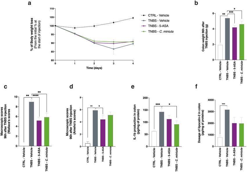
www.nature.com/scientificreports/
Figure 1. Oxygen sensitivity and metabolite production in Christensenella minuta. (a) Bacterial counts (CFUs/
mL) after 0, 2, 4, and 24 h of oxygen exposure. (b) Growth over time expressed in terms of optical density
and colony forming units (CFUs). The arrows show the different sampling points. (c) Butyrate and acetate
production at different sampling points.
populations22 and restore gastrointestinal health by reducing tissue damage and levels of pro-inflammatory
cytokines12,23–26. Moreover, in humans, these approaches can correct dysbiosis and decrease quantities of inflam-
matory mediators, leading to r emission27.
In 2017, research showed that the recently described family Christensenellaceae plays a major role in gut
health28: these bacteria serve as keystone species in the development of the symbiotic gut microbiome. Chris-
tensenella is part of the phylum Firmicutes and the order Clostridiales, and its members are strict anaerobic,
Gram-negative, non-sporulating, and non-motile b acteria29. This family has been described as a highly heritable
taxon and serves as a hub in a co-occurrence network that includes other heritable taxa30. Studies have found
that individuals suffering from CD or UC have significantly lower levels of or completely lack Christensenel-
laceae in their intestinal m
icrobiota31,32. Furthermore, in individuals with CD, such decreases in abundance
are highly predictive of flare-ups33. Recently, it was discovered that IBD-related alterations in gut microbiota
contribute to inflammation dynamics as well as the loss of commensal bacteria that are key to restoring balance
and general h omeostasis34. Taken together, these findings suggest a strong link exists between the abundance of
Christensenellaceae and the occurrence of IBDs, indicating that these commensal bacteria could play a central
role in gut physiology.
Here, we hypothesized that Christensenella species have anti-inflammatory properties that protect the intes-
tinal mucosa. To test this hypothesis, we first characterized various properties displayed by the type species
Christensenella minuta DSM 22607, namely its oxygen sensitivity, metabolic profile, and ability to produce
SCFAs. Then, in vitro, we assessed how well the bacterium modulated inflammation in human colonic cells and
protected intestinal barrier integrity during inflammation. Finally, we explored whether C. minuta DSM 22607
could reduce inflammation in vivo in two distinct preclinical colitis models in rodents. We found support for
our hypothesis—this bacterial species may hold promise for microbiome-based IBD biotherapies.
Results
An oxygen tolerant anaerobe that produces short‑chain fatty acids. To better characterize
C. minuta DSM 22607, we first assessed the strain’s oxygen sensitivity. We found that the bacterium was not
extremely oxygen sensitive (EOS) because it could tolerate the presence of oxygen for at least 24 h (Fig. 1a). We
then quantified its production of SCFAs during different growth phases (latent, exponential, and stationary)
(Fig. 1b,c). We observed that C. minuta was able to produce high levels of acetate and moderate levels of butyrate,
as also seen elsewhere29; in contrast, no propionate was generated. Our results show that, during all three growth
phases, SCFA production ratios remained the same: approximately 1 mol of butyrate for each 5 mol of acetate.
The bacterium’s detailed metabolic phenotype. We obtained an extensive phenotypic profile for C. minuta
using Biolog microassays. We performed three independent replicates. We found that C. minuta can metabo-
lize N-acetyl-d-glucosamine, d-arabitol, arbutin, d-cellobiose, dextrin, d-fructose, l-fucose, d-galactose, α-d-
glucose, maltotriose, d-mannitol, d-mannose, 3-methyl-d-glucose, palatinose, salicin, turanose, fumaric acid,
pyruvic acid, l-phenylalanine, 2′-deoxyadenosine, inosine, and uridine (Table 1).
An ability to limit IL‑8 production and inhibit NF‑κB activation in HT‑29 cells. Using human
HT-29 cells, we analyzed how well C. minuta and its supernatant could modulate TNF-α induced secretion of
IL-8 and thus limit inflammation. IL-8 is a major proinflammatory cytokine, and, therefore, bacteria capable of
inhibiting its secretion have anti-inflammatory effects35. Both the supernatant (Fig. 2a) and the bacterium itself
(Fig. 2b) demonstrated anti-inflammatory properties: IL-8 production decreased by around 50% in both cases.
This level of inhibition resembles that associated with 5-ASA, a compound commonly used to treat IBDs. When
co-incubation occurred at different multiplicities of infection (MOIs), we observed a dose–response effect,
where percent inhibition ranged from 0% (MOI 10) to 50% (MOI 50) (Fig. 2b). Similar results were obtained
with 2% (v/v), 10% (v/v), and 20% (v/v) of the supernatant. We also explored C. minuta’s effects on the NF-κB
pathway, which plays a key role in inflammation by regulating immune r esponses36, including IL-8 p roduction37.
The bacterial supernatant decreased NF-κB activation by 40%, an effect similar to that of the control NF-κB
Scientific Reports | (2021) 11:11494 | https://doi.org/10.1038/s41598-021-90885-1 2
Vol:.(1234567890)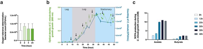
www.nature.com/scientificreports/
Target compound C. minuta Target compound C. minuta Target compound C. minuta
Water − Turanose + 3-Methyl-d-glucose +
N-Acetyl-d-galactosamine − Acetic acid − α-Methyl-d-galactoside −
N-Acetyl-d-glucosamine + Formic acid − β-Methyl-d-galactoside −
N-Acetyl-β-d-mannosamine − Fumaric acid + α-Methyl-d-glucoside −
Adonitol − Glyoxylic acid − β-Methyl-d-glucoside –
Amygdalin − α-Hydroxybutyric acid − Palatinose +
d-Arabitol + β-Hydroxybutyric acid − d-Raffinose −
Arbutin + Itaconic acid − l-Rhamnose −
d-Cellobiose + α-Ketobutyric acid – Salicin +
α-Cyclodextrin − α-Ketovaleric acid − d-Sorbitol −
β-Cyclodextrin − D,l-Lactic Acid − Stachyose −
Dextrin + l-Lactic acid − Sucrose −
Dulcitol − d-Lactic acid methyl ester − d-Trehalose −
i-Erythritol − d-Malic acid − Glycyl-l-aspartic acid −
d-Fructose ++ l-Malic acid − Glycyl-l-glutamine −
l-Fucose ++ Propionic acid − Glycyl-l-methionine −
d-Galactose + Pyruvic acid + Glycyl-l-proline −
d-Galacturonic acid – Pyruvic acid methyl ester − l-Methionine −
Gentiobiose − d-Saccharic acid − l-Phenylalanine +
d-Gluconic acid − Succinamic acid − l-Serine −
d-Glucosaminic acid – Succinic acid − l-Threonine −
α-d-Glucose ++ Succinic acid monomethyl ester − l-Valine –
Glucose-1-phosphate − m-Tartaric acid − l-Valine plus l-aspartic acid –
Glucose-6-phosphate − Urocanic acid − 2′-Deoxyadenosine +
Glycerol − l-Alaninamide − Inosine +
D, l-α-Glycerol phosphate − l-Alanine − Thymidine −
m-Inositol − l-Alanyl-l-glutamine − Uridine +
α-d-Lactose − l-Alanyl-l-histidine − Thymidine-5′-monophosphate −
Lactulose – l-Alanyl-l-threonine − Uridine-5′-monophosphate −
Maltose − l-Asparagine − d-Mannose ++
Maltotriose + l-Glutamic acid − d-Melezitose −
d-Mannitol ++ l-Glutamine − d-Melibiose −
Table 1. Metabolic profile of Christensenella minuta strain DSM 22607.
inhibitor BAY 11-7082 (10 µm) (Fig. 2c). In contrast, no effects were observed when the bacterium alone was
used (data not shown). We thus concluded that C. minuta is likely secreting a potent anti-inflammatory effector
into its culture medium.
An ability to maintain barrier integrity in Caco‑2 cells. We assessed whether C. minuta could main-
tain intestinal barrier integrity in an in vitro cell model by measuring transepithelial electrical resistance (TEER)
in Caco-2 cells exposed to TNF-α, which disrupts tight junctions, increases epithelial barrier permeability,
and causes inflammation. Measurements were made immediately prior to and 24 h after TNF-α exposure. We
observed that the TEER ratio remained stable when Caco-2 cells were treated with C. minuta in Dulbecco’s
modified Eagle’s medium (DMEM) for 3 h beforehand (Fig. 3). This result indicates that barrier integrity had
been maintained, seemingly via the anti-inflammatory action of different effectors that protected the intestinal
barrier.
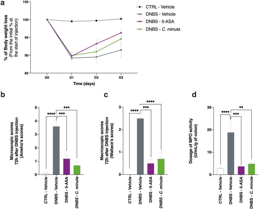
A faculty to prevent and protect against DNBS‑induced colitis. We performed an experiment to
determine whether the anti-inflammatory properties of C. minuta seen in vitro were also observed in vivo. Treat-
ment mice were given daily doses of C. minuta for 14 days. Colitis was then induced by an intrarectal injection of
DNBS, and the mice were euthanized 3 days later. We found that the treatment group tended to gain body mass
faster than the DNBS-vehicle group (Fig. 4a), although this difference was not significant. We also observed a
decrease in the microscopic scores in the treatment group, reflecting restored colonic epithelial structure and
reduced immune cell infiltration (Fig. 4b). A similar pattern was seen in the macroscopic scores (Fig. 4d). To
evaluate the bacterium’s anti-inflammatory effects on colonic tissue, we characterized the activity of myeloperox-
idase (MPO), an enzyme found in the intracellular granules of neutrophils38. We observed that DNBS-induced
inflammation resulted in increased neutrophil infiltration and MPO activity; these effects were significantly less
pronounced in the treatment group gavaged with C. minuta (Fig. 4e).
Scientific Reports | (2021) 11:11494 | https://doi.org/10.1038/s41598-021-90885-1 3
Vol.:(0123456789)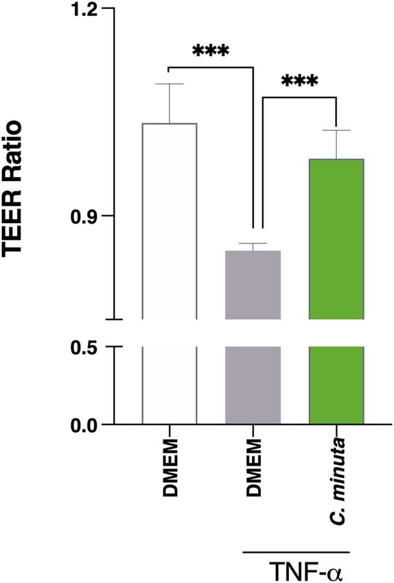
www.nature.com/scientificreports/
Figure 2. Anti-inflammatory properties of Christensenella minuta in vitro. IL-8 production by HT-29 cells
exposed to TNF-α in presence of (a) C. minuta supernatant or (b) C. minuta bacteria. (c) Levels of NF-κB
activation in HT-29 cells transfected with a reporter system and exposed to TNF-α. Results of Mann Whitney U
tests comparing the control groups to the other groups: *p < 0.05, **p < 0.01, and ***p < 0.001.
An ability to prevent and protect against TNBS‑induced colitis. To obtain additional confirmation
of the bacterium’s anti-inflammatory effects in vivo, we repeated the experiment in a second model—TNBS-
induced colitis in rats, known to be more susceptible to inflammation39. Treatment rats were given daily doses of
C. minuta for 14 days. Colitis was then induced by an intrarectal injection of TNBS, and the rats were euthanized
4 days later. Unlike the the mouse model, the treatment group had no effect on body mass gain, compared to
TNBS-vehicle group at the end of the experiment (Fig. 5a). However, colon mass was lower in the treatment
group (Fig. 5b), which indicates that intestinal transit was improved by C. minuta. Remarkably, the macroscopic
scores (i.e., Wallace s cores40; Fig. 5c) provided support for the idea that C. minuta could be as efficient as 5-ASA,
a compound used to treat UC41, in protecting colonic tissue. Moreover, the microscopic scores for the treatment
group showed that inflammatory profiles seemed to be improved at the histological level (Fig. 5d), compared
to what was seen in the TNBS-vehicle group. Furthermore, the C. minuta treatment appeared to induce an
immunomodulatory response by decreasing IL-1β secretion (Fig. 5e). This result specifically indicates that the
TNBS-induced Th1 response was dampened. IL-6 and IL-10 production (of Th2 and Th1 cytokines, respectively)
was not affected by the TNBS injection (data not shown). Finally, we used lipocalin-2 (LCN-2) as a non-invasive
biological marker of intestinal inflammation. The C. minuta treatment tended to decrease the concentration of
LCN-2 in the colon (Fig. 5f). These results validated our in vitro findings, demonstrating the bacterium’s anti-
inflammatory properties in two in vivo colitis models.
Discussion
IBDs are debilitating chronic diseases for which no curative treatments are currently available. Our research is
grounded in the idea that microbiome-based therapies offer an innovative approach to healing intestinal mucosa.
Indeed, there is increasing evidence that some commensal bacteria possess anti-inflammatory properties that
can improve IBD s ymptoms10–12.
In 2012, Morotomi et al.42 described a new family of bacteria, the Christensenellaceae, whose presence was
later found to be correlated with gut microbiota health28. Although this taxon is a subdominant member of the
microbiome, co-occurrence analyses have revealed that it plays a central role within a broader network of herit-
able bacteria in the gut ecosystem30,43. Indeed, individuals with IBDs display drastically lower levels of these bac-
teria in their intestines, suggesting that intestinal inflammation is greater when the abundance of Christensenella
species is lower31,44,45. Consequently, we explored the anti-inflammatory properties of Christensenella minuta
(strain DSM 22607) to determine whether it could be used in IBD treatments.
First, we characterized the bacterium’s metabolic phenotype to gain insight into its overall metabolic capaci-
ties. We found that, while C. minuta is an anaerobe, it was highly tolerant of oxygen, unlike other anaerobic
commensal bacteria, such as Faecalibactium prausnitzii, which are EOS. Indeed, recent research has discovered
that Christensenella occurs in different parts of the human intestine that vary in oxygen concentrations (i.e.,
Scientific Reports | (2021) 11:11494 | https://doi.org/10.1038/s41598-021-90885-1 4
Vol:.(1234567890)www.nature.com/scientificreports/
Figure 3. Effects of Christensenella minuta on intestinal barrier permeability. Polarized monolayers of Caco-2
cells were exposed to TNF-α to disrupt the intestinal barrier. TEER was measured immediately prior to and
24 h after TNF-α exposure. Results of Mann Whitney U tests comparing the DMEM + TNF-α group to the other
groups: *p < 0.05, **p < 0.01, and ***p < 0.001.
the terminal i leum13,44, cecum46, and distal c olon47). These findings support the idea that C. minuta could have
beneficial effects within the upper gastrointestinal tract and, particularly, in the distal ileum, a major site of
inflammation in Crohn’s disease; in contrast, other candidate EOS bacteria only occur in the colon. Since IBDs
are associated with oxidative stress and high levels of ROS5, the ability of C. minuta to tolerate oxygen might
confer resistance to inflammation-induced oxidative stress in the gut. The bacterium might thus be well suited to
creating environmental conditions that allow the establishment of more sensitive anaerobes species. Indeed, the
increasing presence of facultative anaerobes observed in the colon during I BD48 could give a major advantage to
C. minuta as a biotherapy against EOS candidates. The bacterium’s oxygen tolerance could also facilitate its use
in industrial manufacturing processes, a practical consideration if C. minuta’s benefits are to be translated into
microbiome-based clinical treatments.
We also confirmed that C. minuta produces high levels of acetate and moderate levels of butyrate29,49 and
demonstrated that the acetate:butyrate production ratio was 5:1 over all three growth phases. Widely produced
by gut bacteria, SCFAs result from carbohydrate fermentation and, to a lesser extent, from protein f ermentation50.
Interestingly, a number of bacteria have been identified as either acetate or butyrate producers but rarely both51.
SCFAs are crucial compounds since they modulate host pathways through interactions with G-protein-coupled
receptors (GPRs), which are found in colonic, hepatic, muscular, and adipose t issues52. These interactions influ-
ence multiple important functions related to cell differentiation and energy metabolism. The butyrate receptor
GPR109a also occurs in intestinal epithelial cells, adipocytes, and immune cells, where it helps control inflamma-
tion and cell proliferation53. Decreases in SCFA production could have deleterious effects, mainly by influencing
host-microbe interactions34. Consequently, balanced SCFA production is essential to gut homeostasis.
Because SCFAs can affect microbiota-host crosstalk via their immunomodulatory properties54, we ascer-
tained whether C. minuta had an influence on inflammation. We discovered that both C. minuta bacteria and
their its supernatant displayed potent anti-inflammatory properties, decreasing the secretion of IL-8 cytokines
by HT-29 cells following TNF-α induced inflammation. Such anti-inflammatory properties have also been seen
in other bacteria, including Faecalibacterium prausnitzii55, several strains of Lactobacillus56, and Akkermensia
muciniphila24.
Scientific Reports | (2021) 11:11494 | https://doi.org/10.1038/s41598-021-90885-1 5
Vol.:(0123456789)www.nature.com/scientificreports/
Figure 4. Effects of Christensenella minuta on DNBS-induced inflammation in mice. (a) Change in body
mass after colitis was induced; (b) colon microscopic scores; (c) colon macroscopic scores; and (d) levels of
MPO activity. Results of Mann Whitney U tests comparing the DNBS-Vehicle group to the three other groups:
*p < 0.05, **p < 0.01, ***p < 0.001, and ****p < 0.0001.
To further explore the bacterium’s anti-inflammatory effects, we tested the impact of both the bacteria and its
supernatant against HT-29 cells transfected with a NF-κB reporter system known to regulate IL-8 p roduction37.
Only the supernatant decreased NF-κB activation. This result, combined with the findings of the previous experi-
ment, suggest that at least two different bacterial effectors were responsible for the effects observed. Past work
using a variety of commensal and pathogenic microorganisms has shown that bacteria utilize a variety of mecha-
nisms to modulate the canonical NF-κB p athway57. It is possible that butyrate concentrations in the supernatant
(which were about 10 times lower than physiological c oncentrations58) helped inhibit the NF-κB p athway59. It
may also be that other compounds, such as polysaccharides60, peptidoglycans61, and proteins62, were secreted into
the supernatant or exposed on the surface of the bacterial membrane63. Further research is needed to decipher
the underlying mechanisms at work.
We then evaluated how well C. minuta could protect epithelial cells from TNF-α-induced permeability using
a Caco-2 cell line. We found that the bacterium successfully maintained the integrity of the epithelial cell mon-
olayer following induced inflammation. Individuals with IBDs have very low levels of the adhesion molecules
that regulate intestinal permeability64; C. minuta could help restore proper permeability and limit any damage
that has occurred. Recent work has highlighted that Escherischia coli Nissle 1917 could attenuate declines in
TEER induced by TNF-α and IFNγ, notably by inhibiting the NF-κB-mediated activation of the MLCK-P-MLC
signaling pathway65. F. prausnitzii and Roseburia intestinalis have also been found to help reverse impaired
epithelial barrier function by modulating the expression of tight junction proteins and decreasing paracellular
permeability66. It would be worthwhile to decipher the precise mechanism in use by C. minuta.
To ascertain whether C. minuta displayed the same anti-inflammatory properties in vivo, we performed
experiments using two different animal models of colitis: a mouse model of moderate, DNBS-induced colitis
and a rat model of severe, TNBS-induced c olitis39. Based on the macroscopic scores, treatment with C. minuta
Scientific Reports | (2021) 11:11494 | https://doi.org/10.1038/s41598-021-90885-1 6
Vol:.(1234567890)www.nature.com/scientificreports/
Figure 5. Effects of Christensenella minuta on TNBS-induced inflammation in rats. (a) Body mass over the
course of the experiment; (b) colon mass at the end of the experiment; (c) colon microscopic scores; (d) colon
macroscopic scores; colon IL-1β levels (e); and (f) levels of LCN-2, a proxy for colon neutrophil infiltration.
Results of Mann Whitney U tests comparing the TNBS-Vehicle group to other three groups: *p < 0.05, **p < 0.01,
***p < 0.001, and ****p < 0.0001.
significantly limited colon damage in both models. To characterize the bacterium’s immunomodulatory effects,
we assessed neutrophil infiltration in colonic tissues by monitoring MPO activity (in the mice) and LCN-2 levels
(in the rats). In both models, the metrics were lower in C. minuta-treated animals. Similar studies found compara-
ble effects in a mouse model of TNBS-induced colitis using a treatment based on Parabacteroides distasonis67 and
in a mouse model of DNBS-induced colitis using a treatment based on F. prausnitizii68 and different Lactobaccillus
strains56. Taken together, these findings demonstrate that using single-strain colitis treatments could be e ffective6.
It has been shown that IL-8 secretion induces neutrophil activation in inflammed r egions69,70. Given that C.
minuta decreased IL-8 secretion and NF-κB activation in vitro, this signaling pathway could have been involved
in the reduction of neutrophil activation. Furthermore, a decrease in IL-1β was seen in the C. minuta-treated
rats (Fig. 5e). IL-1β signaling is mediated by multiple transcription factors, including NF-κB71. It is possible that
cytokine release in the TNBS-induced colitis model was partially modulated by C. minuta’s secretion of NF-κB
inhibitors. A similar mode of action has been seen in F. prausnitzii in different colitis m odels62, notably via the
release of the microbial anti-inflammatory molecule (MAM). Although the NF-κB signaling pathway serves as
a major line of defense against pathogens, it can have deleterious effects when overactivated due to the increased
production of proinflammatory c ytokines72. Consequently, it is important to determine which molecules help
control pathway activation so that the development of inflammation can be halted. For example, SCFAs such as
butyrate can limit inflammation via their inhibitory a ction73.
In conclusion, our study is the first to show that C. minuta displays strong immunomodulatory properties
in vitro and in vivo. Our findings open the door to intriguing new research questions. Although additional
research is obviously needed to better understand the bacterium’s effects and their underlying mechanisms,
our work underscores that C. minuta holds promise for treating IBDs and merits further study with a view to
developing single-strain biotherapies.
Scientific Reports | (2021) 11:11494 | https://doi.org/10.1038/s41598-021-90885-1 7
Vol.:(0123456789)www.nature.com/scientificreports/
Methods
Culturing the bacteria. Christensenella minuta (DSM 22607) was cultured in Gifu anaerobic medium
(GAM broth, HyServe) in an anaerobic chamber (5%/5%/90% C O2, H2, N2) kept at 37 °C. Granulated agar
(15 g/L, Difco) was added when necessary. Bacterial cultures were centrifuged at 2500×g and then resuspended
in 1X Dulbecco’s phosphate-buffered saline (DPBS, Gibco). We then employed these cultures in the in vitro
and in vivo experiments described below. To establish the growth curves, cultures were followed for their entire
growth cycle (up to 54 h). We used a spectrophotometer (Ultrospec 10) to measure optical density (OD600) and
thus estimate bacterial counts. Samples of cultures were collected at different timepoints and centrifuged at
4000×g at 4 °C for 15 min. We recovered the supernatants and stored them at − 20 °C until we could measure
short-chain fatty acid (SCFA) concentrations.
Characterizing short‑chain fatty acid concentrations. Bacterial supernatants were deproteinized
overnight at 4 °C via the addition of phosphotungstic acid (10% [v/v]); Sigma). We then centrifuged the result-
ing samples for 15 min at 12,000×g. Concentrations of SCFAs were determined using a gas chromatograph (GC;
Agilent 6890 N Network) equipped with a split-splitless injector (GC Agilent 7890B), a flame-ionization detec-
tor, and a capillary column (15 m × 0.53 mm × 0.5 µm) packed with SP 1000 (Nukol; Supelco 25,236). The flow
rate of hydrogen, the carrier gas, was 10 mL/min; the temperatures of the injector, column, and detector were
200 °C, 100 °C, and 240 °C, respectively. We used 2-ethylbutyrate as the internal standard in our samples and
employed a panel of SCFA standards. Two replicates were performed for each sample. We collected the SCFA
data and integrated the peaks using the GC’s default software (Agilent). To determine the final concentrations of
SCFAs, the supernatants were weighed before and after protein precipitation to obtain the appropriate multipli-
cation factor (i.e., the supernatant to sample mass ratio).
Assessing oxygen sensitivity. To evaluate C. minuta’s sensitivity to oxygen, we used bacteria from the
cultures described a bove55. Briefly, we grew C. minuta for 48 h in a liquid medium. Then, we took 10-µL samples
of different concentrations of the bacteria (range of final concentrations: 104–109 CFU/mL) and deposited them
on Petri dishes. The dishes were placed outside of the anaerobic chamber and exposed to oxygen for 2, 4, and
24 h.
Establishing a metabolic profile. We established a metabolic profile for C. minuta using AN Micro-
Plate™ technology (Biolog) in accordance with the manufacturer’s instructions. Briefly, cultures were streaked
twice on Biolog Universal Anaerobe Agar (BUA; Biolog) supplemented with 5% (w/v) defibrinated sheep blood
(Alliance Bio Expertise). We allowed growth to occur for 4 days at 37 °C under anaerobic conditions. Bacteria
were swabbed and transferred into prereduced anaerobic inoculating fluid until 65% transmittance was reached.
Then, 100 mL of this bacterial suspension was used to inoculate each well of AN MicroPlates™ under anaerobic
conditions. We incubated the plates for 24 h at 37 °C under anaerobic hydrogen-free conditions using a GENbox
and anaerobic jar system (bioMérieux). Color shifts in each well were evaluated visually and via optical density
measurements made at 590 nm (FLUOstar Omega, BMG Labtech).
Culturing eukaryotic cells. We obtained the human colon adenocarcinoma cell line HT-29 from the Euro-
pean Collection of Authenticated Cell Cultures (ECACC; Sigma). Cells were grown in McCoy’s 5A medium
(Gibco) supplemented with 10% (v/v) fetal bovine serum (FBS; Gibco) and 1% (v/v) penicillin/streptomycin
(P/S; Sigma). The cultures were maintained at 37 °C under conditions of 5% CO2 until 80% confluence was
reached. We obtained the Caco-2 cell line from the American Type Tissue Collection (ATCC®) and maintained it
in Dulbecco’s modified Eagle’s medium (DMEM) supplemented with glutaMAX™ (Gibco), 20% heat-inactivated
FBS, and 1% non-essential amino acids (Gibco). Cells were kept at 37 °C under conditions of 10% C
O2 until 80%
confluence was reached.
Characterizing immunomodulation in HT‑29 cells. First, we seeded HT-29 cells into 24-well plates
(3 × 10⁵ cells per well). After 24–48 h, confluence was reached, and the complete medium was replaced by a McCoy’s
5A medium supplemented with 2% (v/v) FBS. After 24 h, we replaced this medium with McCoy’s 5A medium
supplemented with 2% (v/v) FBS and TNF-α (5 ng/mL, InvivoGen) to which we added either (1) supernatant
obtained from the culture medium during C. minuta’s stationary phase (at concentrations of 2%, 10%, or 20%)
or (2) bacteria (at MOIs of 10, 50, or 100—ratio of bacteria to eukaryotic cells). PBS glycerol was used as a
control. After 6 h of coincubation, supernatants were obtained from the cell cultures and stored at -80 °C until
interleukine-8 (IL-8) concentrations could be quantified. The latter process was performed using a Human IL-8
ELISA MAX Standard Set (BioLegend); absorbance was measured at 450 nm using a FLUOstar® Omega micro-
plate reader (BMG Labtech).
Characterizing immunomodulation in HT‑29 cells transfected with a NF‑κB luciferase reporter
vector. HT-29 cells at a density of 3 ×
105 cells per well were reverse-transfected with 200 ng pRelA-
luc and 10 ng pRL-TK using X-tremeGENE HP DNA Transfection Reagent (Roche) in 24-well plates. Briefly, we
prepared the transfection reagent:DNA complex as follows: we added the appropriate quantity of diluted plas-
mids to serum and antibiotics-free medium (final volume: 50 µL). The mixture was gently combined, and the
transfection reagent was added at a ratio of 3:1. We gently mixed the transfection complex, which was then incu-
bated for 15 min at room temperature. We subsequently seeded the complex with fresh cells (i.e., still in suspen-
sion). The plates were incubated for 24 h, and the medium was then remplaced by McCoy’s 5A medium supple-
Scientific Reports | (2021) 11:11494 | https://doi.org/10.1038/s41598-021-90885-1 8
Vol:.(1234567890)www.nature.com/scientificreports/
mented with 2% (v/v) FBS. After 24 h of serum starvation, the medium was removed and replaced by McCoy’s
5A medium supplemented with 2% (v/v) FBS and 5 ng/mL of TNF-α (InvivoGen) and either (1) supernatant
obtained from the culture medium during C. minuta’s stationary phase (concentration: 10%) or (2) bacteria and
culture medium (10%) as a control. After 6 h of coincubation, we washed the cells twice with cold 1X PBS and
lyzed them via exposure to 50 µL of Passive Lysis Buffer (Promega) for 15 min at room temperature under condi-
tions of gentle shaking. Next, the lysates were transferred to microtubes. The Dual-Luciferase® Reporter Assay
System (Promega) was used largely in accordance with the manufacturer’s instructions; a few minor modifica-
tions were made. Briefly, we transferred 2 × 20 µL samples of the lysates to white 96-well plates; then, we added 50
µL of LAR II solution to each well. First, we quantified levels of firefly luciferase activity. Then, we added 50 µL of
Stop & Glo® Reagent, and we quantified levels of Renilla luciferase activity using a FLUOstar® Omega microplate
reader (BMG Labtech). NF-κB activity was quantified via the ratio of firefly activity to Renilla activity.
Assessing effects on intestinal permeability by measuring transepithelial electrical resist‑
ance. We used the Caco-2 cell line to determine whether C. minuta could affect the epithelial barrier, as pre-
viously described74. Briefly, Caco-2 cells were grown on Transwell® inserts. When optimal transepithelial electri-
cal resistance (TEER) values were reached (REMS AutoSampler, World Precision Instruments), fresh DMEM
was added. Then, the C. minuta treatment (bacteria at MOI 40) or the control (PBS 1X) was applied to the apical
compartment of the cells. Three hours later, 100 ng/mL of TNF-α (Peprotech) was added to the basal compart-
ment of the cells. TEER was measured just before and 24 h after the treatments. The results were normalized (i.e.,
relative to basal TEER).
Assessing effects on DNBS‑induced colitis in mice. We assessed the effects of a C. minuta treat-
ment on DNBS-induced colitis in mice. We obtained 40 7-week-old male C57BL/6J mice from the Janvier
Lab and maintained them under specific pathogen-free (SPF) conditions in the animal facilities of the French
National Research Institute for Agriculture, Food, and Environment (IERP Experimental Unit, INRAE). They
were housed in cages of five. Our experiments were performed in accordance with European Union legisla-
tion on animal welfare and were approved by COMETHEA, our local committee on animal experimentation
(n°16744-201807061805486) and in compliance with the ARRIVE relevant guidelines. After a 7-day acclimation
period, the 40 mice were divided into 4 groups (n = 10 mice/group): the vehicle control group (no inflamma-
tion; CTRL-Vehicle), the inflamed control group (inflammation induced; DNBS-Vehicle), the treatment group
(inflammation induced, treatment with C. minuta; DNBS-C. minuta), and the anti-inflammatory control group
(inflammation induced, treatment with 5-ASA; DNBS-5-ASA). For two weeks, we gave the vehicle and inflamed
control mice an oral dose of PBS (150 µL) containing 16% (v/v) glycerol and the treatment mice were given an
oral dose of C. minuta (109 CFU/mL). The anti-inflammatory control mice were given an oral dose of 5-ASA
(100 mg/kg; Sigma) from the day of the DNBS injection. Gavages were performed daily until the end of the
experiment. Then, we anesthetized the mice using an intraperitoneal injection of 0.1% ketamine and 0.06%
xylazine; we subsequently gave them an intrarectal injection of DNBS (175 mg/kg) dissolved in 30% ethanol
(w/v). The vehicle control group received an intrarectal injection of 30% ethanol. Three days after the injections,
the mice were euthanized. During the experiment, we measured body mass daily. Colon microscopic scores
(Ameho), macroscopic scores (Wallace), and myeloperoxidase (MPO) activity levels were characterized as pre-
viously described75.
Assessing effects on TNBS‑induced colitis in rats. We assessed the effects of a C. minuta treatment
on TNBS-induced colitis in rats. We used Sprague Dawley rats and performed this research at an accredited
contract research organization (Intestinal Biotech Development, Lille) in accordance with governmental regula-
tions and in compliance with the ARRIVE relevant guidelines. The rats were divided into 4 different groups. For
the first 14 days of the experiment, the vehicle (CTRL-Vehicle) and inflamed control rats (TNBS-vehicle) were
gavaged with an oral dose of PBS (150 µL) containing 1% (v/v) glycerol. The rats were given an oral dose of C.
minuta (109 CFU/mL) (TNBS-C. minuta); 5-ASA granules, the anti-inflammatory control group, were mixed
into the rats’ food. Then, we anesthetized the rats for 2 h and administered an intrarectal injection of TNBS
(80 mg/kg) dissolved in 40% ethanol (w/v) to induce colitis. The rats were euthanized four days after the injec-
tion, and the effects of the treatment and controls were assessed, as was colon mass. During the experiment, we
measured body mass daily. Colon microscopic and macroscopic scores (Ameho and Wallace, respectively) were
characterized as previously d escribed40,76. We quantified inflammation by assessing the levels of the proinflam-
matory cytokines IL-1β and IL-6 and the anti-inflammatory cytokine IL-10 (eBioscience); the level of lipocalin-2
(LCN-2) (Cliniscience) in the colon was determined using ELISA. Briefly, a 1-cm stretch of the distal colon was
recovered and homogenized (50 mg/mL) in Tris–HCl buffer containing protease inhibitors (Sigma) and ceramic
beads (diameter: 1.4 and 2.8 mm) using a Precellys tissue homogenizer. Samples were centrifuged for 20 min,
and the supernatant was frozen at − 80 °C.
Statistical analysis. All results were expressed as means ± standard error of the mean (SEM). We per-
formed non-parametric statistical analyses—two-sided Mann Whitney U tests—using GraphPad Prism (v. 8.2.1;
GraphPad Software). We employed an alpha level of 0.05.
Received: 28 February 2021; Accepted: 11 May 2021
Scientific Reports | (2021) 11:11494 | https://doi.org/10.1038/s41598-021-90885-1 9
Vol.:(0123456789)www.nature.com/scientificreports/
References
1. McDowell, C., Farooq, U. & Haseeb, M. Inflammatory bowel disease. In StatPearls (StatPearls Publishing, 2021).
2. Glassner, K. L., Abraham, B. P. & Quigley, E. M. M. The microbiome and inflammatory bowel disease. J. Allergy Clin. Immunol.
145, 16–27 (2020).
3. Banks, C., Bateman, A., Payne, R., Johnson, P. & Sheron, N. Chemokine expression in IBD Mucosal chemokine expression is
unselectively increased in both ulcerative colitis and Crohn’s disease. J. Pathol. 199, 28–35 (2003).
4. Zhu, Y. et al. CXCL8 chemokine in ulcerative colitis. Biomed. Pharmacother. 138, 111427 (2021).
5. Bourgonje, A. R. et al. Oxidative stress and redox-modulating therapeutics in inflammatory bowel disease. Trends Mol. Med. 26,
1034–1046 (2020).
6. Flynn, S. & Eisenstein, S. Inflammatory bowel disease presentation and diagnosis. Surg. Clin. North Am. 99, 1051–1062 (2019).
7. van der Sloot, K. W. J., Weersma, R. K., Alizadeh, B. Z. & Dijkstra, G. Identification of environmental risk factors associated with
the development of inflammatory bowel disease. J. Crohns Colitis 14, 1662–1671 (2020).
8. Graham, D. B. & Xavier, R. J. Pathway paradigms revealed from the genetics of inflammatory bowel disease. Nature 578, 527–539
(2020).
9. Na, Y. R., Stakenborg, M., Seok, S. H. & Matteoli, G. Macrophages in intestinal inflammation and resolution: A potential therapeutic
target in IBD. Nat. Rev. Gastroenterol. Hepatol. 16, 531–543 (2019).
10. Frank, D. N. et al. Molecular-phylogenetic characterization of microbial community imbalances in human inflammatory bowel
diseases. Proc. Natl. Acad. Sci. USA 104, 13780–13785 (2007).
11. Peterson, D. A., Frank, D. N., Pace, N. R. & Gordon, J. I. Metagenomic approaches for defining the pathogenesis of inflammatory
bowel diseases. Cell Host Microbe 3, 417–427 (2008).
12. Sokol, H. et al. Faecalibacterium prausnitzii is an anti-inflammatory commensal bacterium identified by gut microbiota analysis
of Crohn disease patients. Proc. Natl. Acad. Sci. USA 105, 16731–16736 (2008).
13. Sokol, H. et al. Prominence of ileal mucosa-associated microbiota to predict postoperative endoscopic recurrence in Crohn’s
disease. Gut 69, 462–472 (2020).
14. Bhaskaran, N. et al. Role of Short Chain Fatty Acids in Controlling Tregs and Immunopathology During Mucosal Infection. Front.
Microbiol. 9, 1995 (2018).
15. Zhuang, X. et al. Systematic review and meta-analysis: Short-chain fatty acid characterization in patients with inflammatory bowel
disease. Inflamm. Bowel Dis. 25, 1751–1763 (2019).
16. Kilinçarslan, S. & Evrensel, A. The effect of fecal microbiota transplantation on psychiatric symptoms among patients with inflam-
matory bowel disease: An experimental study. Actas Esp. Psiquiatr. 48, 1–7 (2020).
17. Saez-Lara, M. J., Gomez-Llorente, C., Plaza-Diaz, J. & Gil, A. The role of probiotic lactic acid bacteria and bifidobacteria in the
prevention and treatment of inflammatory bowel disease and other related diseases: A systematic review of randomized human
clinical trials. BioMed Res. Int. 2015, 505878 (2015).
18. Voth, E. & Khanna, S. Fecal microbiota transplantation for treatment of patients with recurrent Clostridioides difficile infection.
Expert Rev. Anti Infect. Ther. https://doi.org/10.1080/14787210.2020.1752192 (2020).
19. Levy, A. N. & Allegretti, J. R. Insights into the role of fecal microbiota transplantation for the treatment of inflammatory bowel
disease. Ther. Adv. Gastroenterol. https://doi.org/10.1177/1756284819836893 (2019).
20. Tan, P., Li, X., Shen, J. & Feng, Q. Fecal microbiota transplantation for the treatment of inflammatory bowel disease: An update.
Front. Pharmacol. 11, 1409 (2020).
21. Gagliardi, A. et al. Rebuilding the gut microbiota ecosystem. Int. J. Environ. Res. Public. Health 15, 1679 (2018).
22. Hill, C. et al. The International Scientific Association for Probiotics and Prebiotics consensus statement on the scope and appropri-
ate use of the term probiotic. Nat. Rev. Gastroenterol. Hepatol. 11, 506–514 (2014).
23. Martín, R. et al. Faecalibacterium prausnitzii prevents physiological damages in a chronic low-grade inflammation murine model.
BMC Microbiol. 15, 67 (2015).
24. Zhai, R. et al. Strain-specific anti-inflammatory properties of two akkermansia muciniphila strains on chronic colitis in mice.
Front. Cell. Infect. Microbiol. 9, 239 (2019).
25. Bach Knudsen, K. E. et al. Impact of diet-modulated butyrate production on intestinal barrier function and inflammation. Nutrients
10, 1499 (2018).
26. Zhang, X.-F. et al. Clinical effects and gut microbiota changes of using probiotics, prebiotics or synbiotics in inflammatory bowel
disease: A systematic review and meta-analysis. Eur. J. Nutr. https://doi.org/10.1007/s00394-021-02503-5 (2021).
27. Basso, P. J., Câmara, N. O. S. & Sales-Campos, H. Microbial-based therapies in the treatment of inflammatory bowel disease: An
overview of human studies. Front. Pharmacol. 9, 1571 (2019).
28. Mancabelli, L. et al. Identification of universal gut microbial biomarkers of common human intestinal diseases by meta-analysis.
FEMS Microbiol. Ecol. 93, fix153 (2017).
29. Morotomi, N. et al. Evaluation of intestinal microbiotas of healthy Japanese adults and effect of antibiotics using the 16S ribosomal
RNA gene based clone library method. Biol. Pharm. Bull. 34, 1011–1020 (2011).
30. Goodrich, J. K. et al. Human genetics shape the gut microbiome. Cell 159, 789–799 (2014).
31. Pascal, V. et al. A microbial signature for Crohn’s disease. Gut 66, 813–822 (2017).
32. Pittayanon, R. et al. Differences in gut microbiota in patients with vs without inflammatory bowel diseases: A systematic review.
Gastroenterology 158, 930-946.e1 (2020).
33. Braun, T. et al. Individualized dynamics in the gut microbiota precede Crohnʼs disease flares. Am. J. Gastroenterol. 114, 1142–1151
(2019).
34. Lee, M. & Chang, E. B. Inflammatory bowel diseases (IBD) and the microbiome-searching the crime scene for clues. Gastroenterol-
ogy 160, 524–537 (2021).
35. Kechaou, N. et al. Identification of one novel candidate probiotic Lactobacillus plantarum strain active against influenza virus
infection in mice by a large-scale screening. Appl. Environ. Microbiol. 79, 1491–1499 (2013).
36. Nennig, S. E. & Schank, J. R. The role of NFkB in drug addiction: Beyond inflammation. Alcohol Alcohol. Oxf. Oxfs. 52, 172–179
(2017).
37. Lee, B. C., Kim, S. H., Choi, S. H. & Kim, T. S. Induction of interleukin-8 production via nuclear factor-κB activation in human
intestinal epithelial cells infected with Vibrio vulnificus. Immunology 115, 506–515 (2005).
38. Odobasic, D., Kitching, A. R. & Holdsworth, S. R. Neutrophil-mediated regulation of innate and adaptive immunity: The role of
myeloperoxidase. J. Immunol. Res. https://doi.org/10.1155/2016/2349817 (2016).
39. LaCroix-Fralish, M. L., Austin, J.-S., Zheng, F. Y., Levitin, D. J. & Mogil, J. S. Patterns of pain: Meta-analysis of microarray studies
of pain. Pain 152, 1888–1898 (2011).
40. Wallace, J. L., MacNaughton, W. K., Morris, G. P. & Beck, P. L. Inhibition of leukotriene synthesis markedly accelerates healing in
a rat model of inflammatory bowel disease. Gastroenterology 96, 29–36 (1989).
41. Bokemeyer, B., Hommes, D., Gill, I., Broberg, P. & Dignass, A. Mesalazine in left-sided ulcerative colitis: Efficacy analyses from
the PODIUM trial on maintenance of remission and mucosal healing. J. Crohns Colitis 6, 476–482 (2012).
Scientific Reports | (2021) 11:11494 | https://doi.org/10.1038/s41598-021-90885-1 10
Vol:.(1234567890)www.nature.com/scientificreports/
42. Morotomi, M., Nagai, F. & Watanabe, Y. Description of Christensenella minuta gen. nov., sp. nov., isolated from human faeces,
which forms a distinct branch in the order Clostridiales, and proposal of Christensenellaceae fam nov. Int. J. Syst. Evol. Microbiol.
62, 144–149 (2012).
43. Brooks, A. W., Priya, S., Blekhman, R. & Bordenstein, S. R. Gut microbiota diversity across ethnicities in the United States. PLoS
Biol. 16, e2006842 (2018).
44. Zakrzewski, M. et al. IL23R -protective coding variant promotes beneficial bacteria and diversity in the ileal microbiome in healthy
individuals without inflammatory bowel disease. J. Crohns Colitis 13, 451–461 (2019).
45. Kummen, M. et al. The gut microbial profile in patients with primary sclerosing cholangitis is distinct from patients with ulcerative
colitis without biliary disease and healthy controls. Gut 66, 611–619 (2017).
46. Moreno-Indias, I. et al. Insulin resistance is associated with specific gut microbiota in appendix samples from morbidly obese
patients. Am. J. Transl. Res. 8, 5672 (2016).
47. Yuan, C., Graham, M., Staley, C. & Subramanian, S. Mucosal microbiota and metabolome along the intestinal tracts reveals loca-
tion specific relationship. Mystems https://doi.org/10.1101/454496 (2018).
48. Rigottier-Gois, L. Dysbiosis in inflammatory bowel diseases: The oxygen hypothesis. ISME J. 7, 1256–1261 (2013).
49. Ruaud, A. et al. Syntrophy via interspecies H2 transfer between Christensenella and Methanobrevibacter underlies their global
cooccurrence in the human gut. MBio 11, e03235-19 (2020).
50. den Besten, G. et al. The role of short-chain fatty acids in the interplay between diet, gut microbiota, and host energy metabolism.
J. Lipid Res. 54, 2325–2340 (2013).
51. Flint, H. J., Duncan, S. H., Scott, K. P. & Louis, P. Links between diet, gut microbiota composition and gut metabolism. Proc. Nutr.
Soc. 74, 13–22 (2015).
52. Canfora, E. E., Jocken, J. W. & Blaak, E. E. Short-chain fatty acids in control of body weight and insulin sensitivity. Nat. Rev. Endo-
crinol. 11, 577–591 (2015).
53. Parada Venegas, D. et al. Short chain fatty acids (SCFAs)-mediated gut epithelial and immune regulation and its relevance for
inflammatory bowel diseases. Front. Immunol. 10, 277 (2019).
54. Thorburn, A. N. et al. Evidence that asthma is a developmental origin disease influenced by maternal diet and bacterial metabolites.
Nat. Commun. 6, 7320 (2015).
55. Martín, R. et al. Functional Characterization Of Novel Faecalibacterium prausnitzii strains isolated from healthy volunteers: A
step forward in the use of F. prausnitzii as a next-generation probiotic. Front. Microbiol. 8, 1226 (2017).
56. Torres-Maravilla, E. et al. Identification of novel anti-inflammatory probiotic strains isolated from pulque. Appl. Microbiol. Bio-
technol. 100, 385–396 (2016).
57. Johannessen, M., Askarian, F., Sangvik, M. & Sollid, J. E. Bacterial interference with canonical NFκB signalling. Microbiology 159,
2001–2013 (2013).
58. Rosignoli, P. et al. Protective activity of butyrate on hydrogen peroxide-induced DNA damage in isolated human colonocytes and
HT29 tumour cells. Carcinogenesis 22, 1675–1680 (2001).
59. Lenoir, M. et al. Butyrate mediates anti-inflammatory effects of Faecalibacterium prausnitzii in intestinal epithelial cells through
Dact3. Gut Microbes 12, 1–16 (2020).
60. Danne, C. et al. A large polysaccharide produced by Helicobacter hepaticus induces an anti-inflammatory gene signature in
macrophages. Cell Host Microbe 22, 733-745.e5 (2017).
61. Macho Fernandez, E. et al. Anti-inflammatory capacity of selected lactobacilli in experimental colitis is driven by NOD2-mediated
recognition of a specific peptidoglycan-derived muropeptide. Gut 60, 1050–1059 (2011).
62. Breyner, N. M. et al. Microbial anti-inflammatory molecule (MAM) from Faecalibacterium prausnitzii shows a protective effect
on DNBS and DSS-induced colitis model in mice through inhibition of NF-κB pathway. Front. Microbiol. 8, 114 (2017).
63. Piqué, N., Berlanga, M. & Miñana-Galbis, D. Health benefits of heat-killed (tyndallized) probiotics: An overview. Int. J. Mol. Sci.
20, 2534 (2019).
64. Michielan, A. & D’Incà, R. Intestinal permeability in inflammatory bowel disease: Pathogenesis, clinical evaluation, and therapy
of leaky gut. Mediators Inflamm. 2015, 628157 (2015).
65. Guo, S. et al. Escherichia coli Nissle 1917 protects intestinal barrier function by inhibiting NF-κB-mediated activation of the
MLCK-P-MLC signaling pathway. Mediators Inflamm. 2019, 5796491 (2019).
66. Mohebali, N., Ekat, K., Kreikemeyer, B. & Breitrück, A. Barrier protection and recovery effects of gut commensal bacteria on dif-
ferentiated intestinal epithelial cells in vitro. Nutrients 12, 2251 (2020).
67. Cuffaro, B. et al. In vitro characterization of gut microbiota-derived commensal strains: Selection of parabacteroides distasonis
strains alleviating TNBS-induced colitis in mice. Cells 9, 2104 (2020).
68. Martín, R. et al. The Commensal Bacterium Faecalibacterium prausnitzii is protective in DNBS-induced chronic moderate and
severe colitis models. Inflamm. Bowel Dis. 20, 417–430 (2014).
69. Bickel, M. The role of interleukin-8 in inflammation and mechanisms of regulation. J. Periodontol. 64, 456–460 (1993).
70. de Oliveira, S. et al. Cxcl8 (IL-8) mediates neutrophil recruitment and behavior in the zebrafish inflammatory response. J. Immunol.
Baltim. Md 1950(190), 4349–4359 (2013).
71. Chaudhry, S. I. et al. Autocrine IL-1β-TRAF6 signalling promotes squamous cell carcinoma invasion through paracrine TNFα
signalling to carcinoma-associated fibroblasts. Oncogene 32, 747–758 (2013).
72. Peng, C., Ouyang, Y., Lu, N. & Li, N. The NF-κB signaling pathway, the microbiota, and gastrointestinal tumorigenesis: Recent
advances. Front. Immunol. 11, 1387 (2020).
73. Lee, C. et al. Sodium butyrate inhibits the NF-kappa B signaling pathway and histone deacetylation, and attenuates experimental
colitis in an IL-10 independent manner. Int. Immunopharmacol. 51, 47–56 (2017).
74. Chamignon, C. et al. Evaluation of the probiotic properties and the capacity to form biofilms of various lactobacillus strains.
Microorganisms 8, 1053 (2020).
75. Barone, M. et al. A versatile new model of chemically induced chronic colitis using an outbred murine strain. Front. Microbiol. 9,
565 (2018).
76. Ameho, C. K. et al. Prophylactic effect of dietary glutamine supplementation on interleukin 8 and tumour necrosis factor α pro-
duction in trinitrobenzene sulphonic acid induced colitis. Gut 41, 487–493 (1997).
Acknowledgements
This work has benefited from the facilities and expertise of @BRIDGe (Université Paris-Saclay, INRAE, Agro-
ParisTech, GABI, 78350 Jouy-en-Josas, France). We wish to thank the staff of the INRAE Infectiology of Fishes
and Rodents Facility (IERP-UE907, Jouy-en-Josas Research Center, France) in which animal experiments have
been performed. IERP Facility belongs to the National Distributed Research Infrastructure for the Control of
Animal and Zoonotic Emerging Infectious Diseases through In Vivo Investigation. R.M and P.L. would like to
especially thank Patrizia Brigidi and Silvia Maravolta for introducing them in Christensenella´s world.
Scientific Reports | (2021) 11:11494 | https://doi.org/10.1038/s41598-021-90885-1 11
Vol.:(0123456789)www.nature.com/scientificreports/
Author contributions
C.K., K.L.C., K.R., K.T., C.M. and F.C. performed the experiements. G.R., P.L., S.C. and R.M. conceived and
supervised the project. C.K. wrote the first draft of the manuscript. G.R., P.L., S.C. and R.M. corrected the manu-
script. All the authors have read and accepted the final version of the manuscript.
Competing interests
CK, KLC, KR, CM, GR and SC are employees of Ysopia Bioscience, during the conduct of the study; The other
authors declare no competing interests.
Additional information
Correspondence and requests for materials should be addressed to R.M.
Reprints and permissions information is available at www.nature.com/reprints.
Publisher’s note Springer Nature remains neutral with regard to jurisdictional claims in published maps and
institutional affiliations.
Open Access This article is licensed under a Creative Commons Attribution 4.0 International
License, which permits use, sharing, adaptation, distribution and reproduction in any medium or
format, as long as you give appropriate credit to the original author(s) and the source, provide a link to the
Creative Commons licence, and indicate if changes were made. The images or other third party material in this
article are included in the article’s Creative Commons licence, unless indicated otherwise in a credit line to the
material. If material is not included in the article’s Creative Commons licence and your intended use is not
permitted by statutory regulation or exceeds the permitted use, you will need to obtain permission directly from
the copyright holder. To view a copy of this licence, visit http://creativecommons.org/licenses/by/4.0/.
© The Author(s) 2021
Scientific Reports | (2021) 11:11494 | https://doi.org/10.1038/s41598-021-90885-1 12
Vol:.(1234567890)You can also read