INTRA- AND INTERFAMILY PHENOTYPIC DIVERSITY IN PAIN SYNDROMES ASSOCIATED WITH A GAIN-OF-FUNCTION VARIANT OF NAV1.7
←
→
Page content transcription
If your browser does not render page correctly, please read the page content below
Estacion et al. Molecular Pain 2011, 7:92
http://www.molecularpain.com/content/7/1/92
                                                                                                                                   MOLECULAR PAIN

 RESEARCH                                                                                                                                      Open Access

Intra- and interfamily phenotypic diversity in pain
syndromes associated with a gain-of-function
variant of NaV1.7
Mark Estacion1†, Chongyang Han1†, Jin-Sung Choi1,7†, Janneke GJ Hoeijmakers2, Giuseppe Lauria3, Joost PH Drenth4,
Monique M Gerrits5, Sulayman D Dib-Hajj1, Catharina G Faber2, Ingemar SJ Merkies2,6 and Stephen G Waxman1*

  Abstract
  Background: Sodium channel NaV1.7 is preferentially expressed within dorsal root ganglia (DRG), trigeminal
  ganglia and sympathetic ganglion neurons and their fine-diamter axons, where it acts as a threshold channel,
  amplifying stimuli such as generator potentials in nociceptors. Gain-of-function mutations and variants (single
  amino acid substitutions) of NaV1.7 have been linked to three pain syndromes: Inherited Erythromelalgia (IEM),
  Paroxysmal Extreme Pain Disorder (PEPD), and Small Fiber Neuropathy (SFN). IEM is characterized clinically by
  burning pain and redness that is usually focused on the distal extremities, precipitated by mild warmth and
  relieved by cooling, and is caused by mutations that hyperpolarize activation, slow deactivation, and enhance the
  channel ramp response. PEPD is characterized by perirectal, periocular or perimandibular pain, often triggered by
  defecation or lower body stimulation, and is caused by mutations that severely impair fast-inactivation. SFN
  presents a clinical picture dominated by neuropathic pain and autonomic symptoms; gain-of-function variants have
  been reported to be present in approximately 30% of patients with biopsy-confirmed idiopathic SFN, and
  functional testing has shown altered fast-inactivation, slow-inactivation or resurgent current. In this paper we
  describe three patients who house the NaV1.7/I228M variant.
  Methods: We have used clinical assessment of patients, quantitative sensory testing and skin biopsy to study these
  patients, including two siblings in one family, in whom genomic screening demonstrated the I228M NaV1.7 variant.
  Electrophysiology (voltage-clamp and current-clamp) was used to test functional effects of the variant channel.
  Results: We report three different clinical presentations of the I228M NaV1.7 variant: presentation with severe facial
  pain, presentation with distal (feet, hands) pain, and presentation with scalp discomfort in three patients housing
  this NaV1.7 variant, two of which are from a single family. We also demonstrate that the NaV1.7/I228M variant
  impairs slow-inactivation, and produces hyperexcitability in both trigeminal ganglion and DRG neurons.
  Conclusion: Our results demonstrate intra- and interfamily phenotypic diversity in pain syndromes produced by a
  gain-of-function variant of NaV1.7.

Introduction                                                                        physiological attributes of NaV1.7 include slow closed-
Sodium channel NaV1.7 is preferentially and abundantly                              state inactivation, which permits activation of the chan-
expressed within dorsal root ganglia (DRG) [1,2], tri-                              nel in response to small, slow depolarizations close to
geminal ganglia [3] and sympathetic ganglion neurons                                resting potential [5]. Na V 1.7 thus acts as a threshold
[1,2], and their fine-diameter axons [4]. The                                       channel, amplifying stimuli such as generator potentials
                                                                                    in nociceptors, thereby setting their gain [6].
* Correspondence: stephen.waxman@yale.edu                                             Gain-of-function mutations and variants (single amino
† Contributed equally                                                               acid substitutions) of NaV1.7 have been linked to three
1
 Department of Neurology, Yale University School of Medicine, New Haven,            pain syndromes. Inherited erythromelalgia (IEM) is
CT 06510, and Center for Neuroscience and Regeneration Research, Veterans
Affairs Medical Center, West Haven, CT 06516, USA                                   characterized clinically by burning pain and redness that
Full list of author information is available at the end of the article

                                      © 2011 Estacion et al; licensee BioMed Central Ltd. This is an Open Access article distributed under the terms of the Creative Commons
                                      Attribution License (http://creativecommons.org/licenses/by/2.0), which permits unrestricted use, distribution, and reproduction in
                                      any medium, provided the original work is properly cited.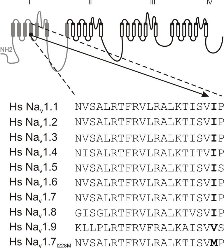
Estacion et al. Molecular Pain 2011, 7:92                                                                    Page 2 of 10
http://www.molecularpain.com/content/7/1/92

is usually focused on the distal extremities, precipitated    complaints that started at age 32, when he experienced
by mild warmth and relieved by cooling, and is caused         excruciating pain in his teeth and jaw triggered by cold
by NaV1.7 mutations that hyperpolarize activation, slow       and heat, which could radiate to the temporomandibular
deactivation, and enhance the channel ramp response           joint, and pain behind both eyes, especially when look-
[7]. Paroxysmal extreme pain disorder (PEPD) is charac-       ing at bright light. The oral mucosa, lips and tongue
terized by perirectal, periocular or perimandibular pain,     were not affected. Multiple tooth extractions did not
often triggered by defecation or lower body stimulation       provide pain relief. He subsequently developed myalgia,
[8], and has been linked to Na V 1.7 mutations that           with muscle pain persisting for 5-6 days after light phy-
severely impair fast-inactivation [9]. Small Fiber Neuro-     sical activity. The pain was aggravated by cold tempera-
pathy (SFN), which involves thinly myelinated and             ture and relieved by warmth. Sometimes the feet were
unmyelinated peripheral nerve fibers [10,11], presents a      also swollen. This patient suffered from stomach cramps
clinical picture that is characteristically dominated by      and diarrhea for more than 35 years, and from dry
neuropathic pain and autonomic symptoms [12],                 mouth and eyes and reduced urinary sensation and
together with preservation of normal strength, tendon         intermittent hesitation for several years. The patient was
reflexes, and vibration sense, and normal nerve conduc-       severely disabled and unable to work due to these com-
tion studies (NCS), which rule out large fiber involve-       plaints. Acetaminophen made the pain bearable, while
ment. The diagnosis of SFN can be confirmed by                short trials of NSAIDs and antidepressants did not pro-
demonstration of reduced intraepidermal nerve fiber           vide relief. Physical examination showed no abnormal-
density (IENFD) on skin biopsy and/or abnormal quan-          ities. Laboratory investigations, nerve conduction studies
titative sensory testing (QST) [13,14]. No apparent           and chest X-ray were normal. IENFD (1.6/mm; age- and
cause for SFN can be identified in 24% to 93% of cases        gender-matched normal values ≥ 3.5/mm [18]) was
in published patient series, and these cases are termed       abnormal. QST revealed abnormal warm and cold
idiopathic I-SFN [10,15,16]. Faber et al., recently           thresholds of the right foot. SCN9A gene analyses
reported that gain-of-function variants (single amino         demonstrated the variant, c.684C > G; NaV1.7/I228M.
acid substitutions) of voltage-gated sodium channel           The patient was diagnosed with Na V 1.7-related SFN.
Na V 1.7 are present in approximately 30% of patients         The patient’s two sons, aged 27 and 29, were found to
with biopsy-confirmed I-SFN [17].                             house the I228M substitution, but did not have any
  Distal (feet, and in some cases, hands) burning or          complaints at the time of study.
stabbing pain or paraesthesias are the initial symptoms          The I228M variant substitutes a highly conserved resi-
in most patients with I-SFN, and facial pain is rare.         due near the C-terminus of the S4 segment in domain I
Most of the eight patients with SFN described earlier by      (DI/S4, Figure 1). All human sodium channels except
Faber et al., [17] fit this clinical picture, and presented   NaV1.9 carry an isoleucine at this position [19], and this
with pain in the feet and in some cases the hands early       residue is invariant in all Na V 1.7 orthologues from
in their course, but did not manifest facial pain [17]. In    mammalian species (data not shown). The conservation
contrast, one patient in this series presented with severe    of the I228 residue among sodium channels suggests
pain in the teeth, jaw, and behind the eyes. This patient     that the I228M substitution might alter the properties of
(patient 8 in Faber et al., 2011) harbored the Na V 1.7       the NaV1.7 channels (Figure 1). This substitution was
variant c.684C > G (I228M) [17]; functional properties        not found in a control panel of DNA from 100 healthy
of this variant have not been previously reported. We         Dutch (Caucasian) individuals (200 chromosomes).
subsequently studied the sister of this patient, who          However, I228M is listed as a natural SNP in one data-
houses the same variant (c.684C > G (I228M) in                base (Craig Venter Human Genome), but with no break-
NaV1.7) and suffers from a different syndrome of pain         down of major/minor allele frequency, and has been
and redness of the hands and feet triggered by warmth,        reported as being associated with Dravet syndrome [20]
and have encountered an additional patient housing the        and in < 0.3% of a control population (5/576 control
same NaV 1.7 variant with pain over the scalp. In this        chromosomes).
study we report these three different clinical presenta-
tions of the I228M NaV1.7 variant, and demonstrate the        Patient 2
effects of the NaV1.7/I228M channels on excitability in       This patient, who is the sister of Patient 1, gave a his-
both trigeminal ganglion and DRG neurons.                     tory of burning pain and redness of hands and feet, trig-
                                                              gered by rising temperature and exercise and relieved by
Results                                                       cooling, beginning at age 36 years. She also reported
Patient 1                                                     increased perspiration, gastrointestinal complaints and
This patient, described as patient number 8 in Faber          hot flashes. Her medical history revealed recurrent urti-
et al., (2011) [17] is a 51-year-old male, referred with      caria attacks, psoriatic arthritis and hypothyroidism for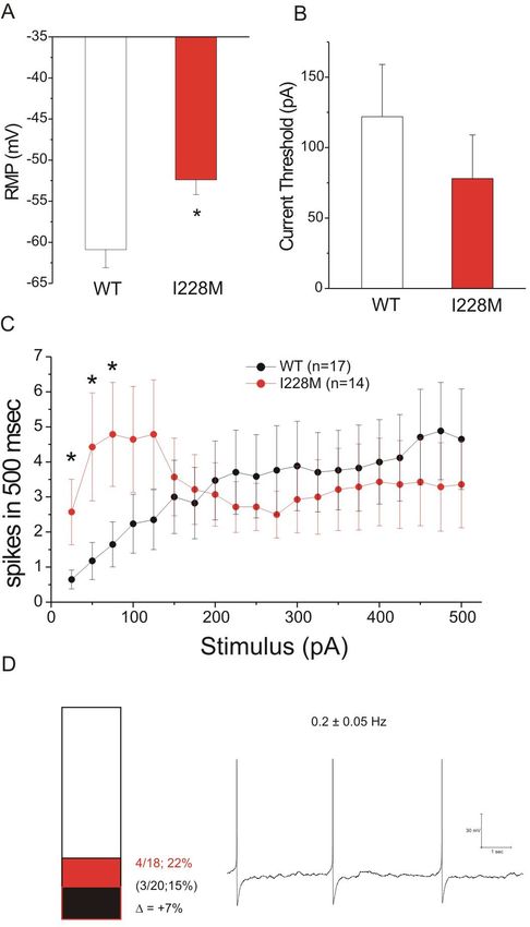
Estacion et al. Molecular Pain 2011, 7:92 Page 3 of 10
http://www.molecularpain.com/content/7/1/92
In addition, the patient reported severe perspiration
since puberty and intermittent difficulties with micturi-
tion. No other dysautonomic symptoms were noted.
Ibuprofen did not relieve the pain. The family history
was negative. Neurological examination was unremark-
able. Laboratory investigations, a chest X-ray and nerve
conduction studies were normal. Quantitative sensory
testing showed abnormal thresholds for warmth and
cold sensation of the dorsum of the right foot. A skin
biopsy demonstrated an INFD of 5.2 per mm, which
was lower than the reported normative values (≤ 5.7/
mm) [18]. The patient was diagnosed as having I-SFN.
SCN9A gene analysis demonstrated the same variant,
c.684C > G; p. I228M, as in Patients 1 and 2.
Functional Analysis
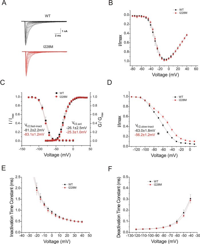
Voltage-clamp analysis
Voltage-clamp analysis of I228M variant channels fol-
lowing expression in HEK293 cells (Figure 2) demon-
strated impaired slow-inactivation (Figure 2D). Current
densities (WT: 432 ± 90 pA/pF, n = 9; I228M: 357 ± 70
pA/pF n = 13), activation V1/2 (WT: -26.1 ± 2.5 mV, n
Figure 1 Schematic of I228M mutation. Sequence alignment of
= 9; I228M: -25.3 ± 1.0 mV, n = 13), and fast-inactiva-
DI/S4 from human sodium channels. The charge-conserved
substitution in DI/S4 replaces a highly conserved isoleucine residue tion V1/2 (WT: -81.2 ± 2.2 mV, n = 8; I228M: -83.1 ±
at the cytoplasmic end of the S4 helix. I228 is conserved in all 1.2 mV, n = 12), for HEK293 cells transfected with WT
human sodium channels except for NaV1.9 which has a conservative or I228M channels were not significantly different (Fig-
substitution, valine, at the corresponding position. ure 2A, B, C). The time constants for fast-inactivation
(Figure 2E) and deactivation (Figure 2F) were not signif-
icantly different for I228M versus WT channels. Persis-
which she is adequately treated. Physical examination tent current (non-inactivating component at 0 mV),
showed no abnormalities other than red discolored measured in CsF-based pipette solution (WT: 0.42% ±
hands. Laboratory investigations, nerve conduction stu- 0.12%, n = 8; I228M: 0.67% ± 0.20%, n = 12) and in
dies and chest X-ray were normal. Quantitative sensory aspartate-based pipette solution (WT: 0.41% ± 0.08%, n
testing showed no abnormalities. IENFD was 8 per mm = 14; I228M: 0.41% ± 0.15%, n = 13) were not signifi-
(normal values ≥ 5.7 per mm) [18]. DNA analysis cantly different for I228M versus wild-type channels.
showed the same substitution in the SCN9A gene as Slow-inactivation was impaired for I228M channels (Fig-
found in her brother. On the basis of the clinical history ure 2D), with a depolarized V1/2 (WT: -63.0 ± 1.8 mV, n
and findings, the patient was diagnosed as having prob- = 10; I228M: -56.2 ± 1.2 mV, n = 14; p < 0.05). The off-
able SFN. set for slow-inactivation (non-inactivating component at
10 mV) was not significantly larger for I228M compared
Patient 3 to wild-type channels (WT: 7.8% ± 1.3%, n = 10; I228M:
This 46-year-old woman presented with a red discolora- 7.8% ± 1.3%, n = 14). Impaired slow-inactivation would
tion of the occiput. Three months later the red area be expected to increase the number of channels avail-
expanded and was noted to be associated with a tin- able for activation at potentials positive to -100 mV,
gling, burning and warm sensation over the scalp. After including potentials close to resting potential of DRG
washing of the hair, the redness increased for ~one neurons.
hour. The patient also complained that the structure of
her hair changed, becoming dryer and more fragile. She Current-Clamp Analysis: DRG Neurons
noted improvement in these complaints with warm tem- I228M had strong functional effects on DRG neurons,
peratures (such as during a visit to the Caribbean) and which were clearly rendered hyperexcitable by these
with fever. Cold had no specific influence. A year fol- channels (Figure 3). I228M produced a 4.8 mV depolar-
lowing onset of scalp symptoms, a red discoloration of izing shift in resting membrane potential of transfected
the toes of both feet developed, together with paraesthe- neurons (WT: -58.5 ± 1.4 mV, n = 22; I228M: -53.7 ±
sias and a burning sensation and tingling in both hands. 1.7 mV, n = 12; p < 0.05). While I228M did not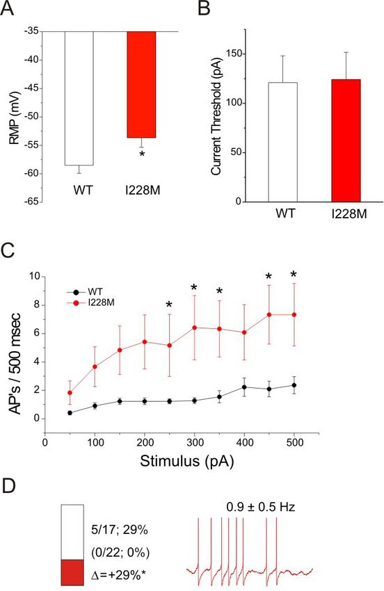
Estacion et al. Molecular Pain 2011, 7:92                                                                                                    Page 4 of 10
http://www.molecularpain.com/content/7/1/92

 Figure 2 Voltage-clamp properties ofNa V 1.7/I228Mchannels in HEK293 cells. Electrophysiological analysis of I228M variant: (A)
 Representative current traces recorded from HEK293 cells expressing wild type Nav1.7 (WT) (top) or I228M (bottom) channels, evoked by voltage
 steps (100 mec) from -80 to +40 mV in 5 mV increments, from a holding potential of -120 mV. (B) Normalized I-V curves for WT and I228M
 expressing cells. (C) Activation and steady-state fast-inactivation for WT (black squares) and I228M (red circles). Fast-inactivation was examined
 using a series of 500 msec prepulses from -140 to 0 mV followed by test pulses to -10 mV. Left inset: midpoint values for fast inactivation (V1/2,
 fast-inact) of WT (black) and I228M (red). Right inset: midpoint values for activation (V1/2, act) of WT (black) and I228M (red). (D) Steady-state slow-
 inactivation of WT (black squares) and I228M (red circles). Slow-inactivation was assessed using a 20 msec pulse to -10 mV after a 30 second
 prepulse to potentials from -130 to 10 mV followed by a 100 msec pulse to -120 mV to remove fast-inactivation. Inset: midpoint values of slow-
 inactivation (V1/2, slow-inact) (WT: black; I228M: red); *p < 0.05. V1/2 represents voltage midpoint, I/Imaxrepresents normalized current, and G/Gmax
 represents normalized conductance for fast-activation, slow-inactivation, and activation. (E) The kinetics of inactivation were analyzed by fitting
 data with a single exponential function for WT and I228M currents. (F) The kinetics of deactivation for WT and I228M expressing cells were
 obtained by holding the cells at -120 mV and tail currents were generated by a brief 0.5 ms depolarization to -20 mV followed by a series of
 repolarizations ranging from -120 to -40 mV. The closing rate of the channels was obtained by fitting the tail currents with a single exponential
 function.
Estacion et al. Molecular Pain 2011, 7:92                                                                            Page 5 of 10
http://www.molecularpain.com/content/7/1/92

                                                                      proportion of spontaneously firing cells (5 of 17 [29%]
                                                                      vs 0 of 22 [0%] for cells transfected with WT channels)
                                                                      (p < 0.05); mean frequency of spontaneous activity in
                                                                      cells transfected with I228M was 0.9 ± 0.5 Hz (n = 5).

                                                                      Current Clamp Analysis: Trigeminal Ganglion Neurons
                                                                      The I228M variant produced hyperexcitability in trigem-
                                                                      inal ganglion neurons (Figure 4). I228M produced an
                                                                      8.5 mV depolarizing shift in resting membrane potential
                                                                      (WT: -60.9 ± 2.2 mV, n = 17; I228M: -52.4 ± 1.8 mV, n
                                                                      = 14; p < 0.05). I228M produced a 36% reduction in
                                                                      current threshold to 200-millisecond stimuli (WT: 122
                                                                      ± 37 pA, n = 13; I228M: 78 ± 31 pA, n = 12). Trigem-
                                                                      inal ganglion neurons transfected with I228M tended to
                                                                      fire multiple action potentials (at a frequency nearly
                                                                      four-fold higher than in cells transfected with wild-type
                                                                      channels) in response to 500 msec stimuli close to
                                                                      threshold (25 to 125 pA, with the difference being statis-
                                                                      tically significant between 25 and 75 pA), although at
                                                                      stimulus levels of ≥ 2X threshold, the number of action
                                                                      potentials falls, approaching that of cells transfected
                                                                      with wild-type channels (Figure 4C). I228M produced a
                                                                      trend toward an increase in the proportion of sponta-
                                                                      neously firing cells (4 of 18 [22%] vs 3 of 20 [15%] for
                                                                      cells transfected with WT channels) that did not reach
                                                                      statistical significance; mean frequency of spontaneous
                                                                      activity in cells transfected with I228M was 0.2 ± 0.05
                                                                      Hz (n = 4).

                                                                      Discussion
 Figure 3 Current-clamp properties of DRG neurons transfected         In this study we describe three patients (two siblings,
 with I228M. Excitability of DRG neurons expressing I228M: (A) RMP
 of DRG neurons expressing WT (-58.5 ± 1.4, n = 22) or I228M (-53.7
                                                                      and a third, unrelated patient) housing the I228M var-
 ± 1.7, n = 12); *p < 0.05. (B) Current threshold of DRG neurons      iant of sodium channel Na V 1.7. One of these patients
 expressing WT (121 ± 27, n = 22) or I228M (124 ± 28, n = 12) to      displayed a clinical phenotype that included pain in the
 200 msec stimuli. (C) Comparison of mean firing frequency in DRG     face as well as in other parts of the body together with
 neurons expressing WT and I228M across a range of current            autonomic symptoms, with the diagnosis of SFN con-
 injections from 50 to 500pA; *p < 0.05. (D) Bar graph showing the
 proportion of spontaneous firing cells for DRG neurons expressing
                                                                      firmed by demonstration of reduced IENFD on skin
 I228M (red) and WT channels (black); numbers to the right of the     biopsy, and abnormal QST. The second patient gave a
 bar graph show values for WT (lower value in parentheses) and        history of distal extremity pain and redness, triggered by
 I228M (upper value); *p < 0.05. The recording on the right shows     warmth and relieved by cooling. While these symptoms
 spontaneous firing (10 seconds) of representative DRG neuron         are commonly reported in IEM [7,21], she also reported
 expressing I228M; the numbers above the trace show average ±
 standard deviation frequency of spontaneous action potentials. APs
                                                                      autonomic symptoms including increased perspiration,
 = action potentials.                                                 gastrointestinal complaints and hot flashes, which are
                                                                      not characteristic of IEM. The third patient initially
                                                                      experienced discomfort and vasomotor instability over
decrease the current threshold (WT: 121 ± 27 pA, n =                  the occiput, which progressed to involve the distal extre-
22; I228M: 124 ± 28 pA, n = 12), it produced on aver-                 mities, together with abnormal perspiration, intermittent
age a higher firing frequency at all stimulus intensities,            difficulties with micturition; skin biopsy and QST in this
even close to current threshold, and increased the num-               patient were both abnormal, confirming the diagnosis of
ber of action potentials evoked by 500-millisecond depo-              SFN.
larizing stimuli at higher stimulus intensities nearly                  Because facial pain was a prominent part of the clini-
four-fold, with the change being statistically significant            cal picture in one of the patients described in this
at many of the intensities tested, from 50 to 500 pA.                 paper, we assessed the effect of the I228M mutation on
I228M also produced a significant increase in the                     excitability of trigeminal ganglion neurons. Our current-
Estacion et al. Molecular Pain 2011, 7:92 Page 6 of 10
http://www.molecularpain.com/content/7/1/92
assessed in trigeminal ganglion neurons. We previously
reported that the A1632E Na V 1.7 mutation, from a
patient who displayed a mixed clinical phenotype with
features of both IEM and PEPD, produces hyperexcit-
ability in trigeminal ganglion neurons [22]. The A1632E
mutation, however, produced hyperexcitability in these
cells over the entire range of stimulus intensities, while
I228M produces hyperexcitability only at low stimulus
intensities. Whether other gain-of-function mutations of
NaV1.7 have similar effects on trigeminal ganglion neu-
rons remains to be determined.
The I228M substitution is located within the fourth
transmembrane segment (S4) within domain I of the
Na V 1.7 channel. The S4 in each of the domains of
sodium channels is an amphiphatic helix which is char-
acterized by a repeat motif of positively charged amino
acids at every third position [19]. Non-charge-conserved
mutations, S211P and F216S, in DI/S4 have been linked
to IEM, and have been shown to shift voltage-depen-
dence of activation in a hyperpolarizing direction, mak-
ing it easier to open the mutant channels [23,24]. The
I228M substitution does not change the number of
charges in the S4 segment, and reasonably conserves the
hydrophobic nature of the side-chain of this residue,
and thus might not have been predicted to have a func-
tional effect. A link to function, however, is suggested
by the conservation of the I228 residue at the equivalent
position in all voltage-gated sodium channels sequenced
to date (Figure 1); I228 is substituted by the other
branched side-chain residue, valine, in Na V 1.9. The
functional effect of I228M might be related to the proxi-
mity of the I228 residue to the cytoplasmic end of the
S4 segment, which could alter the local structure of the
helix in a subtle manner affecting slow-inactivation but
Figure 4 Current-clamp properties of trigeminal ganglion
not activation. Notably, while the I228M variant pro-
neurons transfected with I228M. Excitability of trigeminal
ganglion neurons expressing I228M: (A) RMP of trigeminal ganglion duced hyperexcitability in both DRG and trigeminal
neurons expressing WT (-60.9 ± 2.2, n = 17) or I228M (-52.4 ± 1.8, n ganglion neurons, only two of the three patients
= 14); *p < 0.05. (B) Current threshold of DRG neurons expressing described here reported cranial pain, and it was experi-
WT (122 ± 37, n = 13) or I228M (78 ± 31, n = 12) to 200 msec enced in the jaw and eyes in one, while it was focused
stimuli; p < 0.05. (C) Comparison of mean firing frequency in
on the scalp in the other.
trigeminal ganglion neurons expressing WT and I228M across a
range of current injections from 25 to 500 pA; *p < 0.05. (D) Bar Our results demonstrate phenotypic diversity in the
graph showing the proportion of spontaneous firing cells for pain syndromes associated with the I228M substitution
trigeminal neurons expressing I228M (red) and WT channels (black); in the NaV1.7 channel in three different patients. Two
numbers to the right of the bar graph show mean values for WT of these patients were from the same family, which also
(lower value in parentheses) and I228M (upper value). The recording
includes patient 1’s two asymptomatic sons who carry
on the right shows spontaneous firing (10 seconds) of
representative trigeminal neuron expressing I228M; the numbers the I228M NaV1.7 variant. Both of these asymptomatic
above the trace show average ± standard deviation frequency of carriers are younger than the age of onset of the three
spontaneous action potentials. APs = action potentials. patients presented, and whether they will develop pain
in the future is unclear. We have previously noted dif-
ferent ages of onset and different degrees of pain, and
clamp analysis demonstrated that the I228M variant an asymptomatic carrier in members of a single family,
depolarizes resting membrane potential, reduces current all housing the G616R NaV1.7 mutation [25]. Whether
threshold and enhances repetitive firing in these cells. this phenotypic variability is due to modifier genes, epi-
The effect of only one other NaV1.7 mutation has been genetic factors, and/or environmental factors is not yetEstacion et al. Molecular Pain 2011, 7:92 Page 7 of 10 http://www.molecularpain.com/content/7/1/92 clear. The minor allele of the NaV1.7 R1150W variant, by medical ethics committees at Yale University and which is known to produce hyperexcitability in DRG Maastricht University Medical Center. All aspects of the neurons [26], has been associated with increased pain study were explained and a written informed consent scores in a number of acquired pain syndromes obtained prior to study. (osteoarthritis, compressive radiculopathies, traumatic All three patients met strict eligibility criteria for a limb amputation), suggesting that environmental factors study on SFN as described by Faber et al. [17]. Subjects may, at least in some individuals, act as triggers or were excluded from the study if there was a history or increase risk of developing pain [27]. detection after screening of illnesses known to cause Most peripheral neuropathies present in a “stocking SFN, including impaired glucose tolerance, diabetes mel- glove” distribution with sensory abnormalities and pain litus, hyperlipidemia, liver/kidney/thyroid dysfunction, first appearing in the most distal parts of the limbs (feet, monoclonal-gammopathy, connective tissue disorders, then hands). It has traditionally been held that longer amyloidosis, sarcoidosis, Fabry’s disease (alpha-galactosi- nerve fibers, or the cells giving rise to them, are affected dase, in females combined with GLA-gene sequencing), before shorter fibers or the cells giving rise to them. A celiac disease, HIV, alcohol abuse, hemochromatosis, B6 number of potential mechanisms have been invoked for intoxication, anti-phospholipid syndrome neurotoxic this length-dependent mode of progression of neuropa- drugs (e.g., chemotherapy) [17]. thy, including impairment of axoplasmic transport [28], increased probability of demyelination along longer Clinical characterization nerve fibers [29], or a higher probability of impairment Skin biopsy of calcium homeostasis along longer nerve fibers Punch biopsy (10 cm above lateral malleolus) specimens [30,31]. However, the present results show that the were fixed (2% paraformaldehyde-lysine-sodium period- NaV1.7 I228M variant, which impairs slow-inactivation, ate at 4°C), cryprotected and stored at -80°C in 20% gly- produces physiological changes in primary afferent neu- cerol before sectioning (50 μm) [13]. The numbers of rons (trigeminal ganglion neurons) that innervate the individual nerve fibers crossing the dermal-epidermal relatively proximal sensory field of the face and scalp, as junctions were analyzed by bright-field microscopy well as DRG neurons. While we do not know whether (Olympus BX50 stereology workstation, PlanApo oil- there was degeneration of small fibers innervating the objective 40×/NA = 1.0) in each of three sections, face or scalp in these patients, both exhibited degenera- immunostained with polyclonal rabbit antiprotein-gene- tion of the relatively long axons, as demonstrated by product-9.5 antibody (PGP9.5; Ultraclone, Wellow, Isle- reduced IENFD on skin biopsy from the leg. of-Wight, UK). Linear quantification of intraepidermal In summary, our results demonstrate phenotypic nerve fiber density (IENF/mm) was compared with age- diversity in pain syndromes associated with the I228M and gender-adjusted normative values [18,32]. gain-of-function variant of NaV1.7. Importantly, variabil- Quantitative sensory testing (QST) ity in clinical presentation was present not only when QST, performed in accordance with previous guidelines comparing patients from different families, but also for [33], using a TSA-2001 (Medoc, Ramat-Yishai, Israel) patients within a single family. Our findings also instrument, assessed thresholds at the dorsum of both demonstrate that the I228M variant can increase excit- feet and thenar eminences, using ascending/descending ability of trigeminal ganglion as well as DRG neurons. (warm/cool) thermal ramp stimuli delivered through a While the mechanism(s) responsible for this phenotypic thermode [34]. Heat pain modality was also examined. diversity remain unexplained, our findings suggest that Results were compared with reported normative values clinical studies, in patients who are carriers of functional [35]. Measurements were considered abnormal when Z- variants of sodium channels, should be designed to take values exceeded 2.5. A sensory modality was classified phenotypic variability, even within single families, into as abnormal if results of both method-of-limits and account. method-of-levels were abnormal [36]. Materials and methods SCN9A sequence analysis Patients Exon screening The three patients initially studied were part of a cohort Genomic DNA was extracted from 300 μL whole blood of patients aged ≥ 18 years with idiopathic SFN, seen at using Puregene genomic DNA isolation kit (Gentra-Sys- Maastricht University Medical Center Neurological tems, Minneapolis). All SCN9A coding exons and flank- Clinic, with a clinical diagnosis of SFN between 2006 ing intronic sequences, and exons encoding 5’ and 3- and 2009; this series excluded patients in whom, after a untranslated sequences within the complementary DNA, careful work-up, a cause for SFN was identified. A sister were amplified and sequenced as described previously of patient 1 was also studied. This study was approved [37]. Genomic sequences were compared with reference
Estacion et al. Molecular Pain 2011, 7:92                                                                Page 8 of 10
http://www.molecularpain.com/content/7/1/92

Nav1.7 cDNA (NM_002977.3) to identify sequence var-         Neuron Nucleofector Solution and program G-013, as
iations [38] using Alamut Mutation-Interpretation Soft-     described previously [39]. The ratio of sodium channel
ware (Interactive-Biosoftware; Rouen, France). A control    to GFP constructs was 10:1. The transfected neurons
panel of DNA from 100 healthy Dutch (Caucasian) indi-       were allowed to recover for 5 minutes at 37°C in 0.5 ml
viduals (200 chromosomes) was also screened.                of Ca 2+ -free DMEM containing 10% fetal calf serum.
Plasmids                                                    The cell suspension was then diluted with DRG media
The human Nav1.7-AL insert (carrying the adult exon 5,      containing 1.5 mg/ml bovine serum albumin and 1.5
E5A, and Long loop1), converted to become TTX-R             mg/ml trypsin inhibitor, 80 μl was plated on 12 mm cir-
(hNa v 1.7 R /AL; designated WT hereinafter) by Y362S       cular poly-D-lysine/laminin precoated coverslips (BD
substitution [39], has been previously described [39].      Biosciences, Bedford, MA) and the cells incubated at 37°
The I228M mutation was introduced into WT using             C in 5% CO2 for 30 min. DRG media (1 ml/well), sup-
QuickChange XL II site-directed mutagenesis according       plemented with 50 ng/ml each of mNGF (Alomone
to manufacturer recommendations (Stratagene).               Labs, Jerusalem, Israel) and GDNF (Peprotec, Rocky
  The full-length inserts of the different clones were      Hill, NJ), was then added and the cells maintained at
sequenced at the Howard Hughes Medical Institute/           37°C in a 5% CO2 incubator.
Keck Biotechnology Center at Yale University. Sequence      Electrophysiology
analysis used BLAST (National Library of Medicine) and      Whole-cell voltage-clamp recordings in HEK293 cells
Lasergene (DNAStar, Madison, WI), and confirmed the         were carried out at 20 ± 1°C using a peltier temperature
inserts to be devoid of un-intended mutations.              controller of the recording chamber. The extracellular
Transient transfection of HEK293 cells                      solution contained (in mM): 140 NaCl, 3 KCl, 1 MgCl2,
Transient transfections of the hNa V 1.7 together with      1 CaCl2, and 10 HEPES, pH 7.3 with NaOH (adjusted to
hb1 and hb2 constructs into HEK293 cells were per-          320 mOsm with dextrose). The pipette solution con-
formed using Optifect (Invitrogen) following the recom-     tained (in mM): 140 CsF, 10 NaCl, 2 MgCl2, 1 EGTA,
mended protocol by manufacturer. Recordings were            10 HEPES, pH 7.3 with CsOH (adjusted to 310 mOsm
performed 20-30 hours after transfection.                   with dextrose). Patch-pipettes had a resistance of 1-3
Primary sensory neuron isolation and transfection           MΩ when filled with pipette solution. The calculated
Dorsal root ganglia (DRG) and trigeminal ganglia from       junction potential (JPcalc included in pCLAMP soft-
adult Sprague Dawley rat pups (P0-P5) were isolated         ware) of 9 mV was not compensated. Upon achieving
and then cultured using the same protocol for both.         the whole-cell recording configuration, the pipette and
Dissected ganglia were placed in ice-cold oxygenated        cell capacitance were manually minimized using the
complete saline solution (CSS), which contained (in         Axopatch 200B (Molecular Devices, Union City, CA)
mM) 137 NaCl, 5.3 KCl, 1 MgCl2, 25 sorbitol, 3 CaCl2,       compensation circuitry. To reduce voltage errors, 80-
10 N-2-hydroxyethylpiperazine-N’-2-ethanesulfonic acid      90% series resistance and prediction compensation was
(HEPES); pH 7.2. They were then transferred to an oxy-      applied. Cells were excluded from analysis if the pre-
genated, 37°C CSS solution containing 1.5 mg/ml Col-        dicted voltage error exceeded 3 mV. The recorded cur-
lagenase A (Roche Applied Science, Indianapolis, IN)        rents were digitized at a rate of 50 kHz after passing
and 0.6 mM EDTA and incubated with gentle agitation         through a low-pass Bessel filter setting of 10 kHz. The
at 37°C for 20 min. This solution was then exchanged        Axopatch 200B data were digitized using pCLAMP soft-
with an oxygenated, 37°C CSS solution containing 1.5        ware (version 10) and a Digidata 1440A interface (Mole-
mg/ml Collagenase D (Roche Applied Science, Indiana-        cular Devices). Linear leak and residual capacitance
polis, IN), 0.6 mM EDTA and 30 U/ml papain                  artifacts were subtracted out using the P/N method.
(Worthington Biochemical, Lakewood, NJ) and incu-           The Na+ current recordings were initiated after a 5 min-
bated with gentle agitation at 37°C for 20 min. The         ute equilibration period once whole-cell configuration
solution was then aspirated and the ganglia triturated in   was achieved.
DRG media (DMEM/Fl2 (1:1) with 100 U/ml penicillin,           Data analysis was performed using Clampfit (Molecu-
0.1 mg/ml streptomycin (Invitrogen, Carlsbad, CA) and       lar Devices) and Origin (Microcal Software, Northhamp-
10% fetal calf serum (Hyclone, Logan, UT), which con-       ton, MA). To generate activation curves, cells were held
tained 1.5 mg/ml bovine serum albumin (Sigma-Aldrich,       at -120 mV and stepped to potentials of -80 to 40 mV
St. Louis, MO) and 1.5 mg/ml trypsin inhibitor (Roche       for 100 msec. Peak inward currents obtained from acti-
Applied Science, Indianapolis, IN).                         vation protocols were converted to conductance values
  Either WT or I228M variant channels were transiently      using the equation, G = I/(Vm - ENa), for which G is the
transfected into the DRG or trigeminal ganglion neu-        conductance, I is the peak inward current, V m is the
rons, along with enhanced-GFP, by electroporation with      membrane potential step used to elicit the response and
a Nucleofector II (Amaxa, Gaithersburg, MD) using Rat       ENa is the reversal potential for sodium (determined for
Estacion et al. Molecular Pain 2011, 7:92                                                                                        Page 9 of 10
http://www.molecularpain.com/content/7/1/92

each cell using the x-axis intercept of a linear fit of the      data collection. Input resistance was determined by the
peak inward current responses). Conductance data were            slope of a line fit to hyperpolarizing responses to cur-
normalized by the maximum conductance value and fit              rent steps of 10-35 pA. Threshold was determined by
with a Boltzmann equation of the form G = G min +                the first action potential elicited by a series of depolariz-
(Gmax-Gmin)/(1 + exp[(V1/2 -Vm)/k)], where V1/2 is the           ing current injections that increased in 5 pA increments.
midpoint of activation and k is a slope factor. The              The number of action potentials elicited in response to
kinetics of inactivation were assessed by fitting the fall-      depolarizing current injections of 500 msec duration
ing phase of the currents with a single exponential func-        was also measured. After-hypolarization currents, and
tion. To generate steady-state fast-inactivation curves,         amplitude and width of action potentials were not for-
cells were stepped to inactivating potentials of -140 to         mally analyzed in this study. Data are expressed as
10 mV for 500 msec followed by a 20 msec step to -10             means ± standard error (SEM). Statistical significance
mV. The protocol for slow-inactivation consisted of a 30         was determined by Student’s t-test, Mann-Whitney test
second step to potentials varying from -120 to 10 mV,            (firing frequency) or z-test (frequency of spontaneous
followed by a 100 msec step to -120 mV to remove fast-           firing).
inactivation and a 20 msec step to -10 mV to elicit a
test response. Peak inward currents obtained from
                                                                 Acknowledgements
steady-state fast-inactivation and slow-inactivation pro-        This work was supported in part by grants from the Rehabilitation Research
tocols were normalized by the maximum current ampli-             Service and Medical Research Service, Department of Veterans Affairs; and by
tude and fit with a Boltzmann equation of the form I =           the ‘profileringsfonds’ University Hospital Maastricht. The Center for
                                                                 Neuroscience & Regeneration Research is a Collaboration of the Paralyzed
I min + (I max -I min )/(1 + exp[(V m - V 1/2 )/k)], where V m   Veterans of America with Yale University. We thank Rene te Morsche, Lynda
represents the inactivating pre-pulse membrane poten-            Tyrrell, Lawrence Macala, Peng Zhao and Palak Shah for dedicated
tial and V1/2 represents the midpoint of inactivation. For       assistance.
deactivation the cells were held at -120 mV and tail cur-        Author details
rents were generated by a brief 0.5 ms depolarization to         1
                                                                  Department of Neurology, Yale University School of Medicine, New Haven,
-20 mV followed by a series of repolarizations ranging           CT 06510, and Center for Neuroscience and Regeneration Research, Veterans
                                                                 Affairs Medical Center, West Haven, CT 06516, USA. 2Department of
from -120 to -40 mV. The closing rate of the channels            Neurology, University Medical Center Maastricht, Maastricht, the Netherlands.
was obtained by fitting the tail currents with a single          3
                                                                  Neuromuscular Diseases Unit, IRCCS Foundation, “Carlo Besta”, Milan, Italy.
                                                                 4
exponential function.                                             Department of Gastroenterology and Hepatology, Radboud University
                                                                 Nijmegen Medical Center, Nijmegen, the Netherlands. 5Department of
   Whole-cell current-clamp recordings from isolated             Clinical Genetics, University Medical Center Maastricht, Maastricht, the
DRG or trigeminal ganglion neurons were performed                Netherlands. 6Department of Neurology, Spaarne Hospital, Hoofddorp, the
using the Axopatch 200B amplifier, digitized using the           Netherlands. 7College of Pharmacy, Catholic University of Korea, Bucheon,
                                                                 South Korea.
Digidata 1440A interface and controlled using pCLAMP
software. The bath solution for current-clamp record-            Authors’ contributions
ings contained (in mM): 140 NaCl, 3 KCl, 2 MgCl2 , 2             ME acquired electrophysiological data, completed data analysis, and
                                                                 participated in writing the manuscript. CH acquired electrophysiological
CaCl2, and 10 HEPES, pH 7.3 with NaOH (adjusted to               data, completed data analysis, and participated in writing the manuscript.
315mOsm with dextrose). The pipette solution con-                JSC acquired electrophysiological data and completed data analysis. JGJH
tained (in mM): 140 KCl, 0.5 EGTA, 5 HEPES, and 3                provided clinical assessment of patients. GL participated in study design and
                                                                 manuscript editing. JPHD provided genomic assessment of patients. MMG
Mg-ATP, pH 7.3 with KOH (adjusted to 300 mOsm                    provided genetic analysis of patients. SDDH participated in study design,
with dextrose). The junction potential between these             data analysis and manuscript editing. CGF provided overall project
two solutions given by JPcalc was 5 mV but no correc-            management, participated in study design, and writing of the manuscript.
                                                                 ISJM provided overall project management, participated in study design, and
tion was applied for current-clamp experiments. Record-          writing of the manuscript. SGW provided overall project management,
ings were performed on transfected presumptive                   participated in study design, data analysis, and writing of the manuscript. All
nociceptive neurons based on the morphology of small             authors have read and approved the final manuscript.
diameter (20-28 μm) round cell bodies that also exhib-           Competing interests
ited GFP fluorescence. All recordings were performed             The authors declare that they have no competing interests.
between 40 hr and 50 hr post-transfection 20 ± 1°C.
                                                                 Received: 20 September 2011 Accepted: 2 December 2011
Coverslips were transferred to a perfusable chamber              Published: 2 December 2011
(Warner Instruments, Hamden, CT) and all recordings
were initiated within an hour. Whole-cell configuration          References
was obtained in voltage-clamp mode before proceeding             1. Toledo-Aral JJ, Moss BL, He ZJ, Koszowski AG, Whisenand T, Levinson SR,
                                                                     Wolf JJ, Silos-Santiago I, Halegoua S, Mandel G: Identification of PN1, a
to the current-clamp recording mode. Cells with stable               predominant voltage-dependent sodium channel expressed principally
(< 10% variation) resting membrane potentials (RMPs)                 in peripheral neurons. Proc Natl Acad Sci USA 1997, 94(4):1527-1532.
more negative than -35 mV and overshooting action                2. Rush AM, Dib-Hajj SD, Liu S, Cummins TR, Black JA, Waxman SG: A single
                                                                     sodium channel mutation produces hyper- or hypoexcitability in
potentials (> 85 mV RMP to peak) were used for further
Estacion et al. Molecular Pain 2011, 7:92 Page 10 of 10
http://www.molecularpain.com/content/7/1/92
different types of neurons. Proc Natl Acad Sci USA 2006, Characterization of a novel sodium channel mutation. J Physiol 2010,
103(21):8245-8250. 588(Pt 11):1915-1927.
3. Sangameswaran L, Fish LM, Koch BD, Rabert DK, Delgado SG, Ilnicka M, 24. Choi JS, Dib-Hajj SD, Waxman SG: Inherited erythermalgia: limb pain from
Jakeman LB, Novakovic S, Wong K, Sze P, et al: A novel tetrodotoxin- an S4 charge-neutral Na channelopathy. Neurology 2006, 67(9):1563-1567.
sensitive, voltage-gated sodium channel expressed in rat and human 25. Choi JS, Cheng X, Foster E, Leffler A, Tyrrell L, Te Morsche RH, Eastman EM,
dorsal root ganglia. J Biol Chem 1997, 272(23):14805-14809. Jansen HJ, Huehne K, Nau C, et al: Alternative splicing may contribute to
4. Persson AK, Black JA, Gasser A, Cheng X, Fischer TZ, Waxman SG: Sodium- time-dependent manifestation of inherited erythromelalgia. Brain 2010,
calcium exchanger and multiple sodium channel isoforms in intra- 133(Pt 6):1823-1835.
epidermal nerve terminals. Mol Pain 2010, 6:84. 26. Estacion M, Harty TP, Choi JS, Tyrrell L, Dib-Hajj SD, Waxman SG: A sodium
5. Cummins TR, Howe JR, Waxman SG: Slow closed-state inactivation: a channel gene SCN9A polymorphism that increases nociceptor
novel mechanism underlying ramp currents in cells expressing the hNE/ excitability. Ann Neurol 2009, 66(6):862-866.
PN1 sodium channel. J Neurosci 1998, 18(23):9607-9619. 27. Reimann F, Cox JJ, Belfer I, Diatchenko L, Zaykin DV, McHale DP, Drenth JP,
6. Waxman SG: Neurobiology: a channel sets the gain on pain. Nature 2006, Dai F, Wheeler J, Sanders F, et al: Pain perception is altered by a
444(7121):831-832. nucleotide polymorphism in SCN9A. Proc Natl Acad Sci USA 2010,
7. Dib-Hajj SD, Cummins TR, Black JA, Waxman SG: Sodium channels in 107(11):5148-5153.
normal and pathological pain. Annu Rev Neurosci 2010, 33:325-347. 28. Sabin TD: Classification of peripheral neuropathy: the long and the short
8. Fertleman CR, Ferrie CD, Aicardi J, Bednarek NA, Eeg-Olofsson O, Elmslie FV, of it. Muscle Nerve 1986, 9(8):711-719.
Griesemer DA, Goutieres F, Kirkpatrick M, Malmros IN, et al: Paroxysmal 29. Waxman SG, Brill MH, Geschwind N, Sabin TD, Lettvin JY: Probability of
extreme pain disorder (previously familial rectal pain syndrome). conduction deficit as related to fiber length in random-distribution
Neurology 2007, 69(6):586-595. models of peripheral neuropathies. J Neurol Sci 1976, 29(1):39-53.
9. Fertleman CR, Baker MD, Parker KA, Moffatt S, Elmslie FV, Abrahamsen B, 30. Viader A, Golden JP, Baloh RH, Schmidt RE, Hunter DA, Milbrandt J:
Ostman J, Klugbauer N, Wood JN, Gardiner RM, et al: SCN9A mutations in Schwann cell mitochondrial metabolism supports long-term axonal
paroxysmal extreme pain disorder: allelic variants underlie distinct survival and peripheral nerve function. J Neurosci 2011,
channel defects and phenotypes. Neuron 2006, 52(5):767-774. 31(28):10128-10140.
10. Lacomis D: Small-fiber neuropathy. Muscle Nerve 2002, 26(2):173-188. 31. Baloh RH: Mitochondrial dynamics and peripheral neuropathy.
11. Lauria G: Small fibre neuropathies. Curr Opin Neurol 2005, 18(5):591-597. Neuroscientist 2008, 14(1):12-18.
12. Stewart JD, Low PA, Fealey RD: Distal small fiber neuropathy: results of 32. Bakkers M, Merkies IS, Lauria G, Devigili G, Penza P, Lombardi R,
tests of sweating and autonomic cardiovascular reflexes. Muscle Nerve Hermans MC, van Nes SI, De Baets M, Faber CG: Intraepidermal nerve fiber
1992, 15(6):661-665. density and its application in sarcoidosis. Neurology 2009,
13. Lauria G, Hsieh ST, Johansson O, Kennedy WR, Leger JM, Mellgren SI, 73(14):1142-1148.
Nolano M, Merkies IS, Polydefkis M, Smith AG, et al: European Federation 33. Shy ME, Frohman EM, So YT, Arezzo JC, Cornblath DR, Giuliani MJ,
of Neurological Societies/Peripheral Nerve Society Guideline on the use Kincaid JC, Ochoa JL, Parry GJ, Weimer LH: Quantitative sensory testing:
of skin biopsy in the diagnosis of small fiber neuropathy. Report of a report of the Therapeutics and Technology Assessment Subcommittee
joint task force of the European Federation of Neurological Societies of the American Academy of Neurology. Neurology 2003, 60(6):898-904.
and the Peripheral Nerve Society. Eur J Neurol 2010, 17(7):903-912, e944- 34. Reulen JP, Lansbergen MD, Verstraete E, Spaans F: Comparison of thermal
909. threshold tests to assess small nerve fiber function: limits vs. levels. Clin
14. Tesfaye S, Boulton AJ, Dyck PJ, Freeman R, Horowitz M, Kempler P, Lauria G, Neurophysiol 2003, 114(3):556-563.
Malik RA, Spallone V, Vinik A, et al: Diabetic neuropathies: update on 35. Yarnitsky D, Sprecher E: Thermal testing: normative data and repeatability
definitions, diagnostic criteria, estimation of severity, and treatments. for various test algorithms. J Neurol Sci 1994, 125(1):39-45.
Diabetes Care 2010, 33(10):2285-2293. 36. Hoitsma E, Drent M, Verstraete E, Faber CG, Troost J, Spaans F, Reulen JP:
15. Devigili G, Tugnoli V, Penza P, Camozzi F, Lombardi R, Melli G, Broglio L, Abnormal warm and cold sensation thresholds suggestive of small-fiber
Granieri E, Lauria G: The diagnostic criteria for small fibre neuropathy: neuropathy in sarcoidosis. Clin Neurophysiol 2003, 114:2326-2333.
from symptoms to neuropathology. Brain 2008, 131(Pt 7):1912-1925. 37. Drenth JP, te Morsche RH, Guillet G, Taieb A, Kirby RL, Jansen JB: SCN9A
16. Bednarik J, Vlckova-Moravcova E, Bursova S, Belobradkova J, Dusek L, mutations define primary erythermalgia as a neuropathic disorder of
Sommer C: Etiology of small-fiber neuropathy. J Peripher Nerv Syst 2009, voltage gated sodium channels. The Journal of investigative dermatology
14(3):177-183. 2005, 124(6):1333-1338.
17. Faber CG, Hoeijmakers JG, Ahn HS, Cheng X, Han C, Choi JS, Estacion M, 38. Klugbauer N, Lacinova L, Flockerzi V, Hofmann F: Structure and functional
Lauria G, Vanhoutte EK, Gerrits MM, et al: Gain of function Na(V) 1.7 expression of a new member of the tetrodotoxin-sensitive voltage-
mutations in idiopathic small fiber neuropathy. Annals of neurology 2011. activated sodium channel family from human neuroendocrine cells. The
18. Lauria G, Bakkers M, Schmitz C, Lombardi R, Penza P, Devigili G, Smith AG, EMBO journal 1995, 14(6):1084-1090.
Hsieh ST, Mellgren SI, Umapathi T, et al: Intraepidermal nerve fiber density 39. Dib-Hajj SD, Choi JS, Macala LJ, Tyrrell L, Black JA, Cummins TR,
at the distal leg: a worldwide normative reference study. J Peripher Nerv Waxman SG: Transfection of rat or mouse neurons by biolistics or
Syst 2010, 15(3):202-207. electroporation. Nat Protoc 2009, 4(8):1118-1126.
19. Catterall WA, Goldin AL, Waxman SG: International Union of
Pharmacology. XLVII. Nomenclature and structure-function relationships doi:10.1186/1744-8069-7-92
of voltage-gated sodium channels. Pharmacol Rev 2005, 57(4):397-409. Cite this article as: Estacion et al.: Intra- and interfamily phenotypic
20. Singh NA, Pappas C, Dahle EJ, Claes LR, Pruess TH, De Jonghe P, diversity in pain syndromes associated with a gain-of-function variant
Thompson J, Dixon M, Gurnett C, Peiffer A, et al: A role of SCN9A in of NaV1.7. Molecular Pain 2011 7:92.
human epilepsies, as a cause of febrile seizures and as a potential
modifier of Dravet syndrome. PLoS Genet 2009, 5(9):e1000649.
21. Drenth JP, Waxman SG: Mutations in sodium-channel gene SCN9A cause
a spectrum of human genetic pain disorders. J Clin Invest 2007,
117(12):3603-3609.
22. Estacion M, Dib-Hajj SD, Benke PJ, Te Morsche RH, Eastman EM, Macala LJ,
Drenth JP, Waxman SG: NaV1.7 gain-of-function mutations as a
continuum: A1632E displays physiological changes associated with
erythromelalgia and paroxysmal extreme pain disorder mutations and
produces symptoms of both disorders. J Neurosci 2008,
28(43):11079-11088.
23. Estacion M, Choi JS, Eastman EM, Lin Z, Li Y, Tyrrell L, Yang Y, Dib-Hajj SD,
Waxman SG: Can robots patch-clamp as well as humans?You can also read