Phylogenetic study documents different speciation mechanisms within the Russula globispora lineage in boreal and arctic environments of the ...
←
→
Page content transcription
If your browser does not render page correctly, please read the page content below
Caboň et al. IMA Fungus 2019, 10:5
https://doi.org/10.1186/s43008-019-0003-9                                                                                   IMA Fungus

 RESEARCH                                                                                                                                        Open Access

Phylogenetic study documents different
speciation mechanisms within the Russula
globispora lineage in boreal and arctic
environments of the Northern Hemisphere
Miroslav Caboň1, Guo-Jie Li2, Malka Saba3,4,8, Miroslav Kolařík5, Soňa Jančovičová6, Abdul Nasir Khalid4,
Pierre-Arthur Moreau7, Hua-An Wen2, Donald H. Pfister8 and Slavomír Adamčík1*

  Abstract
  The Russula globispora lineage is a morphologically and phylogenetically well-defined group of ectomycorrhizal
  fungi occurring in various climatic areas. In this study we performed a multi-locus phylogenetic study based on
  collections from boreal, alpine and arctic habitats of Europe and Western North America, subalpine collections from
  the southeast Himalayas and collections from subtropical coniferous forests of Pakistan. European and North
  American collections are nearly identical and probably represent a single species named R. dryadicola distributed
  from the Alps to the Rocky Mountains. Collections from the southeast Himalayas belong to two distinct species:
  R. abbottabadensis sp. nov. from subtropical monodominant forests of Pinus roxburghii and R. tengii sp. nov. from
  subalpine mixed forests of Abies and Betula. The results suggest that speciation in this group is driven by a climate
  disjunction and adaptation rather than a host switch and geographical distance.
  Keywords: Ectomycorrhizal fungi, Biogeography, Climate, Disjunction, Evolutionary drivers, New taxa

INTRODUCTION                                                                          dispersal and gene flow between boreal-arctic species of
Russula is a cosmopolitan genus of basidiomycetes com-                                the Russulaceae (Russula and Lactarius) was common. A
prising hundreds of species with a mainly agaric habit of                             phylogenetic study of subsection Xerampelinae (Adamčík
the basidiomes (Looney et al. 2016). Members of the                                   et al. 2016a) has demonstrated that R. subrubens and R.
genus are important ectomycorrhizal (ECM) partners in                                 clavipes occur not only in distant areas of Europe, but
forest ecosystems and are also used as commercially                                   that they also inhabit a wide range of ecosystems in
traded edible fungi (Looney et al. 2018). A study of global                           very different climatic areas (from the temperate belt
datasets of Russula shows only small overlaps in species                              to boreal-arctic-alpine regions).
diversity between geographically distant areas in the                                    Little is known about the phylogeography of ECM
Northern Hemisphere (Looney et al. 2016). Current stud-                               fungi in Eurasia. For a long time, European Russula spe-
ies suggest that Russula species reported from multiple                               cies were believed to be present in East Asia. Hongo
continents are mainly boreal-arctic taxa. Bazzicalupo et al.                          (1960), for example, listed only four native Asian species
(2017) found that 17 of 72 Russula species from the                                   among 40 Russulas reported from Japan, and 31 species
Northwest of the USA (mostly from boreal-arctic areas)                                reported from the country were originally described
matched a sequence of European barcoded taxa. Accord-                                 from Europe; the remaining species were from North
ingly, Geml et al. (2012) suggested that long distance                                America. Cao et al. (2013) demonstrated a 3–6% ITS
                                                                                      nrDNA sequence divergence between Chinese collec-
* Correspondence: slavomir.adamcik@savba.sk                                           tions and the most similar European species R. virescens,
1
 Department of Cryptogams, Institute of Botany, Plant Science and                     but despite the generally accepted 97% sequence similar-
Biodiversity Centre, Slovak Academy of Sciences, Dúbravská cesta 9, SK-845            ity cut-off value for the recognition of species (Ryberg
23 Bratislava, Slovakia
Full list of author information is available at the end of the article                2015), they still hesitated to describe them as a new

                                        © The Author(s). 2019 Open Access This article is distributed under the terms of the Creative Commons Attribution 4.0
                                        International License (http://creativecommons.org/licenses/by/4.0/), which permits unrestricted use, distribution, and
                                        reproduction in any medium, provided you give appropriate credit to the original author(s) and the source, provide a link to
                                        the Creative Commons license, and indicate if changes were made. The Creative Commons Public Domain Dedication waiver
                                        (http://creativecommons.org/publicdomain/zero/1.0/) applies to the data made available in this article, unless otherwise stated.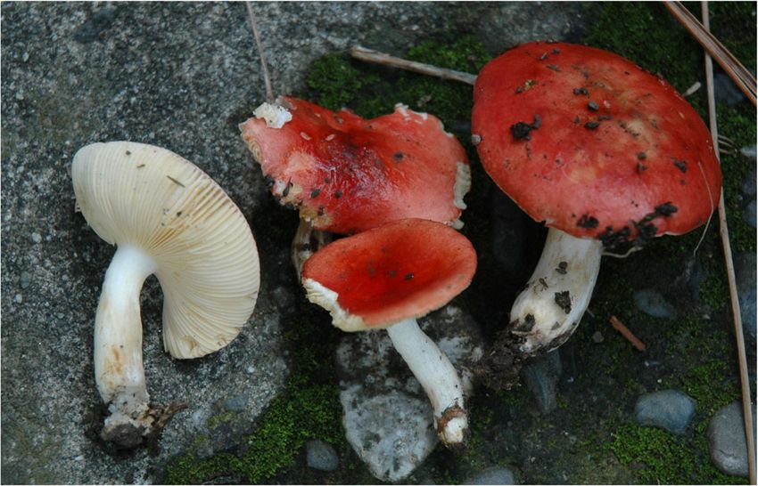
Caboň et al. IMA Fungus 2019, 10:5                                                                                 Page 2 of 16

species. More recently, there has been a considerable in-         contribute to the evolution of ECM fungi using the
crease of new Russula species described from Southeast            Russula globispora lineage. This lineage belongs to the
Asia. For example, 24 new Russula species have been               subsection Maculatinae and is morphologically defined
described from China in the period 2005–16 (Li et al.             by a slightly acrid taste of the flesh, yellow spore print,
2016), and an additional eight new species have been              yellow-brownish spots on the basidiomata and spores with
recognized there in 2017, which indicates an increasing           isolated large warts or spines (Adamčík & Jančovičová
annual rate (Das et al. 2017a, b). However, only two new          2013, Adamčík et al. 2016b). In the R. globispora lineage,
Russula taxa have so far been described from higher ele-          R. globispora and R. dryadicola are the only species re-
vations of the montane belt (2500–4000 m a.s.l.) of the           ported from boreal, arctic or alpine habitats of Europe
southeast Tibetan Plateau: R. wangii and R. amethystina           (Sarnari 1998). The aim of this study was to analyse phylo-
subsp. tengii (Li et al. 2016). Accordingly, in the area of       genetic relationships among collections and available
the southeast Himalayas, most taxa have been described            sequence data of the R. globispora lineage originating from
from lower elevations of up to 2300 m (Joshi et al. 2012),        coniferous subtropical, mixed boreal and alpine forests of
and only R. shingbaensis and R. thindii have been found           Europe, the southeast Himalayas, and North America.
in subalpine coniferous forests (elevations above 3000 m)
with Abies densa (Das et al. 2014).                               MATERIAL AND METHODS
  The montane belt of the Himalayan Mountains has                 Sampling
developed an autonomous flora differing from Eurasian             We analysed 10 collections of Russula globispora and R.
boreal taiga forests because of isolation by distance and         dryadicola from various habitats across Europe, including
climatic disjunctions (Miehe et al. 2015). The montane            temperate deciduous forests, boreal mixed forests and al-
coniferous forests that flank the Himalayas at their              pine habitats. Asian material from the south Himalayan
southeast to southwest edge are not connected to the              Mountains is represented by four morphologically similar
Siberian boreal taiga forests. The closest areas with a           collections from subtropical coniferous rainforests of Pinus
connection to Eurasian taiga forests are the Tian Shan            roxburghii in Pakistan, and four other similar collections
and Altai Mountains to the northwest and Inner                    from montane mixed forests of China associated with Abies
Mongolia to the northeast. Singer (1938) reported 33              spectabilis and Betula delavayi. Three representatives of
Russula taxa from the montane, subalpine to alpine belts          subsection Cupreinae, identified and sequenced by us, were
of the Altai and four Russula species from similar areas          used as the outgroup. This study is supplemented by se-
of Tian Shan. To his Asiatic collections from the area,           quences with high (99%) similarity retrieved from the Gen-
he assigned 21 European Russula names and described               bank (https://www.ncbi.nlm.nih.gov) and UNITE (https://
three new species, five new subspecies, four new varieties        unite.ut.ee) databases. All samples and sequences used are
and one new form. His alpine species R. oreina is consid-         listed in Additional file 1: Table S1.
ered a synonym of the European R. pascua (Knudsen &
Borgen 1992a) and his R. mesospora (associated with               Molecular analysis
Betula) a synonym of the European R. intermedia (Ruotsa-          Total genomic DNA was extracted from dried material
lainen & Vauras 1994). Russula citrinochlora, described           using a variety of protocols (Eberhardt 2012, Adamčík et
from the subalpine belt of the Altai Mountains, has also          al. 2016a). In addition to these protocols, the EZNA Fun-
been reported from Greenland (Knudsen & Borgen                    gal DNA Mini Kit (Omega) was used following the manu-
1992b) and Fennoscandia (Marstad 2004). Only a few Rus-           facturer’s recommendations, but with prolonged
sula species have been reported from Inner Mongolia (e.g.         incubation time of up to 1 h after addition of the
Tian et al. 2014) and only one, R. jilinensis (Li et al. 2012),   RNA-lytic enzyme. Three molecular markers were ampli-
has been described as new from the region.                        fied: (1) the internal transcribed spacer regions of riboso-
  The assembly of phylogenetic community structure and            mal DNA (ITS); (2) the partial mitochondrial small
species pool scaling of forest communities in East Asia           subunit of ribosomal DNA (mtSSU); and (3) the region
revealed the important role of intercontinental migration         between domains six and seven of the nuclear gene encod-
during the Neogene and Quaternary in the formation of             ing the second largest subunit of RNA polymerase II
species diversity in the area (Feng et al. 2015). Study of the    (rpb2). The ITS region was amplified using the primers
global biogeographic distribution of Alnus-associated             ITS1F–ITS4 (White et al. 1990, Gardes & Bruns 1993).
ECM fungi (Põlme et al. 2013) indicated co-dispersal of           The mtSSU region was amplified using the primer pair
hosts and their mycobionts, but at a regional scale, geo-         MS1 and MS2 (White et al. 1990). The rpb2 was amplified
graphic distance or disjunctions may also largely account         using the primers A-Russ-F–frpb2-7CR (Matheny 2005,
for the observed diversity (Bahram et al. 2012).                  Caboň et al. 2017). All three molecular markers were
  In this study we attempted to test the hypothesis that          amplified with Hot Start Firepol Polymerase (Solis Bio-
geographic distance, a host switch or a climatic disjunction      dyne, Tartu, Estonia) using cycling protocols according to
Caboň et al. IMA Fungus 2019, 10:5                                                                            Page 3 of 16

Caboň et al. (2017). The PCR products were purified           a warm aqueous KOH solution to dissolve the gelatinous
using Exo-Sap enzymes (Thermo Fisher Scientific, Wil-         matrix and improve tissue dissociation. All tissues were
mington, DE) or the Qiaquick PCR Purification Kit (Qia-       also examined in Cresyl blue to verify the presence of
gen, Hilden, Germany). Samples were sequenced by the          ortho- or metachromatic reactions as explained in Buyck
Seqme company (Dobříš, Czech Republic).                       (1989). The trama and cystidia were examined in a sulfo-
                                                              vanillin solution. Acidoresistant incrustations of primor-
Phylogenetic analysis                                         dial hyphae were stained with carbolfuchsin and observed
Raw sequences were edited in Geneious version R10             in distilled water after incubation for a few seconds in a
(Kearse et al. 2012). Intra-individual polymorphic sites      10% solution of HCl (cf. Romagnesi 1967). Spores were
having more than one signal were marked with NC-              scanned with an Artray Artcam 300MI camera and mea-
IUPAC ambiguity codes. All three single-locus datasets        sured by Quick Micro Photo version 2.1 software with an
were aligned by MAFFT version 7 using the strategy            accuracy of 0.1 μm. Spore measurements excluded orna-
E-INS-i (Katoh & Standley 2013), manually improved            mentation and their line drawings were made using
in Geneious version R10 (Kearse et al. 2012) and              enlarged, scanned pictures. The Q value indicates the
concatenated into one multi-locus dataset using Sea-          length/width ratio of the spores. Spore ornamentation
View version 4.5.1 (Gouy et al. 2010). The multi-locus        density was estimated following Adamčík & Marhold
dataset was analysed using two different methods:             (2000). Estimates of the density of cystidia estimates
Bayesian inference (BI) and the maximum likelihood            follow Buyck (1991). Statistics for the measurements of
(ML) approach. For the ML analysis, the concatenated          microscopic characteristics were based on 30 measure-
alignment was loaded as a PHYLIP file into RAxMLGUI           ments per specimen and are based on all examined mater-
v. 1.2 (Silvestro & Michalak 2012) and analysed as a          ial of the described species. The range of measured values
partitioned dataset under the GTR + I model with 1000         is expressed as the mean ± standard deviation; presented
bootstrap iterations. In addition, the GTR + G model          in parentheses are the 5th and 95th percentiles.
was used as recommended by Stamatakis (2008). Phylo-
genetic analyses generated using both models resulted         RESULTS
in trees with identical topology and very similar boot-       Phylogenetic analysis
strap support. Thus, only the GTR + G + I analysis is         In total, 54 sequences were newly generated and the
presented. For the BI analysis, the dataset was divided       final dataset was supplemented by 28 published se-
into six partitions: ITS, mtSSU, intronic region 7 of         quences, including 20 ITS sequences with high sequence
rpb2, and the 1st, 2nd and 3rd codon positions of rpb2.       similarity to Russula dryadicola retrieved from public
The best substitution model for each partition was            databases (Additional file 1: Table S1). They altogether
computed jointly in PartitionFinder v. 1.1.1 (Lanfear et      correspond to 42 collections in the tree (Fig. 1), and 38
al. 2012). The BI was computed independently twice in         of them are placed in the R. globispora clade with full
MrBayes version 3.2.6 (Ronquist et al. 2012) with four        support. Three collections of R. globispora from temper-
MCMC chains for 10,000,000 iterations until the stand-        ate deciduous forests are placed at the basal position of
ard deviation of split frequencies fell below the 0.01        the clade. Collections from all other habitats (coniferous
threshold. The convergence of runs was visually               and boreal forests or arctic-alpine habitats) are clustered
assessed using the Trace function in Tracer version 1.6       in a clade with moderate ML and full BI support (70/1).
(Rambaut et al. 2013).                                        This clade is composed of three well supported clades
                                                              with various branch length, each corresponding to sam-
Morphological analysis                                        ples from different habitats and geographic areas. Our
Micromorphological characteristics were observed using        collections from Pinus roxburghii mono-dominant for-
an Olympus CX-43 microscope with an oil-immersion             ests of the southwest Himalayan Mountains (Pakistan)
lens at a magnification of 1000×. All drawings of micro-      are grouped in a fully supported clade and below we de-
scopic structures, with the exception of spores, were         scribe them as a new species bearing the name R. abbot-
made with a ‘camera lucida’ using an Olympus U-DA             tabadensis. Another well supported species clade is
drawing attachment at a projection scale of 2000×. The        formed by collections from mixed montane forests of
contents of hymenial cystidia and pileocystidia were          the southeast Himalayas (China) and corresponds to the
illustrated as observed in Congo red preparations from        second new species, R. tengii, described below. The third
dried material, with the exception of some pileocystidia      clade groups samples that originated from a large geo-
for which the contents are indicated schematically (by plus   graphic area of Europe and Alaska, collected in boreal,
signs). Spores were observed on the lamellae stained with     arctic and alpine habitats. Alaskan samples retrieved from
Melzer’s reagent. All other microscopic observations were     the Genbank database received strong support (81/0.96),
made in ammoniacal Congo red after a short treatment in       but they are represented only by ITS sequences and differ
Caboň et al. IMA Fungus 2019, 10:5                                                                                                    Page 4 of 16

 Fig. 1 Phylogram generated by Maximum Likelihood (RAxML) analysis based on combined sequence data of ITS, mtSSU and rpb2 for five species
 of Russula subsect. Maculatinae and three outgroup sequences. Maximum likelihood bootstrap support values greater than 50% and Bayesian
 posterior probabilities greater or equal to 0.90 are indicated above or below the nodes. More details about all sequences used in this study are
 presented in Additional file 1: Table S1

from other samples within this clade of European ori-                        Under the microscope, the most important differences
gin only in 1–2 parsimony-informative positions. All                       were observed in the pileipellis. Russula abbottabadensis
European collections correspond morphologically and                        has long (on average longer than 29.5 μm), narrow (on
ecologically to the concept of R. dryadicola.                              average to 3 μm), cylindrical terminal cells of hyphae in
                                                                           the pileipellis near the pileus margin with usually
Morphological analysis                                                     branched subterminal cells and narrower (on average to
All four species in the Russula globispora clade are mor-                  5 μm), prevailingly 1–2-celled pileocystidia. Russula ten-
phologically defined by yellow-brownish spots on the sur-                  gii has shorter (on average to 29.5 μm) terminal cells of
face of the basidiomata, yellow spore print and relatively                 hyphae in the pileipellis near the pileus margin, less
large spores, which are characters that correspond to the                  branched subterminal cells, and wider (on average more
circumscription of R. subsect. Maculatinae. In addition,                   than 5 μm) and more septate (mainly 2- or more-celled)
all studied members of the R. globispora clade have large                  pileocystidia. Russula dryadicola is well characterized by
isolated spines on the spores and a weakly acrid to mild                   wider (on average more than 3.5 μm), frequently fusi-
taste that distinguishes them from other members of the                    form or lanceolate terminal cells of hyphae in the pilei-
subsection. In the field, R. abbottabadensis is easily recog-              pellis near the pileus margin that are often moniliform.
nized by the bright red colours on the pileus showing only                   There are no other species known from southeast
a little discolouration in spots near the centre, whereas the              Himalayas that remind morphologically or in sequence
other two species usually have dull pink to violet colours                 R. globispora lineage. However, according to our know-
and are strongly discoloured near the centre. The basidio-                 ledge (K. Wisitrassameewong, personal communication),
mata of R. dryadicola are more robust and larger.                          there are probably more undescribed members of the
Caboň et al. IMA Fungus 2019, 10:5 Page 5 of 16
lineage from South Korea and possibly other areas of slightly eccentric, cylindrical to fusiform, often curved,
Southeast Asia. Our detailed Russula descriptions based solid, rugulose; white, with early-developing rusty spots at
on robust statistical support of several measurements the base, when old slightly yellowing throughout. Context
from multiple collections will undoubtedly serve as a firm, softer in the stipe, white, slowly turning yellow-
good base for their morphological delimitation and sup- brownish when wounded. Smell rather strong, of hot
port the phylogeny based species hypothesis. bread, somewhat honey-like at the stipe base. Taste mild to
slightly acrid. Spore print yellow (IVa at the scale of
TAXONOMY Romagnesi 1967 according to Biscoletto & Ostellari 2005).
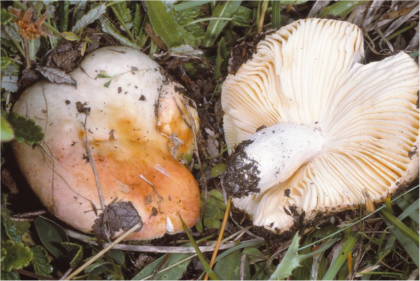
Russula dryadicola Singer ex R. Fellner & Landa, Bib-
lioth. Mycol. 150: 34 (1993); Figs. 2, 3, 4. Spores (7.9–)8.7–9.5–10.2(− 11) × (6.9–)7.6–8.2–8.8(− 9.5)
μm, subglobose to broadly ellipsoid, Q = (1.08–)1.12–1.16–
Type: Switzerland: Ticino, Olivone, Lucomagno, alt. 1.23(− 1.29), ornamentation of large, distant [(2–)3–5(− 6)
1950 m, among Dryas sp. and Juniperus on limestone, 15 in a 3 μm diam circle] amyloid warts, (0.7–)0.8–1(− 1.1)
August 1995, G. Lucchini (IB 1997/0750 – neotype, μm high; occasionally fused in pairs [0–1(− 2) fusions
designated by Sarnari 1998). in the circle], line connections very rare and short (0–1
in the circle), warts mainly isolated; suprahilar plage large,
Description: Basidiomata medium-sized but robust. Pi- amyloid. Basidia (41–)48.5–54–59(− 65) × (12.5–)13–13.9–
leus 40–130 mm diam, first hemispherical, soon appla- 15(− 16) μm, 4-spored, clavate, pedicellate; basidiola first
nate, with age slightly depressed; margin never striated cylindrical, then clavate, ca 4–12 μm wide. Subhyme-
or tuberculate; pileus cuticle thick, hardly separable, nium pseudoparenchymatic. Lamellar trama mainly
sometimes slightly rugulose, greasy when moist, then composed of large sphaerocytes. Pleurocystidia dispersed,
shiny; colour first uniformly whitish, soon becoming 450–700/mm2, (70–)75.5–86.2–97(− 130) × (9–)10.5–11.6–
salmon-orange near the margin, when mature some- 12.5(− 14.5) μm, fusiform or clavate, pedicellate, apically
times with copper-red, pink-reddish ‘vesca’, or even acute to subacute, without or with 5–10(− 24) μm long ap-
purplish red tints irregularly developing near the centre, pendage, thin-walled or occasionally with slightly thickened
under sun exposure usually remaining pale, butter yellow walls (to 0.75 μm thick), contents heteromorphous, banded,
to golden yellow, irregularly rusty yellow spotted. Lamellae slowly turning red-brown in sulfovanillin. Cheilocystidia
rather distant, ca 60 reaching the stipe, adnexed, often abundant, usually protrude more than half of their length
forming a collar near the stipe, broad, ventricose, occasion- above densely arranged marginal cells, smaller than
ally forked, first pale cream, when mature light yellow; edge pleurocystidia, (39–)46–55.5–65(− 85) × (5.5–)6.5–7.7–
even, concolourous. Stipe 40–70 × 15–25 mm, central or 9(− 10.5) μm, usually clavate, occasionally fusiform,
Fig. 2 Russula dryadicola (LIP PAM95082603), field appearance. Bar = 1 cm. Photo: P.-A. MCaboň et al. IMA Fungus 2019, 10:5 Page 6 of 16 Fig. 3 Russula dryadicola (IB 1997/0750, neotype). Microscopic features of the pileipellis. a. Pileocystidia near the pileus centre. b. Pileocystidia near the pileus margin. c. Hyphal terminations in the pileus centre. d. Hyphal terminations near the pileus margin. Contents of cystidia are as observed in Congo red. Bars = 10 μm. Drawings: S.A. & S.J pedicellate, apically mainly obtuse, contents usually horizontally oriented, 2.5–5(− 10) μm wide hyphae. banded and yellow-pigmented. Marginal cells smaller and Acid-resistant incrustations absent, but the contents of narrower than basidiola on gill sides, subcylindrical, flexu- pileocystidia stain red after carbol fuchsin treatment. ous, often also slightly moniliform, occasionally nodulous, Hyphal terminations near the pileus margin slender, dis- (14–)20–26.3–32.5(− 45) × (3–)4.5–5.3–6.5(− 7.5) μm, api- tinctly moniliform, often slightly flexuous, thin-walled, cally obtuse or subacute. Pileipellis orthochromatic in Cre- with terminal cells (12.5–)18–25.6–33(− 40) × (2.5–)3– syl blue, sharply delimited from the underlying 3.8–4.5(− 5) μm, mainly cylindrical, occasionally subulate sphaerocytes of the context, strongly gelatinized in all or lanceolate, apically usually constricted to ca 1.5– parts, 140–150 μm deep, vaguely divided in a 65–90 μm 2.5 μm, but not attenuated; subterminal cells branched or deep suprapellis of ascending or erect, but near the surface not, usually equally wide and long, often with lateral nod- often repent, relatively densely packed hyphae, gradually ules and rarely also with lateral branches. Hyphal termina- transitioning into a dense, 65–80 μm deep subpellis of tions near the pileus centre less moniliform and usually intricate, irregularly oriented, but near the trama cylindrical, with terminal cells (12–)18–25.3–33(−
Caboň et al. IMA Fungus 2019, 10:5                                                                                                        Page 7 of 16

 Fig. 4 Russula dryadicola (IB 1997/0750, neotype). Microscopic features of the hymenium. a. Basidia and basidiola. b. Marginal cells. c.
 Basidiospores in Melzer’s reagent. d. Cheilocystidia. e. Pleurocystidia. Contents of cystidia are represented as observed in Congo red. Bars = 10 μm,
 spores = 5 μm. Drawings: S.A. & S.J

40) × 2.5–3.4–4(− 5) μm, apically obtuse and rarely                          Additional material examined: Italy: Südtirol: Sexten,
constricted. Pileocystidia near the pileus margin nu-                        Fischleintal, near Zsigmondyhütte, alt. ca 2400 m, 5 Aug.
merous, 1–1.9–3 celled, thin-walled, terminal cells                          2006, U. Peintner & I. Göschl (IB 2002/0432). – Finland:
(19–)28–54–80(− 106) × (4–)4.5–5.7–6.5(− 7.5) μm, cy-                        Enontekion Lappi: Kilpisjärvi, Saana, near the biological
lindrical, obtuse, contents heteromorphous, granular or                      station, mountain birch forest, 16 Aug. 1990, J. Ruotsalai-
banded, often with yellow refringent pigments, hardly                        nen (TURA 151632); Etelä-Häme, Raikonkulma, Raikko, E
react in sulfovanillin, near the pileus centre with often                    of Kivijärvi lake, Kalkkimäki, herb rich forest with Picea,
clavate and wider terminal cells, (21–)25–36.1–                              Pinus, Betula, Salix caprea, Populus tremula on calcareous
47.5(− 58.5) × (4–)4.5–6.1–8(− 9) μm; subterminal cells                      soil, 22 Aug. 2003, J. Vauras JV20125 (TURA152390). –
usually equally large, occasionally longer or wider.                         France: Savoie: Bourg-Saint-Maurice, Arc 2000, vers col.
Cystidioid hyphae observed only in the upper part of the                     des Frettes, alpine pasture with Dryas octopetala, on dolo-
subpellis, absent in the pileus trama. Clamp connections                     mite, 25 Aug. 1998, P.-A. Moreau PAM98082511 (LIP);
absent in all parts.                                                         ibid., 29 Aug. 2000, P.-A. Moreau PAM00082907 (LIP);
Caboň et al. IMA Fungus 2019, 10:5 Page 8 of 16
Fig. 5 Russula tengii (HMAS262728, holotype), field appearance. Bar = 1 cm. Photo: G.-J.L.
ibid., 26 Aug. 1995, P.-A. Moreau PAM95082603 (LIP); light brownish vinaceous to light russet vinaceous, when
ibid., 4 Sept. 1994, P.-A. Moreau PAM94090402 (LIP). – old brick-red, and madder-brown when dry, near the pi-
Sweden: Uppsala, Nåsten, 23 Sept. 2004, A. Taylor leus centre mustard-yellow to aniline-yellow when old and
AT2004140 (UPS). dry, variegated with dark blackish red and dark yellow
spots. Lamellae relatively dense, 10–16 per cm near the
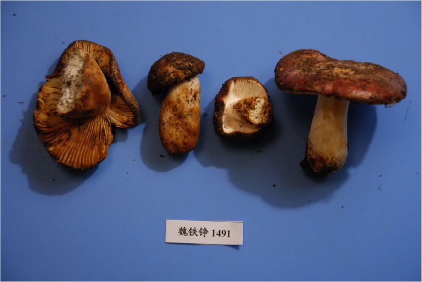
Russula tengii G.J. Li & H.A. Wen, sp. nov; Figs. 5, 6, pileus margin, adnate-emarginated, 4–6 mm broad, occa-
7. sionally forked near the pileus margin, rarely near the
stipe, slightly anastomosed, without lamellulae, first
MycoBank No.: MB823538. creamy to yellowish, when mature baryta yellow to buff
yellow, pale yellow-orange when dry; edge even, conco-
Etymology: Named in honour of Shu Chün Teng (1902– lourous. Stipe 35–47 × 12–15 mm, central or slightly ec-
70) for his contribution to mycology. centric, cylindrical, often swollen toward the base, solid
to hollow when mature, smooth; white, soon with a buff
Diagnosis: Basidiomata small to medium sized, thick- yellow tint, especially near the base. Context 3–7 mm
fleshed, similar to R. dryadicola in pileus colour and pres- thick at the attachment of lamellae to the stipe, com-
ence of yellow-brownish spots on surface of pileus and pact but fragile in gills, white, slightly turning pale
stipe, from which differs by mainly cylindrical, apically creamy or yellowish when wounded or old. Smell indis-
obtuse and not constricted, in average up to 3.5 μm wide tinct. Taste mild. Spore print yellow (IVc–IVd accord-
hyphal terminations in pileipellis near the pileus margin ing to Romagnesi 1967).
and pileocystidia that are mainly two and more celled.
Spores (8.1–)8.9–9.7–10.5(− 11.3) × (6.9–)7.5–8.2–8.9(− 9.5)
Type: China: Xizang Autonomous Region: Riwoqe μm, broadly ellipsoid, Q = (1.14–)1.15–1.18–1.21(− 1.24), or-
County, 31°12′ N 96°36′ E, alt. 3741 m, 24 July 2010, namentation of large, moderately distant [(3–)4–6(− 7)
T.Z. Wei 1491 (HMAS262728 – holotype). in a 3 μm diam circle] amyloid warts, (0.6–)0.8–1(− 1.2)
μm high; occasionally fused in pairs [0–2(− 3) fusions
Description: Basidiomata small to medium-sized, robust. in the circle], line connections very rare and short [0–2(− 3)
Pileus 28–78 mm diam, first hemispherical, then convex in the circle], warts mainly isolated; suprahilar plage large,
to applanate, sometimes slightly depressed; margin first amyloid. Basidia (51–)54–58.1–62(− 66) × (13–)13.5–14.7–
slightly deflexed, when mature straight to slightly inflexed, 15.5(− 16.5) μm, 4-spored, clavate, pedicellate; basidiola first
sometimes undulate, indistinctly striate; pileus cuticle cylindrical, then clavate, ca 5–13 μm wide. Subhymenium
slightly viscous when wet; colour near the pileus margin pseudoparenchymatic. Lamellar trama mainly composedCaboň et al. IMA Fungus 2019, 10:5 Page 9 of 16 Fig. 6 Russula tengii (HMAS262728, holotype). Microscopic features of the pileipellis. a. Pileocystidia near the pileus centre. b. Pileocystidia near the pileus margin. c. Hyphal terminations in the pileus centre. d. Hyphal terminations near the pileus margin. Contents of cystidia are represented as observed in Congo red. Bars = 10 μm. Drawings: S.A. & S.J of large sphaerocytes. Pleurocystidia dispersed to moder- narrower than basidiola on gill sides, subcylindrical, flexu- ately numerous, ca 600–800/mm2, (73–)79.5–88.7–98(− ous, often also slightly moniliform, occasionally nodulous, 112) × (9–)10–11.1–12.5(− 14.5) μm, fusiform or clavate, (14–)22–29.2–36.5(− 41) × (2.5–)3–3.9–4.5(− 5.5) μm, api- usually pedicellate, apically subacute to obtuse, mainly with cally obtuse or slightly constricted. Pileipellis ortho- 1–8(− 13) μm long appendage, usually with slightly thick- chromatic in Cresyl blue, sharply delimited from the ened walls (ca 0.5 μm thick), contents heteromorphous, underlying sphaerocytes of the context, strongly gela- banded, slowly turning red-brown in sulfovanillin. Cheilo- tinized in all parts, 160–200 μm deep, vaguely divided cystidia abundant, (39–)44.5–54.5–65(− 81) × (5–)6.5– in a 55–80 μm deep suprapellis of erect but often irregu- 7.4–8.5(− 9.5) μm, usually clavate, occasionally fusiform, larly oriented, near the surface loosely arranged hyphae, pedicellate, apically mainly obtuse, occasionally with a gradually transitioning into a dense, 100–130 μm deep short (2–6 μm) appendage, few with slightly thickened subpellis of intricate, irregularly oriented, but near the walls (to 0.5 μm thick), contents usually banded, often dis- trama horizontally oriented, (2–)2.5–5(− 6.5) μm wide persed and yellow-pigmented. Marginal cells smaller and hyphae. Acid-resistant incrustations absent, but the
Caboň et al. IMA Fungus 2019, 10:5                                                                                                     Page 10 of 16

 Fig. 7 Russula tengii (HMAS262728, holotype). Microscopic features of the hymenium. a. Basidia and basidiola. b. Marginal cells. c. Basidiospores
 in Melzer’s reagent. d. Cheilocystidia. e. Pleurocystidia. Contents of cystidia are represented as observed in Congo red; crosses schematically
 indicate the contents of some cystidia. Bars = 10 μm, spores = 5 μm. Drawings: S.A. & S.J

contents of pileocystidia stain red after carbol fuchsin                    5.4–6(− 7) μm, cylindrical, obtuse or apically slightly con-
treatment. Hyphal terminations near the pileus margin                       stricted, contents heteromorphous, granular or banded,
slender, distinctly moniliform, thin-walled, with terminal                  often with yellow refringent pigments, in sulfovanillin
cells (15–)21–26.8–32.5(− 39) × 2.5–3.1–3.5(− 4) μm,                        turning slowly to grey-brown; near the pileus centre with
mainly cylindrical, occasionally subulate, rarely lanceolate,               often clavate and wider terminal cells, (15–)18–36.3–54(−
apically usually obtuse, not frequently constricted; subter-                71) × (4–)5–6.4–8(− 9) μm, apically obtuse to rounded;
minal cells branched or not, usually equally wide and long,                 subterminal cells usually equally large, occasionally longer,
occasionally with lateral nodules and rarely also with lat-                 rarely wider. Cystidioid hyphae with heteromorphous
eral branches. Hyphal terminations near the pileus centre                   contents and yellowish pigments present in the subpellis,
similar in shape but occasionally also distinctly clavate,                  dispersed or absent in the pileus trama. Clamp connec-
with terminal cells (15–)19–24–29(− 39) × 2.5–3.4–4(− 5)                    tions absent in all parts.
μm, apically obtuse and rarely constricted. Pileocystidia
near the pileus margin numerous, 1–2.5–4(− 5) celled,                       Additional material examined: China: Xizang Autono-
thin-walled, terminal cells (18–)24.5–42.7–61(− 79) × 4.5–                  mous Region: Maizhokunggar County, Riduo Township,
Caboň et al. IMA Fungus 2019, 10:5                                                                                             Page 11 of 16

 Fig. 8 Russula abbottabadensis (FH00304589, holotype), field appearance. Bar = 1 cm. Photo: M.S.

29°38′ N 92°26′ E, Abies-Betula forest, 6 Aug. 2012, W.L.                   red, vivid red or vivid reddish orange, rarely with light
Lu 100 (HMAS244255); ibid., Gesang village, 29°48′ N,                       orange-yellow spots near the centre. Lamellae adnexed or
92°39′ E, Abies-Betula forest, 16 Aug. 2012, G.J. Li, D.                    adnate-emarginate, brittle, without lamellulae; when
Zhao & W. Li 12,163 (HMAS251829); ibid., 16 Aug. 2012,                      young white to pale yellow, when mature yellow; edge
G.J. Li, D. Zhao & S. Qi 12,358 (HMAS264837).                               even, concolourus. Stipe 16–35 × 7–12 mm, central, cla-
                                                                            vate or cylindrical, longitudinally rugulose, not pruinose,
Russula abbottabadensis Saba & Adamčík, sp. nov;                            white, occasionally with pink flush, with yellowish brown
Figs. 8, 9, 10.                                                             or rusty spots, solid. Context compact, white, slowly turn-
                                                                            ing yellow-brownish when wounded or old. Smell indis-
MycoBank No.: MB823541;                                                     tinct. Taste mild to slightly acrid. Spore print yellow.

Etymology: referring to the district of Abbottabad, the                     Spores (8.3–)9.1–9.9–10.6(− 11.4) × (6.9–)7.6–8.3–9(− 9.7)
area of origin of the samples under study.                                  μm, broadly ellipsoid, Q = (1.14–)1.17–1.19–1.21(− 1.23), or-
                                                                            namentation of large, moderately distant [3–6(− 7) in a
Diagnosis: Basidiomata small to medium sized, pileus red,                   3 μm diam circle] amyloid warts, (0.8–)0.9–1.2(− 1.5) μm
stipe with yellowish brown rusty spots, taste mild to                       high; occasionally fused in pairs or short chains [0–2(− 3)
slightly acrid, spore print yellow, spores large, similar as in             fusions in the circle], line connections very rare and short
R. globispora but with occasionally fused spines, Hyphal                    [0–1(− 2) in the circle], warts mainly isolated; suprahi-
terminations in pileipellis near the pileus margin with nar-                lar plage large, amyloid. Basidia (35–)36.5–39–41(−
row (in average up to 3 μm), often attenuated terminal                      42) × (12–)12.5–13.9–15(− 15.5) μm, 4-spored, broadly
cells and mainly branched subterminal cells, pileocystidia                  clavate, pedicellate; basidiola first cylindrical, then clavate,
narrow (in average up to 5 μm) and usually 1–2 celled.                      ca 4–12 μm wide. Subhymenium pseudoparenchymatic.
                                                                            Lamellar trama mainly composed of large sphaerocytes.
Type: Pakistan: Khyber Pakhtoon Khaw: Abbottabad,                           Pleurocystidia moderately numerous, ca 950–1150/mm2,
Shimla, 34°10′27.62“ N, 73°12’18.13” E, alt. 1297 m,                        (44–)52–60.6–69(− 89) × (9.5–)11–12.3–13.5(− 14.5) μm,
under Pinus roxburghii, 14 Sept. 2012, M. Saba & A.N.                       fusiform or clavate, pedicellate, apically usually subacute,
Khalid MSM#0072 (FH00304589 – holotype).                                    often with short 1–2(− 4) μm long appendage, thin-walled,
                                                                            contents heteromorphous-banded, turning dark brown-
Description: Basidiomata small to medium-sized, rela-                       black in sulfovanillin. Cheilocystidia abundant, (32–)44.5–
tively thin-fleshed. Pileus 13–73 mm diam, first convex,                    54.1–63.5(− 82) × (7–)8–9.5–11(− 13) μm, clavate, pedicel-
then plano-convex to applanate, sometimes slightly                          late, apically mainly obtuse-rounded, contents banded or
centrally depressed; margin first deflexed, when ma-                        granulose. Marginal cells smaller, but usually similar to
ture usually inflexed, entire, hardly striated; pileus                      basidiola on gill sides, subcylindrical or clavate, (8–)10–
cuticle matt when dry, sometimes viscid and shiny                           16.6–23(− 35) × (3.5–)4–5.4–6.5(− 9) μm, apically obtuse.
near the centre; colour moderate red, strong red, deep                      Pileipellis orthochromatic in Cresyl blue, not sharply
Caboň et al. IMA Fungus 2019, 10:5 Page 12 of 16 Fig. 9 Russula abbottabadensis (FH00304589, holotype). Microscopic features of the pileipellis. a. Pileocystidia near the pileus centre. b. Pileocystidia near the pileus margin. c. Hyphal terminations in the pileus centre. d. Hyphal terminations near the pileus margin. Contents of cystidia are represented as observed in Congo red. Bars = 10 μm. Drawings: S.A. & S.J delimited from the underlying sphaerocytes of the context, constricted or attenuated, less frequently obtuse; subter- strongly gelatinized in all parts, 165–225 μm deep, minal cells mainly branched, often flexuous and intricate. vaguely divided in 60–75 μm deep suprapellis of erect, Hyphal terminations near the pileus centre similar, with densely arranged hyphae, gradually transitioning into a terminal cells (26–)29.5–38–46.5(− 55) × (2–)2.5–2.8–3(− 100–120 μm deep subpellis of intricate, relatively 3.5) μm, apically usually constricted or attenuated. Pileocys- loose, irregularly oriented, but near the trama horizon- tidia near the pileus margin numerous, 1–1.6–2(− 3) tally oriented and dense, 1.5–3 μm wide hyphae. celled, thin-walled, narrow, terminal cells (16–)22–45– Acid-resistant incrustations absent, but contents of 68(− 91) × (3–)3.5–4.6–5.5(− 6) μm, cylindrical, apically pileocystidia stain red after carbol fuchsin treatment. obtuse, contents abundant heteromorphous, banded, Hyphal terminations near the pileus margin slender, turning black-brown in sulfovanillin; near the pileus often slightly moniliform or flexuous, thin-walled, with ter- centre with often shorter terminal cells, (12–)14–29.1– minal cells (18–)25.5–32.9–40.5(− 50) × (2–)2.5–2.9–3.5(− 44(− 70) × (3.5–)4–4.8–5.5(− 6) μm; subterminal cells usu- 4) μm, cylindrical or subulate, apically frequently ally equally wide but often distinctly longer. Cystidioid
Caboň et al. IMA Fungus 2019, 10:5                                                                                                       Page 13 of 16

 Fig. 10 Russula abbottabadensis (FH00304589, holotype). Microscopic features of the hymenium. a. Basidia and basidiola. b. Marginal cells. c.
 Basidiospores in Melzer’s reagent. d. Cheilocystidia. e. Pleurocystidia. Contents of cystidia are represented as observed in Congo red. Bars = 10 μm,
 spores = 5 μm. Drawings: S.A. & S.J

hyphae present only in the upper part of the subpellis, ab-                  DISCUSSION
sent in the pileus trama. Clamp connections absent in all                    This study confirms that Russula dryadicola is a well-de-
parts.                                                                       fined species in accordance with Fellner & Landa (1993),
                                                                             contrary to some previous studies that treat it as an infra-
Additional material examined: Pakistan: Khyber Pakhtoon                      specific taxon of R. maculata (Singer 1926, Knudsen & Bor-
Khaw: Abbottabad, Shimla, 34°10′27.62“ N 73°12’18.13” E,                     gen 1992b). The species was first reported as R. maculata
alt. 1297 m, under Pinus roxburghii, 14 Sept. 2012, M. Saba                  subsp. alpina from the Alps (Singer 1925, 1926) and con-
& A.N. Khalid MSM#0073 (LAH310071); ibid., 4 Aug. 2014,                      sidered an alpine-arctic element distributed in Greenland,
M. Saba & A.N. Khalid MSM#00151 (LAH310099); Man-                            Scandinavia, mountains of Central Europe and the Ural
sehra Batrasi, 34°23′42.94“ N 73°18’53.03” E, alt. 1113 m,                   Mts (Kühner 1975, Knudsen & Borgen 1992b). The
under Pinus roxburghii, 3 Aug. 2014, M. Saba & A.N. Kha-                     related European species Russula globispora differs in
lid MSM#0078 (FH00304558).                                                   having larger spores and, based on this, collections
Caboň et al. IMA Fungus 2019, 10:5 Page 14 of 16 from mixed boreal forests (mainly with Picea and Russula tengii is represented only by collections Betula) in southern Finland were reported by J. Ruot- from mixed subalpine forests of the southwest Hima- salainen and J. Vauras under the name R. globispora layan Mountains in China. This type of habitat falls (Sarnari 1998). We have studied seven collections sent within the known ecological amplitude of R. dryadi- by J. Vauras from similar boreal habitats in Finland, cola. The existence of allopatric boreal-arctic species and they all morphologically correspond to R. dryadi- within the R. globispora clade suggests that glaciation cola. In our phylogenetic tree, boreal collections are events and geographic and climate disjunctions were represented by TURA 152390 and TU 101835, and the evolutionary drivers of speciation in the group on they are clustered with other alpine and arctic col- the Eurasian scale. It is possible that the common an- lections of R. dryadicola. This species is an example cestor of R. dryadicola and R. tengii migrated via of alpine-arctic habitats being incorrectly applied for available corridors for movement to newly emerging species delimitation and identification. Similarly, R. environments during climatic changes in the Quater- nuoljae, previously described and recorded from al- nary (Donoghue 2008), but we do not know the ori- pine and arctic habitats, was recently reported also ginal area of this ancestor species. from boreal forests, and R. subrubens, previously re- The collections of R. abbottabadensis under study ported only from lowlands of the temperate belt vege- originate from subtropical coniferous forests of Pinus tated by willows, was synonymized with the alpine-arctic roxburghii in northeast Pakistan at the foothills of the R. chamitae (Adamčík et al. 2016a). As new morpho- Himalayas. Coniferous trees seem to be obvious hosts logical characters of R. dryadicola, differentiating it of this species, and they possibly also host R. dryadi- from R. globispora (the latter species is re-described cola and R. tengii, which grow in mixed boreal for- in Adamčík & Jančovičová 2013), we introduce here ests. The long branch of this species and lack of shorter (on average to 33 μm long) and often fusiform support for R. globispora suggest missing data on terminal cells of hyphae in the pileipellis near the pi- possibly unrepresented species of the R. globispora leus margin, narrower pileocystidia (on average to 6.5 lineage from other environments and regions of wide; Fig. 3) and narrower pleurocystidia (on average Eurasia. It is also possible that gene flow and species to 12.5 μm wide; Fig. 4). Alaskan samples from boreal migrations between Southeast Asia and Europe were forests have nearly identical ITS sequences and any multidirectional and that R. abbottabadensis may decision about their taxonomic status requires sup- represent another lineage of so far unknown origin. port of more DNA loci or other molecular support. Both Asian species described here represent, to our There are also two ITS sequences from environmental knowledge, the first reports of the R. globispora samples associated with the alpine sedge Kobresia lineage from Southeast Asia and we hope that this myosuroides; these sequences are of unspecified origin study will boost more studies from different parts of and have apparently been sampled for the Niwot the continent to answer questions on origin and spe- Ridge project (http://niwot.colorado.edu) dealing with ciation pattern of this Russula group. research of permanent plots in the Colorado alpine Looney et al. (2016) suggested that host switching ecosystem in the Rocky Mountains (USA). Neverthe- plays major role for species diversification of the less, this affinity of North American and European genus Russula especially at the rank of clades. How- collections suggests a wide distribution of R. dryadi- ever, the speciation mechanism of Russulaceae and cola in Eurasian taiga and tundra habitats, as demon- even ECM fungi in general is not well understood es- strated by Geml et al. (2012). North American ITS pecially within closely related species lineages. This sequences from environmental DNA samples, placed study suggests that host switch did not contribute to in this study in the R. globispora lineage, are the first speciation of studied species of R. globispora lineage reports of the group from America, but not a single and more probable drivers in the evolution of R. ten- basidiome has been collected in that region to date. gii and R. abbottabadensis were climate disjunctions The BLAST result of the ex-holotype ITS sequence and adaptations. of R. tengii shows 99% identity (under 100% query coverage) with the sequence of the R. dryadicola col- CONCLUSIONS lection TURA 151632. This is an example of high We recognised three species of R. globispora lineage similarity in the ITS region within the R. globispora that either grow in mixed forests in cold climate clade. However, all three species recognized in our (boreal, montane, arctic or alpine) or are associated multi-locus phylogenetic analysis (Fig. 1) received with conifers in mountains. Russula dryadicola is good support (BS ≥ 65, PP ≥ 0.98). This is analogous widely distributed in alpine, arctic and boreal areas to the high ITS sequence similarity within the R. of Eurasia and Northern America, although North clavipes species complex (Adamčík et al. 2016a). American collections show little genetic divergence.
Caboň et al. IMA Fungus 2019, 10:5                                                                                                                  Page 15 of 16

This species proved the hypothesis of widely                                   Publisher’s Note
distributed ECM species in hemiboreal areas (Geml                              Springer Nature remains neutral with regard to jurisdictional claims in
                                                                               published maps and institutional affiliations.
et al. 2012). Collections from south Himalayas
represent two species distinct by its morphology,                              Author details
                                                                               1
genetics, climate and ecological preferences; they are                          Department of Cryptogams, Institute of Botany, Plant Science and
                                                                               Biodiversity Centre, Slovak Academy of Sciences, Dúbravská cesta 9, SK-845
described here as R. abbottabadensis and R. tengii.                            23 Bratislava, Slovakia. 2State Key Laboratory of Mycology, Institute of
The results suggest that climatic disjunction and                              Microbiology, Chinese Academy of Sciences, No 3 1st Beichen West Road,
isolation by distance were drivers of speciation                               Chaoyang District, Beijing 100101, China. 3Department of Plant Sciences,
                                                                               Quaid-i-Azam University, Islamabad 45320, Pakistan. 4Department of Botany,
within the lineage. The host range and distribution                            University of the Punjab, Quaid-e-Azam Campus, Lahore 54590, Pakistan.
limits of the studied species are not sufficiently                             5
                                                                                Laboratory of Fungal Genetics and Metabolism, Institute of Microbiology,
known yet and the research in this area may con-                               Czech Academy of Sciences, Vídeňská 1083, CZ-142 20 Praha, Czech
                                                                               Republic. 6Department of Botany, Faculty of Natural Sciences, Comenius
tribute to understanding of evolutionary processes of                          University in Bratislava, Révová 39, SK-811 02 Bratislava, Slovakia. 7Laboratoire
ECM fungi.                                                                     IMPECS, Fac. Pharma. Lille, Université de Lille, F-59000 Lille, France. 8Farlow
                                                                               Reference Library and Herbarium of Cryptogamic Botany, Harvard University,
                                                                               Cambridge, MA 02138, USA.
Additional file
                                                                               Received: 30 April 2019 Accepted: 1 May 2019
 Additional file 1: Table S1. List of samples with collection details and
 GenBank numbers of corresponding DNA sequences. Sequences starting
 with MG are published first in this study. (DOCX 33 kb)                       References
                                                                               Adamčík S, Caboň M, Eberhardt U, Saba M, Hampe F, et al. A (2016b) a molecular
                                                                                    analysis reveals hidden species diversity within the current concept of
Ethics approval and consent to participate
                                                                                    Russula maculata (Russulaceae, Basidiomycota). Phytotaxa 270: 71–88
Not applicable.
                                                                               Adamčík S, Jančovičová S (2013) Type studies in Russula subsection Maculatinae
                                                                                    and affiliated taxa: four species as interpreted by Henri Romagnesi. Sydowia
Adherence to national and international regulations                                 65:201–222
All specimens analysed in this study have been collected with respect to the   Adamčík S, Marhold K (2000) Taxonomy of the Russula xerampelina group. I.
laws of specimen’s country of origin and they are deposited in public               Morphometric study of the Russula xerampelina group in Slovakia.
herbaria. The authors declare that they have no conflict with Nagoya                Mycotaxon 76:463–479
Protocol compliances.                                                          Adamčík S, Slovák M, Eberhardt U, Ronikier A, Jairus T et al (2016a) Molecular
                                                                                    inference, multivariate morphometrics and ecological assessment are applied
Consent for publication                                                             in concert to delimit species in the Russula clavipes complex. Mycologia 108:
Not applicable.                                                                     716–730
                                                                               Bahram M, Põlme S, Koljalg U, Zarre S, Tedersoo L (2012) Regional and local
Availability of data and materials                                                  patterns of ectomycorrhizal fungal diversity and community structure along
The sequences generated and/or analysed during the current study are                an altitudinal gradient in the Hyrcanian forests of northern Iran. New
available in the GenBank repository, [https://www.ncbi.nlm.nih.gov/genbank/         Phytologist 193:465–473
]. All analysed specimens are deposited in public herbaria indicated in        Bazzicalupo AL, Buyck B, Saar I, Vauras J, Carmean D, Berbee ML (2017) Troubles
Additional file 1: Table S1.                                                        with mycorrhizal mushroom identification where morphological
                                                                                    differentiation lags behind barcode sequence divergence. Taxon 66:791–810
Competing interests                                                            Biscoletto A, Ostellari C (2005) Russula dryadicola (Singer) Fellner & Landa, primo
The authors declare that they have no competing interests.                          ritrovamento in Italia. Rivista di Mycologia 4:301–307
                                                                               Buyck B (1989) Valeur taxonomique du bleu de crésyl pour le genre Russula.
                                                                                    Bulletin Trimestriel de la Societé Mycologique de France 105:1–6
Funding
                                                                               Buyck B (1991) The study of microscopic features in Russula 2. Sterile cells of the
The majority of the study has been funded by the Slovak grant APVV 15–
                                                                                    hymenium. Russulales News 1:62–85
0210. The field work and molecular analyses of M.S., A.N.K. and D.H.P. were
                                                                               Caboň M, Eberhardt U, Looney BP, Hampe F, Kolařík M et al (2017) New insights
supported by a project under Phase II, Batch I, Indigenous PhD fellowships
                                                                                    in Russula subsect. Rubrinae: phylogeny and the quest for synapomorphic
programme for 5000 scholars financed by the Higher Education Commission
                                                                                    characters. Mycological Progress 16:877–892
(HEC), Islamabad, Pakistan, and through the International Research Support
                                                                               Cao Y, Zhang Y, Yu Z, Mi F, Liu C et al (2013) Structure, gene flow, and
Initiative Program (IRSIP).
                                                                                    recombination among geographic populations of a Russula virescens ally
                                                                                    from southwestern China. PLoS One 8:e73174
Acknowledgements                                                               Das K, Dowie NJ, Li GJ, Miller SL (2014) Two new species of Russula (Russulales)
Gareth Griffith is acknowledged for his comments to final version of the            from India. Mycosphere 5:612–622
manuscript. We thank the reviewers of this manuscript for important            Das K, Ghosh A, Bhatt RP, Chakraborty D, Hofstetter V, Buyck B (2017a) Fungal
remarks.                                                                            biodiversity profiles 41–50. Cryptogamie, Mycologie 38:527–547
                                                                               Das K, Ghosh A, Chakraborty DF, Li J, Qiu L et al (2017b) Fungal biodiversity
Authors’ contributions                                                              profiles 31–40. Cryptogamie, Mycologie. Mycologie 38:353–406
SA designed the study, the main conceptual ideas and wrote the paper with      Donoghue MJ (2008) A phylogenetic perspective on the distribution of plant diversity.
support from MC. MC designed molecular part of the study, performed                 Proceedings of the National Academy of Sciences, USA 105(Suppl. 1):11549–11555
molecular and phylogenetic analyses. Both SA and MC contributed to the         Eberhardt U (2012) Methods for DNA barcoding of fungi. In: Kress WJ, Erickson DL (eds)
data interpretations. SJ prepared line drawings, both with SA performed             DNA barcodes methods and protocols. Humana Press, New York, pp 183–205
morphological measurements. GJL, MS, ANK, PAM, HAW, DHP contributed            Fellner R, Landa J (1993) Some species of Cortinariaceae and Russulaceae in the
with collecting the samples, macromorphological descriptions and provided           alpine belt of the Belaer Tatras. Bibliotheca Mycologica 150:33–37
the study with sequences of extra-european species. MK provided critical       Feng G, Mi X, Eiserhardt WL, Jin G, Sang W et al (2015) Assembly of forest
feedback to the study design and paper. All authors revised and approved            communities across East Asia insights from phylogenetic community
the final manuscript.                                                               structure and species pool scaling. Scientific Reports 5:9337
Caboň et al. IMA Fungus 2019, 10:5 Page 16 of 16
Gardes M, Bruns TD (1993) ITS primers with enhanced specificity for Singer R (1938) Contribution a l’étude des Russules (1). 3. Quelques Russules
basidiomycetes-application to the identification of mycorrhizae and rusts. américaines et asiatiques. Bulletin de la Société Mycologique de France Fr 54:
Molecular Ecology 2:113–118 132–177
Geml J, Timling I, Robinson CH, Lennon N, Nusbaum HC et al (2012) An arctic Stamatakis A (2008) The RAxML 7.0.4 manual. https://web.natur.cuni.cz/~vlada/
community of symbiotic fungi assembled by long-distance dispersers: moltax/RAxML-Manual.7.0.4.pdf
phylogenetic diversity of ectomycorrhizal basidiomycetes in Svalbard based Tian H, Liu T, Lian J, Li G, Ba T (2014) Identification and classification of four
on soil and sporocarp DNA. Journal of Biogeography 39:74–88 Russula species from Inner Mongolia based on morphology and ITS
Gouy M, Guindon S, Gascuel O (2010) SeaView version 4: a multiplatform sequencing. Acta Edulis Fungi 4:646
graphical user interface for sequence alignment and phylogenetic tree White TJ, Bruns T, Lee S, Taylor J (1990) Amplification and direct sequencing of
building. Molecular Biology and Evolution 27:221–224 fungal ribosomal RNA genes for phylogenetics. In: Innis MA, Gelfand DH,
Hongo T (1960) The Agaricales of Japan I-3. Russulaceae. Acta Phytotaxonomica Sninsky JJ, White TJ (eds) PCR Protocols: a guide to methods and
Geobotanica 18:129–146 applications. Academic Press, San Diego, pp 315–322
Joshi S, Bhatt RP, Stephenson SL (2012) The current status of the family
Russulaceae in the Uttarakhand Himalaya, India. Mycosphere 3:486–501
Katoh K, Standley DM (2013) MAFFT multiple sequence alignment software,
version 7: improvements in performance and usability. Molecular Biology and
Evolution 30:772–780
Kearse M, Moir R, Wilson A, Stones-Havas S, Cheung M et al (2012) Geneious
basic: an integrated and extendable desktop software platform for the
organization and analysis of sequence data. Bioinformatics 28:1647–1649
Knudsen H, Borgen T (1992a) New and rare taxa of Russula from Greenland.
Persoonia 14:509–517
Knudsen H, Borgen T (1992b) Russulaceae in Greenland. In: Arctic and Alpine
Mycology (Laursen GA, Ammirati JF, eds) 1: 216–244. Seattle: University of
Washington Press
Kühner R (1975) Agaricales de la zone Alpine. Genre Russula Pers, ex S. F. Gray.
Bulletin Trimestriel de la Societé Mycologique de France 91:313–390
Lanfear R, Calcott B, Ho SY, Guindon S, Lanfear R et al (2012) PartitionFinder:
combined selection of partitioning schemes and substitution models for
phylogenetic analyses. Molecular Biology and Evolution 29:1695–1701
Li GJ, Hyde KD, Zhao RL, Hongsanan S, Abdel-Aziz FA et al (2016) Fungal
diversity notes 253–366: taxonomic and phylogenetic contributions to fungal
taxa. Fungal Diversity 78:1–237
Li GJ, Li SF, Liu XZ, Wen HA (2012) Russula jilinensis sp. nov. (Russulaceae) from
Northeast China. Mycotaxon 120:49–58
Looney BP, Meidl P, Piatek MJ, Miettinen O, Martin FM et al (2018) Russulaceae: a
new genomic dataset to study ecosystem function and evolutionary
diversification of ectomycorrhizal fungi with their tree associates. New
Phytologist 218:54–65
Looney BP, Ryberg M, Hampe F, Sánchez-García M, Matheny PB (2016) Into and
out of the tropics: global diversification patterns in a hyperdiverse clade of
ectomycorrhizal fungi. Molecular Ecology 25:630–647
Marstad P (2004) Russula in the Nordic countries. Tønsberg: P Marstad
Matheny PB (2005) Improving phylogenetic inference of mushrooms with RPB1
and RPB2 nucleotide sequences (Inocybe; Agaricales). Molecular Phylogenetics
and Evolution 35:1–20
Miehe G, Miehe S, Böhner J, Bäumler R, Ghimire SK et al (2015) Vegetation
ecology. In: Miehe G, Pendry CA, Chaudhary R (eds) An Introduction to the
natural history, ecology and human environment of the Himalayas Nepal.
Royal Botanic Garden, Edinburgh, pp 385–472
Põlme S, Bahram M, Yamanaka T, Nara K, Dai YC et al (2013) Biogeography of
ectomycorrhizal fungi associated with alders (Alnus spp.) in relation to biotic
and abiotic variables at the global scale. New Phytologist 198:1239–1249
Rambaut A, Suchard MA, Xie D, Drummond AJ (2013) Tracer. Version 1.6. http://
beast.bio.ed.ac.uk/software/tracer/
Romagnesi H (1967) Les Russules d’Europe et d'Afrique du Nord. Bordas, Paris
Ronquist F, Teslenko M, van der Mark P, Avres DL, Darling A et al (2012) MrBayes
3.2: efficient Bayesian phylogenetic inference and model choice, across a
large model space. Systematic Biology 61:539–542
Ruotsalainen J, Vauras J (1994) Novelties in Russula : R. olivobrunnea, R. intermedia
and R. groenlandica. Karstenia 34:21–34
Ryberg M (2015) Molecular operational taxonomic units as approximations of
species in the light of evolutionary models and empirical data from Fungi.
Molecular Ecology 24:5770–5777
Sarnari M (1998) Monographia Illustrata del Genere Russula in Europa, vol 1.
Associazione Micologica Bresadola, Trento
Silvestro D, Michalak I (2012) raxmlGUI: a graphical front-end for RAxML.
Organismal Diversity and Evolution 12:335–337
Singer R (1925) Zur Russula-Forschung. Zeitschrift für Pilzkunde 5:73–80
Singer R (1926) Monographie der Gattung Russula. Hedwigia 66:153–260You can also read