RESEARCH PROJECTS 2021 - (HONOURS, MASTERS AND PHD) DEPARTMENT OF OPTOMETRY AND VISION SCIENCES - MELBOURNE SCHOOL OF HEALTH SCIENCES
←
→
Page content transcription
If your browser does not render page correctly, please read the page content below
Department of Optometry
                             and Vision Sciences

Research Projects
2021
(Honours, Masters and PhD)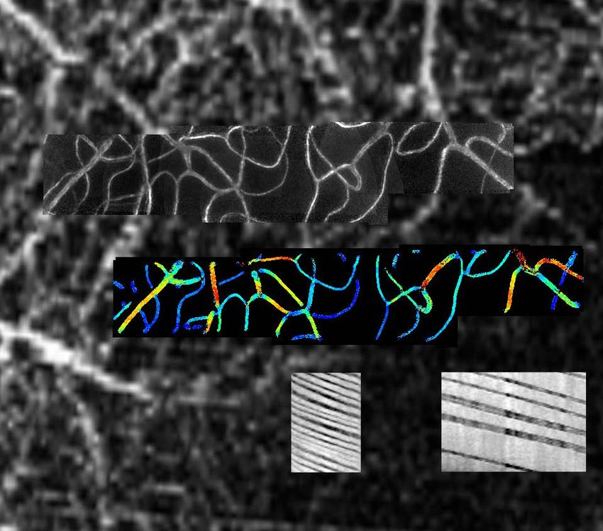
Contents
       Clinical Psychophysics Unit         4

       Optological Laboratory              6

       The Retinal Observatory             8

       Ocular Physiology Laboratory        10

       Ocular Biomarker Laboratory         12

       Corneal and Ocular                  14
       Immunology Laboratory
       Anterior Eye, Clinical Trials and   16
       Research Translation Unit
       Visual and Cognitive                18
       Neuroscience Laboratory
       Vision Optimisation Laboratory 20
       Contact us                          back cover

2 Department of Optometry and Vision Sciences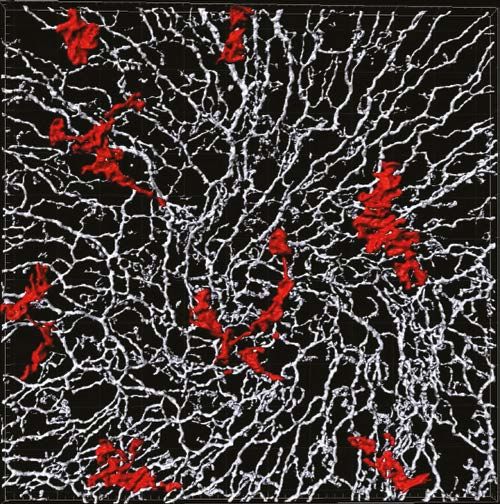
Research in the Department of
Optometry and Vision Sciences
The Department of Optometry and Vision Sciences is based in the Faculty of Medicine, Dentistry & Health Sciences (MDHS). Vision
science research is multidisciplinary in nature and spans topics from understanding the fundamental workings of the living retina
on the microscopic scale to evaluating the viability of crowdsourcing for research. No matter what your major, there are vision
research pathways for you. In this brochure we highlight some of the projects available for research students. If you have a passion
for vision science that is not covered specifically in this project set, please contact our researchers to discuss further.

For students who have completed an undergraduate degree a research pathway though an Honours or Master of Biomedical
Science is an appropriate research path. For students with a BSc (Hons) or BBiomed (Hons) further scientific training through a
three to four year PhD or a two year Master of Philosophy would be appropriate.

You can also contact the Departmental Honours and Master of Biomedical Science Coordinator, Prof Trichur Vidyasagar on
+61383447004 trv@unimelb.edu.au or the Departmental Graduate Researcher Coordinator for PhD and Master of Philosophy
related queries, A/Prof Andrew Anderson on +61390359916 aaj@unimelb.edu.au .

       For further information
       Honours
       http://mdhs-study.unimelb.edu.au/degrees/honours/overview

       Masters of Biomedical Science
       https://study.unimelb.edu.au/find/courses/graduate/master-of-biomedical-science/

       Masters of Philosophy
       https://study.unimelb.edu.au/find/courses/graduate/master-of-philosophy-mdhs-biomedical-science/

       PhD
       https://study.unimelb.edu.au/find/courses/graduate/doctor-of-philosophy-medicine-dentistry-and-health-sciences

       Department of Optometry and Vision Sciences
       https://healthsciences.unimelb.edu.au/departments/optometry-and-vision-sciences

                                                                                                             Research Projects 2020    3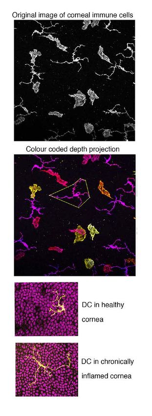
Clinical Psychophysics Unit

Laboratory Head                                                  Project 1: Understanding motion perception in peripheral
                                                                 vision
Name: Professor Allison McKendrick
Email: allisonm@unimelb.edu .au                                  Peripheral vision is very sensitive to visual motion cues. These
Ph: 83447005                                                     cues are used to identify objects in our periphery and to
                                                                 segment them from other background features. This project
Project Supervisors                                              will explore which stimulus features are important to the ability
                                                                 to segment moving objects in our peripheral vision, as well as
Name: Dr Bao Nguyen                                              studying whether individual differences in simple aspects of
Email: bnguyen@unimelb.edu.au                                    motion perception predict individual ability to identify moving
Ph: 9035 9979                                                    objects on noisy backgrounds.
Website: https://healthsciences.unimelb.edu.au/research-
groups/optometry-and-vision-sciences-research/clinical-
psychophysics-unit

Lab blog: http://uomcpulab.wordpress.com/

Summary of lab interests: Our research aims to better
understand normal visual processing and damage due to
disease. We have specific interests in the study of glaucoma,
migraine, and the process of normal ageing. Our applied aims
include developing better clinical tests for the assessment of
vision loss; determining methods of preventing visual damage,
and improving understanding of the consequences of vision loss
on performance in natural visual environments and day-to-day
tasks. Our current studies use a variety of methods including
visual psychophysics (testing visual performance), human
electrophysiology and brain and ocular imaging. Our work
is highly collaborative with colleagues from ophthalmology,
psychology, physiotherapy, neurology and neuroimaging.

                                                                 PhD student, Juan Sepulveda, measuring motion cues used to identify
                                                                 human movement.

4 Department of Optometry and Vision Sciences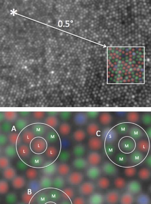
Project 2: Outer retinal structure and function in people          Project 3: Does caffeine influence perceptual eye dominance
who suffer episodic migraines                                      plasticity?
Migraine headaches can be associated with visual dysfunction       Caffeine is a widely used psychostimulant that is associated
at the time of an attack, but also in between migraine attacks.    with increased acetylcholine in the brain. Acetylcholine is a
This study will consider whether there is evidence for anomalies   neuromodulator that plays an important role in the processing
in outer retinal structure and function. This project will be      of visual information. In particular, acetylcholine and
an analytical study of data collected in young and otherwise       the cholinergic system are thought to be involved in adult brain
healthy people with normal vision who suffer from episodic         plasticity, which can be measured by temporary patching of
migraines, compared to people who do not regularly get             one eye for a few hours. A recent study showed that perceptual
headaches. This study will contribute to our understanding of      eye dominance plasticity is reduced with pharmacological
how migraine may impact on the visual system.                      administration of donepezil (an acetylcholine enzyme
                                                                   inhibitor) in healthy human observers. Here, we test whether
                                                                   temporarily manipulating caffeine levels has a similar effect on
                                                                   perceptual eye dominance plasticity.

Retina of an individual who suffers from migraines

                                                                   Binocular rivalry stimuli, commonly used to infer visual system plasticity. The
                                                                   red and green stimuli are presented to each eye separately, with the combined
                                                                   percept swapping regularly from one to the other (rivalrous percept).

                                                                   Recent related publications from our team:
                                                                   1.	Chan YM, Pitchaimuthu K, Wu Q-Z, et al. Relating excitatory
                                                                       and inhibitory neurochemicals to visual perception: A
                                                                       magnetic resonance study of occipital cortex between
                                                                       migraine events. PLoS One 2019;1-13.

                                                                   2.	Nguyen BN, Hew SA, Ly J, et al. Acute caffeine ingestion
                                                                       affects surround suppression of perceived contrast. J
                                                                       Psychopharmacol 2018;32:81-88.

                                                                   3.	Sepulveda JA, Anderson AJ, Wood JM, McKendrick AM.
                                                                       Differential aging effects in motion perception tasks for
                                                                       central and peripheral vision. J Vis. 2020;20(5):8.

                                                                   4.	Nguyen BN, McKendrick AM. Foveal and parafoveal contrast
                                                                       suppression are different: mechanisms revealed by the
                                                                       study of healthy ageing. Journal of Vision 2016;16(3):10.

                                                                                                                     Research Projects 2020          5
Optological Laboratory

Laboratory Head                                          The Optological Laboratory non-invasively investigates how
                                                         the human eye and brain function, both in normal observers
Name: Assoc. Prof. Andrew Anderson
                                                         and those with eye disease. Although our understanding
Email: aaj@unimelb.edu.au
                                                         of neuroscience has been greatly enhanced through
Ph: +613 9035 9916
                                                         electrophysiological recordings from individual neurons and
https://healthsciences.unimelb.edu.au/research-groups/   through computer imaging of gross neural activity across the
optometry-and-vision-sciences-research/optological-      brain, such information only tells us part of how the brain and
laboratory                                               eye work. Ultimately, we also need to understand how the eye
                                                         and brain behave in response to various forms of information,
                                                         and to ascertain what functional limits exist in processing such
                                                         information. By combining results from a range of studies –
                                                         including electrophysiological, imaging and behavioural studies
                                                         – a more complete understanding of neuroscience be achieved.

                                                         Our laboratory uses a range of techniques to determine how
                                                         the eye and brain behave, many of which can be classed under
                                                         the general heading of psychophysical methods. Sometimes
                                                         our investigations involve visual targets used in clinical tests of
                                                         vision, allowing us to better understand how such tests work
                                                         and allowing more effective clinical tests to be developed.
                                                         Other investigations use customised visual stimuli and special
                                                         experimental protocols to examine how the eye transmits
                                                         information to the brain, and also how the brain processes this
                                                         information in order to make decisions. The laboratory is well
                                                         equipped to undertake a wide range of behavioural experiments
                                                         and so can address a broad range of behavioural questions,
                                                         both in the clinical and basic sciences.

6 Department of Optometry and Vision Sciences
Project 1: Do the mechanisms that prevent our noticing small     Selected Publications:
eye movements improve our ability to judge small movements       Park ASY, Bedggood PA, Metha AB, Anderson AJ (2019). The
in the world?                                                    influence of perceptual stabilisation on perceptual grouping of
Even when we stare intently at a small target, our eyes are      temporally asynchronous stimuli. Vision Res 160:1-9.
constantly in motion. This results in images that continuously   Sepulveda JA, Anderson AJ, Wood JM, McKendrick AM (2020).
move on our retina. Powerful perceptual stabilisation            Differential aging effects in motion perception tasks for central
mechanisms prevent our noticing this motion, however. Whilst     and peripheral vision. J Vis 20(5):8.
this means our world doesn’t appear to incessantly jiggle
                                                                 Mahjoob M, Anderson AJ (2019). Contrast discrimination under
around, does this actually improve our ability to see things?
                                                                 task-induced mental load. Vision Res 165:84-89.
This project will investigate whether perceptual stabilization
mechanisms improve our ability to do a very common task –        Anderson AJ, Chaurasia AK, Sharma A, Gupta A, Gupta S,
making fine judgement of relative motion between objects in      Khanna A, Gupta V (2019). Comparison of rates of fast and
the world.                                                       catastrophic visual field loss in three glaucoma subtypes. Invest
                                                                 Ophthalmol Vis Sci 60(1):161-167.

                                                                                                          Research Projects 2020     7
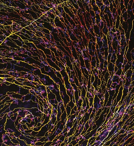
The Retinal Observatory
(Imaging cellular structure and function in the living human retina)

Laboratory Head                                                     Project 1: Keeping neurons alive in the living human eye:
                                                                    single red and white blood cell flow through retinal capillary
Name: Associate Professor Andrew Metha
                                                                    networks
Email: ametha@unimelb.edu.au
Ph: 9035 9783                                                       With newly-developed adaptive optics (AO) retinal imaging, we
                                                                    can now visualize the finest capillaries in the eye and watch the
Project Supervisors                                                 passage of single red and white blood cells through vascular
                                                                    networks. These are the networks that keep your retina
Name: Dr Phillip Bedggood                                           healthy, and which fail in diseases such age-related macular
Email: pabedg@unimelb.edu.au                                        degeneration and diabetes. The fine details of blood flow
Ph: 9035 9979                                                       patterns have not yet been fully documented because blood
                                                                    flows very quickly - and also because the retina is designed to
https://healthsciences.unimelb.edu.au/                              be transparent, making it hard to obtain high contrast images
research-groups/optometry-and-vision-sciences-research/             without risking light damage. With the recent lifting of these
imaging-retinal-cells-human-unit/                                   technical issues, a novel project emerges to characterize
individual-photoreceptors-in-the-human-eye                          aspects of normal flow such as: cell deformability during flow;
Our broad research aim is to understand the fundamental             variation in flow velocity through different parts of the network;
workings of the living retina on the microscopic scale: how         and the influence of the cardiac cycle on flow pulsatility.
this works normally and how this becomes compromised in             This project would suit Honours Students who wish to learn
sight-debilitating diseases such as diabetes. We combine a          about and apply optical and image processing skills to
range of novel investigative tools including high-spatiotemporal    questions of basic human physiology with immediate clinical
resolution non-invasive retinal imaging, psychophysics, and         applicability.
computational modelling.

Our current research projects make use of high speed, multi-
spectral adaptive optics to visualize the smallest neurons and
blood vessels that is possible to see in in living human eyes. We
study the dynamics of flow and oxygen exchange at the level
of individual red blood cells, and the cascade of optical and
physiological events that occur when a photoreceptor interacts
with light.

This requires a multi-disciplinary approach and so we welcome
motivated students across all fields (e.g. Mathematics, Physics,
Computer Science, Engineering, Biology, Psychology), who
are interested in contributing to our innovative program of
research.

                                                                    Tracking single-file blood cell flow though the retinal capillary network

8 Department of Optometry and Vision Sciences
Project 2: Wiring the retina for human vision - a single-cell          Project 3: Methods to improve the measurement of visual
behavioural approach                                                   performance
The normal human retina is tiled with a mosaic of about 110            Measurements of human visual performance are important
million rods and 6 million cone photoreceptors of 3 types that         both to understand the basic science behind vision and for
are sensitive to long (L), middle (M) and short (S) wavelengths        diagnosis of blinding eye diseases. The methods currently used
of light. These 116 million photoreceptors converge to a mere 1        to measure visual performance in the clinic and laboratory
million axons that form the optic nerve connecting the eye and         are time-consuming, which limits the amount of information
brain. The retina itself is responsible for much more than image       that can be gained in a given test session. This honours project
detection, but is involved in substantial processing of visual         will evaluate the use of alternate testing strategies designed
information as well!                                                   to improve test efficiency, and determine whether such
                                                                       improvements can be obtained whilst avoiding the introduction
Text BoxUsing psychophysical methods to record behavioral              of inaccuracy or bias. Specifically, the project asks whether: 1)
responses to stimulating either single cells or specific               the reported degree of certainty of participant’s responses be
cell arrangements, an exciting ARC-funded project exists               used to determine visual threshold more quickly than assessing
to establish precisely how signals from 3 types of cone                the accuracy of responses alone; and 2) the degree to which
photoreceptor are organised within the receptive fields of             cueing participants to direct their attention to a smaller part
retinal ganglion cells whose fibers exit the eye, and how this         of the visual field can improve the reliability of their responses.
impacts the information conveyed for spatial and colour                This information may have immediate clinical applicability
perception.                                                            for improving standard clinical perimetry (visual field testing)
                                                                       for diseases such as glaucoma and maculopathy, and also for
                                                                       making more efficient laboratory investigations of precise
                                                                       retinal cell sensitivity.

                                                                       Recent related publications from our team:
                                                                       1.	Bedggood, P., & Metha, A. (2020). Adaptive optics imaging
                                                                           of the retinal microvasculature. Clinical Experimental
                                                                           Optometry, 103(1), 112-12).

                                                                       2.	Duan, A., Bedggood, P. A., Metha, A. B., & Bui, B. V. (2017).
                                                                           Reactivity in the human retinal microvasculature measured
                                                                           during acute gas breathing provocations. Scientific reports,
                                                                           7(1), 2113.

                                                                       3.	Bedggood, P., & Metha, A. (2013). Optical imaging of human
                                                                           cone photoreceptors directly following the capture of light.
                                                                           PloS one, 8(11).

Targeting single cone photoreceptors to investigate inputs to midget
ganglion cells

                                                                                                                Research Projects 2020    9
Ocular Physiology Laboratory

Laboratory Head
Name: Bang Bui
Email: bvb@unimelb.edu.au
Ph: +61 3 83447006

Project Supervisors:
Dr Christine Nguyen
Email: christine.nguyen@unimelb.edu.au
Ph: +61 3 9035 3186

Dr Vickie Wong
Email: vickie.wong@unimelb.edu.au
Ph: +61 3 8344 1484                                                  Project 2: Studies of retinal vascular autoregulation
                                                                     The retina and brain are the most highly energy demanding tissues
Da Zhao                                                              in the body. In the retina, the need for optical clarity and many
Email: da.zhao@unimelb.edu.au                                        neurons for good vision comes at the expense of fewer blood
Ph: +61 3 8344 1484                                                  vessels as well as the lack of ways to store energy. The retina is
                                                                     completely dependent on a stable blood supply to deliver oxygen
https://healthsciences.unimelb.edu.au/research-groups/
                                                                     and glucose. In the retina there are three major vascular beds
optometry-and-vision-sciences-research/ocular-physiology-
                                                                     (superficial, intermediate and deep) that entirely locally controlled
lboratory
                                                                     to respond to normal fluctuations in blood and eye pressure. This
Summary of lab interests: Our laboratory is interested in            control system is known as vascular autoregulation and involves
understanding the causes of retinal and optic nerve injury in        not only the cell lining the blood vessel walls (endothelial cells) but
diabetes and glaucoma. We are also interested in developing          also neurons and supporting glial cells (astrocytes and microglia).
new ways to clinically detect eyes at risk of vision loss from       The failure of autoregulation has been implicated in retinal disease
these conditions.                                                    such as diabetes and glaucoma. In these studies, we will employ
                                                                     optical coherence tomography imaging to assess autoregulation in
Project 1: Understanding how pressure affects ganglion cells         animal models of retinal diseaseresponse to changes in pressure.
in glaucoma                                                          response to changes in pressure.
Our investigations of glaucoma hope to shed light on how the
cells that connect the eye to the brain, the retinal ganglion
cells are able to adapt to changes in their local environment.
When such adaptation mechanisms fail ganglion cells undergo
programmed cell death. Ganglion cells have to cope with
constant changes in the pressures in and around the eye;
intraocular pressure, blood pressure and intracranial pressure. As
the eye gets older the capacity to cope with stress is diminished,
but at the moment we dont understand why this occurs. In order
to study how ageing and other risk factors impact the capacity for
retinal ganglion cells to cope with stress we have developed both
acute and chronic model of intraocular pressure elevation. We
will study ganglion cell responses to stress by quantifying their
function and relating this to changes in dendritic morphology and
expression of membrane pressure sensors.

The figure top left shows a ganglion cell in a mouse eye, that we
                                                                     Higher eye pressure changes the blood vessels in the living eye,
can co-stain for synapses to better understand why ganglion cells
                                                                     which we measure using optical coherence tomography. By
are affected by high eye pressure.
                                                                     looking at the vessel at different retinal depths we can see that
                                                                     the superficial vessels (blue) respond differently to intermediate
                                                                     (green) and deeper (red) vessels.

10 Department of Optometry and Vision Sciences
Project 3: Developing a clinical test of vascular autoregulation Recent related publications from our team:
As we better understand how blood vessels in the eye work to 1. Grant ZL, Whitehead L, Wong VHY, He Z, Yan RY, Miles AR,
supply blood when needed, we use this information to help us Benest AV, Bates DO, Prahst C, Bentley K, Bui BV, Symons
develop better clinical tests for detecting blood vessels that RC, Coultas L. Blocking endothelial apoptosis revascularises
don’t work as they should. Optical coherence tomography the retina in a model of ischemic retinopathy. J Clin Invest.
angiography can be used in the laboratory as well as in the 2020:127668.
clinic. By using a flickering light stimulus we challenge the blood
vessels to dilate, in order to get more blood to support the 2. Zhao D, He Z, Wang L, Fortune B, Lim JKH, Wong VHY,
increased communication between retinal neurons that occurs Nguyen CTO, Bui BV. Response of the Trilaminar Retinal
with lights repeatedly turning on and off. Using this approach Vessel Network to Intraocular Pressure Elevation in Rat Eyes.
we may be able to identify eyes that have regions of blood Invest Ophthalmol Vis Sci. 2020 ;61(2):2.
vessels that do not respond as they should. We believe that this
3. Zhao D, Wong VHY, Nguyen CTO, Jobling AI, Fletcher
will help us detect earlier those eyes that might go on to develop
EL, Vingrys AJ, Bui BV. Reversibility of Retinal Ganglion Cell
vision loss.
Dysfunction From Chronic IOP Elevation. Invest Ophthalmol
Vis Sci. 2019;60(12):3878-3886.
4. Liu G, Cull G, Wang L, Bui BV. Hypercapnia Impairs
Vasoreactivity to Changes in Blood Pressure and Intraocular
Pressure in Rat Retina. Optom Vis Sci. 2019;96(7):470-476.
5. Li F, Hung SSC, Mohd Khalid MKN, Wang JH, Chrysostomou
V, Wong VHY, Singh V, Wing K, Tu L, Bender JA, Pébay A, King
AE, Cook AL, Wong RCB, Bui BV, Hewitt AW, Liu GS. Utility of
Self-Destructing CRISPR/Cas Constructs for Targeted Gene
Editing in the Retina. Hum Gene Ther. 2019;30(11):1349-
1360.
Optical coherence tomography angiography can help us to discern
key blood vessel layers in the retina.
Research Projects 2020 11Ocular Biomarker Laboratory
Laboratory Head Project 1: Imaging Parkinson’s disease in the eye
Dr Christine Nguyen Diagnosis of Parkinson’s disease is a difficult and lengthy
Email: christine.nguyen@unimelb.edu.au process. A hallmark of Parkinson’s is alpha-synuclein deposits
Ph: +61 3 9035 3186 in the brain but the skull makes these difficult to detect.
Interestingly, in our lab and others, alpha-synuclein has been
identified in the retina, an outpouching of the central-nervous-
Project Supervisors: system. The aim of this project is to provide proof-of-principle
A/Prof Bang Bui that it is possible to image alpha-synuclein in the mouse
Email: bvb@unimelb.edu.au retina. Given the clear optics the eye, we will fluorescently tag
Ph: +61 3 8344 7006 an antibody and directly image them in living animals. The
capacity to develop early, specific biomarkers for PD is pivotal
Prof Cassandra Szoeke for development of treatments.
Email: cszoeke@unimelb.edu.au
Ph: +61 3 8344 1835
Project 2: The retina as a window to Alzheimer’s disease: a
Dr Vickie Wong prospective study
Email: vickie.wong@unimelb.edu.au Retinal ganglion cells and their axonal projections form part of
Ph: +61 3 8344 1484 the central nervous system and are uniquely suited to direct
visualisation and imaging. In Alzheimer’s disease, reports using 3D
Dr Da Zhao retinal scans (optical coherence tomography) have suggested that
Email: da.zhao@unimelb.edu.au the retinal nerve fibre layer is thinned in patients with advanced
dementia. More recently, there has been suggestion that the inner
https://healthsciences.unimelb.edu.au/research-groups/ plexiform layers of the retina are thickened in people with early
optometry-and-vision-sciences-research/ocular-biomarker- Alzheimer’s. This project is part a longitudinal study known as
laboratory the Women in Healthy Ageing Project and will correlate optical
coherence tomography measures to mental health status from
Summary of lab interests: The eye affords a unique
depression to dementia. In this manner the project will evaluate
opportunity to gain insights into what is occurring in the brain.
whether the time course of retinal measurements is potentially
It is the only place in the body where neurons and blood vessels
useful as a topographical biomarker for Alzheimer’s disease.
can be directly visualised. Moreover, neurological diseases
such as Alzheimer’s disease, Parkinson’s disease and multiple
sclerosis have been shown to exhibit changes in the eye which
can be measured with currently available clinical tools and
emerging technologies.
Alpha-synuclein imaging in the eye A. Retinal photograph B. following injection of a fluorescently labeled tag, alpha-synuclein (green spots) can be visualised.
C. A combination image where some retinal detail and tagged alpha-synuclein can be simultaneously viewed
12 Department of Optometry and Vision SciencesProject 3: A marker for Parkinson's disease? ON- OFF- Project 4: Examining neuroinflammation in a model of
electroretinography assessment Parkinson’s disease
Emerging evidence indicates that changes to the electrical Neuroinflammation is central to the pathophysiology of
response from the retina (electroretinogram) may reflect changes Parkinson’s disease, however it is challenging to measure in
in cortical disease such as Parkinson’s disease. Studies have vivo. Assessment in peripheral systems (such as blood) may
shown dampening of the electroretinogram in Parkinson's disease be indicative but are limited due to the distinct inflammatory
patients that reverse with current gold standard treatment with pathways found within the central nervous system. It is
levadopa. Indeed, dopamine has multiple roles in the retina established from Parkinson’s disease human post‐mortem
including light adaptation and ON-OFF bipolar cells responses but substantia nigra tissue, that microglia become activated
light adaptation can be time-consuming and ON- OFF- responses and release specific proinflammatory cytokines that lead to
have been difficult to measure. This study aims to apply a novel neurodegeneration. The eye is an out‐pouching of the brain and
analysis approach recently shown to differentiate ON and OFF literature indicates that 3 dimensional scans of the retina show
bipolar cell responses to electroretinography recordings from thickening which is typical of active inflammation. What has not
patients and animal models with Parkinson's disease. Such an been examined are inflammatory markers which correspond to
approach will aid understanding of whether assessment of the ON- these changes. This project aims to examine this link in a mouse
OFF- system is an informative marker for Parkinson's diseas. model of Parkinson's disease.
Recent related publications from our team:
1. Nguyen CT, Hui F, Charng J, Velaedan S, Van Koeverden
A, Lim JK, He Z, Wong VHY, Vingrys AJ, Bui BV, Ivarsson M
(2017). Retinal biomarkers provide “insight” into cortical
pharmacology and disease. Pharmacology and Therapeutics.
175: 151-177.
2. Lim JK, Li QX, He Z, Vingrys, AJ, Wong, VHY, Currier N. Mullen
J, Bui BV, Nguyen, CT (2016). The Eye as a Biomarker for
Alzheimer's Disease. Front Neurosci 10, 536.
3. Habiba U, Merlin S, Lim JKH, Wong VHY, Nguyen CT, Morley
JW, Bui BV, Tayebi M. Age-Specific Retinal and Cerebral
Immunodetection of Amyloid-beta Plaques and Oligomers
in a Rodent Model of Alzheimer's Disease. J Alzheimers Dis
2020
4. Shahandeh A, Bui BV, Finkelstein DI, Nguyen CT. Therapeutic
Functional and structural assessment of retinal health in animals models of
applications of chelating drugs in iron metabolic disorders
neurodegenerative disease of the brain and retina. J Neurosci Res 2020
Research Projects 2020 13Corneal and Ocular Immunology
Laboratory Head
Name: Dr Holly Chinnery
Email: holly.chinnery@unimelb.edu.au
Ph: 9035 6445
Summary of lab interests: We investigate the structural,
physiological and immunological interplay between immune
cells and other non-immunological structures such as sensory
nerves and epithelial cells in the cornea during homeostasis
and disease. Techniques used in our lab include in vivo clinical
imaging of the cornea, ex vivo confocal microscopy and 3D
image reconstruction and molecular biology and protein
assays. We also collaborate closely with Dr Laura Downie, who
leads the Anterior Eye, Clinical Trials and Research Translation
Unit in DOV.
Corneal nerves forming a whorl pattern in the central epithelium
Project 1: Time course of sensory nerve recovery after
epithelial injury
Following corneal epithelial injury, the regeneration of the
corneal nerves is a slow process, often taking months to recover.
However, despite this slow recovery process, the cornea
appears structurally normal, with the epithelial cells and tissue
architecture appearing clear and healthy. We propose that this
is due to differential rates of recovery of different nerve plexi.
This project will quantify the regeneration rates of nerves
and measure neuropeptide secretion in distinct regions of
the cornea after injury. Techniques include animal handling,
clinical imaging, confocal microscopy, protein assays, 3D image
Corneal macrophages living just beneath the corneal epithelial nerve plexus. reconstruction and image analysis. This project would be
suitable for Honours, Masters or PhD student.
14 Department of Optometry and Vision SciencesProject 2: Correlating immune cell morphometry with
markers of cellular activation in the mouse cornea.
Recent clinical studies have demonstrated using in vivo confocal
microscopy that corneal immune cells change their shape and
size in response to local and systemic inflammatory diseases.
It is unclear how the changes in cell shape and size relate to
function and maturation. In this project, mouse models of
corneal inflammation will be used to correlate morphological
changes in immune cell populations with alterations in cell
surface markers indicative of cell activation. These findings will
provide clinically translatable information that will shed light on
the functional relevance of morphological changes to immune
cells in the human cornea. This project will involve animal
handling, clinical imaging, ex vivo confocal microscopy, flow
cytometry and 3D image reconstruction and image analysis.
This project would be suitable for Honours, Masters or PhD students.
Recent related publications from our team:
1. Jiao, H., L. E. Downie, X. Huang, M. Wu, S. Oberrauch,
R. J. Keenan, L. H. Jacobson and H. R. Chinnery. 2020.
"Novel alterations in corneal neuroimmune phenotypes
in mice with central nervous system tauopathy." J
Neuroinflammation 17(1): 136.
2. Wu, M., L. E. Downie, L. M. Grover, R. J. A. Moakes, S. Rauz,
A. Logan, H. Jiao, L. J. Hill and H. R. Chinnery. 2020. "The
neuroregenerative effects of topical decorin on the injured
mouse cornea." J Neuroinflammation 17(1): 142
3. Jiao H, Naranjo Golborne C, Dando S, McMenamin PG,
Downie LE & Chinnery HR. 2019. Topographical and
morphological differences of corneal dendritic cells during
steady state and inflammation. Accepted for publication in
Ocular Immunology and Inflammation .
4. Downie LE, Naranjo Golborne C, Chen M…Chinnery HR et
al. Recovery of the sub-basal nerve plexus and superficial
nerve terminals after corneal epithelial injury in mice. Exp
Eye Res 2018; 171: 92-100.
Resident immune cells (dendritic cells and macrophages) in mouse cornea.
Shape and size analysis reflect functional alterations in immune cells.
Research Projects 2020 15Anterior Eye, Clinical Trials and
Research Translation Unit
Laboratory Head
Name: A/Prof Laura Downie BOptom, PhD
Email: ldownie@unimelb.edu.au
Ph: 9035 3043
Project Supervisors:
Name: Dr Holly Chinnery BSc(Hons), PhD
Email: holly.chinnery@unimelb.edu.au
Ph: 9035 6445
http://healthsciences.unimelb.edu.au/research2/optometry- Figure 1: After non-invasive collection, human tears are analysed using a
range of cutting-edge techniques, to quantify parameters such as
and-vision-sciences-research/aect-and-rt-unit
viscoelasticity, protein composition and lipid content. These studies are the
foundation for developing novel lab-on-a-chip tests for ocular and systemic
Summary of lab interests: The Anterior Eye, Clinical Trials and disease.
Research Translation unit adopts an integrated and innovative
approach to research that combines laboratory, clinical and Research translation: This research focuses on improving
implementation science, as a basis for improving patient patient outcomes by identifying, synthesising and promoting
outcomes. Our team possess advanced expertise in anterior implementation of the best-available evidence in eye care practice.
eye disease (including the development and translation
We are currently undertaking several projects that are developing
of novel ocular diagnostic devices and therapeutics) and
new clinical tools and digital platforms to support evidence-
research translation (to develop and test interventions to
based practice, in areas such as dry eye disease and age-related
improve research dissemination and its implementation
macular degeneration. We have developed a free, online
in practice). Our collaborators include industry, national
platform (in collaboration with A/Prof Michael Pianta) called
and international research groups (including researchers in
CrowdCARE, (Crowdsourcing Critical Appraisal of Research
neurology, endocrinology, immunology, neuroscience and
Evidence crowdcare.unimelb.edu.au), which uses crowdsourcing
chemical engineering), and the Corneal and Ocular Immunology
to support evidence-based practice. CrowdCARE has the capacity
laboratory (led by Dr Chinnery) on projects combining pre-
to redefine how clinicians discover and use appraised research
clinical and clinical science.
evidence, through its capacity to: teach critical appraisal, enable
Anterior eye biomarkers of ocular and systemic disease: This critically appraised research to be shared amongst a global
program of research investigates using the anterior eye, in interdisciplinary community, and facilitate contributions and
particular tears (Figure 1), to provide novel insights into human access to an evolving stream of appraised research.
health. We combine sophisticated clinical techniques with
Project 1: Understanding the dynamics of corneal immune
laboratory-based studies to characterise tear film responses in
cells in the human eye
ocular and systemic disease. These investigations are the basis
for developing new diagnostic and prognostic tests to inform
the management of clinical conditions. Some of our recent
studies have identified new tear biomarkers for diabetes, dry
eye disease and contact lens discomfort, leading to patents
and subsequent projects focussed on the commercialisation of
these discoveries.
16 Department of Optometry and Vision SciencesFigure 2: Laser-scanning confocal microscopy images from the central Project 2: Is crowdsourcing a valid approach to evaluating
cornea of a person with peripheral neuropathy, at (A) baseline and (B) after research quality?
eight weeks of oral prednisolone treatment, showing a reduction in the
density of putative dendritic cells (white trapezoid-shaped cell bodies).
In vivo confocal microscopy (Figure 2) is a high-resolution
imaging technique that permits direct visualisation of corneal
nerves and immune cells (dendritic cells) in the living human
eye. The cornea is the only tissue in the body that permits
this non-invasive, in vivo observation of peripheral nerves
and immune cells. Corneal dendritic cells are known to be a Before we ‘trust’ a research study, we need to consider how it
dynamic cell population, however there is currently a lack of has been performed, and evaluate its potential weaknesses
understanding with respect to how their density change over and/or biases. This process, called critical appraisal, enables us
time. This project will investigate the fundamental dynamics to assess the quality of a scientific paper. This is a potentially
of dendritic cell responses in the healthy cornea, in order to time-consuming task, which is an established barrier to it being
provide insight into the repeatability of this metric, as a marker routinely performed. Using data contributed to our online
of corneal inflammation. crowdsourced critical appraisal platform (CrowdCARE), the
This project will involve participant recruitment, clinical major aim of this project is to evaluate the quality of the data
examinations, and digital image analysis. It is suitable for generated using crowdsourcing, with a focus on the appraisal of
Honours, Masters and PhD students. laboratory-based studies.
This project will engage researchers and students to contribute
critical appraisals, and involve considerable data evaluation
and statistical analysis. It is suitable for Honours and Masters
students.
Recent related publications from our team:
1. McDonnell A, Lee JH, Makrai E, Yeo LY, Downie LE. Tear film
extensional viscosity is a novel potential biomarker of dry
eye disease. Ophthalmology 2019;126(8):1196-8.
2. Downie LE, Wormald R, Evans J, et al. Analysis of a
systematic review about blue light-filtering intraocular
lenses for retinal protection. JAMA Ophthalmology
2019;137(6):694-7.
3. Gad A, Vingrys AJ, Wong CY, Jackson DC, Downie LE. Tear
film inflammatory cytokine upregulation in contact lens
discomfort. Ocul Surf 2019;17(1):89-97.
4. Pianta MJ, Makrai E, Verspoor KM, Cohn TA, Downie LE.
Crowdsourcing critical appraisal of research evidence
(CrowdCARE) was found to be a valid approach to assessing
clinical zresearch quality. J Clin Epidemiol 2018;104:8-14.
5. Downie LE, Gad A, Wong CY, et al. Modulating contact
lens discomfort with anti-inflammatory approaches: a
randomized controlled trial. Invest Ophthalmol Vis Sci
2018;59(8):3755-66
Research Projects 2020 17Visual and Cognitive
Neuroscience Laboratory
Laboratory Head Project 2: Neural Mechanisms of Top-down Attention and
predictive coding
Professor Trichur Vidyasagar MBBS, PhD
Email: trv@unimelb.edu.au How does the brain manage to attend to a specific object or
region of visual space when it is confronted with innumerable
Project Supervisors: objects? How are we able to pick out a face in a large crowd,
often so effortlessly? Such focussing of attention is known to
Dr Ekaterina Levichkina PhD involve some specific areas of the brain, but how these interact
Email: ele@unimelb.edu.au with each other has been largely unknown. In these experiments
on trained macaques, we record from multiple brain areas
Dr Yamni Mohan PhD implicated in visual attention, in order to characterise the
Email: mohan.y@unimelb.edu.au distributed processing that occurs with attention. With these
https://healthsciences.unimelb.edu.au/research-groups/ experiments, we also seek to test an influential new
optometry-and-vision-sciences-research/visual-and- model that suggests that the brain makes conscious or
cognitive-neuroscience-laboratory unconscious predictions about what it expects to see in the
external world and updates these expectations using any
Summary of lab interests: Our laboratory is interested in mismatches with the sensory inputs.
understanding the neural basis of visual perception, attention
and memory.
Project 1: Functional microcircuitry of the visual cortex
Different areas of the cerebral cortex have fairly
similar morphological structures regardless of their specific
functions, suggesting that there is a universal cortical
microcircuit which is involved in transforming the inputs.
Understanding this microcircuit is important to understanding
how the brain makes sense of the external world. In our lab, we
examine the microcircuit of the primary visual cortex in
anaesthetised cats and macaques, to shed new light on this
problem. In these studies, we use a combination of single
electrodes, multi-electrode arrays and optical imaging of
intrinsic signals to examine the cortical inputs, responses of
individual neurons and groups of neurons, to shed new light on
this problem.
18 Department of Optometry and Vision SciencesProject 3: Visual attention, Reading and Dyslexia. Publications of the team relevant to current interests:
1. Archer K, Pammer K & Vidyasagar TR (2020). A temporal
The basic cause of specific reading disability, commonly
sampling basis for visual processing in developmental
known as dyslexia, has been a matter of intense debate for
dyslexia. Front Human Neurosci., Vol. 14, Article No.213. doi:
decades. Reading is a relatively recent activity in human history
10.3389/fnhum.2020.00213.
and so it is very unlikely that humans have evolved a specific
brain region or circuitry devoted to reading, but we probably 2. Mohan YS, Jayakumar J, Lloyd EKJ, Levichkina E &
use for reading brain functions that evolved for a different Vidyasagar TR (2019). Diversity of feature selectivity in
purpose. Our lab has been working on the idea that one such macaque visual cortex arising from limited number of
critical brain function is the visuo-spatial attention network broadly-tuned input channels, Cerebral Cortex, 29, 5255-5268.
usually used in focussing attention at a visual field location for
object identification. We recently found the visual attention 3. Vidyasagar TR & Levichkina E (2019). An Integrated Neuronal
efficiency to differ substantially between people and it is related Model of Claustral Function in Timing the Synchrony
both to reading speeds and to the functional size of the primary Between Cortical Areas. Front Neural Circuits, Vol. 13:3. doi:
visual cortex. We are now exploring these relationships further 10.3389/fncir.2019.00003.
using visual psychophysics and functional brain imaging in
the dyslexic population and also comparing reading of scripts 4. Kermani M, Verghese, A, Vidyasagar TR (2018) Attentional
written from left to right (as in English) with those written from asymmetry between the visual hemifields is related to
right to left (as in Farsi). habitual direction of reading and its implications for debate
on cause and effects of dyslexia. Dyslexia. 24(1):33-432.
5. Levichkina E, Saalmann YB, Vidyasagar TR (2017) Coding of
spatial attention priorities and object features in the macaque
lateral intraparietal cortex. Physiological Reports. 5(5).
6. Vidyasagar TR, Eysel UT (2015) Origins of
feature selectivities and maps in the mammalian primary
visual cortex. Trends in Neurosciences. 38(8), 475-485.
7. Jayakumar J, Roy S, Dreher B, Martin P & Vidyasagar
TR (2013). Multiple pathways carry signals from short
wavelengths-sensitive ("blue") cones to the middle temporal
(MT) area of the macaque, J. Physiol (Lond), 591, 339-352.
8. Vidyasagar TR & Pammer K (2010). Dyslexia: a deficit in
visuo-spatial attention, not in phonological processing.
Trends Cognitive Sci., 14(2):57-63.
9. Saalmann YB, Pigarev IN, Vidyasagar TR (2007) Neural
mechanisms of visual attention: how top-down feedback
highlights relevant locations. Science 316(5831), 1612-1615.
Research Projects 2020 19Vision Optimisation Laboratory Laboratory Head Name: Dr Lauren Ayton Email: layton@unimelb.edu.au Ph: 8344 3441 www.linkedin.com/in/drlaurenayton Summary of lab interests: In recent years, there have been a number of interventions developed for vision loss and blindness. From gene therapy to bionic eyes, all treatments require thorough evaluation of visual function pre- and post- intervention, as well as an understanding of the impact of the treatments on a person’s life. Our team works on clinical vision and psychosocial assessments of people who receive such vision interventions. We have developed and run clinical studies for retinal prostheses (bionic eyes), gene therapy, and other low vision aids (including sensory substitution). Our aim is to ensure that every person is able to make the most of the vision they have. The lab maintains strong collaborations with engineers (University of Melbourne, Bionics Institute, Swinburne University, Cornell University USA), neurologists (Harvard University, USA), ophthalmologists (Royal Victorian Eye and Ear Hospital, Centre for Eye Research Australia), visual function experts (Oxford University, UK) and basic scientists (University of Melbourne) to assist in the development of new treatments. Currently, we are collaborating on a project to develop Australia’s first ocular gene therapy for an inherited retinal disease; designing new software algorithms for electronic and audio-based low vision aids; developing novel imaging techniques to identify raised intracranial pressure and running natural history studies to identify biomarkers of retinal degenerative disease. 20 Department of Optometry and Vision Sciences
Project 1: Development of New Vision Tests for Vision                 Recent related publications from our team:
Restoration Clinical Trials                                           1.	Ayton LN, Rizzo JF, Bailey I et al. The Harmonization of
This project, in collaboration with clinicians at the University of       Outcomes and Vision Endpoints in Vision Restoration Trials
Oxford and ophthalmologists from the Centre for Eye Research              (HOVER) consensus document. Transl Vis Sci Technol 2020;
Australia, will develop and validate new methods of measuring             In press.
low vision in patients who may be eligible for treatments such
                                                                      2. Kvansakul J, Hamilton L, Ayton LN, McCarthy CD, Petoe MA.
as gene therapy and stem cells. At present, there is a lack of
                                                                         Sensory augmentation to adi training with retinal
gold standard test protocols for low vision testing, and this
                                                                         prostheses. Journal of Neural Engineering 2020; In press.
project will provide important data on the validity of new tests.
For example, one avenue of interest in this area is colour vision     3. Ayton LN, Blamey PJ, Guymer RH, Luu CD, Nayagam DAX,
measures.                                                                Sinclair NC, Shivdasani MN, Yeoh J, McCombe MF, Briggs
                                                                         RJ, Opie NL, Villalobos J, Dimitrov PN, Varsamidis M, Petoe
Project 2: Natural History of Inherited Retinal Diseases
                                                                         MA, McCarthy CD, Walker JG, Barnes N, Burkitt AN,
A large study in our group is focusing on the collection of data         Williams CE, Shepherd RK, Allen PJ, for the Bionic Vision
on the natural history of inherited retinal diseases in Australia        Australia Research Consortium. First-in-human trial of a
and New Zealand. A number of research projects into imaging              novel suprachoroidal retinal prosthesis. PLOS One 2014;
biomarkers, genotype/phenotype correlations and visual                   9(12): e115239.
function measures in this population are available.
                                                                      4. Bentley SA, O’Hare F, Murphy GC, Finger RP, Luu CD, Keeffe
Project 3: Evaluation of Advanced Low Vision Technologies                JE, Guymer RH, Ayton LN. Psychosocial assessment of
Historically, low vision aids were low-tech, such as magnifying          potential retinal prosthesis recipients. Clinical and
glasses and high-powered spectacle lenses. However,                      Experimental Optometry 2019; 102(5):506-12.
recent advances have led to a proliferation in high-tech
alternatives, such as the iPhone, text-to-speech software and         5. Finger RP, McSweeney SC, Deverell LA, O’Hare F, Bentley
spectacle-mounted camera systems. This research program                  SA, Luu CD, Guymer RH, Ayton LN. Developing an
is investigating the efficacy and uptake of these technologies,          instrumental activities of daily living tool as part of the
and comparing to the more traditional options for patients with          Low Vision Assessment of Daily Activities (LoVADA) protocol.
conditions such as age-related macular degeneration.                     Investigative Ophthalmology and Vision Science 2014;
                                                                         55(12): 8458- 8466

                                                                                                            Research Projects 2020     21
Notes 22 Department of Optometry and Vision Sciences
Notes

        Research Projects 2020   23
healthsciences.unimelb.edu.au/
departments/optometry-and-vision-sciences
If you are interested in vision Honours Coordinator
research, here is your road map: Prof Trichur Vidyasagar
Step 1: Inform yourself of the research + 61 3 8344 7004
going on in the Department of Optometry
and Vision Sciences from this brochure trv@unimelb.edu.au
and/or from the webpages of the different
research labs (https://healthsciences.
unimelb.edu.au/research-groups/ Master of Biomedical Science
optometry-and-vision-sciences-research) Coordinator
Step 2: Contact one or more lab heads Prof Trichur Vidyasagar
for potential supervision in the area/s you
may be interested in, after emailing them + 61 3 8344 7004
with your latest statement of results.
trv@unimelb.edu.au
Step 3: After mutual tentative agreement
for potential supervision, go to the faculty
website for application to either Honors Graduate Researcher Coordinator
or Masters (https://study.unimelb.edu. A/Prof Andrew Anderson
au/fac/courses/undergraduate/bachelor-
+61 3 9035 9916
of-biomedicine-degree-with-honours or
https://healthsciences.unimelb.edu.au/
research-groups/optometry-and-vision- aaj@unimelb.edu.au
sciences-research respectively)
healthsciences.unimelb.edu.au/
If you think you are eligible to apply for a departments/optometry-and-vision-
Research Higher Degree, there is either sciences
an MPhil of PhD option that you may like
to consider. (https://study.unimelb.
edu.au/find/courses/graduate/master-
of-philosophy-mdhs-health-sciences/
or https://study.unimelb.edu.au/find/
courses/graduate/doctor-of-philosophy-
medicine-dentistry-and-health-sciences/
respectively)
Research Projects 2021
Intellectual property Disclaimer Authorised by:
Copyright in this publication is owned by the The University endeavours to ensure that Professor Allison McKendrick,
University and no part of it may be reproduced information contained in this publication is current September 2020
without the permission of the University. and correct at the time of printing. However, the CRICOS Provider Code: 00116K
University may change details relating to its courses
For further information, refer to: from time to time (such as subjects offered, fees Printed on paper from responsible sources.
unimelb.edu.au/governance/statutes or academic staff). You should not rely on this
Statement on Privacy Policy publication to make any decision about making or
accepting any application to study at the University.
When dealing with personal or health information
Before doing so, you should contact the school
about individuals, the University of Melbourne is
or faculty directly to ensure that the relevant
obliged to comply with the Information Privacy Act
information is current and correct. This does not
2000 and the Health Records Act 2001.
affect any rights you may have under the Australian
For further information, refer to: Consumer Law.
unimelb.edu.au/unisec/privacyYou can also read