Ocular pigmentation in humans, great apes, and gibbons is not suggestive of communicative functions - Nature
←
→
Page content transcription
If your browser does not render page correctly, please read the page content below
www.nature.com/scientificreports
OPEN Ocular pigmentation in humans,
great apes, and gibbons
is not suggestive of communicative
functions
Kai R. Caspar1*, Marco Biggemann1, Thomas Geissmann2 & Sabine Begall1
Pigmentation patterns of the visible part of the eyeball, encompassing the iris and portions of the
sclera, have been discussed to be linked to social cognition in primates. The cooperative eye hypothesis
suggests the white sclera of humans to be a derived adaptive trait that enhances eye-mediated
communication. Here, we provide a comparative analysis of ocular pigmentation patterns in 15
species of hominoids (humans, great apes & gibbons) that show marked differences in social cognition
and quantify scleral exposure at the genus level. Our data reveals a continuum of eye pigmentation
traits in hominoids which does not align with the complexity of gaze-mediated communication in the
studied taxa. Gibbons display darker eyes than great apes and expose less sclera. Iridoscleral contrasts
in orangutans and gorillas approach the human condition but differ between congeneric species.
Contrary to recent discussions, we found chimpanzee eyes to exhibit a cryptic coloration scheme
that resembles gibbons more than other apes. We reevaluate the evidence for links between social
cognition and eye pigmentation in primates, concluding that the cooperative eye hypothesis cannot
explain the patterns observed. Differences in scleral pigmentation between great apes and humans
are gradual and might have arisen via genetic drift and sexual selection.
Eyes are importantly involved in human non-verbal communication1. Glancing (i.e., eye orientation/eye gaze;
opposed to gazing, i.e., head orientation) in particular can facilitate social communication, for instance as a ref-
erential cue to inform observers about one’s attentional focus2,3. It is commonly assumed that glance-mediated
communication is far more sophisticated in humans than it is in other primates, if present there at all. In an
influential paper, Kobayashi and Kohshima4 argued that this difference in communicative behavior is mirrored
by the morphology of the human eye, a hypothesis popularized beforehand by Morris5. Humans exhibit an
almost complete depigmentation of the sclera and overlying conjunctiva, creating the white of the eye, which
contrasts with the darker iris. Beside its conspicuous coloration, large portions of scleral surface are exposed in
the human eye due to its marked horizontally extended outline. The latter characteristic might have originally
abitats4. Kob-
evolved to facilitate wide-angle glancing and thereby to extend humans’ visual field in terrestrial h
4
ayashi and K ohshima emphasized how ocular morphology differs between humans and non-human apes (from
here on “apes”, if not specified otherwise) and argued that the traits of the human eye would be uniquely suited
to enable effective glance-based communication. This idea was further developed and termed the cooperative
eye hypothesis by Tomasello et al.3 who characterized the human eye as a social tool to convey intentions, guide
actions and mediate joint attention (see a lso6). Darker, less conspicuous eyes on the other hand would conceal
glance direction in order to mask intentions, which was hypothesized to be advantageous in the more competi-
tive social environments assumed for a pes4. Given that, human and non-human primate pigmentation patterns
would serve contrary adaptive purposes. However, the assumption of such a clear dichotomy between ape and
human eyes has been contested.
It has become increasingly apparent that ocular pigmentation in most great ape species is far more variable
than assumed by Kobayashi and Kohshima4, who predominately studied few individuals per species in their
sample (e.g., n = 2 for bonobos, n = 5 for orangutans). In Western gorillas (Gorilla gorilla) and bonobos (Pan
paniscus), scleral pigmentation appears to be particularly plastic, ranging from plain black to fully w hite7,8.
A small-scale study also found predominantly light sclerae in Sumatran orangutans (Pongo abelii)9. Bornean
1
Department of General Zoology, University of Duisburg-Essen, Universitaetsstraße 5, 45141 Essen,
Germany. 2Anthropological Institute, University Zurich-Irchel, Winterthurerstraße 190, 8057 Zurich,
Switzerland. *email: kai.caspar@uni-due.de
Scientific Reports | (2021) 11:12994 | https://doi.org/10.1038/s41598-021-92348-z 1
Vol.:(0123456789)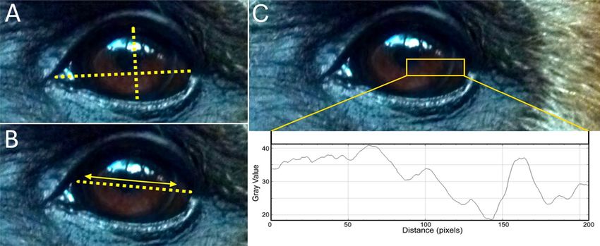
www.nature.com/scientificreports/
orangutans (Pongo pygmaeus), chimpanzees (Pan troglodytes) and Eastern gorillas (Gorilla beringei) were instead
reported to almost consistently display dark s clerae7–9, but see e.g.,10 for exceptions), so that great ape scleral
coloration does not appear to follow a clear phylogenetic pattern. Additionally, it has been shown that the amount
of visible sclera does not differ significantly between gorillas and humans in averted gaze situations, which are
of particular communicative value, demonstrating a greater than previously assumed continuity in this ocular
trait as well7. In orangutans, scleral exposure during averted glancing is markedly lower, approximating that of
humans during forwardly directed glancing11. Data on scleral exposure in varying gaze situations is so far lack-
ing for other primates.
In a recent study, Perea García et al.8 revisited the topic of ape and human eye coloration by comparatively
quantifying iridoscleral contrasts in humans, bonobos, and chimpanzees. Despite pronounced differences in
ocular coloration, the grayscale brightness of the iris when compared to the sclera (relative iris luminance = RIL)
did not differ significantly between the studied species. Chimpanzees have light, amber-colored irises that con-
trast with their typically black sclerae, inversing the pattern found in humans and the majority of bonobos.
Perea García et al.8 stated that owing to comparable RIL values, ocular pigmentation patterns in the three species
are equally conspicuous and suggested that chimpanzees, bonobos and humans share effective eye-mediated
communication.
However, there is only very little experimental support for the hypothesis that great apes or non-human pri-
mates in general rely on conspecifics’ glancing as a communicative cue ( compare12). Laboratory studies on rhesus
macaques (Macaca mulatta) monitoring eye motion showed that these monkeys respond to conspecific glancing
by reflexively aligning their own field of vision, just like humans do13. By now, comparable experimental data on
apes are not available, but it has been shown that chimpanzees can respond to human glances in a similar f ashion2.
Therefore, reflexive glance following can be expected to be shared by a variety of primates, although it remains
speculative to which degree this information is used by them in social situations. For example, chimpanzees
typically fail to exploit glancing as a referential cue in forced-choice tasks despite their ability to reflexively fol-
low human g lances2,14 (but s ee3). In any case, it is obvious how different patterns of ocular contrast and scleral
exposure might facilitate glance cueing in a variety of situations if a particular species is able to decode relevant
information9.
Apart from that, Perea García et al.8 hypothesized the depigmented sclera of humans and bonobos to be an
evolutionary byproduct for selection against aggression. In that, it would mirror the pleiotropy-induced domes-
tication syndrome described for companion a nimals15 (see a lso16, and17). They also implied that such selection
against aggression might explain the divergent scleral pigmentation patterns among gorilla and orangutan spe-
cies, suggesting a link between social patterns and ocular pigmentation. Although this byproduct hypothesis
contradicts the assumption that communicative demands chiefly drive the evolution of eye color in apes, it is
compatible with the idea of depigmented eyes being particularly effective in communicative tools. Still, limited
data make it hard to convincingly state a correlation between ocular (de)pigmentation, the expression of RIL,
and socio-cognitive functions.
To clarify these issues, it would be desirable to place ocular pigmentation in great apes into a broader evo-
lutionary perspective. Of particular interest for such comparisons are the small apes or gibbons (family Hylo-
batidae, genera Hoolock, Hylobates, Nomascus, Symphalangus), which form the sister group to the human and
great ape clade (family Hominidae). Different from great apes, gibbons are morphologically, ecologically, and
socially rather uniform. All species are specialized canopy-dwellers that live in small family g roups18. Despite
their phylogenetic position and resulting relevance to understand the evolution of hominoid cognitive traits,
gibbons are widely ignored in primate behavioral research (excluding acoustic communication), so that little is
known about their socio-cognitive traits19,20. However, there currently is consensus that glancing does not carry
noteworthy communicative value to t hem21,22. In line with this, gibbon gaze following is less sophisticated than
in their large-bodied relatives. Small apes follow the head orientation of both conspecifics and humans but do so
in a reflexive way, while great apes deduce referential information from gaze, as indicated by double checking and
habituation to repeated gazing events23. A comparison of great and small apes could therefore aid in identifying
correlates of eye-mediated communication.
As described above, it has been argued that eye coloration in the genera Homo and Pan represents an adapta-
tion for optimized glance c ueing8 and that ocular coloration is tightly correlated with socio-cognitive functions
among great apes and humans3,4,9. This notion implies that primates that are not expected to utilize glance cues
should show less conspicuous ocular contrasts, representing an unspecialized ancestral state9. Gibbon eyes indeed
appear unspecialized based on a small mixed-species dataset presented by Kobayashi and Kohshima4. Compared
to great apes, scleral portions of the gibbon eyeball remain largely unexposed during direct glancing, as their eyes
show a circular rather than an elliptical lid excision4. The uniformly colored iris is filling out the visible part of
the eye almost completely. Potential iridoscleral contrast, a trait not previously quantified in gibbons, is therefore
significantly concealed. Instead, conspicuous facial fur patterns are present in many gibbon species that might
effectively indicate head but not eye o rientation24.
Here, we provide data on eye contours as well as ocular pigmentation for a large sample of hylobatids to
draw quantitative comparisons with hominids. For this, we also quantified iridoscleral contrasts in gorillas and
orangutans, providing the first comprehensive overview of ocular pigmentation patterns across the hominoid
radiation. Following the cooperative eye hypothesis, we predicted that gibbon eyes would be less conspicuous than
hominid eyes. We further expected small ape species to display uniform patterns of ocular coloration (i.e. little
variation between species), since gaze following and other potentially eye-mediated behaviors appear not to vary
between hylobatid taxa. Finally, we assessed eye contours and the amounts of sclera exposed during forwardly
directed and averted glancing in three gibbon genera and each hominid genus. Again, we hypothesized consistent
differences between small and great apes based on the preliminary results of Kobayashi and Kohshima4, with the
small apes exposing less sclera than their large-bodied relatives.
Scientific Reports | (2021) 11:12994 | https://doi.org/10.1038/s41598-021-92348-z 2
Vol:.(1234567890)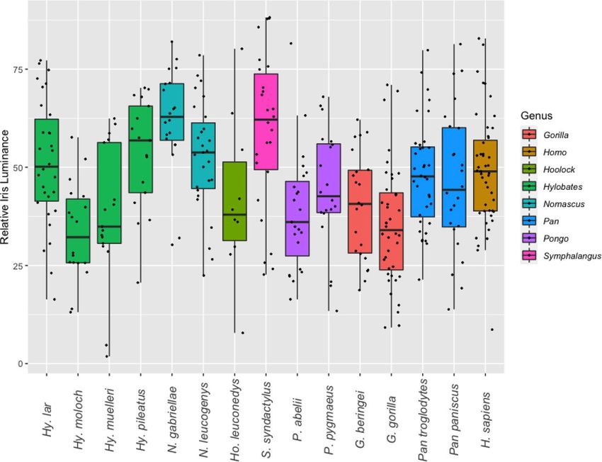
www.nature.com/scientificreports/
Species n % of type I phenotype HC RIL
Hoolock leuconedys 10 0 30.5 41.8
Hylobates lar 30 46 34.5 51.2
Hylobates moloch 18 0 39.0 33.5
Hylobates muelleri 17 0 41.5 38.0
Hylobates pileatus 17 12 30.2 53.4
Nomascus gabriellae 20 30 17.3 62.0
Nomascus leucogenys 27 0 24.1 52.7
Symphalangus syndactylus 29 38 31.2 59.4
Gorilla beringei 22 41 56.7 40.2
Gorilla gorilla 40 85 81.7 35.2
Homo sapiens* 49 100 84.4** 48.7
Pan paniscus* 24 88 61.4 46.4
Pan troglodytes* 36 11 33.3 48.0
Pongo abelii 23 87 83.7 38.6
Pongo pygmaeus 24 79 48.0 45.6
Table 1. Summary of data on ape ocular pigmentation patterns. Type I phenotype describes eyes in which the
sclera is lighter than the iris. HC and RIL correspond to species means. *Data derive from Perea García et al.8.
**n = 47.
Materials and methods
We conducted an extensive internet search for pictures of great apes and gibbons from wild as well as captive
environments and pooled images found online with private digital photographs (see Suppl. Table 1). In case of
data gathered online, information on the identity, sex and location of photographed apes was derived from the
source websites to avoid repeated sampling of particular individuals. If available, international studbooks were
used to check whether zoo-housed individuals were born in the wild or in captivity. Species level classification
and nomenclature were adopted from Burgin et al.25. Our species selection was dictated by the availability of
photographs, which forced us to exclude specific ape lineages, such as Tapanuli orangutans (P. tapanuliensis)
and the northerly distributed species of crested gibbons (N. concolor, N. hainanus, and N. nasutus). Within
hylobatids, we pooled data from recently diverging populations to reach larger sample sizes for the respective
groups. As a consequence, Nomascus siki was grouped under N. leucogenys, N. annamensis under N. gabriellae,
Hoolock tianxing under Ho. leuconedys, and Hylobates abbotti and Hy. funereus under Hy. muelleri (see Suppl.
Tables 1 and 2 for taxonomic identities of each subject). Because ocular pigmentation and the shape of the eye
contour can differ between juvenile and mature apes, we restricted our sampling to pictures of adults. Subadult
animals were included for some gibbon species but only if they had already developed adult pelage traits (e.g.,
after entering the pale color phase in female gibbons of the genera Hoolock and Nomascus).
We followed the methodology of Mayhew and Gómez7, Perea García9, and Perea García et al.8 in using
ImageJ26 to take quantitative measurements from the digital images we gathered.
Quantifying ocular luminance in great and small apes. Ocular pigmentation approximated by lumi-
nance (= gray values) was quantified for gorillas (G. beringei, n = 22; G. gorilla, n = 40), orangutans (P. abelii,
n = 23; P. pygmaeus, n = 24), crested gibbons (N. gabriellae, n = 20; N. leucogenys, n = 27), dwarf gibbons (Hy. lar,
n = 30; Hy. moloch, n = 18; Hy. muelleri, n = 17; Hy. pileatus, n = 17), hoolock gibbons (Ho. leuconedys, n = 10), and
siamangs (S. syndactylus, n = 29), summing up to 277 individuals (Table 1). Additional data on ocular pigmenta-
tion traits in adult bonobos, chimpanzees and humans was derived from Perea García et al.8, resulting in a total
of seven hominid and eight hylobatid species representing all of the eight genera of extant apes ( ntotal = 386).
To be included in the sample, picture resolution had to be high enough to unequivocally distinguish between
pupil, iris, and sclera in at least one eye of the photographed subject. We noticed that all gibbon species, as well
as orangutans and gorillas, display a thin gray line encircling the peripheral iris, which appears as a salient
demarcation to the sclera (see Fig. 1). We were unable to find discussion of this trait in the literature. While this
structure is superficially reminiscent of the arcus s enilis27 commonly found in aging humans, it probably has
different physiological underpinnings. We only sampled pictures on which this demarcation line was visible,
to clearly distinguish iridial and scleral portions of the visible eyeball. In case the aforementioned criteria were
met, pictures capturing both direct and averted gaze were included.
We used the plot profile function in ImageJ (Fig. 1) to retrieve luminance values from the images9. Only traits
from one eye per subject were quantified. In doing so, we chose the better illuminated eye for measurements. In
case both eyes appeared equally well visible (direct gaze conditions), we selected the one with higher contrast
values to further mitigate shadow-induced biases.
We quantified gray scale luminance values to quantify ocular contrasts and classified eyes into two phenotypic
groups following Perea García et al.8. In type 1 eyes, the sclera is (at least partially) lighter than the iris (e.g.,
humans), while the opposite is true for type 2 eyes (e.g., most chimpanzees). Dependent on eye type, we chose
either the highest or lowest grayscale luminance values from either portion of the eye to achieve the highest
Scientific Reports | (2021) 11:12994 | https://doi.org/10.1038/s41598-021-92348-z 3
Vol.:(0123456789)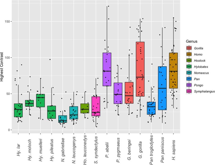
www.nature.com/scientificreports/
Figure 1. Visualization of ocular measurements exemplified on a white-handed gibbon (Hylobates lar).
(A) Measurements of width and height of the eye taken to calculate a ratio (width-height ratio, WHR). (B)
Measurements of the width of visible sclera and iris to calculate a ratio (scleral size index, SSI). (C) Deriving
ocular luminance measurements (HC, RIL) from gray values in ImageJ using the plot profile function. Photo
credit: Thomas Geissmann.
possible contrast difference. Subsequently, these values were used to calculate the absolute (highest ocular con-
trast = HC) and relative (relative iris luminance = RIL) differences between scleral and iridal luminance per
eye. RIL reflects the percentage of grayscale luminance shown by the darker portion of the eye (sclera or iris)
in comparison to the lighter one, which per definition is assumed to represent 100% luminance28. Higher HC
values indicate greater conspicuousness, while the opposite is the case for RIL. Procedures were adopted from
Perea García9 and Perea García et al.8. Reflections mirrored in the eye as well as the demarcation line between
iris and sclera were carefully avoided. We also quantified the gray value slope between iris and s clera9 for each
individual but did not incorporate these data into our analyses (Suppl. Table 1).
Besides interspecific comparisons, we attempted to compare pigmentation patterns in wild and captive-
bred individuals. This way we could test for potential biases resulting from an animal’s rearing background. It
is known that the physiology and morphology of zoo animals can significantly differ from wild conspecifics in
various ways29, and an effect on eye pigmentation should be considered possible. However, we only considered
our samples sufficient for such a comparison in Gorilla gorilla (ncaptive-bred = 23; nwildborn = 17) and Hylobates lar
(ncaptive-bred = 13; nwildborn = 9).
Quantifying ocular shape and sclera exposure in hominoids. Ocular shape was quantified at the
genus level for all genera of extant hominoids except for Hoolock. The latter was omitted due to a lack of suitable
photographs. A set of images from 20 individuals representing either direct (eyes oriented forwardly towards
the camera; n = 10) or sideways averted glances (n = 10) was analyzed for each genus, respectively. Congeneric
species were grouped because preliminary screenings did not reveal notable differences. This resulted in a total
sample of 140 pictures (Suppl. Table 2). In all these photos, subjects consistently faced the camera with fully
opened eyes to reduce effects of the angle of photography on the measurements. While pictures of non-human
primates were collected as described beforehand, all photos of humans derived from the private archives of the
authors. All persons pictured gave consent for the photos to be used in this study. The human subjects were of
diverse Eurasian descent. For nonhuman species, a trained observer (KRC) assigned photos into the “direct
glance” and “averted glance” categories but a naïve coder (SB) scored them into said categories as well, allowing
us to compute Cohen’s Kappa as a measure of inter-rater reliability.
The width-height ratio (WHR) and the exposed sclera size index (SSI) of eyes were quantified, following
the procedures of Kobayashi and Kohshima4 and Mayhew and Gómez7 to allow for meaningful comparisons
(Fig. 1). WHR is a measure to approximate eye shape, while SSI indicates the amount of exposed sclera. WHR
was calculated from all images in the dataset for the respective genus, while SSI was calculated for direct and
averted glance images independently to approximate changes in visible scleral surface during averted glancing.
In case both eyes of an individual were clearly visible (n = 137), measurements from the left and right eye were
averaged (Suppl. Table 2).
Statistical analysis. All statistics were performed in R
30. After log-transformation, normal distribution
of data was assessed by applying the Shapiro–Wilk test and homogeneity of variances was checked by running
the Bartlett test. Parametric data were compared via ANOVA, while non-parametric data were analyzed using
Wilcoxon’s rank sum test. Tukey’s Honest Significant Differences was employed as a post-hoc test correcting for
multiple comparisons subsequent to ANOVA, while a Bonferroni correction was applied to address this issue
for Wilcoxon tests. Differences in RIL and HC between wildborn and captive-bred individuals were tested by
Scientific Reports | (2021) 11:12994 | https://doi.org/10.1038/s41598-021-92348-z 4
Vol:.(1234567890)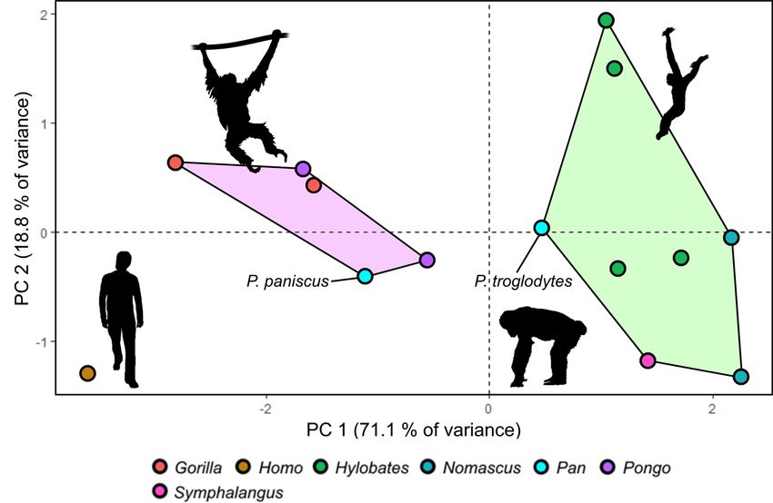
www.nature.com/scientificreports/
Genus WHR n SSI (direct) n SSI (averted) n
Hylobates 1.60 (0.13) 20 1.15 (0.06) 10 1.46 (0.13) 10
Nomascus 1.57 (0.14) 20 1.19 (0.09) 10 1.36 (0.08) 10
Symphalangus 1.70 (0.17) 20 1.24 (0.06) 10 1.46 (0.07) 10
Gorilla 2.17 (0.24) 20 1.71 (0.08) 10 2.21 (1.30) 10
Homo 2.75 (0.43) 20 2.0 (0.31) 10 2.30 (0.27) 10
Pan 1.93 (0.16) 20 1.46 (0.11) 10 1.82 (0.16) 10
Pongo 1.87 (0.15) 20 1.52 (0.10) 10 1.76 (0.28) 10
Table 2. Median values (± SD) of width-height ratio (WHR) and scleral size index (SSI) during direct and
averted gaze in seven hominoid genera.
employing the Welch two sample t-test (Hy. lar) or the Wilcoxon rank sum test (G. gorilla). All tests were per-
formed at an α-level of 0.05.
We visualized phylogenetic patterns, quantified phylogenetic signals (Pagel’s λ, phylosig function) and
computed maximum likelihood ancestral state estimates (fastAnc function) with the phytools package version
0.7–7031. The hominoid phylogeny used was derived from the 10kTrees w ebsite32.
To visualize differences in the quantified ocular traits, a principal component analysis (PCA) was run on the
species mean values for HC, RIL, and the species-specific proportion of type 1 eyes in the population (Type)
as well as on the genus medians for SSI during averted glancing and WHR (see Tables 1, 2). Due to the lack of
SSI and WHR measurements, Hoolock was omitted from the analysis. SSI and WHR data of congeneric species
were assumed to be equal.
Results
Qualitative assessment of ocular pigmentation in great and small apes. In small apes, the sclera
was found to be predominately darker than the iris (type 2 phenotype; n = 135 of 168; 80%), but notable inter-
specific variation was found (Table 1). In some species, the type 2 pattern was recovered for all individuals (Ho.
leuconedys, Hy. moloch, Hy. muelleri, N. leucogenys). The highest prevalence of light sclerae (type 1 phenotype)
was found in siamangs (S. syndactylus, n = 11 of 29; 38%) and white-handed gibbons (Hy. lar, n = 14 of 30,
46%). In most of these individuals, however, scleral depigmentation was moderate, leading to a medium to light
brown sclera, which often was only minimally lighter than the iris. Of all sampled hylobatids, only four white-
handed gibbons displayed advanced bilateral depigmentation of the sclera, resulting in a mottled white appear-
ance, vaguely resembling the human condition. The small ape sclera, if not depigmented, appears dark brown to
almost black in all species. Iris color was found to be mostly dark to chestnut brown (see Fig. 2). However, in Hy.
moloch and Hy. muelleri, the iris has a dark amber color, similar to that of chimpanzees (Fig. 2).
The sclerae of gorillas and orangutans tended to be, at least in parts, lighter than their irises, displaying vari-
ous degrees of depigmentation. Eastern gorillas were an exception to this trend. Here the inverse pattern (G.
beringei, n = 13 of 22, 59%) was found to be more abundant but both occured at high frequencies. In Western
gorillas and orangutans, light sclerae were predominately found (G. gorilla, n = 34 of 40, 85%; P. abelii, n = 20 of
23, 87%; P. pygmaeus, n = 19 of 24, 79%). However, just as in many gibbon species, depigmentation in Bornean
orangutans was often low, leading to predominately brownish instead of white sclerae in this species. In both
gorillas and orangutans, the portions of the sclera immediately surrounding the iris typically (but not always)
remained pigmented, creating a dark frame of variable thickness.
Gorillas deviated from both orangutans and gibbons in frequently displaying clearly asymmetric depigmen-
tation patterns. In approximately one quarter of Eastern (n = 6 of 22; 27%) and Western gorillas (n = 10 of 40;
25%), conspicuous depigmentation was restricted to just one eye. This pattern was only noted for one Sumatran
orangutan and was absent in the Bornean species. In gibbons, such asymmetries were also rare, occurring in
white-handed gibbons (n = 4 of 30; 13%), pileated gibbons (n = 2 of 17; 12%) and siamangs (n = 2 of 29; 7%).
Quantitative assessment of ocular pigmentation in great and small apes. At the family level, we
found a greater HC (Wilcoxon test: p < 0.001; Fig. 3) but lower RIL (Wilcoxon test: p < 0.001; Fig. 4) in hominids
compared to hylobatids.
Humans, Sumatran orangutans and Western gorillas exhibited the highest mean HC values, followed by
bonobos, Eastern gorillas, Bornean orangutans and chimpanzees (Table 2). Chimpanzees were the only homi-
nids for which the mean HC was recovered to lay within the range of variation of hylobatid species means.
Among small apes, Bornean and Javan gibbons exhibited the highest HC values while the lowest were found
among crested gibbons. Concerning RIL, species means for hominids fell within the range of hylobatid variation
(Fig. 4). The lowest RIL in the sample was found in Javan gibbons, while the highest was recovered for Southern
yellow-cheeked gibbons. Among hominids, humans displayed the highest RIL, while the lowest was found in
Western gorillas (Table 2).
Reflecting these findings, we found a significant but moderate phylogenetic signal for HC among the homi-
noid sample (Pagel’s λ = 0.565, p = 0.026). RIL on the other hand was not found to correlate with phylogeny
(Pagel’s λ < 0.001, p = 1), which is mirrored by the inconsistent distribution of the trait among the studied taxa.
Maximum likelihood ancestral state estimates of HC and RIL for each node within our hominoid phylogeny
Scientific Reports | (2021) 11:12994 | https://doi.org/10.1038/s41598-021-92348-z 5
Vol.:(0123456789)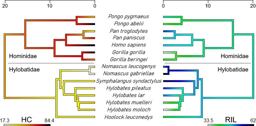
www.nature.com/scientificreports/

                                            Figure 2.  Typical external appearance of the eye in representatives of the seven non-human ape genera
                                            (superfamily Hominoidea). Note the difference in scleral exposure between gibbons (A–F) and great apes
                                            (G–L) (A) Southern white-cheeked gibbon (Nomascus (leucogenys) siki). (B) Southern yellow-cheeked gibbon
                                            (Nomascus gabriellae). (C) Siamang (Symphalangus syndactylus). (D) East Bornean gray gibbon (Hylobates
                                            funereus). (E) White-handed gibbon (Hylobates lar). (F) Gaoligong hoolock gibbon (Hoolock tianxing).
                                            (G) Eastern gorilla (Gorilla beringei). (H) Western gorilla (Gorilla gorilla). (I) Bonobo (Pan paniscus). (J)
                                            Chimpanzee (Pan troglodytes). (K) Sumatran orangutan (Pongo abelii). (L) Bornean orangutan (Pongo
                                            pygmaeus). Photo credit: A, E—Miriam Lindenmeier (used with permission); K—Kai R. Caspar; all remaining
                                            pictures—Thomas Geissmann.

          Scientific Reports |   (2021) 11:12994 |              https://doi.org/10.1038/s41598-021-92348-z                                                  6

Vol:.(1234567890)
www.nature.com/scientificreports/
Figure 3. Variation of highest ocular contrast (HC) in the eight extant hominoid genera at species level.
Figure. 4. Variation of relative iris luminance (RIL) in the eight extant hominoid genera at species level.
Scientific Reports | (2021) 11:12994 | https://doi.org/10.1038/s41598-021-92348-z 7
Vol.:(0123456789)www.nature.com/scientificreports/
Figure 5. Visualization of phylogenetic patterns in species means of highest ocular contrast (HC, left) and
relative iris luminance (RIL, right) in the ape superfamily (Hominoidea). Note the discrepancy between the
two measures and the secondary acquisition of a gibbon-like HC in chimpanzees (Pan troglodytes). Time scales
correspond to million years before present. Maximum likelihood ancestral state estimates at the nodes of the
trees are provided in Supplementary Table 3.
Hoolock Hylobates Hylobates Hylobates Hylobates Symphalangus Nomascus Nomascus Gorilla Gorilla Homo Pan Pan Pongo Pongo
Species leuconedys lar moloch muelleri pileatus syndactylus gabriellae leucogenys beringei gorilla sapiens paniscus troglodytes abelii pygmaeus
Hoolock leuconedys 1.000 1.000 1.000 1.000 1.000 0.289 1.000 0.973 0.01 0.001 1.000 1.000 0.001 0.648
Hylobates lar 0.942 1.000 1.000 1.000 1.000 0.018 1.000 0.129 0.000 0.000 0.755 1.000 0.000 0.355
Hylobates moloch 0.99 0.012 1.000 1.000 1.000 0.000 0.039 1.000 0.05 0.000 1.000 1.000 0.000 1.000
Hylobates muelleri 1.000 0.249 1.000 1.000 1.000 0.002 0.071 1.000 0.129 0.000 1.000 1.000 0.012 1.000
Hylobates pileatus 0.859 1.000 0.014 0.2 1.000 0.457 1.000 0.039 0.000 0.000 0.656 1.000 0.000 0.102
Symphalangus syndactylus 0.138 0.811 0.000 0.001 0.996 0.243 1.000 0.113 0.000 0.000 0.527 1.000 0.000 0.093
Nomascus gabriellae 0.055 0.49 0.000 0.000 0.934 1.000 1.000 0.000 0.000 0.000 0.001 0.005 0.000 0.000
Nomascus leucogenys 0.852 1.000 0.005 0.137 1.000 0.962 0.761 0.001 0.000 0.000 0.025 1.000 0.000 0.001
Gorilla beringei 1.000 0.424 0.99 1.000 0.345 0.002 0.001 0.25 1.000 0.074 1 0.073 0.886 1.000
Gorilla gorilla 0.997 0.002 1.000 1.000 0.005 0.000 0.000 0.001 0.996 1.000 1 0.000 1.000 1.000
Homo sapiens 0.99 1.000 0.024 0.417 1.000 0.302 0.113 1.000 0.643 0.003 1 0.000 1.000 0.01
Pan paniscus 1.000 0.999 0.326 0.928 0.984 0.175 0.063 0.983 0.99 0.244 1.000 1.000 1.000 1.000
Pan troglodytes 0.998 1.000 0.081 0.663 0.998 0.227 0.082 0.998 0.866 0.026 1.000 1.000 0.000 0.145
Pongo abelii 1.000 0.19 0.999 1.000 0.165 0.000 0.000 0.095 1.000 1.000 0.333 0.926 0.61 0.15
Pongo pygmaeus 1.000 0.973 0.687 0.996 0.911 0.081 0.027 0.894 1.000 0.677 0.998 1.000 1.000 0.997
Table 3. Species-level comparison of highest ocular contrast (HC) and relative iris luminance (RIL) between
hominoids. Values of the upper triangular matrix (orange) correspond to statistical results for HC, those of the
lower one (blue) to RIL. Bold values indicate significant results (HC: Bonferroni-corrected Wilcoxon tests; RIL:
Tukey’s Honest Significant Differences).
are provided in Supplementary Table 3 and are visualized in Fig. 5. Importantly, hominids, when compared to
hylobatids, maintained a contrasting coloration throughout their evolutionary history. A comparatively high
HC (63.6; 95% CI: 44–83.3) was also estimated for the common ancestor of the Pan-Homo clade, suggesting the
dark eyes of chimpanzees to be a recently evolved trait.
Results from interspecies comparisons in HC and RIL are summarized in Table 3. For HC, only crested gib-
bons (genus Nomascus) exhibited values that significantly differed from other hylobatids, their eyes being notably
dark. This genus also included the only gibbon species that significantly deviated from the patterns found in
chimpanzees, bonobos and Bornean orangutans. Human HC differed significantly from all species in the sample
except for Sumatran orangutans, bonobos, and gorillas, species which also tend to exhibit depigmented sclerae.
Therefore, the pattern of contrasts found in the human eye is not unique. Comparisons of RIL did not produce
similarly comprehensible patterns. While some species did not show significant differences to any others in the
sample (Eastern hoolock, bonobo), Western gorillas did so in comparison to seven species, including humans,
chimpanzees, and a range of small apes. There was a moderate but significant negative correlation between HC
and RIL (Pearson’s r =− 0.54, p = 0.04).
No significant differences in RIL and HC could be detected between captive-born and wild-born Western
gorillas (Wilcoxon test: W ≥ 129; p ≥ 0.07) or white-handed gibbons (t-test: t ≥ -0.511; p ≥ 0.62).
Scientific Reports | (2021) 11:12994 | https://doi.org/10.1038/s41598-021-92348-z 8
Vol:.(1234567890)www.nature.com/scientificreports/
Figure 6. Results of PCA on quantified ocular traits in apes. (A) Visualization of PCA results. Chimpanzees
and gibbons form a cluster highlighted in green. Gorillas, bonobos and orangutans cluster together in a
morphospace colored in light purple. Humans are separated from all other hominoids. The orangutan silhouette
is in public domain, others were created by KRC.
Quantifying ocular shape and sclera exposure in hominoids. Inter-rater reliability for the scoring
of glance direction was strong throughout, but agreement was slightly higher for the great ape (59/60; κ = 0.96,
p < 0.001) than for the gibbon sample (56/60; κ = 0.86; p < 0.001). Mean SSI was found to be consistently smaller
in hylobatids compared to those of hominids, with a particularly pronounced difference occurring in the averted
gaze condition (Table 2, Suppl. Table 4; Bonferroni-corrected Wilcoxon test: p < 0.001 for all hominid-hylobatid
species pairs in both glance conditions). In WHR, Symphalangus did not differ significantly from either Pan or
Pongo (p > 0.13), while all other interfamilial comparisons of WHR yielded marked differences (p < 0.001). Small
ape genera did not deviate significantly from each other in either direct or averted gaze SSI (p > 0.3) and the only
significant difference in WHR among small apes was found between Symphalangus and Nomascus (p = 0.04),
with the latter displaying lower WHR values. Within the Hominidae, Homo deviated significantly from all other
genera in direct glance SSI and WHR (p ≤ 0.01). However, in averted glance SSI, Homo only differed significantly
from Pan and Pongo (p < 0.05) but not from Gorilla (p = 1). No notable differences were found between Pan and
Pongo in any of the measurements observed (p = 1 for all comparisons). Gorilla significantly deviated from all
other hominids in WHR and direct glance SSI, but only from Pan regarding averted SSI (p < 0.01).
Principal component analysis of ocular traits. PCA grouped hominoids into three groups based on
ocular traits (Fig. 6). The first two principal components of the PCA encompassed 89.8% of the total variance
in the sample, exhibiting eigenvalues of 3.55 and 0.94, respectively. Variable contributions are visualized in Sup-
plementary Fig. 1. The first group is constituted by hylobatids and chimpanzees, which clustered together in the
PCA morphospace (Fig. 6). Bonobos grouped together with gorillas and orangutans, forming a second cluster.
Finally, humans fell far outside of the range of variance of all the other hominoid genera. A second PCA omitting
RIL, which is a problematic variable (see “Discussion” section), resulted in a similar pattern (Suppl. Figures 2 and
3), with the first and second principal component encompassing even more of the total variance in the sample
(94.3%). In this PCA, gorillas were situated closer to humans than in the first one, diffusing the cluster consti-
tuted by bonobos, gorillas and orangutans.
Discussion
General discussion of results. Although we found only few consistent differences in ocular traits separat-
ing small and great apes, some predictions of the cooperative eye hypothesis could be confirmed. Importantly, SSI
was found to be consistently lower in gibbons compared to other hominoids and this difference became even
more pronounced in averted gaze situations. This finding supports the conclusions of Kobayashi and Kohshima4
that were drawn from a small-scale dataset and further demonstrates that gibbon eyes are indeed far less suited
to convey glance signals than those of great apes and humans. Regarding the width-height ratio of the eyes, hylo-
Scientific Reports | (2021) 11:12994 | https://doi.org/10.1038/s41598-021-92348-z 9
Vol.:(0123456789)www.nature.com/scientificreports/
batids also displayed lower mean values than hominids. Nevertheless, siamangs approach the great ape genera
Pan and Pongo in WHR and were found not to differ significantly from them in that regard. Still, this did not
result in notably higher SSI values in siamangs compared to other gibbons, nor to equal SSI when compared to
these great apes (Table 2). Our results on scleral exposure and eye outlines in human and great ape eyes match
the data from previous s tudies4,11. In particular, we replicated the finding of Mayhew and Gómez7 that humans
and gorillas do not differ in the degree of scleral exposure during averted glancing but do so in the direct glance
condition. The reason for this disparity lies probably in the horizontally widened outline of the human eye. It
causes rotations of the human eyeball to have less of an effect on scleral exposure when compared to other homi-
noids. Accordingly, the relative difference in the amount of visible sclera between direct and averted glance SSI
shown by humans is exceeded by all great apes as well as by the gibbon genus Hylobates (Table 2).
Pigmentation patterns followed a roughly similar pattern to SSI among hominoids, but they were not con-
gruent. Hylobatids displayed less contrasted eyes than their large-bodied relatives, as indicated by values for
highest ocular contrast (HC), which were found to moderately correlate with hominoid phylogeny. As with the
comparatively small amounts of exposed sclera in the gibbon eye, this again demonstrates a greater signaling
value of hominid compared to hylobatid eyes. At the species level however, this notion cannot be generalized as
chimpanzees were found to exhibit mean HC values in the range of gibbons, rendering their eyes similarly incon-
spicuous. Chimpanzee HC differed significantly from that of hominids with strongly contrasted eyes (humans,
Sumatran orangutans and Western gorillas) but not from the ones of siamangs, dwarf gibbons, and hoolocks.
As exemplified by our results on Western gorillas and white-handed gibbons, wildborn and captive-bred apes
do not differ in ocular contrasts, making biases through imbalanced sampling of natural and captive popula-
tions unlikely. Still, it should be pointed out that our methodology does not capture the full extent of scleral
pigmentation patterning. For instance, pronounced local scleral depigmentation will yield similar results to a
fully depigmented sclera, despite obvious phenotypic differences and related effects on glance direction signaling
(compare Fig. 2C). This constitutes an important limitation of our method, which is also insensitive to asym-
metric expressions of pigmentation. Merging quantitative analyses with a qualitative scoring of pigmentation
patterns (compare7 could constitute a way of overcoming this limitation in future studies. Another potential
shortcoming of our, as well as of all previous approaches so far, is that only differences in ocular contrasts but
not in hues were quantified (see33) or scored to approximate salience. This could have led to an underestimation
of conspicuousness, particular in species with dark sclerae.
Relative iris luminance (RIL) was recovered as a trait that varied independent of phylogeny. Importantly, we
could not find support for the assumption that low RILs are reliable indicators of more conspicuously colored
eyes8, despite a moderate negative correlation of the two traits within our sample. The lowest average RIL value
found was that of the Javan gibbon (Hylobates moloch, mean RIL: 33.5) which shows an amber-colored iris and
dark brown sclera, rendering its eyes obviously far less conspicuous than the ones of, for instance, humans (mean
RIL: 48.7), which were found to have a significantly higher RIL. This fact points out a major issue in the usage
of RIL as a meaningful measure of ocular pigmentation. If gray values for sclera and iris are both low and differ
little from one another, resulting RIL values may still equal or range below those obtained from eyes that show
salient contrasts between these regions. This insensitivity makes RIL an unsuitable proxy for the conspicuous-
ness of ocular pigmentation patterns. However, it might still be used to analyze intraspecific pigmentation
patterns in groups with uniformly colored sclerae such as humans, for which the measure has originally been
established28. Given that HC is a more faithful proxy of general ocular conspicuousness, we propose to rely on
HC rather than on RIL to quantify ocular pigmentation in future comparative studies. For this, large sample sizes
are recommended to counteract effects of differing lighting regimes in the analyzed pictures. Perea García et al.8
have argued based on RIL that chimpanzee, bonobo and human eyes are equally suited to convey gaze signals.
However, for reasons just pointed out, this argument does not hold. In ocular pigmentation, chimpanzees’ dark
eyes resemble the ones of gibbons more than the human or even the average bonobo condition, arguing against
a human-like social signaling function of chimpanzee eye coloration (compare Fig. 4). Above that, scleral expo-
sure in Pan is the lowest among African apes, differing significantly from both humans and gorillas in averted
and direct glancing situations. For these reasons, chimpanzee eyes are notable for being, on average, the least
conspicuous of all hominids. Low RIL values alone fail to diagnose species that employ glance cueing or even
sophisticated gaze cueing, as exemplified by several gibbon species in our sample.
Our results highlight differences in scleral depigmentation rates in hominids compared to hylobatids. The
dataset of Kobayashi and K ohshima4 would suggest the dark-eyed gibbon pattern to be the plesiomorphic one.
Therefore, more contrastingly colored eyes would be expected to have evolved in hominids after their split from
hylobatids. However, this hypothesis can be challenged, given the small species sample sizes in that study together
with the fact that its assumptions were later shown not to hold for most hominids7–9. From just our own experi-
ence, we can anecdotally report the presence of light sclerae in multiple species of Old World and New World
monkeys (Suppl. Figure 4). Studies on the frequency and phylogenetic distribution of this trait in primates other
than apes are necessary to sufficiently characterize the ancestral state for hominoid eye pigmentation.
It is difficult to discern what underlies patterns of ocular appearance among the two hominoid families.
Considering the cooperative eye hypothesis, it might be tempting to suggest that brighter and more exposed eyes
in great apes reflect the more sophisticated gaze following behavior in this group when compared to g ibbons23.
However, the hypothesis fails to explain the derived chimpanzee phenotype and cannot account for the great
variability of hominid ocular contrasts. Furthermore, differences in SSI and WHR between hylobatids and
hominids might be more parsimoniously explained by scaling effects deriving from differences in body size
instead of by communicative demands4.
It is notable that each great ape genus encompasses species that markedly differ in their ocular pigmentation
patterns (compare8,9). Perea García et al.8 suggested that scleral depigmentation in apes might be an evolutionary
byproduct of greater social tolerance, induced by pleiotropic genes controlling neural crest development and
Scientific Reports | (2021) 11:12994 | https://doi.org/10.1038/s41598-021-92348-z 10
Vol:.(1234567890)www.nature.com/scientificreports/
mirroring patterns found in domesticated mammals. Indeed, the dark-eyed Bornean orangutans and Eastern
gorillas are less tolerant towards unfamiliar conspecifics than their congeners in the wild (van Schaik 1999;
Cooksey et al. 2020). Nevertheless, it remains to be clarified whether this is a consequence of intrinsic behavioral
predispositions rather than extrinsic factors relating to habitat characteristics. Between bonobos and chim-
panzees, such intrinsic predispositions are far better characterized than in other ape taxa and point to marked
physiological differences underlying behavioral disparities within the genus Pan34,35. Comparisons of socially
tolerant and despotic monkey species could further test for a correlation between scleral pigmentation and
aggressiveness. Yet, specific pleiotropic effects affecting scleral but not general skin or fur coloration appear to
be yet undescribed in mammals (compare e.g.,15), making the link a speculative one at the moment. It is also
unclear how the social tolerance hypothesis might apply to the hominid family in general when compared to
hylobatids or specifically to white-handed gibbons. Although the latter show the highest scleral depigmentation
rates among small apes, there is no evidence to suggest them to exhibit decreased levels of aggression towards
unfamiliar conspecifics when compared to other species.
To sum up, the cooperative eye hypothesis might fit the family-level patterns we describe but loses its explana-
tory power at the species level. It is further important to point out that the human eye is on average not more
saliently contrasted than that of gorillas, bonobos and Sumatran orangutans, further challenging its validity. The
occurrence of at least locally depigmented sclerae in varying portions of the total population likely is an ancient
hominid trait but its biological significance remains obscure. The view that extensive scleral depigmentation
is exclusive to the human lineage4 or that it might even be diagnostic for the species Homo sapiens6 cannot be
maintained. Finally, reliance on RIL as an indicator of ocular conspicuousness may give rise to misleading results.
Is ocular pigmentation linked to specific socio‑cognitive traits? Previous research on scleral pig-
mentation in primates has highlighted a potential connection between cognitive traits such as social glance
cueing and conspicuous ocular pigmentations3,4,6,8. Whether these characters are correlated is not yet clear, how-
ever, because humans are the only primate species that evidently combines them (see below). Deducing glance
cueing and other cognitive abilities from eye coloration alone may quickly lead into an adaptationist pitfall.
Even if primate species converge in ocular pigmentation, the ways in which these species perceive conspecifics’
eyes may differ. For example, Western gorillas and Sumatran orangutans exhibit ocular contrasts resembling the
human condition. Yet, their viewing patterns of conspecific’s faces and particularly eyes, is more reminiscent of
chimpanzees than humans, they exhibit pronounced gaze avoidance in diverse social contexts and evidence of
conspecific glance cue exploitation is, to our knowledge, absent11,36.
The hypothesis that dark eyes evolved to mask glance direction in competitive social environments must be
critically reevaluated as well. Kobayashi and K ohshima4 proposed that all non-human primates would exhibit
dark sclerae to benefit from “gaze camouflage”, but this idea remains hypothetical rather than empirical. This
assumption is not based on any experimental evidence and does not address how this trait would be advantageous
to primates with low SSI and across the extreme diversity of social, activity, and foraging regimes that the 91
species they studied encompass. In line with this, the additional notion that dark sclerae would lower predation
risk via gaze concealment1,4 is purely speculative as well. Kobayashi and Kohshima4 also ignore the relevance
of other facial ornaments for gaze/glance communication ( compare11,37 and the great variety of cooperative
behaviors in non-human primates that are often linked to gaze following (compare38). Dark sclerae appear to
be a plesiomorphic primate t rait4, and disentangling its evolutionary roots will require accounting for other
mammals or even more distantly related vertebrate groups. We therefore discard both the glance cueing and
cryptic gaze hypotheses of eye coloration as too simplistic to be helpful in interpreting the evolution of ocular
pigmentation in primates. Our dataset could be expanded to test, whether ocular morphology indeed correlates
with specific cognitive traits across primate groups. For this, however, there must be agreement on the defini-
tions of the cognitive characteristics studied. Currently, this is not the case for primate gaze or glance cueing.
So far, most information on gaze and glance cue understanding in apes derive from studies in which human
experimenters signal to an ape subject in a captive s etting3,14,21,22. It is important to note that responsiveness to
human eye orientation by habituated animals does not equate with the usage of glance cues among conspecifics,
as for instance the successful exploitation of human glances by Californian sea lions (Zalophus californianus)
demonstrates39. The evidence that apes utilize conspecifics’ glancing independent from head orientation to
inform their actions is meager. Many studies assume that apes’ head direction and eye orientation align with
each other for the most part, despite evidence of the contrary12. Equating head and eye direction in interpreta-
tions of glancing behavior has at times led to confusion. For example, Perea García et al.8 cite studies that either
did not differentiate between the t wo40,41 or that approximated gaze by head orientation a lone42 to support the
notion that glance cues are relevant to chimpanzee communication. We are not aware of studies that unequivo-
cally show exploitation of conspecific glance cues in any ape species within a social context. An effect of ocular
contrast on such behaviors would need to be demonstrated further. Because humans can reliably deduce the
glance direction of chimpanzees, it can be hypothesized that from a perceptual perspective, conspecifics might
do so as well, despite their cryptically colored e yes12. The additional effect of an increased iridoscleral contrast
on gaze salience might well be comparatively minor. On a different note, it should be discussed how exactly the
inclusion of glance cues could enhance apes’ communicative repertoire in the wild. What referential informa-
tion could glance cues convey in a naturalistic setting that head orientation cannot? In the absence of evidence
for conspecific glance cueing in great and small apes or an unambiguous link between ocular pigmentation
and cognitive traits relating to gaze/glance following, as well as for reasons of parsimony, we assume that these
characters evolve independently from one another.
Scientific Reports | (2021) 11:12994 | https://doi.org/10.1038/s41598-021-92348-z 11
Vol.:(0123456789)www.nature.com/scientificreports/
Which factors underly the pigmentation of the human eye? The evolutionary trend of scleral
depigmentation in hominids finds its strongest expression in humans. Although rudimentary pigmentation of
the conjunctiva and inconspicuous scleral spots can frequently be found in h umans43, apparent complete scleral
depigmentation approaches 100% in our species4. Why do humans, but not other apes, exhibit such a uniformly
white scleral phenotype? The assumption that this trait evolved to facilitate glance or gaze cueing is problematic.
First, as already pointed out, its occurrence among mammals that do or do not show sophisticated gaze following
or glance cueing has not been sufficiently investigated. Above that, available evidence suggests that humans can
reliably assess the glance direction of chimpanzees from a distance of 2–10 m12 as well as that of human models
with artificially modified scleral colors44. Although it took naïve participants significantly longer to assess the
glance direction of the latter compared to natural human eyes, the time differences only encompassed fractions
of a second and no differences in the accuracy of deducing glance direction from normal human models and
those with matched iris and scleral colors were found44. The relevance of the depigmented sclera for human com-
munication might therefore be overstated, especially when compared to other morphological (e.g., the widened
horizontal outline of the visible eyeball) or physiological ocular traits of our species (e.g., emotional tearing,
particularly in infants), which are not shared by other hominoids7,45).
What requires an explanation is perhaps not that the human sclera simply exhibits depigmentation but that
its expression is uniformly extreme across individuals and populations. The marked variability of scleral color in
other hominid genera, which includes complete scleral depigmentation7, makes a similar phenotypic diversity
in human ancestors appear likely. Following that, the human pattern does not necessarily require an adaptive
explanation but may simply result from genetic drift acting on ancestral trait variability. It is also reasonable
to assume that sexual selection has contributed to the evolution of human eye pigmentation, not excluding
but possibly complementing effects of genetic drift. As we have shown, other apes do not only exhibit strong
interindividual differences in scleral pigmentation but at times also asymmetric scleral coloration, particularly
gorillas (compare a lso7). Both may point to a relaxed evolutionary pressure on ocular appearance in great apes
compared to humans.
It has been demonstrated that scleral brightness strongly affects the attractiveness of human eyes and that it
can act as an indicator of individual age46,47. A negative effect of age on scleral brightness has also been shown for
chimpanzees and bonobos8. Thereby, uniformly light, salient sclerae should contribute to a juvenilized appearance
of the face, which complies to general sexual selection pressures for neotenic facial traits in humans compared
to other hominids, particularly in f emales6,48–50. Similarly, the symmetrical scleral depigmentation pattern in
our species is in line with that human facial attractiveness is generally enhanced by increased symmetry51. Light
sclerae in humans could take over signaling functions that are absent in other apes, for example because they
pay less attention to or even prefer to avoid gazing into each other’s eyes, e.g., gorillas and orangutans11,36; or
because correlates of scleral brightness such as youthfulness do not increase sexual attractiveness in these spe-
cies, e.g., chimpanzees52. Distinct selection pressure on ocular appearance in humans is also indicated by iris
color variability. Although there is at least one other primate genus, spider monkeys, in which two distinct iris
color morphs co-occur in the adults of some species (Ateles fusciceps and A. hybridus53, A. paniscus—personal
observation), humans appear to be unique among non-domesticated mammals in the diversity of iridal hues
found in the global population, particularly Western Asia and E urope54. Other apes display uniform, species-
specific iridal coloration. Given that humans exhibit notable eye color preferences in the context of mate choice55,
it can be assumed that iris color represents a sexually selected ocular trait that differentiates our species from
non-human apes, just as it might be the case with the plain white sclera.
Conclusion
Our data add to growing evidence suggesting a graded evolution of hominoid ocular coloration instead of a clear
dichotomy between human and non-human primate eyes. Still, the evolutionary drivers behind the recovered
trend of scleral depigmentation in hominids, which peaked in humans and reversed in chimpanzees, remain
unidentified. However, the great intra- and interspecific variability of hominoid eye pigmentation opens possibili-
ties for further comparative research which should include both the great apes and gibbons as well as a range of
outgroup taxa. The classic cooperative eye hypothesis that proposes an evolutionary link between primate ocular
morphology and social signaling, needs to be experimentally revisited and scrutinized. In order to uphold it,
a clear relevance to ocular pigmentation traits for communication among conspecifics in nonhuman primates
must be demonstrated.
Received: 31 March 2021; Accepted: 9 June 2021
References
1. Langton, S. R. H., Watt, R. J. & Bruce, V. Do the eyes have it? Cues to the direction of social attention. Trends Cogn. Sci. 4(2), 50–59
(2000).
2. Povinelli, D. J. & Eddy, T. J. Chimpanzees: joint visual attention. Psychol. Sci. 7(3), 129–135 (1996).
3. Tomasello, M., Hare, B., Lehmann, H. & Call, J. Reliance on head versus eyes in the gaze following of great apes and human infants:
the cooperative eye hypothesis. J. Hum. Evol. 52(3), 314–320 (2007).
4. Kobayashi, H. & Kohshima, S. Unique morphology of the human eye and its adaptive meaning: comparative studies on external
morphology of the primate eye. J. Hum. Evol. 40(5), 419–435 (2001).
5. Morris, D. Bodywatching: A Field Guide to the Human Body (Jonathan Cape, 1985).
6. Hare, B. Survival of the friendliest: Homo sapiens evolved via selection for prosociality. Annu. Rev. Psychol. 68(1), 155–186 (2017).
7. Mayhew, J. A. & Gómez, J.-C. Gorillas with white sclera: a naturally occurring variation in a morphological trait linked to social
cognitive functions. Am. J. Primatol. 77(8), 869–877 (2015).
Scientific Reports | (2021) 11:12994 | https://doi.org/10.1038/s41598-021-92348-z 12
Vol:.(1234567890)You can also read