Hippocampal Neuropathology of Domoic Acid-Induced Epilepsy in California Sea Lions (Zalophus californianus) - OHSU
←
→
Page content transcription
If your browser does not render page correctly, please read the page content below
R E S EA R C H A R T I C L E

Hippocampal Neuropathology of Domoic Acid–
Induced Epilepsy in California Sea Lions (Zalophus
californianus)
Paul S. Buckmaster,1,2* Xiling Wen,1 Izumi Toyoda,1 Frances M.D. Gulland,3 and William Van Bonn3
1
  Department of Comparative Medicine, Stanford University, Stanford, California 94305
2
  Department of Neurology & Neurological Sciences, Stanford University, Stanford, California 94305
3
  The Marine Mammal Center, Sausalito, California 94965

ABSTRACT                                                                  ing was measured stereologically. Chronic DA sea lions
California sea lions (Zalophus californianus) are abun-                   displayed hippocampal neuron loss in patterns and
dant human-sized carnivores with large gyrencephalic                      extents similar but not identical to those reported previ-
brains. They develop epilepsy after experiencing status                   ously for human patients with temporal lobe epilepsy.
epilepticus when naturally exposed to domoic acid. We                     Similar to human patients, hippocampal sclerosis in sea
tested whether sea lions previously exposed to DA                         lions was unilateral in 79% of cases, mossy fiber sprout-
(chronic DA sea lions) display hippocampal neuropathol-                   ing was a common neuropathological abnormality, and
ogy similar to that of human patients with temporal                       somatostatin-immunoreactive axons were exuberant in
lobe epilepsy. Hippocampi were obtained from control                      the dentate gyrus despite loss of immunopositive hilar
and chronic DA sea lions. Stereology was used to esti-                    neurons. Thus, hippocampal neuropathology of chronic
mate numbers of Nissl-stained neurons per hippocam-                       DA sea lions is similar to that of human patients with
pus in the granule cell layer, hilus, and pyramidal cell                  temporal lobe epilepsy. J. Comp. Neurol. 522:1691–
layer of CA3, CA2, and CA1 subfields. Adjacent sec-                       1706, 2014.
tions were processed for somatostatin immunoreactivity
or Timm-stained, and the extent of mossy fiber sprout-                                                             C 2013 Wiley Periodicals, Inc.
                                                                                                                   V

INDEXING TERMS: hippocampal sclerosis; mossy fiber sprouting; somatostatin; stereology; dentate gyrus

   Temporal lobe epilepsy is common in adult humans                       lepsy is common (Potschka et al., 2013), but temporal
(Engel et al., 1997). Typically, patients have a history of               lobe epilepsy is rare (Buckmaster et al., 2002b; Kuwa-
a precipitating brain insult (French et al., 1993). Anti-                 bara et al., 2010). Non-human primate models of tem-
convulsant drugs fail to control seizures in many cases                   poral lobe epilepsy have been developed (Ribak et al.,
(Kwan et al., 2011). With the exception of surgical                       1998; Gunderson et al., 1999) or attempted (Perez-
resection of temporal lobe tissue, current therapies                      Mendes, 2005), but they involve ethical questions and
only suppress seizures and do not cure the epileptic                      practical limitations. California sea lions (Zalophus cali-
condition. Ideally, temporal lobe epilepsy would be pre-                  fornianus) offer a possible alternative.
vented by administering treatments following a brain
insult. No such anti-epileptogenic treatment exists cur-
rently. A variety of rodent models of temporal lobe epi-                  Dr. Van Bonn’s current address: John G. Shedd Aquarium, 1200 S.
                                                                          Lake Shore Drive, Chicago, IL 60605.
lepsy are available to investigate mechanisms of
                                                                          Grant sponsor: National Science Foundation (OCE-131821); Grant
temporal lobe epilepsy and develop new treatments                         sponsor: National Institute of Environmental Health Sciences, National
(Buckmaster, 2004), but differences in the brains of                      Institutes of Health (ES021960); Grant sponsor: National Institute of
                                                                          Neurological Disorders and Stroke, National Institutes of Health
rodents versus humans may contribute to the poor suc-                     (NS039110, NS040276).
cess rate of translating treatments for neurological dis-                    *CORRESPONDENCE TO: Paul Buckmaster, 300 Pasteur Drive, R321
                                                                          Edwards Building, Department of Comparative Medicine, Stanford Univer-
orders to human patients (Kola and Landis, 2004).                         sity, Stanford, CA 94305. E-mail: psb@stanford.edu
There is a paucity of large animal models of temporal                     Received October 2, 2013; Revised November 21, 2013;
lobe epilepsy. In dogs, for example, spontaneous epi-                     Accepted November 21, 2013.
                                                                          DOI 10.1002/cne.23509
                                                                          Published online November 25, 2013 in Wiley            Online   Library
C 2013 Wiley Periodicals, Inc.
V                                                                         (wileyonlinelibrary.com)

                                 The Journal of Comparative Neurology | Research in Systems Neuroscience 522:1691–1706 (2014)              1691
P.S. Buckmaster et al. Sea lions are abundant along the west coast of the sea lions naturally exposed to DA display patterns and United States. They are large carnivores with gyrence- extents of hippocampal neuron loss and synaptic reor- phalic brains approximately one-fourth the size of ganization similar to that reported previously for human human brains (Bininda-Emonds, 2000; Montie et al., patients with temporal lobe epilepsy. 2010). Sea lions develop epilepsy after consuming domoic acid (DA), the amnesic shellfish poisoning toxin identified in a Montreal area incident when over 100 MATERIALS AND METHODS humans became ill after eating tainted mussels (Perl Subjects were sea lions (Zalophus californianus) that et al., 1990). Natural DA exposure also affects other stranded along the central California coast in 2010 and species including birds (Work et al., 1993), otters were admitted to The Marine Mammal Center in Sausa- (Kreuder et al., 2003), dolphins (Torres de la Riva et al., lito, California for rehabilitation but did not respond to 2009), and whales (Doucette et al., 2012). During sea- treatment and were euthanized due to poor clinical prog- sonal blooms, DA-producing Pseudo-nitzschia algae are nosis. Stranded sea lions were collected under a Letter consumed and concentrated by grazing planktivorous of Authorization from the National Marine Fisheries Serv- fish, such as anchovies, which then are eaten by sea ice to The Marine Mammal Center. Sex and age determi- lions that absorb the toxin through the gut, develop nation was based on published criteria (Greig et al., neurological signs including status epilepticus, and 2005): pup (0–1 years), yearling (1–2 years), juvenile become stranded on beaches where they may be male (2–4 years), subadult male (4–8 years), juvenile or observed and reported to The Marine Mammal Center subadult female (2–5 years), adult male (81 years), and in Sausalito, California, which rescues stranded animals adult female (51 years). Control subjects (44% female) for rehabilitation and eventual release (Scholin et al., were pups (n 5 3), yearlings (n 5 1), juveniles (n 5 1), 2000). Some sea lions that survive initial DA toxicosis subadults (n 5 2), or adults (n 5 2) that were euthanized appear to recover following treatment, but many because of pneumonia, malnutrition, cancer, or severe develop spontaneous recurrent seizures, fail to thrive shark bite wounds. Subjects known or suspected of pre- upon release, and restrand in poor condition requiring vious exposure to DA were identified by previously euthanasia (Goldstein et al., 2008). described criteria (Goldstein et al., 2008; Thomas et al., It seems likely that DA-exposed sea lions develop 2010), which included intermittent seizures (at least 2 temporal lobe epilepsy. DA is a potent ligand of weeks apart and/or at least 2 weeks following admission kainate-type glutamate receptors (Debonnel et al., to The Marine Mammal Center), unusual behaviors, 1989a,b; Stewart et al., 1990; Tasker et al., 1991) and stranding individually (not in clusters during blooms of to a lesser degree AMPA receptors (Larm et al., 1997). Pseudo-nitzschia algae, like acute DA-exposed animals), DA generates unusually long-lasting (non-desensitizing) and/or hippocampal atrophy evident by MRI. channel activation (Zhang et al., 2008) and strongly These “chronic DA” animals were yearling (n 5 1), excites hippocampal neurons (Zaczek and Coyle, 1982; juvenile (n 5 2), and subadult (n 5 3), but mostly adults Sari and Kerr, 2011), some of which express high levels (n 5 7) and mostly females (71%). DA toxicosis is com- of kainate receptors (K€unig et al., 1995). At high doses, mon in adult females, probably because their normal DA was found to cause hippocampal damage in labora- migration patterns increase the likelihood of exposure tory animals (Sutherland et al., 1990; Tryphonas and (Gulland et al., 2002; Goldstein et al., 2008). Five Iverson, 1990; Strain and Tasker, 1991). Human chronic DA sea lions were admitted in status epilepti- patients who died during the Montreal DA incident (Tei- cus, three of which had DA in their feces or urine. telbaum et al., 1990) and a survivor who later devel- Detection of DA in bodily fluids is not always possible oped temporal lobe epilepsy displayed hippocampal because plasma half-time is short (Truelove and Iver- neuron loss (Cendes et al., 1995). Sea lions that died son, 1994). Four of the five sea lions that were admit- after DA exposure displayed histological evidence of ted in status epilepticus were observed to have later excitotoxicity in the hippocampus (Silvagni et al., spontaneous seizures. Another sea lion was included in 2005). Magnetic resonance imaging (MRI) reveals hip- the chronic DA group because of intermittent seizures. pocampal atrophy in sea lions that recover from DA tox- Six others were included because of hippocampal atro- icosis (Goldstein et al., 2008). However, hippocampal phy, two of which also were observed to have sponta- neuron loss in DA-exposed sea lions has not been neous seizures. Two sea lions were not initially quantified and compared with data from human suspected of being chronic DA animals, were not tested patients. Furthermore, tissue from DA-exposed sea lions for DA exposure or hippocampal atrophy, and were has not been evaluated for epilepsy-related synaptic euthanized within 9 days of admission, but were added reorganization. The present study addressed whether to the chronic DA group because they displayed 1692 The Journal of Comparative Neurology | Research in Systems Neuroscience
Hippocampal neuropathology of sea lion epilepsy

hippocampal sclerosis histologically. The minimum dura-             Sections were processed for somatostatin immunocy-
tion between possible or known DA exposure and the               tochemistry by using an established protocol (Buckmas-
first observed spontaneous convulsive seizure was                ter et al., 2002b). Briefly, sections were incubated in
26 6 7 days (mean 6 SEM). The minimum duration                   polyclonal rabbit antiserum to somatostatin for 40
between possible or known DA exposure and euthana-               hours at 4 C (Table 1). The somatostatin antiserum had
sia was 52 6 14 days.                                            been tested with a radioimmunoassay by the manufac-
   Immediately after euthanasia by barbiturate over-             turer and was reported to cross-react with
dose, hippocampi were isolated from brains and placed            somatostatin-14, 228, 225, and [Des-Ala1]-somatosta-
in 0.37% sodium sulfide for 30 minutes before transfer           tin, but not [D-Trp8]-somatostatin, prosomatostatin-32,
to 4% formaldehyde in 0.1 M phosphate buffer (PB; pH             somatostatin analog RC-160, somatostatin analog
7.4) at 4 C for 2 days. After equilibration in 30%              (CTOP-NH2), substance P, neuropeptide Y (NPY; por-
sucrose in 0.1 M PB, hippocampi were frozen and sec-             cine), vasoactive intestinal protein (VIP), insulin
tioned (40 lm) from the septal pole to the temporal              (human), or glucagon (human). The antiserum stains the
pole. Beginning at a random point near the septal pole,          appropriate pattern of cellular morphology and distribu-
a 1-in-72 series of sections was mounted on slides and           tion in rats (Buckmaster and Dudek, 1997), dogs (Buck-
Nissl-stained with 0.25% thionin (10–14 sections/hippo-          master et al., 2002a), monkeys (Austin and
campus). Adjacent series of sections were processed              Buckmaster, 2004), and wild-type mice but not somato-
for somatostatin immunoreactivity and Timm staining.             statin knockout mice (Buckmaster et al., 2002a).
   An investigator blinded to subject group counted              Between extensive rinses, sections were exposed to a
Nissl-stained neurons in the granule cell layer, hilus,          biotinylated secondary antibody and streptavidin conju-
and pyramidal cell layer of CA3, CA2, and CA1. The               gated to horseradish peroxidase. Immunopositive struc-
hilus was defined by its border with the granule cell            tures were visualized by diaminobenzidine reaction
layer and straight lines drawn from the tips of the gran-        product. Sections from control and chronic DA sea lions
ule cell layer to the proximal end of the CA3 pyramidal          were processed together in the same solutions.
cell layer (Fig. 1A). The border between the CA3 and                Timm staining was developed in slide-mounted
CA2 pyramidal cell layer was determined by the distal            sections for 75 minutes in 120 ml 50% gum arabic,
end of granule cell axons, labeled black in the stratum          20 ml 2 M citrate buffer, 60 ml 0.5 M hydroquinone,
lucidum of CA3 in adjacent Timm-stained sections. The            and 1 ml 19% silver nitrate. After rinsing, Timm-
transition from CA2 to CA1 was identified by dispersion          stained sections were exposed to 5% sodium thiosul-
of the pyramidal cell layer. The border between CA1              fate for 4 minutes before dehydration and coverslip-
and the subiculum was identified by the point at which           ping with DPX. Mossy fiber sprouting was evaluated
superficial CA1 pyramidal cells ceased being contigu-            by an investigator blinded to subject group using an
ous. Isolated hippocampi did not include the subiculum           established protocol (Buckmaster and Lew, 2011).
in its entirety, so subicular neurons were not counted.          Briefly, NIH ImageJ was used to measure the percent-
Numbers of neurons per hippocampus were estimated                age of the area of the granule cell layer plus the
by using the optical fractionator method (West et al.,           molecular layer that displayed black Timm staining in
1991). Dissector height was total section thickness. For         a 1-in-72 series of sections. The granule cell layer
other sampling parameters, see the summary in Table              plus the molecular layer was outlined, and area
2, which shows mean coefficients of error much smaller           measurements were used to calculate the percentage
than coefficients of variation, indicating sufficient sam-       that was stained black. Timm-positive areas were
pling within hippocampi.                                         selected by adjusting with a darkness threshold tool.
   Granule cell layer thickness was measured in control             Only brightness and contrast were adjusted in digital
sea lions and other mammals. Nissl-stained sections              images.
(30–40 lm thick) of hippocampi from previous studies
were available from macaque monkeys (Austin and
Buckmaster, 2004), squirrel monkeys (Lyons et al.,               RESULTS
2010), dogs (Buckmaster et al., 2002b), rats (Thind                 The hippocampus of control sea lions (Fig. 1A) con-
et al., 2010), and mice (Buckmaster and Lew, 2011).              tained over 6 million neurons (Table 2). Sea lions
From control animals of each species a hippocampal               appeared to have a relatively small proportion of neu-
section from the mid-septotemporal level was selected,           rons in the granule cell layer compared with other
and the height of the granule cell layer was measured            mammals. To test what might be contributing to this
at a representative straight length of the internal part         difference, the height of the granule cell layer was
of the granule cell layer.                                       measured in control sea lions and from the comparable

                                               The Journal of Comparative Neurology | Research in Systems Neuroscience   1693
P.S. Buckmaster et al. Figure 1. Nissl-stained hippocampi from a control (A) and chronic domoic acid (DA) sea lions (B–D). All sections from similar septotempo- ral levels. A: Lines indicate border between the hilus (h) and CA3 field. g, granule cell. B–D: Increasing levels of neuron loss in three chronic DA sea lions. All were admitted in status epilepticus with DA toxicity and were euthanized 22–53 days later. E,F: Higher magnifica- tion views of hilar regions indicated by rectangles in A and B, respectively. Scale bar 5 1 mm in C (applies to A–D); 100 lm in E (applies to E,F). part of hippocampi in other mammals, including maca- control sea lions (56 6 2 lm [mean 6 SEM]) was less que monkeys, squirrel monkeys, dogs, rats, and mice than that of all other species, and only 69–71% of mac- (Fig. 2). The average height of the granule cell layer in aques (79 6 4 lm, P 5 0.002, analysis of variance 1694 The Journal of Comparative Neurology | Research in Systems Neuroscience
Hippocampal neuropathology of sea lion epilepsy
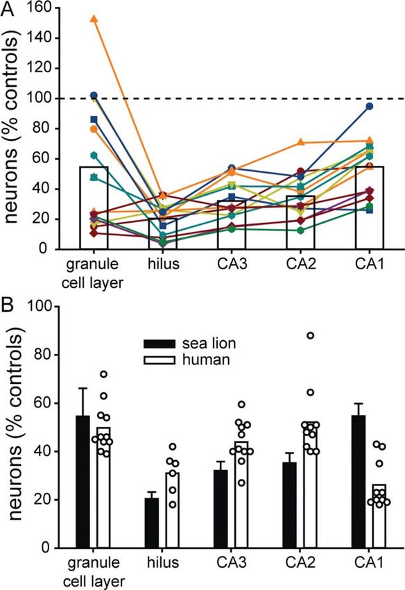
[ANOVA] with the Holm–Sidak method), squirrel mon- test). Compared with controls, average neuron loss in
keys (80 6 6 lm, P 5 0.019), and dogs (81 6 7 lm, affected hippocampi of chronic DA sea lions was sub-
P 5 0.009). Average height of the granule cell layer in stantial in all hippocampal subfields (Table 2), most
mice (57 6 4 lm) was significantly less than that of severe in the hilus (20% of controls, P < 0.05, ANOVA
macaques and dogs (P < 0.02). Average height of the on ranks with Dunn’s method), least severe in the gran-
granule cell layer in rats (73 6 4 lm) was not signifi- ule cell layer (54%, not significantly different) and CA1
cantly different from that of any other species. In addi- pyramidal cell layer (55%, P < 0.05), and intermediate in
tion to a thinner layer, granule cells of control sea lions the CA2 (35%, P < 0.05) and CA3 pyramidal cell layers
appeared less densely packed. These findings suggest (32%, P < 0.05) (Fig. 3A).
that sea lions have proportionally fewer granule cells Neuron loss in chronic DA sea lions was most vari-
than other mammals in part because the granule cell able in the granule cell layer. The coefficient of varia-
tion for granule cell layer neuron counts in affected
layer is thinner.
hippocampi from chronic DA sea lions was 0.798.
All chronic DA sea lions displayed significant hippo-
Some affected hippocampi in chronic DA sea lions dis-
campal neuron loss, but to varying degrees (Fig. 1B–D).
played severe granule cell loss with values only approxi-
To compare neuron loss across hippocampal subfields,
mately 20% of controls, whereas others displayed no
averages were calculated from “affected” hippocampi,
granule cell loss. In other subregions of the hippocam-
which had neuron numbers in any subfield at least 2
pus, neuron loss was more consistent. When granule
standard deviations below average control values. When
cell loss occurred, it appeared as thinning of the layer
this criterion was used, significant neuron loss was evi-
and sometimes gaps. Bilayers of granule cells, as
dent in the left hippocampus of four chronic DA sea
reported in some cases of human temporal lobe epi-
lions, the right hippocampus of seven, and bilaterally in
lepsy (Houser, 1990; Thom et al., 2002; Bl€umcke et al.,
three. Although right-sided neuron loss was more fre-
2009), were not observed in chronic DA sea lions.
quent, the difference was not significant (P > 0.05, v2
All of the male chronic DA sea lions were young (one
yearling, two juveniles, and one subadult), whereas
TABLE 1.
females were mostly older (two subadults and eight
Primary Antibody Used in This Study
adults). In the granule cell layer, hilus, and CA3 field,
Antigen Immunogen Manufacturer Dilution neuron loss was less severe in the younger males com-
Somatostatin-14 Synthetic Peninsula 1:4,000 pared with the older females (Table 3). Affected hippo-
somatostatin-14 Laboratories campi in male chronic DA sea lions had an almost
(Belmont, CA), normal average number of granule cells (95% of con-
rabbit polyclonal,
#IHC 8001 trols), whereas females had only 39% of controls (male
vs. female, P 5 0.02, t test). The average number of
TABLE 2.
Parameters and Results of Stereological Analysis of Hippocampal Neuron Numbers in Control and Chronic DA Sea Lions
Known or Suspected of Previous Exposure to Domoic Acid
Granule cell layer Hilus CA3 CA2 CA1
Counting grid 200 3 200 lm 200 3 200 lm 400 3 400 lm 200 3 200 lm 500 3 500 lm
Counting frame 20 3 20 lm 50 3 50 lm 50 3 50 lm 50 3 50 lm 50 3 50 lm
Cells counted/hippocampus 260 6 16 207 6 16 231 6 15 227 6 15 283 6 14
(mean 6 SEM)
Coefficient of variation 0.44 0.56 0.48 0.36 0.49
Mean coefficient of error1 0.06 0.10 0.08 0.10 0.06
Neurons/hippocampus of 2,120,000 6 333,000 6 1,350,000 6 332,000 6 2,310,000 6
control sea lions (n 5 9 animals, 350,000 14,000 170,000 98,000 300,000
18 hippocampi)(mean 6 SEM)
Neurons/hippocampus of 1,150,000 6 68,100 6 9,4003 433,000 6 117,000 6 1,260,000 6
chronic DA sea lions (n 5 14 animals, 250,0002 51,0003 14,0003 120,0003
17 affected hippocampi)(mean 6 SEM)
Neurons/hippocampus of chronic 2,470,000 6 349,000 6 1,570,000 6 386,000 6 2,670,000 6
DA sea lions (n 5 11 animals, 140,000 12,000 60,000 27,000 110,000
11 unaffected hippocampi)(mean 6 SEM)
1
Calculated according to West et al. (1991).
2
Only different from unaffected hippocampi, P < 0.05, ANOVA on ranks with Dunn’s method.
3
Significantly different from controls and unaffected hippocampi in chronic DA sea lions.
The Journal of Comparative Neurology | Research in Systems Neuroscience 1695P.S. Buckmaster et al. Figure 2. A: Dentate gyrus granule cell layer thickness in different mammals. All images and measurements obtained from a straight region of the internal part of the granule cell layer (g) at the mid-septotemporal level of the hippocampus. h, hilus; m, molecular layer. B: Granule cell layer thickness values (mean 6 SEM). Number of animals indicated at base of bars. *Average granule cell layer thickness was greater in macaques, squirrel monkeys, and dogs compared with sea lions (P < 0.02, ANOVA with Holm–Sidak method). Scale bar 5 50 lm in A. hilar neurons was 31% of controls in males, whereas it that in human patients with temporal lobe epilepsy, was only 16% in females (P 5 0.01). The average num- results were compiled from previously published studies ber of neurons in CA3 was 44% of controls in males, of human tissue that had been obtained after temporal whereas it was only 27% in females (P 5 0.04). lobectomy (Babb et al., 1989; Kim et al., 1990; Sass In control sea lions, granule cells, hilar neurons, and et al., 1990; Lee et al., 1995; Mathern et al., 1995, CA2 pyramidal cells were distributed relatively evenly in 1996, 1997; Bahh et al., 1999; de Lanerolle et al., sections along the septotemporal axis (Fig. 4A). CA1 2003; Bl€umcke et al., 2007) or autopsy (Thom et al., pyramidal cells were least numerous at the septal pole 2005). Substantial neuron loss was evident in all hippo- and most numerous at the extreme end of the temporal campal subfields of patients with temporal lobe epi- pole. CA3 pyramidal cells were increasingly more lepsy and chronic DA sea lions compared with controls numerous in the temporal third of the hippocampus. (Fig. 3B). In sea lions neuron loss was more severe in Neuron loss in affected hippocampi of chronic DA sea the hilus, CA3, and CA2 subfields compared with lions occurred relatively evenly along the septotemporal humans. In humans neuron loss was more severe in axis in all regions (Fig. 4C–F) except in the granule cell CA1. Sea lions and humans displayed similar levels of layer (Fig. 4B). In the granule cell layer of chronic DA granule cell loss. sea lions, neuron numbers were 81% of controls in the Neuron loss was unilateral in 11/14 chronic DA sea septal half of the hippocampus but only 48% of controls lions (Fig. 5A,B). Similar to human patients with tempo- in the temporal third. ral lobe epilepsy (Thom et al., 2005), in chronic DA sea To test whether the extent and pattern of hippocam- lions, neurons were spared in hippocampi contralateral pal neuron loss in chronic DA sea lions was similar to to affected hippocampi (excluding bilateral cases). 1696 The Journal of Comparative Neurology | Research in Systems Neuroscience
Hippocampal neuropathology of sea lion epilepsy

Average numbers of neurons in each subfield of contra-                   different from controls but significantly higher than
lateral hippocampi of chronic DA sea lions were 105–                     affected hippocampi (P < 0.05 ANOVA on ranks with
117% of control values (Table 2) and not significantly                   Dunn’s method).
                                                                            To test whether the frequency of unilateral neuron
                                                                         loss in chronic DA sea lions was similar to that in
                                                                         human patients with temporal lobe epilepsy, results
                                                                         were compiled from previously published human MRI
                                                                         studies (King et al., 1995; Barr et al., 1997; Briellmann
                                                                         et al., 1999) or autopsy findings (Sano and Malamud,
                                                                         1953; Margerison and Corsellis, 1966; Thom et al.,
                                                                         2005). Unilateral or asymmetric atrophy or hippocampal
                                                                         neuron loss is reported to occur in 63–91% of human
                                                                         patients (81%, average). In the present study 79% of
                                                                         sea lions had unilateral hippocampal neuron loss, which
                                                                         is similar to previous reports that used MRI to detect
                                                                         hippocampal atrophy (Thomas et al., 2010; Montie
                                                                         et al., 2012) or histology to screen for hippocampal
                                                                         damage in chronic DA sea lions (Goldstein et al., 2008)
                                                                         and found asymmetric or unilateral damage in 68–87%
                                                                         of cases (76%, average). These findings suggest that
                                                                         unilateral hippocampal neuropathology is common in
                                                                         human patients with temporal lobe epilepsy and in
                                                                         chronic DA sea lions.
                                                                            In affected hippocampi of chronic DA sea lions, neu-
                                                                         ron loss in the hilus most clearly illustrated laterality of
                                                                         lesions (Fig. 5C). In addition to frequently being unilat-
                                                                         eral, hilar neuron loss consistently was severe, not
                                                                         graded. In affected hippocampi of chronic DA sea lions,
                                                                         hilar neurons were only 4–36% of the average control
                                                                         value. To compare the pattern and extent of hilar neu-
                                                                         ron loss with another animal model of temporal lobe
                                                                         epilepsy, results from a previous study were plotted
Figure 3. Neuron loss in hippocampal subregions. A: Neurons per          that used similar methods to estimate numbers of hilar
affected hippocampus in chronic domoic acid (DA) sea lions               neurons per hippocampus in control and epileptic
(n 5 14 animals, 17 hippocampi) normalized to average control
                                                                         kainate-treated rats (Buckmaster and Dudek, 1997).
values (n 5 nine animals, 18 hippocampi). Each line-scatter plot is
from an individual chronic DA sea lion. Bars indicate averages. B:       Like chronic DA sea lions, epileptic kainate-treated rats
Neurons per affected hippocampus for chronic DA sea lions                display hilar neuron loss (average 48% of controls).
(mean 6 SEM) and neuron densities reported in previously pub-            However, in kainate-treated rats, hilar neuron loss is
lished studies for patients with temporal lobe epilepsy (Babb            bilateral and graded, not unilateral and “all or none” as
et al., 1989; Kim et al., 1990; Sass et al., 1990; Lee et al., 1995;     in chronic DA sea lions (Fig. 5D).
Mathern et al., 1995, 1996, 1997; Bahh et al., 1999; de Lanerolle
                                                                            Immunostaining was used to test whether g-
et al., 2003; Thom et al., 2005; Bl€umcke et al., 2007). Symbols
indicate results from individual studies. Bars indicate averages.        aminobutyric acid (GABA)ergic interneuron axon sprout-
                                                                         ing might have occurred in the dentate gyrus of chronic

                                                      TABLE 3.
    Hippocampal Neuron Numbers in Male and Female Sea Lions Known or Suspected of Previous Exposure to Domoic Acid1
                                Granule cell layer            Hilus               CA3                  CA2                  CA1
Male (n 5 4 animals,         2,000,000 6 560,000       103,000 6 9,000     597,000 6 82,000     143,000 6 35,000 1,550,000 6 270,000
  4 hippocampi)
Female (n 5 10 animals,        820,000 6 190,0002       54,000 6 10,0002 368,000 6 52,0002 106,000 6 14,000 1,150,000 6 120,000
  13 affected hippocampi)
1
Values represent neurons/hippocampus (mean 6 SEM).
2
P < 0.05, t test.

                                                       The Journal of Comparative Neurology | Research in Systems Neuroscience    1697
P.S. Buckmaster et al. Figure 4. Sea lion hippocampal neurons quantified along the septotemporal axis. A: Neurons per section along the septotemporal axis of the hippocampus in controls (n 5 9 animals, 18 hippocampi). B–F: Neurons per section along the septotemporal axis of the hippocampus in the granule cell layer (B), hilus (C), and pyramidal cell layer of CA3 (D), CA2 (E), and CA1 (F) in controls (n 5 9 animals, 18 hippocampi) and affected hippocampi in chronic domoic acid (DA) sea lions (n 5 14 animals, 17 hippocampi). Values indicate mean 6 SEM. DA sea lions. Abundant somatostatin-positive hilar neu- Timm staining was used to test whether mossy fiber rons were evident in control sea lions but few in sprouting occurred in chronic DA sea lions. Control sea affected hippocampi of chronic DA sea lions (Fig. 6). lions (n 5 9 animals, 18 hippocampi) displayed black Despite fewer hilar neurons, somatostatin- Timm staining primarily in the hilus of the dentate gyrus immunoreactive axons were more evident in affected and stratum lucidum of CA3 (Fig. 7A,C). Affected hippo- hippocampi of chronic DA sea lions than in controls. campi in chronic DA sea lions (n 5 12 animals, 15 1698 The Journal of Comparative Neurology | Research in Systems Neuroscience
Hippocampal neuropathology of sea lion epilepsy

Figure 5. Laterality of hippocampal neuron loss in chronic domoic acid (DA) sea lions. A,B: Hippocampi of a chronic DA sea lion admitted
in status epilepticus with DA toxicity and euthanized 26 days later. Neuron loss was substantial in the right (B) but not left hippocampus
(A). g, granule cell layer; h, hilus. C: Hilar neurons per right and left hippocampus in control and chronic DA sea lions. D: Hilar neurons
per right and left hippocampus in control and chronic kainate (KA)-treated rats reported previously (Buckmaster and Dudek, 1997).

hippocampi, with two animals omitted because of poor                    DISCUSSION
Timm staining) displayed an extra band of black Timm                       The main findings of the present study are that after
staining in the granule cell layer and especially in the                natural exposure to domoic acid (DA), sea lions fre-
inner third of the molecular layer, indicating aberrant                 quently develop unilateral hippocampal neuron loss,
mossy fiber sprouting (Fig. 7B,D). The average percent-                 hilar somatostatin-immunoreactive neuron loss but axon
age of the granule cell layer plus the molecular layer                  sprouting by surviving cells, and mossy fiber sprouting.
that was Timm-positive was abnormally elevated at all                   These neuropathological abnormalities are similar to
septotemporal levels except the extreme end of the                      those of human patients with temporal lobe epilepsy.
septal pole (Fig. 7E). The extent of mossy fiber sprout-
ing was greater in the septal half than in the temporal
end of the hippocampus. The difference might be attrib-                 Sea lion hippocampal anatomy compared
utable to more severe loss of granule cells in the tem-                 with other species
poral hippocampus (Fig. 4B). Loss of granule cells                         Qualitatively, the cytoarchitecture of the sea lion hip-
would be expected to reduce numbers of mossy fibers                     pocampus is similar to that of other mammals, but
and extent of Timm staining. The average amount of                      there are quantitative differences. The hippocampus of
black Timm staining in the granule cell layer plus the                  control sea lions contained over 6 million neurons,
molecular layer per affected chronic DA sea lion hippo-                 which is over three times more than rats (West et al.,
campus was 4.0-fold higher than controls and 2.6-fold                   1991) and 17% of humans (West et al., 1994). Com-
higher than contralateral chronic DA sea lion hippo-                    pared with other mammals, sea lions have a smaller
campi (P < 0.05, ANOVA on ranks with Dunn’s method)                     proportion of neurons in the granule cell layer. In rats
(Fig. 7F). Contralateral hippocampi of chronic DA sea                   (West et al., 1991) and humans (West et al., 1994),
lions (n 5 11 animals, 11 hippocampi) displayed slightly                granule cells account for 64% and 50% of hippocampal
(1.5-fold) but not significantly higher levels of black                 neurons, respectively, but in sea lions only 33%. The dif-
Timm staining in the granule cell layer plus the molecu-                ference is specific to the granule cell layer. For exam-
lar layer compared with controls.                                       ple, not surprisingly, sea lions have two to three times

                                                      The Journal of Comparative Neurology | Research in Systems Neuroscience         1699
P.S. Buckmaster et al.
Figure 6. Somatostatin immunoreactivity in sea lion hippocampi. A,B: Sections from the mid-septotemporal level of the hippocampus in a
control (A) and an epileptic sea lion suspected of previous DA exposure at least 28 days previously (B). Somatostatin-positive cell bodies
are abundant in the hilus (h) and proximal CA3 pyramidal cell layer (CA3) in the control but not the chronic DA sea lion. However,
somatostatin-positive fibers are abundant in the hilus, granule cell layer (g), and molecular layer (m) of the chronic DA sea lion. Scale
bar 5 250 lm in B (applies to A,B).
more hilar neurons than smaller carnivores (Mitchell hemisphere-specific activity. However, this unusual EEG
et al., 1999; Buckmaster et al., 2002b) but unexpect- feature is not necessary for focal damage, because in
edly only a fifth as many granule cells (Amrein and Slo- humans (Fujikawa et al., 2000) and non-human prima-
mianka, 2010). Average granule cell layer thickness tes (Meldrum et al., 1974), who do not display marked
was significantly lower in control sea lions compared interhemispheric EEG asymmetry during sleep, status
with control macaque monkeys, squirrel monkeys, and epilepticus commonly causes asymmetrical or unilateral
dogs, all of which have substantially smaller body sizes. hippocampal neuron loss. One possible mechanism for
Thus, sea lions appear to have fewer granule cells at unilateral lesions in sea lions and primates but bilateral
least in part because of their relatively thin granule cell lesions in rodents is a difference in hippocampal com-
layer. Granule cell layer thickness is only one of many missures, which are more developed in rodents than
parameters that can affect the total number of granule primates (Amaral et al., 1984). Tract-tracing studies
cells, and more work is needed to identify the mecha- have not been reported for sea lions, but cats, another
nisms underlying this species difference. It is not carnivore, have extensive interhemispheric hippocampal
known whether other marine mammals also have pro- connectivity (Jouandet et al., 1985), suggesting that
portionally fewer granule cells, and the functional con- other factors account for unilateral lesions in sea lions.
sequences of fewer granule cells are unclear. Human brains contain many more neurons than
rodent brains, but the average numbers of synapses
Unilateral lesions after systemic exposure received per neuron are roughly similar (DeFelipe et al.,
Unilateral hippocampal neuropathology is common in 2002), indicating that, on average, neurons in human
human patients with temporal lobe epilepsy and in brains must be less interconnected. One speculative
chronic DA sea lions. It is unclear why status epilepti- possibility is that generally reduced connectivity in spe-
cus following systemic exposure to a toxin causes uni- cies with large brains (including sea lions) limits seizure
lateral lesions. In rodents, status epilepticus after spread resulting in more focal seizures and more focal
exposure to systemic DA (and other convulsants) damage. Similar unilateral or asymmetric patterns of
causes bilateral damage in the hippocampus and other hippocampal damage in sea lions (Goldstein et al.,
brain regions (Colman et al., 2005). California sea lions 2008; Thomas et al., 2010; Montie et al., 2012), non-
are members of the Otariidae family of pinnipeds, who human primates (Meldrum et al., 1974), and humans
like dolphins can sleep while swimming and display (Fujikawa et al., 2000) suggest that common patho-
interhemispheric electroencephalographic (EEG) asym- physiological mechanisms cause hippocampal neuron
metry during some stages of sleep (Lyamin et al., loss in species with large brains that experience status
2008), suggesting a possible predisposition for epilepticus. Although temporal lobe epilepsy in humans
1700 The Journal of Comparative Neurology | Research in Systems NeuroscienceHippocampal neuropathology of sea lion epilepsy
Figure 7. A–D: Timm-stained hippocampi from a control (A,C) and a chronic domoic acid (DA) sea lion suspected of DA exposure at least
54 days before euthanasia (B,D). m, molecular layer; g, granule cell layer; h, hilus of the dentate gyrus (A). Asterisks indicate areas shown
at higher magnification in C and D. E,F: Average percentage of the granule cell layer plus molecular layer that displayed black Timm stain-
ing along the septotemporal axis (E) and per hippocampus (F) in control sea lions (n 5 9 animals, 18 hippocampi) and in affected (n 5 12
animals, 15 hippocampi) and contralateral hippocampi (n 5 11 animals, 11 hippocampi) of chronic DA sea lions. Bars indicate SEM.
*P < 0.05, ANOVA on ranks with Dunn’s method. Scale bar 5 500 lm in B (applies to A,B); 50 lm in D (applies to C,D).
The Journal of Comparative Neurology | Research in Systems Neuroscience 1701P.S. Buckmaster et al.
is rarely caused by DA-induced status epilepticus (Cen- quently are affected. Chronic DA sea lions with hippo-
des et al., 1995), the similar pattern of hippocampal campal atrophy sometimes display atrophy of the
damage in sea lions suggests they may be a good parahippocampal gyrus, which contains the entorhinal
model. cortex (Montie et al., 2012). After DA toxicosis in sea
lions, histological lesions occur consistently in the hip-
Hippocampal neuron loss pocampus and sometimes in extra-hippocampal struc-
Substantial loss of hippocampal neurons is common tures, including the amygdala (Silvagni et al., 2005).
in human patients with temporal lobe epilepsy and in Similarly, the entorhinal cortex (Du et al., 1993) and
chronic DA sea lions. As in human patients with tempo- amygdala also are damaged in some human patients
ral lobe epilepsy (Margerison and Corsellis, 1966; de with hippocampal sclerosis (Sano and Malamud, 1953;
Lanerolle et al., 2003; Swartz et al., 2006; Bl€umcke Margerison and Corsellis, 1966).
et al., 2007), chronic DA sea lions displayed individual Hippocampal neuron loss was more severe in older
variation in the pattern and extent of hippocampal neu- female chronic DA sea lions than younger males. Most
ron loss. Constraints of most human studies preclude a DA-exposed sea lions are adult females, which might be
direct comparison of numbers of neurons for entire hip- attributable to the timing of their congregation at rook-
pocampi in humans with temporal lobe epilepsy with eries where frequent blooms of Pseudo-nitzschia algae
that from chronic DA sea lions in the present study. occur (Bejarano et al., 2008; Thomas et al., 2010). Sea
Nevertheless, comparison of neuron density data from lions have a long gestation period, pregnant sea lions
previous human tissue studies with neuron number are exposed to DA, and DA reaches the fetus, which
data from sea lion hippocampi revealed average val- might be exposed to recirculated toxin for extended
ues 55% of controls for every subfield in Ammon’s periods (Brodie et al., 2006). Thus, it has been pro-
horn and the dentate gyrus. posed that the increasing frequency of younger animals,
Patterns of neuron loss were similar but not identical especially males, with signs of previous DA exposure
in human patients with temporal lobe epilepsy and might be attributable to in utero exposure (Goldstein
chronic DA sea lions. The hilus was the site of the most et al., 2008; Ramsdell and Zabka, 2008). Of the four
severe hippocampal neuron loss in chronic DA sea lions, male chronic DA sea lions in the present study, only
which is similar to rodent models of temporal lobe epi- one stranded because of acute DA toxicosis, raising the
lepsy (Mello et al., 1993; Gorter et al., 2001). In most speculative possibility that the other three were
previous studies of tissue from human patients with tem- exposed in utero. DA-exposure histories of wild sea
poral lobe epilepsy, the hilus was combined with proximal lions are incomplete. However, if some of the young
CA3 for cell counting, probably because the border males in the present study had been exposed during
between the two regions is difficult to clearly visualize in gestation, results of the present study suggest that in
Nissl-stained primate hippocampi (Buckmaster and Ama- utero exposure to DA can cause epilepsy to develop
ral, 2001). Some previous studies evaluated the hilus later in life and that hippocampal neuron loss after in
independently and reported that the hippocampal subre- utero exposure is less severe than that caused by adult
gion with the most severe neuron loss was the hilus exposure. Similarly, it has been hypothesized that in
(Sass et al., 1990), CA1 (Mathern et al., 1997; de Laner- utero exposure to low levels of DA that do not cause
olle et al., 2003), or both (Bahh et al., 1999). CA1 cell symptoms in adults might cause temporal lobe epilepsy
loss is less severe in sea lions than in humans and least to develop later in developing humans (Stewart, 2010).
severe in rats (Mello et al., 1993; Gorter et al., 2001). It
is unclear why CA1 pyramidal cells are more vulnerable Synaptic reorganization
in humans. Hilar neuron loss tends to occur in a unilateral The present study revealed temporal lobe epilepsy-
“all or none” pattern in chronic DA sea lions, whereas in related synaptic reorganization in chronic DA sea lions.
rats it is bilateral and graded. In humans, neuron loss fre- Over half of the GABAergic interneurons in the dentate
quently is unilateral and neurons in contralateral hippo- hilus in rats and monkeys express somatostatin (Austin
campi are spared (Thom et al., 2005). Without and Buckmaster, 2004). Numbers of somatostatin-
stereological evaluation of entire human hippocampi, it immunoreactive hilar neurons are reduced substantially
remains unclear whether hilar neuron loss in human in patients with temporal lobe epilepsy (de Lanerolle
patients is graded as in rodent models or is as consis- et al., 1989), but surviving cells sprout axon collaterals
tently severe as in chronic DA sea lions. that become even more exuberant than in controls
It appears that in both chronic DA sea lions and (Mathern et al., 1995). In mouse models of temporal
human patients the hippocampus is the site of most lobe epilepsy, sources of sprouted axons include surviv-
severe neuron loss, although other brain regions fre- ing somatostatin-immunoreactive neurons in the hilus
1702 The Journal of Comparative Neurology | Research in Systems NeuroscienceHippocampal neuropathology of sea lion epilepsy
(Zhang et al., 2009) and CA1 field (Peng et al., 2013). period of time. Unlike laboratory animals, sea lions are
Similarly, chronic DA sea lions displayed loss of hilar wild animals protected legally by the Marine Mammal
somatostatin interneurons but abundant somatostatin- Protection Act. Stranded sea lions cannot be implanted
immunoreactive axons in the dentate gyrus, suggesting with electrodes for chronic EEG recording or held in
synaptic reorganization. captivity for long-term seizure monitoring. Direct meas-
Mossy fiber sprouting is a common neuropathological ures of epileptogenicity, such as seizure frequency,
abnormality in patients with temporal lobe epilepsy probably are not feasible. Nevertheless, indirect meas-
(Sutula et al., 1989; de Lanerolle et al., 1989; Houser ures of anti-epileptogenic treatment efficacy can be
et al., 1990) and in animal models of temporal lobe epi- obtained. One potentially informative parameter is
lepsy (Buckmaster, 2004). Sprouted mossy fibers syn- restranding rate, which is measured by identifying indi-
apse predominantly with granule cells, forming a viduals from flipper tags they receive before release
recurrent excitatory circuit in human patients (Babb (Greig et al., 2005). Chronic DA sea lions restrand at a
et al., 1991; Franck et al., 1995; Zhang and Houser rate of 71% (Goldstein et al., 2008), which is much
1999) and animal models (Represa et al., 1993; Wenzel higher than that of sea lions rehabilitated after strand-
et al., 2000; Buckmaster et al., 2002c). Similarly, mossy ing for other reasons (0.5%) (Gulland et al., 2002).
fiber sprouting was a consistent finding in chronic DA Other parameters include movement, migration, and
sea lions. diving patterns that can be monitored from a satellite
transmitter glued to the fur, which remains attached for
weeks until molting. Chronic DA sea lions exhibit abnor-
Sea lions as a possible model of human mal movement, migration, and diving patterns (Thomas
temporal lobe epilepsy et al., 2010). Thus, experimental anti-epileptogenic
The validity of rodent models generated by systemic treatments could be evaluated in DA-exposed sea lions
treatment with kainic acid and other convulsants is sup- to test whether restranding rates decrease and whether
ported by the development of temporal lobe epilepsy in movement, migration, and diving patterns normalize.
a human (Cendes et al., 1995) and in sea lions after
DA intoxication. Sea lions provide opportunities and CONFLICT OF INTEREST STATEMENT
challenges as a potential animal model of human tem-
The authors have no conflicts of interest.
poral lobe epilepsy. Advantages include their similar
hippocampal neuropathology, human-like size, and avail-
ability. On average, The Marine Mammal Center in Sau- ROLE OF AUTHORS
salito, California admits approximately 70 cases/yr with All authors had full access to all the data in the
acute DA toxicosis (Goldstein et al., 2008). Rates of study and take responsibility for the integrity of the
Pseudo-nitzschia algal blooms along the coast of Califor- data and the accuracy of the data analysis. Study con-
nia (Sekula-Wood et al., 2011) and cases of DA toxico- cept and design: PSB, IT, FMDG, WVB. Acquisition of
sis admitted to The Marine Mammal Center appear to data: PSB, XW, IT, FMDG, WVB. Analysis and interpreta-
be increasing (Greig et al., 2005; Goldstein et al., tion of data, drafting of the manuscript, statistical anal-
2008). Because sea lions are exposed in the wild fol- ysis, obtained funding, and study supervision: PSB.
lowing ingestion of prey containing varying amounts of
DA, doses of DA and exact timing of exposure are LITERATURE CITED
unclear, which is an experimental limitation.
Amaral DG, Insausti R, Cowan WM. 1984. The commissural
Nevertheless, status epilepticus can be recognized, connections of the monkey hippocampal formation.
and the presence of DA in feces and urine can be J Comp Neurol 224:307–336.
detected. Admitted sea lions with acute DA toxicosis Amrein I, Slomianka L. 2010. A morphologically distinct gran-
receive anticonvulsive and supportive therapy for days ule cell type in the dentate gyrus of the red fox corre-
lates with adult hippocampal neurogenesis. Brain Res
to weeks (Gulland et al., 2002), during which time they
1328:12–24.
also could receive experimental anti-epileptogenic treat-
Austin JE, Buckmaster PS. 2004. Recurrent excitation of gran-
ments. Upon discharge, continued treatment becomes ule cells with basal dendrites and low interneuron den-
impossible, and 54% of released status epilepticus sur- sity and inhibitory postsynaptic current frequency in the
vivors ultimately die prematurely or are euthanized dentate gyrus of macaque monkeys. J Comp Neurol 476:
205–218.
(Goldstein et al., 2008). Thus, there is a pressing veteri-
nary need for (and an opportunity to test) anti- Babb TL, Pretorius JK, Kupfer WR, Crandall PH. 1989. Gluta-
mate decarboxylase-immunoreactive neurons are pre-
epileptogenic treatments that could be administered served in human epileptic hippocampus. J Neurosci 9:
shortly after precipitating injuries and for a limited 2562–2574.
The Journal of Comparative Neurology | Research in Systems Neuroscience 1703P.S. Buckmaster et al.
Babb TL, Kupfer WR, Pretorius JK, Crandall PH, Levesque MF. Colman JR, Nowocin KJ, Switzer RC, Trusk TC, Ramsdell JS.
1991. Synaptic reorganization by mossy fibers in human 2005. Mapping and reconstruction of domoic acid-
epileptic fascia dentata. Neuroscience 42:351–363. induced neurodegeneration in the mouse brain. Neuro-
Bahh BE, Lespinet V, Lurton D, Coussemacq M, Le Gal La toxicol Teratol 27:753–767.
Salle G, Rougier A. 1999. Correlations between granule Debonnel G, Beauchesne L, de Montigny C. 1989a. Domoic
cell dispersion, mossy fiber sprouting, and hippocampal acid, the alleged “mussel toxin,” might produce its neu-
cell loss in temporal lobe epilepsy. Epilepsia 40:1393– rotoxic effect through kainate receptor activation: an
1401. electrophysiological study in the rat dorsal hippocampus.
Barr WB, Ashtari M, Schaul N. 1997. Bilateral reductions in Can J Physiol Pharmacol 67:29–33.
hippocampal volume in adults with epilepsy and a history Debonnel G, Weiss M, de Montigny C. 1989b. Reduced neuro-
of febrile seizures. J Neurol Neurosurg Psychiatry 63: excitatory effect of domoic acid following mossy fiber
461–467. denervation of the rat dorsal hippocampus: further evi-
Bejarano AC, VanDola FM, Gulland FM, Rowles TK, Schwacke dence that toxicity of domoic acid involves kainate
LH. 2008. Production and toxicity of the marine biotoxin receptor activation. Can J Physiol Pharmacol 67:904–
domoic acid and its effects on wildlife: a review. Hum 908.
Ecol Risk Assess 14:544–567. DeFelipe J, Alonso-Nanclares L, Arellano JI. 2002. Microstruc-
Bininda-Emonds ORP. 2000. Pinniped brain sizes. Mar Mamm ture of the neocortex: comparative aspects. J Neurocytol
Sci 16:469–481. 31:299–316.
Bl€umcke I, Pauli E, Clusmann H, Schramm J, Becker A, Elger de Lanerolle NC, Kim JH, Robbins RJ, Spencer DD. 1989. Hip-
C, Merschhemke M, Meencke H, Lehmann T, von pocampal interneuron loss and plasticity in human tem-
Deimling A, Scheiwe C, Zentner J, Volk B, Romst€ock J, poral lobe epilepsy. Brain Res 495:387–395.
Stefan H, Hildebrandt M. 2007. A new clinic-pathological de Lanerolle NC, Kim JH, Williamson A, Spencer SS, Zaveri
classification system for mesial temporal sclerosis. Acta HP, Eid T, Spencer DD. 2003. A retrospective analysis of
Neuropathol 113:235–244. hippocampal pathology in human temporal lobe epilepsy:
Bl€umcke I, Kistner I, Clusmann H, Schramm J, Becker AJ, evidence for distinctive patient subcategories. Epilepsia
Elger CE, Bien CG, Merschhemke M, Meencke H, 44:677–687.
Lehmann T, Buchfelder M, Weigel D, Buslei R, Stefan H,
Doucette GJ, Midulski CM, King KL, Roh PB, Wang Z, Leandro
Pauli E, Hildebrandt M. 2009. Towards a clinic-
LF, DeGrasse SL, White KD, De Biase D, Gillett RM,
pathological classification of granule cell dispersion in
Rolland RM. 2012. Endangered North Atlantic right
human mesial temporal lobe epilepsies. Acta Neuropa-
whales (Eubalaena glacialis) experience repeated, concur-
thol 117:535–544.
rent exposure to multiple environmental neurotoxins pro-
Briellmann RS, Jackson GD, Mitchell LA, Fitt GF, Kim SE, duced by marine algae. Environ Res 112:67–76.
Berkovic SF. 1999. Occurrence of hippocampal sclerosis:
Du F, Whetsell WO Jr, Abou-Khalil B, Blumenkopf B, Lothman
is one hemisphere or gender more vulnerable? Epilepsia
EW, Schwarcz R. 1993. Preferential neuronal loss in layer
40:1816–1820.
III of the entorhinal cortex in patients with temporal lobe
Brodie EC, Gulland FMD, Greig DJ, Hunter M, Kaakola J, St.
epilepsy. Epilepsy Res 16:223–233.
Leger J, Leighfield TA, Van Dolah FM. 2006. Domoic acid
causes reproductive failure in California sea lions (Zalo- Engel J Jr, Williamson PD, Wieser H. 1997. Mesial temporal
phus californianus). Mar Mamm Sci 22:700–707. lobe epilepsy. In: Engel J Jr, Pedley TA, eds. A compre-
Buckmaster PS. 2004. Laboratory animal models of temporal hensive textbook. Philadelphia: Lippincott-Raven Press. p
lobe epilepsy. Comp Med 54:473–485. 2417–2426.
Buckmaster PS, Amaral DG. 2001. Intracellular recording and Franck JE, Pokorny J, Kunkel DD, Schwartzkroin PA. 1995.
labeling of mossy cells and proximal CA3 pyramidal cells Physiologic and morphologic characteristics of granule
in macaque monkeys. J Comp Neurol 430:264–281. cell circuitry in human epileptic hippocampus. Epilepsia
Buckmaster PS, Dudek FE. 1997. Neuron loss, granule cell 36:543–558.
axon reorganization, and functional changes in the den- French JA, Williamson PD, Thadani VM, Darcey TM, Mattson
tate gyrus of epileptic kainate-treated rats. J Comp Neu- RH, Spencer SS, Spencer DD. 1993. Characteristics of
rol 385:385–404. medial temporal lobe epilepsy: I. Results of history and
Buckmaster PS, Lew FH. 2011. Rapamycin suppresses mossy physical examination. Ann Neurol 24:774–780.
fiber sprouting but not seizure frequency in a mouse Fujikawa DG, Itabashi HH, Wu A, Shinmei SS. 2000. Status
model of temporal lobe epilepsy. J Neurosci 31:2337– epilepticus-induced neuronal loss in humans without sys-
2347. temic complications or epilepsy. Epilepsia 41:981–991.
Buckmaster PS, Otero-Corchon V, Rubinstein M, Low MJ. Goldstein T, Mazet JAK, Zabka TS, Langlois G, Colegrove KM,
2002a. Heightened seizure severity in somatostatin Silver M, Bargu S, Van Dolah F, Leighfield T, Conrad PA,
knockout mice. Epilepsy Res 48:43–56. Barakos J, Williams DC, Dennison S, Haulena M, Gulland
Buckmaster PS, Smith MO, Buckmaster CL, LeCouteur RA, FMD. 2008. Novel symptomatology and changing epide-
Dudek FE. 2002b. Absence of temporal lobe epilepsy miology of domoic acid toxicosis in California sea lions
pathology in dogs with medically intractable epilepsy. (Zalophus californianus): an increasing risk to marine
J Vet Intern Med 16:95–99. mammal health. Proc R Soc B 275:267–276.
Buckmaster PS, Zhang G, Yamawaki R. 2002c. Axon sprouting Gorter JA, van Vliet EA, Aronica E, Lopes da Silva FH. 2001.
in a model of temporal lobe epilepsy creates a predomi- Progression of spontaneous seizures after status epilepti-
nantly excitatory feedback circuit. J Neurosci 22:6650– cus is associated with mossy fibre sprouting and exten-
6658. sive bilateral loss of hilar parvalbumin and somatostatin-
Cendes F, Andemann F, Carpenter S, Zatorre RJ, Cashman immunoreactive neurons. Eur J Neurosci 13:657–669.
NR. 1995. Temporal lobe epilepsy caused by domoic Greig DJ, Gulland FMD, Kreuder C. 2005. A decade of live Cal-
acid intoxication: evidence for glutamate receptor- ifornia sea lion (Zalophus californianus) strandings along
mediated excitotoxicity in humans. Ann Neurol 37:123– the central California coast: causes and trends, 1991–
126. 2000. Aquat Mamm 31:11–22.
1704 The Journal of Comparative Neurology | Research in Systems NeuroscienceHippocampal neuropathology of sea lion epilepsy
Gulland FMD, Haulena M, Fauquier D, Langlois G, Lander ME, Mathern GW, Babb TL, Leite JP, Pretorius JK, Yeoman KM,
Zabka T, Duerr R. 2002. Domoic acid toxicity in Califor- Kuhlman PA. 1996. The pathogenic and progressive fea-
nia sea lions (Zalophus californianus): clinical signs, treat- tures of chronic human hippocampal epilepsy. Epilepsy
ment and survival. Vet Rec 150:475–480. Res 26:151–161.
Gunderson VM, Dubach M, Szot P, Born DE, Wenzel HJ, Mathern GW, Kuhlman PA, Mendoza D, Pretorius JK. 1997.
Maravilla KR, Zierath DK, Robbins CA, Schwartzkroin PA. Human fascia dentata anatomy and hippocampal neuron
1999. Development of a model of status epilepticus in densities differ depending on the epileptic syndrome and
pigtailed macaque infant monkeys. Dev Neurosci 21: age at first seizure. J Neuropathol Exp Neurol 56:199–
352–364. 212.
Houser CR. 1990. Granule cell dispersion in the dentate gyrus Meldrum BS, Horton RW, Brierley JB. 1974. Epileptic brain
of humans with temporal lobe epilepsy. Brain Res 535: damage in adolescent baboons following seizures
195–204. induced by allylglycine. Brain 97:407–418.
Houser CR, Miyashiro JE, Swartz BE, Walsh GO, Rich JR, Mello LEAM, Cavalheiro EA, Tan AM, Kupfer WR, Pretorius JK,
Delgado-Escueta AV. 1990. Altered patterns of dynorphin Babb TL, Finch DM. 1993. Circuit mechanisms of seiz-
immunoreactivity suggest mossy fiber reorganization in ures in the pilocarpine model of chronic epilepsy: cell
human hippocampal epilepsy. J Neurosci 10:267–282. loss and mossy fiber sprouting. Epilepsia 34:985–995.
Jouandet ML, Lachat J, Garey LJ. 1985. Distribution of the Mitchell TW, Buckmaster PS, Hoover EA, Whalen LR, Dudek
neurons of origin of the great cerebral commissures in FE. 1999. Neuron loss and axon reorganization in the
the cat. Anat Embryol 171:105–120. dentate gyrus of cats infected with the feline immunode-
Kim JH, Guimaraes PO, Shen MY, Masukawa LM, Spencer DD. ficiency virus. J Comp Neurol 411:563–577.
1990. Hippocampal neuronal density in temporal lobe Montie EW, Wheeler E, Pussini N, Battey TWK, Barakos J,
epilepsy with and without gliomas. Acta Neuropathol 80: Dennison S, Colegrove K, Gulland F. 2010. Magnetic res-
41–45. onance imaging quality and volumes of brain structures
King D, Spencer SS, McCarthy G, Luby M, Spencer DD. 1995. from live and postmortem imaging of California sea lions
Bilateral hippocampal atrophy in medial temporal lobe with clinical signs of domoic acid toxicosis. Dis Aquat
epilepsy. Epilepsia 36:905–910. Organ 91:243–256.
Kola I, Landis J. 2004. Can the pharmaceutical industry Montie EW, Wheeler E, Pussini N, Battey TWK, Van Bonn W,
reduce attrition rates? Nat Rev Drug Discov 3:711–715. Gulland F. 2012. Magnetic resonance imaging reveals
Kreuder C, Miller MA, Jessup DA, Lowenstine LJ, Harris MD, that brain atrophy is more severe in older California sea
Ames JA, Carpenter TE, Conrad PA, Mazet JAK. 2003. lions with domoic acid toxicosis. Harmful Algae 20:19–
Patterns of mortality in southern sea otters (Enhydra lut- 29.
ris nereis) from 1998–2001. J Wild Dis 39:495–509. Peng Z, Zhang N, Wei W, Huang CS, Cetina Y, Otis TS, Houser
K€unig G, Hartmann J, Drause F, Deckert J, Heinsen H, CR. 2013. A reorganized GABAergic circuit in a model of
Ransmayr G, Beckmann H, Riederer P. 1995. Regional epilepsy: evidence from optogenetic labeling and stimula-
differences in the interaction of the excitotoxins domoate tion of somatostatin interneurons. J Neurosci 33:14392–
and L-b-oxalyl-amino-alanine with [3H]kainate binding 14405.
sites in human hippocampus. Neurosci Lett 187:107–
Perez-Mendes P, Cinini SM, Medeiros MA, Tufik S, Mello LE.
110.
2005. Behavioral and histopathological analysis of
Kuwabara T, Hasegawa D, Kobayashi M, Fujita M, Orima H. domoic acid administration in marmosets. Epilepsia
2010. Clinical magnetic resonance volumetry of the hip- 46(suppl 5):148–151.
pocampus in 58 epileptic dogs. Vet Radiol Ultrasound
Perl TM, Bedard L, Kosatsky T, Gockin JC, Todd ECD, Remis
51:485–490.
RS. 1990. An outbreak of toxic encephalopathy caused
Kwan P, Schachter SC, Brodie MJ. Drug-resistant epilepsy.
by eating mussels contaminated with domoic acid. N
2011. N Engl J Med 365:919–926.
Engl J Med 322:1775–1780.
Larm JA, Beart PM, Cheung NS. 1997. Neurotoxin domoic
acid produces cytotoxicity via kainate- and AMPA- Potschka H, Fischer A, von R€uden E, H€ulsmeyer V,
sensitive receptors in cultured cortical neurons. Neuro- Baumg€artner W. 2013. Canine epilepsy as a translational
chem Int 31:677–682. model? Epilepsia 54:571–579.
Lee N, Tien RD, Lewis DV, Friedman AH, Felsberg GJ, Crain B, Ramsdell JS, Zabka TS. 2008. In utero domoic acid toxicity: a
Hulette C, Osumi AK, Smith JS, VanLandingham KE, fetal basis to adult disease in the California sea lion
Radtke RA. 1995. Fast spin-echo, MRI-measured hippo- (Zalophus californianus). Mar Drugs 6:262–290.
campal volume: correlation with neuronal density in ante- Repressa A, Jorquera I, Le Gal La Salle G, Ben-Ari Y. 1993.
rior temporal lobectomy patients. Epilepsia 36:899–904. Epilepsy induced collateral sprouting of hippocampal
Lyamin OI, Lapierre JL, Kosenko PO, Mukhametov LM, Siegel mossy fibers: does it induce the development of ectopic
JM. 2008. Electroencephalogram asymmetry and spectral synapses with granule cell dendrites? Hippocampus 3:
power during sleep in the northern fur seal. J Sleep Res 257–268.
17:154–165. Ribak CE, Seress L, Weber P, Epstein CM, Henry TR, Bakay
Lyons DM, Buckmaster PS, Lee A, Wu C, Mitra R, Patel PD, RA. 1998. Alumina gel injections into the temporal lobe
Schatzberg AF. 2010. Stress coping stimulates hippo- of rhesus monkeys cause complex partial seizures and
campal neurogenesis in adult monkeys. Proc Natl Acad morphological changes found in human temporal lobe
Sci U S A 107:14823–14827. epilepsy. J Comp Neurol 401:266–290.
Margerison JH, Corsellis JAN. 1966. Epilepsy and the temporal Sano K, Malamud N. 1953. Clinical significance of sclerosis of
lobes. Brain 89:499–530. the cornu ammonis. Arch Neurol Psychiatry 70:40–53.
Mathern GW, Babb TL, Pretorius JK, Leite JP. 1995. Reactive Sari P, Kerr DS. 2001. Domoic acid-induced hippocampal CA1
synaptogenesis and neuron densities for neuropeptide Y, hyperexcitability independent of region CA3 activity. Epi-
somatostatin, and glutamate decarboxylase immunoreac- lepsy Res 47:65–76.
tivity in the epileptogenic human fascia dentata. J Neuro- Sass KJ, Spencer DD, Kim JH, Westerveld M, Novelly RA,
sci 15:3990–4004. Lencz T. 1990. Verbal memory impairment correlates
The Journal of Comparative Neurology | Research in Systems Neuroscience 1705You can also read