Sequence Memory in the Hippocampal-Entorhinal Region - DoellerLab
←
→
Page content transcription
If your browser does not render page correctly, please read the page content below
Sequence Memory in the Hippocampal–Entorhinal Region

                   Jacob L. S. Bellmund1, Ignacio Polti2, and Christian F. Doeller1,2

Abstract
■ Episodic memories are constructed from sequences of events.            subregion carries temporal information during ongoing behavior.
When recalling such a memory, we not only recall individual events,      The human homologue is recruited during memory recall where
but we also retrieve information about how the sequence of events        its representations reflect the temporal relationships between events
unfolded. Here, we focus on the role of the hippocampal–entorhinal       encountered in a sequence. We further introduce the idea that the
region in processing and remembering sequences of events, which          hippocampal–entorhinal region might enable temporal scaling of
are thought to be stored in relational networks. We summarize evi-       sequence representations. Flexible changes of sequence progression
dence that temporal relations are a central organizational principle     speed could underlie the traversal of episodic memories and mental
for memories in the hippocampus. Importantly, we incorporate novel       simulations at different paces. In conclusion, we describe how the
insights from recent studies about the role of the adjacent entorhinal   entorhinal cortex and hippocampus contribute to remembering
cortex in sequence memory. In rodents, the lateral entorhinal            event sequences—a core component of episodic memory. ■

INTRODUCTION
                                                                         for episodic memory. Theoretical accounts suggest that it
Episodic memories are classically thought to comprise infor-             forms networks of related experiences, for example, the differ-
mation about specific events as well as about where and when             ent elements of an event sequence (Eichenbaum, Dudchenko,
these events occurred (Tulving, 1972, 1983). Recalling such              Wood, Shapiro, & Tanila, 1999; Eichenbaum & Cohen, 1988).
an episodic memory is characterized by a vivid feeling of                Different events occurring in temporal proximity can be
recollection—famously referred to as “mental time travel”                linked together in relational networks (Eichenbaum et al.,
(Tulving, 1983, 2002). However, episodes do not occur in                 1999; Eichenbaum & Cohen, 1988). Associating event details
isolation but rather in a sequence. Think back to an event               with contextual information—constituted, for example, by
that happened earlier today. For example, you might re-                  the time and place at which an event takes place—has been
member meeting your colleague upon arriving at work this                 suggested as a key contribution of the hippocampus to
morning: You had just locked your bike and entered the                   episodic memory (Diana, Yonelinas, & Ranganath, 2007;
building before exchanging a few words with her. Then,                   Eichenbaum, Yonelinas, & Ranganath, 2007). Other accounts
you climbed the stairs to the third floor and made your                  have proposed the role of the hippocampus to lie in the gen-
way to your office. The events that precede and follow an                eration of content-free sequences (Buzsáki & Tingley, 2018;
event are important for defining its temporal context.                   Friston & Buzsáki, 2016). A representation of an event se-
Sequence information such as relative times of occurrence                quence might then emerge from associations of these se-
can support memory for when events took place (Friedman,                 quential states with particular content in neocortical regions
1993). This can happen in concert with distance information              (Friston & Buzsáki, 2016).
like the decaying strengths of memory traces or differences                 How does the brain keep track of how events unfold over
in the accessibility of items that were stored in memory at              time to allow remembrance of event sequences? In keeping
different times. Furthermore, location information in the                with its key role in episodic memory and sequence processing
form of temporal tags or general contextual associations                 in general, we will center this review on the human hippocam-
can be encoded with the event and underlie memory for                    pal formation. Specifically, we will focus on recent neuroimag-
when events occurred (Friedman, 1993). Here, we aim to                   ing work investigating the hippocampus and entorhinal
review the role of the hippocampus and entorhinal cortex                 cortex. We further incorporate insights from intracranial re-
in the formation and retrieval of sequence memories.                     cordings in patients as well as theoretical work and findings
   The hippocampal–entorhinal region, situated in the medial             from animal models of temporal processing. Where a compre-
temporal lobe of the human brain, is thought to be central               hensive description of the different aspects of temporal coding
                                                                         and memory is beyond the scope of this article, we refer the
                                                                         reader to insightful reviews.
1
 Max Planck Institute for Human Cognitive and Brain Sciences,               We begin by summarizing findings that implicate the hip-
Leipzig, Germany, 2 Norwegian University of Science and                  pocampus in the formation and retrieval of sequence mem-
Technology, Trondheim, Norway                                            ories. We argue that correlations of activity patterns reflect

© 2020 Massachusetts Institute of Technology                                           Journal of Cognitive Neuroscience X:Y, pp. 1–13
                                                                                                   https://doi.org/10.1162/jocn_a_01592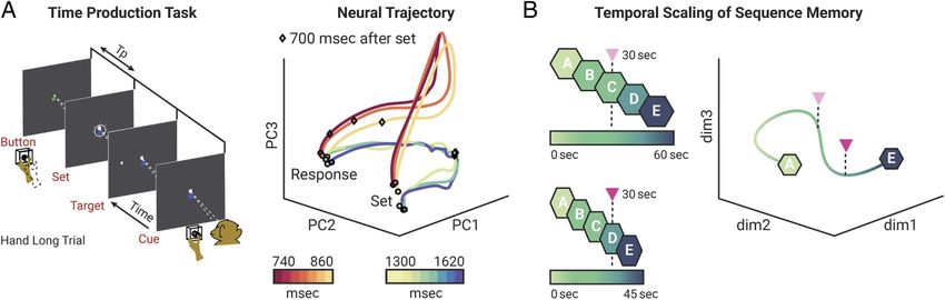
sequence information. Subsequently, we will briefly review (Lieberman, Kyle, Schedlbauer, Stokes, & Ekstrom, 2017;
recent studies suggesting that sequence representations in Ekstrom, Copara, Isham, Wang, & Yonelinas, 2011; Ekstrom
the hippocampal formation include information about the & Bookheimer, 2007) or retrieving the next element to com-
precise duration of the temporal structure of the sequence. plete a learned sequence (Ross et al., 2009) has also been
Next, we shift our focus to the entorhinal cortex as a major observed. Collectively, these findings suggest that both suc-
cortical input to the hippocampus. Importantly, we highlight cessful encoding and retrieval of event sequences recruit the
recent evidence suggesting that the anterior–lateral subre- hippocampus.
gion of the entorhinal cortex is particularly relevant for tem-
poral processing. We thus emphasize the role of the
PATTERN CORRELATIONS REFLECT
entorhinal cortex—in concert with the hippocampus—in se-
SEQUENCE INFORMATION
quence memory. Furthermore, we introduce the idea of tem-
poral scaling, which has been described for sensory and One way by which the hippocampus could support memory
motor timing and the corresponding cortical networks. for temporal relationships is by segregating representations
Specifically, we suggest that the flexible adaptation of se- of events that were originally separated in time. For exam-
quence progression speed, in episodic memory and mental ple, the activity of neuronal ensembles in the medial tempo-
simulation, is supported by the hippocampal–entorhinal re- ral lobe changes over time, resulting in similar population
gion. Finally, we present outstanding questions for future re- signals for nearby points in time and more dissimilar neural
search that emerge from these findings and ideas. patterns at increased time lags (Folkerts, Rutishauser, &
Howard, 2018; Howard, Viskontas, Shankar, & Fried, 2012).
Slowly drifting signals might arise from decaying traces of
SEQUENCE MEMORY AND
prior experiences or because of changing internal states. In
THE HIPPOCAMPUS
models of episodic memory, these signals serve as contextual
The role of the hippocampus in the formation and retrieval representations that allow the tagging of individual memo-
of memories for temporal and sequence information is well ries (Howard, Fotedar, Datey, & Hasselmo, 2005; Howard
established (for reviews, see, e.g., Ranganath & Hsieh, 2016; & Kahana, 2002). The current state of these contextual rep-
Eichenbaum, 2014). Here, we primarily focus on work in hu- resentations could be incorporated into the mnemonic rep-
mans, although the fundamental role of the hippocampus in resentation of an event occurring at a certain moment,
sequence memory is also well documented in animals putatively facilitating memory for how a sequence of events
(Eichenbaum, 2017; Fortin, Agster, & Eichenbaum, 2002; unfolded over time. Consistent with the notion that changing
Kesner, Gilbert, & Barua, 2002). In studies using fMRI, hip- neural patterns relate to sequence memory, hippocampal ac-
pocampal activations have been observed when participants tivity patterns during the encoding of object sequences were
learned stimulus sequences (Ross, Brown, & Stern, 2009; less similar, when the order of object pairs was remembered
Kumaran & Maguire, 2006a) or completed serial RT tasks correctly in a later memory test (Jenkins & Ranganath, 2016).
(Schendan, Searl, Melrose, & Stern, 2003). Likewise, hippo- Furthermore, increased correlations of hippocampal multi-
campal activity is increased for temporal information that is voxel patterns between stimuli were related to remembering
later successfully recalled from memory (Tubridy & these stimuli as being relatively close in time, compared to
Davachi, 2011; Jenkins & Ranganath, 2010). One such study stimuli separated by the same temporal distance that were
analyzed activity during the delay period of a serial order judged to be far apart (Ezzyat & Davachi, 2014; Figure 1A).
working memory task as a function of later memory for However, when participants use associative strategies to en-
when individual stimuli were encountered. After the presen- code stimulus sequences, increased similarity of hippocam-
tation of the stimulus sequence, greater hippocampal activ- pal representations has been reported for items whose order
ity related to more accurate subsequent memory for the was later discriminated correctly (DuBrow & Davachi, 2014,
temporal position of stimuli (Jenkins & Ranganath, 2010). 2017). This points toward an influence of encoding strategies
Consistently, hippocampal activity during encoding pre- on the way hippocampal activity relates to memory for order.
dicted serial recall for items encountered at event bound- Together, these data and theoretical considerations suggest
aries, defined by stimulus category switches, suggesting a that hippocampal activity patterns at encoding support
role for the hippocampus in linking different event se- memory by providing a temporal context representation al-
quences (DuBrow & Davachi, 2016). lowing the encoding of sequence relationships.
These reports of increased hippocampal activity during Interestingly, hippocampal activity patterns also carry in-
the encoding of temporal information that was later remem- formation about temporal context and the temporal relations
bered are complemented by studies of brain activity during of events during retrieval. Paralleling behavioral contiguity ef-
the retrieval of sequence order. For example, when partic- fects during the recall of word lists, reinstatement of medial
ipants recalled the order of scenes from a movie, the hip- temporal lobe activity patterns observed during encoding
pocampus was more strongly engaged than in a baseline accompanies the successful recall of information from mem-
condition or when participants logically inferred the order ory (Folkerts et al., 2018; Yaffe et al., 2014; Howard et al.,
of scene stimuli (Lehn et al., 2009). Likewise, hippocampal in- 2012; Manning, Polyn, Baltuch, Litt, & Kahana, 2011). In these
volvement in retrieving the order of events during navigation studies, neural activity patterns during item recall correlated
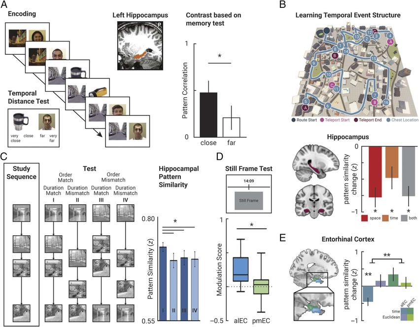
2 Journal of Cognitive Neuroscience Volume X, Number YFigure 1. Sequence memory in the hippocampus and the entorhinal cortex. (A) In an associative learning task, participants saw trial-unique images
of faces or objects paired with scene stimuli defining the context for a sequence of images. In a memory test, they were subsequently asked to
indicate the temporal distances between pairs of images on a 4-point scale. Pattern similarity in the left hippocampus during encoding was higher for
pairs of stimuli subsequently judged to be (very) close together than for those judged to be (very) far apart. Adapted with permission from Ezzyat and
Davachi (2014). (B) Participants navigated a virtual city along a fixed route (blue) to learn where in space, and when during this sequence, they
encountered specific objects (blue circles). Spatial (measured by Euclidean distances and shortest paths) and temporal (measured by walking times)
distances between object pairs were decorrelated using teleporters (pink and purple circles). The similarity of object representations was assessed
before and after learning. Hippocampal representational change correlated with remembered spatial and temporal distances between object pairs.
Adapted from Bellmund et al. (2019) and Deuker et al. (2016), both licensed under CC-BY. (C) While undergoing fMRI scanning, participants
monitored the stimulus order of four-element sequences of scene images. The order of images and the ISIs were varied independently. Hippocampal
pattern similarity was greatest when order and ISI of the test sequence matched the study sequence. Adapted with permission from Thavabalasingam
et al. (2018). (D) After having watched an episode from a TV show, participants saw still frames from the episode and indicated on a timeline when
the presented scene occurred. Activity in the anterior-lateral entorhinal cortex was modulated by the precision of memory recall such that activity was
greater for trials where participants responded most compared to least accurately. This modulation was not observed in the posterior-medial
entorhinal cortex. Adapted with permission from Montchal et al. (2019). (E) In the experiment outlined in B, pattern similarity change, in the
anterior-lateral entorhinal cortex, reflected the temporal relationships of objects from the sequence, which were measured by participants’ walking
times between them. This effect was selective to the anterior-lateral entorhinal cortex. Adapted from Bellmund et al. (2019), licensed under CC-BY.
with those observed during encoding, not only of the recalled In one study, participants navigated a virtual city and learned
item but also of those preceding and following it during when along a fixed route they encountered certain objects
learning (Folkerts et al., 2018; Yaffe et al., 2014; Howard (Deuker, Bellmund, Navarro Schröder, & Doeller, 2016;
et al., 2012; Manning et al., 2011). Figure 1B). In a postlearning fMRI scan, they were presented
with images of these objects in random order. Pattern simi-
The analysis of fMRI multivoxel patterns further suggests larity in the anterior hippocampus correlated negatively with
that hippocampal activity in response to retrieval cues carries the temporal distances by which participants remembered
information about the temporal relationships of memories. the objects to be separated. Relative to a prelearning baseline
Bellmund, Polti, and Doeller 3scan, the representations of pairs of objects that were re- the two event sequences. Specifically, hippocampal multi-
membered as being close in time became more similar, voxel patterns diverged gradually as the narratives unfolded
whereas objects separated by large temporal distances be- (Milivojevic, Varadinov, Grabovetsky, Collin, & Doeller,
came more dissimilar (Deuker et al., 2016). Consistent re- 2016). Notably, regions of the default network can integrate
sults were observed in a study in which participants judged narrative information on timescales of around 30 sec, even
temporal relations of location triads, visited in a delivery task when the hippocampus is severely damaged (Zuo et al.,
(Kyle, Smuda, Hassan, & Ekstrom, 2015). Furthermore, the 2020). However, successful memory encoding and retrieval
relationship between multivoxel pattern similarity in the an- of event sequences might require interactions with the hip-
terior hippocampus and the temporal distances of events pocampus. Consistent with the notion that the hippocampus
was also demonstrated in a study in which participants supports the grouping of related events from a sequence,
viewed photographs of real-life events, which were taken data from statistical learning paradigms demonstrate that
with life-logging devices over the course of a month. This multivoxel pattern representations of events likely to occur
provides encouraging evidence suggesting that the anterior close in time become more similar after repeated expo-
hippocampus represents temporal relations also between sure (Schapiro, Turk-Browne, Norman, & Botvinick,
naturalistic events encountered outside the controlled set- 2016; Schapiro, Kustner, & Turk-Browne, 2012). Activity
ting of laboratory studies (Nielson, Smith, Sreekumar, profiles of the dentate gyrus are sensitive to repetitions
Dennis, & Sederberg, 2015). Furthermore, hippocampal ac- of the same spatio-temporal sequence, in line with a role
tivity patterns have been shown to reflect conjunctions of of the hippocampus in discriminating different sequences
item information and sequence position (Hsieh, Gruber, (Azab, Stark, & Stark, 2014). Together, these data impli-
Jenkins, & Ranganath, 2014). Collectively, these studies sug- cate the hippocampus in both relating elements of a se-
gest that hippocampal activity patterns during retrieval carry quence to each other and distinguishing different
information about the relative sequence position at which sequences separated by event boundaries.
different events were encountered.
One possibility for how such effects could arise is that
INCORPORATING DURATION IN
related events are reactivated when recalling a memory.
HIPPOCAMPAL SEQUENCE PROCESSING
In line with this, during recency judgments, lower classifier
evidence for the visual category of two items has been The above findings establish a central role for the hippocam-
reported, if these items were separated by items from a dif- pus in tracking sequences of events for episodic memory.
ferent compared to the same category in the encoding However, the fidelity with which event sequences are repre-
sequence (DuBrow & Davachi, 2014). Classifier evidence sented in the hippocampus is less clear. Behavioral work
was correlated with multivoxel pattern similarity during suggests that the durations of sequence elements can be
retrieval and encoding of intervening items from the same accurately remembered for events that can be recollected
category. Together with behavioral priming effects, this has (Brunec, Ozubko, Barense, & Moscovitch, 2017). Under
been interpreted as reinstatement of associative links be- most circumstances, events separated by a longer temporal
tween items during memory recall of temporal relations interval are also separated by a larger number of intervening
(DuBrow & Davachi, 2014). Associative retrieval of related events. This makes it difficult to study whether and how in-
memories might explain why hippocampal representations formation about the relative duration of events or the pre-
at retrieval reflect temporal relationships. cise intervals separating sequence elements influences
Despite our lives progressing continuously, we typically hippocampal sequence processing. Nevertheless, recent
remember segregated episodes from our experience. The evidence suggests that the hippocampus processes time in-
Event Horizon Model describes how ongoing experience is tervals and that it potentially incorporates duration informa-
segmented into sequences of discrete events (Radvansky & tion in sequence representations.
Zacks, 2014). Currently active event models influence mne- The discovery of sequentially active cells in rodents is cen-
monic processing and are stored in long-term memory tral to the notion that the hippocampus forms precise repre-
when event boundaries are encountered (Zacks, 2020; sentations of intervals. These cells were described in animals
Radvansky & Zacks, 2014). Brain areas at different levels running in place throughout the delay periods of working
of the cortical hierarchy process event boundaries at varying memory tasks (MacDonald, Lepage, Eden, & Eichenbaum,
timescales (Baldassano et al., 2017). Evidence suggests that 2011; Pastalkova, Itskov, Amarasingham, & Buzsáki, 2008).
event boundaries exert a strong influence on sequence Over the course of the delay, these cells fire in a fixed se-
memory, including memory for the order of events, as well quence. In different repetitions of the interval, the same cells
as estimates of temporal distances and duration (Bangert, become active at approximately the same points in time,
Kurby, Hughes, & Carrasco, 2019; Faber & Gennari, 2015, with respect to the beginning of the delay. Hence, these cells
2017; Ezzyat & Davachi, 2014; for a review, see Clewett, are often referred to as time cells (Eichenbaum, 2014;
DuBrow, & Davachi, 2019). In a study where participants MacDonald et al., 2011). Building on the fine-grained infor-
watched a movie consisting of two interleaved narratives, mation these cell assemblies carry about elapsed time, time
reflecting alternative storylines involving the same characters cells have been suggested as a potential mechanism to incor-
and locations, hippocampal representations differentiated porate temporal information in episodic memory (for a
4 Journal of Cognitive Neuroscience Volume X, Number Yreview, see Eichenbaum, 2014, 2017). Currently, this is a dif- is sensitive to the duration of intervals separating the individual
ficult idea to test as conscious recollection of past experi- elements of sequences (Lee, Thavabalasingam, Alushaj,
ences is extremely difficult, if not impossible, to assess in Çavdaroğlu, & Ito, 2020).
animals. Theoretical accounts have linked properties of time In summary, the hippocampus is involved in encoding and
cells to a scale-invariant compression of memory for time, retrieving temporal information (Lieberman et al., 2017;
which could allow them to serve as a mechanism for temporal DuBrow & Davachi, 2016; Ekstrom et al., 2011; Tubridy &
coding at different timescales (Liu, Tiganj, Hasselmo, & Davachi, 2011; Jenkins & Ranganath, 2010; Lehn et al.,
Howard, 2019; Howard, 2018). Thus, time cells could equip 2009; Ross et al., 2009; Ekstrom & Bookheimer, 2007). This
hippocampal sequence representations with duration is in line with its well-established role in tracking sequences
information. and its sensitivity to event boundaries that separate different
In line with the idea that the hippocampus enables duration sequences of events (Clewett et al., 2019). Hippocampal
encoding, recent studies of amnesic patients with damage to activity patterns reflect the temporal relations of different
the medial temporal lobe suggest that the hippocampus con- events (Deuker et al., 2016; Nielson et al., 2015). One inter-
tributes to duration estimates (Palombo et al., 2020; Palombo esting question concerns how faithful and precise hippocam-
& Verfaellie, 2017; Palombo, Keane, & Verfaellie, 2016). In one pal representations of temporal relations are. On the one
such study, patients watched videos and judged their dura- hand, biases in pattern similarity for events with identical un-
tions in a forced-choice task. For videos of 4 min or more— derlying temporal relationships have been linked to differ-
but not for short videos of 90 sec or less—the patients re- ences in memory for these relations (Jenkins & Ranganath,
sponded less accurately than matched control participants 2016; DuBrow & Davachi, 2014; Ezzyat & Davachi, 2014). On
(Palombo et al., 2016). Next to this contribution to prospec- the other hand, recent work emphasizes that the hippocam-
tive duration estimates in the context of long intervals, dam- pus is also sensitive to the precise timing between events
age to the medial temporal lobe also impairs duration within a sequence (Lee et al., 2020; Palombo et al., 2020;
estimates in the range of seconds, if events are part of a se- Thavabalasingam et al., 2018, 2019).
quence (Palombo et al., 2020). Here, participants watched
pinwheels spin for variable durations. This was followed by
ENTORHINAL CORTEX CONTRIBUTIONS TO
test stimuli moving for the same or a different amount of
SEQUENCE MEMORY
time. Amnesic patients performed worse than matched con-
trols. Notably, this effect only emerged for durations that Most cortical inputs are relayed to the hippocampus via the en-
were part of a sequence of two spins and not for the dura- torhinal cortex (for reviews, see, e.g., Witter, Doan, Jacobsen,
tion of individual events (Palombo et al., 2020). Together, Nilssen, & Ohara, 2017; Witter, Kleven, & Flatmoen, 2017; van
these data suggest hippocampal damage can impair Strien, Cappaert, & Witter, 2009). This raises the possibility that
memory for the duration of event sequences or the in- the entorhinal cortex also contributes to sequence memory.
dividual elements of a sequence. However, its role has long been unclear. An early study ob-
Converging evidence from neuroimaging studies further served that the entorhinal cortex is sensitive to violations of
demonstrates that hippocampal responses to stimulus sequences learned stimulus sequences. For example, it responded more
are sensitive to duration information (Thavabalasingam, O’Neil, strongly to the latter items of a four-item sequence, when these
Tay, Nestor, & Lee, 2019; Thavabalasingam, O’Neil, & Lee, violated expectations based on the initial items of the sequence
2018; Barnett, O’Neil, Watson, & Lee, 2014). Specifically, these (Kumaran & Maguire, 2006b). Entorhinal activity also increased
studies manipulated the duration of intervals separating when encountering entirely unknown and novel sequences,
the elements of image sequences. Contrasting hippocampal whereas the hippocampus responded more selectively to vio-
activity patterns from a study sequence with the subsequent lations of sequence expectations, rather than novelty per se
test sequence revealed higher pattern similarity when stim- (Kumaran & Maguire, 2006b). In line with overlapping repre-
ulus order and ISIs at test matched. Pattern similarity was sentations of successive stimuli from probabilistic sequences,
lower if test sequences consisted of the same stimuli in iden- the entorhinal cortex might extract regularities from repeatedly
tical order, but with changed ISIs (Thavabalasingam et al., encountered sequences (Garvert, Dolan, & Behrens, 2017).
2018; Figure 1C). Follow-up work demonstrated a similar However, how does the entorhinal cortex support the
effect in an adaptation of the paradigm to long-term mem- representation of event sequences for episodic memory?
ory (Thavabalasingam et al., 2019). Participants studied It is conceivable that the entorhinal cortex provides tempo-
four sequences of three images each. The same or differ- ral information for sequence representations. Consistent
ent stimuli were presented with the same or different ISIs. with this, decorrelated activity patterns in the entorhinal cor-
During later recall, individual sequences could be decoded tex during sequence processing have been related to later
from activity patterns in the anterior hippocampus, whereas memory for temporal intervals (Lositsky et al., 2016).
the classification of interval duration and stimulus identity Over the course of 25 min, participants listened to a science
alone did not exceed chance levels. This suggests that the fiction story while undergoing fMRI scanning. In a surprise
anterior hippocampus combines information about stimu- memory test, they later estimated the time that passed be-
lus identity and duration (Thavabalasingam et al., 2019). tween pairs of events from the story, which were sepa-
Together, these studies suggest that the human hippocampus rated by a constant temporal distance. During encoding,
Bellmund, Polti, and Doeller 5multivoxel patterns changed more strongly for events that What contribution does the anterior-lateral entorhinal
participants remembered to be separated by larger time cortex make to sequence memory? Pattern similarity analy-
intervals. Furthermore, activity patterns in the entorhinal ses suggest that it carries information about the temporal
cortex changed particularly slowly in comparison to other relationships between events of a sequence (Bellmund
brain regions (Lositsky et al., 2016). These findings are in et al., 2019). In the experiment, originally described in
line with contextual representations in the entorhinal cor- Deuker et al. (2016) and briefly outlined above, participants
tex that slowly change over time, for example, through learned a sequence of events defined by the objects en-
the encounter of different events (Lositsky et al., 2016; countered when navigating a route through a virtual city
Howard et al., 2005). (Bellmund et al., 2019). Later, participants saw images of
Anatomically, the entorhinal cortex is typically subdivided these objects in random order while undergoing fMRI scan-
into two subregions: the lateral and medial entorhinal cortex ning. Remarkably, the multivoxel pattern similarity between
in rodents (Witter, Doan, et al., 2017; van Strien et al., 2009). object pairs reflected the temporal distance between when
In humans, these correspond to the anterior-lateral and the respective objects were encountered (Figure 1E).
posterior-medial portions of the entorhinal cortex (Maass, Objects encountered at nearby sequence positions became
Berron, Libby, Ranganath, & Düzel, 2015; Navarro representationally more similar compared to objects at dis-
Schröder, Haak, Zaragoza Jimenez, Beckmann, & Doeller, tant sequence positions. This effect was specific to the
2015). Do the entorhinal subregions differentially contrib- anterior-lateral entorhinal cortex and temporal distances.
ute to temporal processing or sequence memory? One re- More specifically, it was neither observed for spatial dis-
cent study suggests that the rodent lateral entorhinal cortex tances, measured by Euclidean distances or the lengths of
in particular carries temporal information during ongoing the shortest paths between positions, nor was it detectable
behavior (Tsao et al., 2018). Neural activity was recorded in the posterior-medial subregion. Importantly, entorhinal
from the entorhinal cortex of navigating rats. For a subset pattern similarity was related to the order in which partici-
of cells in the lateral entorhinal cortex, activity patterns were pants recalled the events during a subsequent and unex-
characterized by ramping activity with firing rates increasing pected memory test. Participants, in whom entorhinal
or decreasing with different time constants (Tsao et al., pattern similarity resembled the sequence structure more
2018). Temporal epochs could be decoded from population closely, tended to recall objects together, which were orig-
activity at multiple timescales, notably with greater accuracy inally encountered in temporal proximity. Furthermore, the
in the lateral compared to the medial entorhinal cortex and timeline of events could be reconstructed from multivoxel
hippocampus. Importantly, this temporal information was patterns in the anterior-lateral entorhinal cortex (Bellmund
suggested to arise through the integration of experience et al., 2019). Together, these findings suggest that anterior-
from ongoing behavior and internal states, rather than from lateral entorhinal cortex forms holistic representations of
an explicit clocking mechanism (Tsao et al., 2018). The lat- the temporal relations between different elements in an
eral entorhinal cortex might thus provide an inherent code event sequence.
for the temporal progression of experience, which could Taken together, the evidence described above allows us
potentially subserve the formation of sequence representa- to view the role of the entorhinal cortex in sequence mem-
tions for episodic memory (Sugar & Moser, 2019; Tsao et al., ory from a new angle. Specifically, these findings suggest the
2018; Tsao, 2017). lateral entorhinal cortex and its human homologue, the
This potential role of the lateral entorhinal cortex in sequence anterior-lateral entorhinal cortex, to be particularly relevant
memory has been investigated in humans. Importantly, recent for memory of event sequences. Ramping cell activity and
neuroimaging studies indeed implicated the anterior-lateral population drift in this region carry precise temporal infor-
subregion of the human entorhinal cortex in sequence mem- mation. Decaying traces of prior experience in the entorhi-
ory (Bellmund, Deuker, & Doeller, 2019; Montchal, Reagh, & nal cortex (Bright et al., 2019; Tsao et al., 2018) could
Yassa, 2019), thus offering a novel perspective on the role of provide information about how long ago prior events oc-
the entorhinal subregions in episodic memory. In one study, curred. Such a mechanism is well suited for episodic mem-
participants were shown snapshots from an episode of a sit- ory because temporal information inherently arises from
com and were asked to indicate when, over the course of the experience rather than requiring repeated exposure or
episode, they had seen each scene (Montchal et al., 2019). training. Consequently, temporal relations can be incorpo-
The anterior-lateral entorhinal cortex, together with a net- rated in mnemonic representations of event sequences. At
work of brain regions including the hippocampus, the medial retrieval, representations of individual elements might thus
pFC, posterior cingulate cortex, and angular gyrus, was reflect temporal interrelations of the events comprising a
more engaged when more accurately recalling the tempo- sequence.
ral position of a scene (Figure 1D). Notably, the posterior-
medial entorhinal cortex was not modulated by memory
TEMPORAL SCALING OF EPISODIC
accuracy (Montchal et al., 2019). These findings suggest
MEMORY SEQUENCES
that anterior-lateral entorhinal cortex activity supports mne-
monic precision for when specific events occurred during a The capacity to represent temporal information embedded
sequence. in the environment extends far beyond sequence memory
6 Journal of Cognitive Neuroscience Volume X, Number Yin the hippocampal–entorhinal region. Precise timing is cru- To support flexible temporal scaling, a neural ensemble in
cial for many behaviors that do not centrally rely on the me- the hippocampal–entorhinal region would need to (1) re-
dial temporal lobe. For example, dancing tango requires the flect the temporal patterns elicited by externally or internally
execution of a motor sequence consisting of different steps. generated sequences of stimuli upon recall and (2) tempo-
Notably, research on sensory and motor timing has uncov- rally scale the retrieved patterns to flexibly replay the se-
ered cellular and network mechanisms underlying temporal quence at different speeds (Figure 2B). Temporal scaling
computations in different brain regions including the BG of a sequence of events can be advantageous in everyday life
and the cerebellum as well as sensory and motor cortices (Boyer, 2008; Schacter, Addis, & Buckner, 2007; Suddendorf
(Paton & Buonomano, 2018). Once the temporal structure & Corballis, 2007). Consider the way from your office to the
of such a sequence is well learned, “temporal scaling”—the exit of the building. Imagine realizing that you lost your keys
flexible adaptation of the speed of a sequence in response to on your way to the office. You might mentally traverse your
internal or external demands—becomes possible (Hardy, memory of arriving at work that day, at a slow pace, to figure
Goudar, Romero-Sosa, & Buonomano, 2018; Remington, out where you may have dropped the keys, in between lock-
Egger, Narain, Wang, & Jazayeri, 2018; Lerner, Honey, ing your bike and entering your office. Conversely, in case of
Katkov, & Hasson, 2014; Gütig & Sompolinsky, 2009). a fire emergency, you need to quickly plan an escape route
Analysis of population activity in the frontal cortex of macaques, to leave the building and therefore need to replay the trajec-
performing a time-interval reproduction task, demonstrates tory at a fast pace.
the scaling effect on the neural level ( Wang, Narain, Temporal compression appears to occur automatically
Hosseini, & Jazayeri, 2018). There were two contrasting aspects when event sequences are retrieved from memory. Behavioral
of the scaling effect. Although the shape of the trajectories in experiments provide evidence that recalled sequences prog-
neural space was similar for trials with the same target interval, ress at a faster rate than the original experience. These stud-
the speed at which activity evolved along the trajectories de- ies suggest that recall is particularly compressed when few
pended on the produced duration (Figure 2A). However, it contextual changes are recalled, such as turns, or when spa-
is unclear if temporal scaling is observed in episodic memory tially coherent images of a route can be mentally replayed
networks in a manner similar to what has been observed in (Arnold, Iaria, & Ekstrom, 2016; Bonasia, Blommesteyn, &
other cognitive domains (Remington, Narain, Hosseini, & Moscovitch, 2016). Conversely, compression is less pronounced
Jazayeri, 2018; Wang et al., 2018; Mello, Soares, & Paton, when details of goal-directed actions—during which con-
2015; Lerner et al., 2014). text is relatively constant—are remembered (Jeunehomme &
Figure 2. Temporal scaling of a sequence memory. (A, left) Time production task. Macaques rested their hand on a button and focused their gaze on
a square fixation spot. A trial started with a color cue indicating the target interval (red: short, 800 msec; blue: long, 1500 msec). After a random delay,
a white ring appeared around the fixation spot indicating the onset (Set) of the production interval (Tp). The offset of Tp was marked by the animal’s
response (a button press). A juice reward was given for accurate responses. (Right) Neural population activity of frontal cortex during Tp projected
onto the first three principal components (PCs). The evolution of the network state over time forms a path from “Set” to “Response.” The trajectory
of population activity follows a path of similar shape for different Tps belonging to the short (warm colors) or long (cold colors) interval. Note that
the progression speed of the trajectory (diamond shows 700 msec after Tp onset) depends on the length of Tp. Adapted from Wang et al. (2018),
licensed under CC-BY. (B, left) Schematic representation of the proposed temporal scaling of an episodic memory sequence. (Top) Sequence of
events (“A”–“E”) progressing over the course of 60 sec (color gradient). (Bottom) The same sequence of events can be compressed in time (45 sec),
allowing an agent to mentally traverse it at a faster speed. The triangles represent 30 sec from the sequence onset. (Right) State-space representation
of a population trajectory during the mental traversal of the sequence of events depicted in the left panel. The low-dimensional population trajectory
is projected onto three dimensions for visualization purposes. “A” and “E” represent the onset and offset of the event sequence, respectively. The
color gradient reflects the sequence progression over time. In an episodic memory network subserving temporal scaling, the traversal of a mental
sequence of events at different paces would be reflected in state space by trajectories with an identical shape but differing speeds. The triangles
represent 30 sec from sequence onset for the original episode (pink) and for the compressed version of it (magenta).
Bellmund, Polti, and Doeller 7D’Argembeau, 2019; Jeunehomme, Folville, Stawarczyk, Van (Ólafsdóttir, Bush, & Barry, 2018; Pfeiffer & Foster, 2015;
der Linden, & D’Argembeau, 2018). Indeed, the temporal Davidson, Kloosterman, & Wilson, 2009; Diba & Buzsáki,
compression of episodic recall is reduced when experience 2007; Lee & Wilson, 2002). Notably, neural correlates of the
is segmented into more fine-grained events (Jeunehomme replay of learned sequences have been observed in humans
& D’Argembeau, 2020). These findings are in line with the (Liu, Dolan, Kurth-Nelson, & Behrens, 2019; Michelmann
role of contextual boundaries in delineating event sequences et al., 2019; Kurth-Nelson, Economides, Dolan, & Dayan,
(Bangert et al., 2019; Clewett et al., 2019; Faber & Gennari, 2016). Recent studies suggest these decoded sequential pat-
2015, 2017). Moreover, neuroimaging studies show varia- terns, which reflect previous nonspatial experiences, may
tions in the amount of temporal compression between par- originate from hippocampal activity (Liu, Dolan, et al., 2019;
ticipants (Chen et al., 2017), and there is evidence suggesting Schuck & Niv, 2019).
that the variable duration of memory reactivation is because The aforementioned evidence supports the idea that cell
of participants skipping sequence elements (Michelmann, assemblies in the hippocampus can reproduce temporally
Staresina, Bowman, & Hanslmayr, 2019). In contrast, dura- structured activity patterns at faster speeds. However, they
tion estimates for recalling a life-threatening situation are ex- do not fully demonstrate that evolving neural ensembles in
panded (Stetson, Fiesta, & Eagleman, 2007). the hippocampal–entorhinal region are temporally scalable
Clues about a potential role of the hippocampus in the scal- because it is unclear whether their speed can be flexibly
ing of neural activity sequences for episodic memory are pro- adapted. During sensorimotor tasks, cell assembly sequences
vided by studies of spatial coding. A sequence of place cells in in the striatum of rats (Gouvêa et al., 2015), or the frontal cor-
the hippocampus will become active as an animal traverses its tex of monkeys (Remington, Narain, et al., 2018; Wang et al.,
environment. Notably, comparable hippocampal sequences 2018), are able to adapt their rate of change in response to
have been observed in various situations and experiments external or internal demands. A similar observation has been
in rodents. Such studies have shown that time-varying activity reported in the medial entorhinal cortex of mice performing
of cell assemblies in the hippocampal–entorhinal region can an instrumental timing task. Specifically, the animals’ re-
be driven by either endogenous (self-organized patterning) or sponse timing correlated with the sequence progression
exogenous (environmental and/or self-motion cues) mecha- speed of a population of neurons in the medial entorhinal
nisms (Buzsáki & Tingley, 2018; Aronov, Nevers, & Tank, cortex (Heys & Dombeck, 2018). Despite a growing amount
2017; Friston & Buzsáki, 2016; Kay et al., 2016; Kraus et al., of studies on human spontaneous replay (Liu, Dolan, et al.,
2015; Modi, Dhawale, & Bhalla, 2014; Kraus, Robinson, 2019; Michelmann et al., 2019; Schuck & Niv, 2019; Kurth-
White, Eichenbaum, & Hasselmo, 2013; MacDonald et al., Nelson et al., 2016), systematic variations in the speed of neu-
2011; Pastalkova et al., 2008; Tsao et al., 2018). The sequential ral activity sequences during replay and memory recall have,
firing of cells during a trajectory through space has been related to the best of our knowledge, not been studied. Hence, it re-
to interval timing (Issa, Tocker, Hasselmo, Heys, & Dombeck, mains to be tested whether it is possible to alter the speed of
2020) as well as to the representation of a sequence of events progression through hippocampal activity sequences in re-
(Friston & Buzsáki, 2016; Buzsáki, Peyrache, & Kubie, 2014; sponse to internal or external demands. Investigating the cor-
Buzsáki & Moser, 2013; Hasselmo, 2009; Byrne, Becker, & respondence between episodic memory retrieval and
Burgess, 2007). In line with the latter, studies using fMRI spontaneous replay could help researchers determine how
demonstrate that hexadirectional activity modulations in memories are both voluntarily and implicitly temporally
the human entorhinal cortex, elicited during active virtual scaled.
navigation (Doeller, Barry, & Burgess, 2010), are also pres- How could the rate of change of an episodic memory se-
ent when participants simulate trajectories through space quence be controlled? One possibility is that, in the same way
during imagined navigation (Bellmund, Deuker, Navarro as running velocity adjusts the transition time between hippo-
Schröder, & Doeller, 2016; Horner, Bisby, Zotow, Bush, campal place cell ensembles within the theta cycle (Maurer,
& Burgess, 2016). Hippocampal activity sequences might Burke, Lipa, Skaggs, & Barnes, 2012), an internally generated
underlie memory for episodes unfolding in time. signal could adjust the rate of change of this evolving neural
To speed up or slow down the progress through an episodic population state (Buzsáki, 2019; Buzsáki & Tingley, 2018).
memory sequence, a neural network needs to first learn the Evidence consistent with this idea comes from studies show-
sequence and then reproduce its temporal structure. Rodent ing that changes in internal states such as attention (Polti,
studies demonstrate that learned sequences of traveled paths, Martin, & van Wassenhove, 2018), emotions (Droit-Volet,
represented by an evolving assembly of hippocampal neu- 2013), or even body temperature (Hancock, 1993) influence
rons, can be reactivated spontaneously during wakeful rest duration estimates. Interestingly, recent work on a time-cell
or sleep (Karlsson & Frank, 2009; Nádasdy, Hirase, Czurkó, model has demonstrated how the temporal scale of a se-
Csicsvari, & Buzsáki, 1999; Wilson & McNaughton, 1994). quence can be modified, resulting in sequences progressing
An important feature of this replay of past experiences is at different speeds (Liu, Tiganj, et al., 2019). Similar temporal
the temporal compression of the reactivated activity patterns. rescaling was observed in a subset of time cells after changes
In rodents, the average speed of a replayed hippocampal se- of the delay duration, although most cells unpredictably
quence at wakeful rest tends to be ∼20 times faster than the changed their activity profile in response to this alteration
experienced one, ranging between 100 and 300 msec (MacDonald et al., 2011). Notably, temporal scaling of
8 Journal of Cognitive Neuroscience Volume X, Number Ysequence memories might be reflected in changes of the could help to generate more precise predictions to dissect
speed at which mnemonic trajectories are traversed their potential interplay in human sequence memory.
(Buzsáki et al., 2014; Buzsáki & Moser, 2013; Hasselmo, We have discussed evidence that the hippocampus sup-
2009; Byrne et al., 2007). ports the representation of temporal relations, particularly
Taken together, the available experimental evidence sug- its anterior portion. However, a better understanding is
gests that the temporal scaling of episodic memory se- needed of the role different hippocampal subfields might
quences can be supported by the population activity of play beyond functional segregation along its long axis.
neurons in the hippocampal–entorhinal region. Future re- Studies in rodents have uncovered changes in the activity
search could build on this new perspective and aim to un- patterns of cells in CA1 and CA2, which might provide tem-
derstand the mechanisms that drive the flexible dynamics of poral context information for memory (Mau et al., 2018;
this mnemonic network. Furthermore, it would help to Mankin, Diehl, Sparks, Leutgeb, & Leutgeb, 2015; Mankin
elucidate how humans are able to voluntarily stretch or et al., 2012; Manns, Howard, & Eichenbaum, 2007). In con-
compress episodic memories and which role the hippo- trast, CA3 activity patterns are comparatively stable over
campus and entorhinal cortex play in the temporal scaling time (Mankin et al., 2012, 2015). Recent evidence in humans
of event sequences. suggests an overlapping representation of items from the
same episode in CA1, whereas CA3 differentiated these
items (Dimsdale-Zucker, Ritchey, Ekstrom, Yonelinas, &
OUTSTANDING QUESTIONS Ranganath, 2018). Given ongoing improvements of high-
resolution neuroimaging techniques, it will be interesting
In this review, we have summarized the established role of to further disentangle the roles of the different hippocampal
the hippocampus in temporal processing and incorporated subfields in human sequence memory.
recent evidence demonstrating contributions of the ento- Recent theoretical work has taken different perspectives
rhinal cortex to sequence memory. In the following, we out- on domain-general coding principles of the entorhinal cor-
line some questions that emerge from this new perspective tex (Mok & Love, 2019; Behrens et al., 2018; Bellmund,
on the medial temporal lobe memory system. The first Gärdenfors, Moser, & Doeller, 2018; Buzsáki & Tingley,
question concerns the way the hippocampus and entorhi- 2018; Stachenfeld, Botvinick, & Gershman, 2017). In the
nal cortex interact when processing sequences of events. medial entorhinal cortex, grid cells are thought to provide
Theoretical work has, for instance, demonstrated that time a distance function for cognitive spaces (Bellmund et al.,
cells in the hippocampus might arise from entorhinal time 2018) and might extract structural information from transi-
ramping cells or decaying traces of prior experience (Liu, tion statistics (Behrens et al., 2018). As alluded to above, this
Tiganj, et al., 2019; Rolls & Mills, 2019; Howard et al., 2014; raises the question of how medial versus lateral entorhinal
Shankar & Howard, 2012), yielding testable predictions for cortices are differentially involved in sequence memory.
future studies. With respect to human memory, elucidat- The lateral entorhinal cortex has been implicated in object
ing commonalities and differences between hippocampal processing (Knierim, Neunuebel, & Deshmukh, 2014; Tsao,
and entorhinal representations poses an intriguing ques- Moser, & Moser, 2013; Deshmukh & Knierim, 2011), a func-
tion along with the challenge of understanding how they tion mirrored in some proposals (Behrens et al., 2018).
emerge during learning. Possibly, traces of prior events in the entorhinal cortex
Although we focused mostly on the lateral entorhinal cor- (Bright et al., 2019; Tsao et al., 2018) allow it to contribute
tex, prior work has also reported temporal coding in the me- information not only about item identities but also about
dial entorhinal cortex. For example, grid cells are sensitive to temporal relationships between different sequence ele-
elapsed time as well as distance while running in place ments to memory. This might explain the involvement of
(Kraus et al., 2015). Furthermore, cell populations sensitive the anterior-lateral entorhinal cortex in recent studies of ep-
to specific time points of a delay, during which the animal isodic memory (Bellmund et al., 2019; Montchal et al.,
was immobile, have also been described in the medial ento- 2019). The medial entorhinal cortex has been suggested
rhinal cortex (Heys & Dombeck, 2018). Whether and how to play a central role in extracting structural information
the medial entorhinal cortex contributes to human memory (Behrens et al., 2018). For example, the sequences of events
of temporal relations remains to be understood. With re- unfolding on your way to work might share a similar struc-
spect to the interplay between the hippocampus and medial ture across days. Notably, prior work has emphasized the
entorhinal cortex, there is conflicting evidence concerning role of a posterior-medial memory network in representing
the relevance of the medial entorhinal cortex for hippocam- shared sequence structure (Cohn-Sheehy & Ranganath,
pal time cells. Whereas one study reported destabilized time 2017; Hsieh & Ranganath, 2015). Future research should
cell sequences after transient optogenetic inactivation of the aim to elucidate how different sequences, which might
medial entorhinal cortex (Robinson et al., 2017), more re- share a similar structure, are represented in the hippocam-
cent work observed no effects of medial entorhinal cortex pus and entorhinal cortex.
lesions on time cells (Sabariego et al., 2019). A better under- We have highlighted contributions of the hippocampus
standing of the influence of the medial entorhinal cortex on and the adjacent entorhinal cortex in sequence memory.
temporal codes in the hippocampus, in animal models, Notably, the hippocampal–entorhinal system supports
Bellmund, Polti, and Doeller 9episodic memory in close connection with cortical net- continuous narrative perception and memory. Neuron,
works, which can integrate episodic information from the 95, 709–721.
Bangert, A. S., Kurby, C. A., Hughes, A. S., & Carrasco, O.
past 30 sec even when the hippocampus is damaged (Zuo (2019). Crossing event boundaries changes prospective
et al., 2020). How temporal integration processes in cortical perceptions of temporal length and proximity. Attention,
areas interact with the hippocampal–entorhinal region to Perception, & Psychophysics.
create sequence memories remains to be understood. Barnett, A. J., O’Neil, E. B., Watson, H. C., & Lee, A. C. H. (2014).
The human hippocampus is sensitive to the durations of events
and intervals within a sequence. Neuropsychologia, 64, 1–12.
Conclusion Behrens, T. E. J., Muller, T. H., Whittington, J. C. R., Mark, S.,
Baram, A. B., Stachenfeld, K. L., et al. (2018). What is a
In this review, we have focused on memory for event se- cognitive map? Organizing knowledge for flexible behavior.
quences. We have summarized influential findings demon- Neuron, 100, 490–509.
strating a role of the hippocampus in both the encoding Bellmund, J. L. S., Deuker, L., & Doeller, C. F. (2019). Mapping
sequence structure in the human lateral entorhinal cortex.
and retrieval of sequence information. We have incorpo- eLife, 8, e45333.
rated novel evidence implicating the entorhinal cortex in Bellmund, J. L. S., Deuker, L., Navarro Schröder, T., & Doeller,
sequence memory to take a new perspective on the C. F. (2016). Grid-cell representations in mental simulation.
hippocampal–entorhinal memory system. Population sig- eLife, 5, e17089.
nals well suited to provide temporal information for episodic Bellmund, J. L. S., Gärdenfors, P., Moser, E. I., & Doeller, C. F.
(2018). Navigating cognition: Spatial codes for human
memory have been discovered in the rodent lateral entorhi- thinking. Science, 362, eaat6766.
nal cortex, and consistent with that, studies in humans have Bonasia, K., Blommesteyn, J., & Moscovitch, M. (2016). Memory
demonstrated the involvement of its homologue region in and navigation: Compression of space varies with route
memory recall. Furthermore, we have discussed the idea of length and turns. Hippocampus, 26, 9–12.
temporal scaling in the context of event sequences and de- Boyer, P. (2008). Evolutionary economics of mental time travel?
Trends in Cognitive Sciences, 12, 219–224.
scribed how flexibly adapting the speed of sequence progres- Bright, I. M., Meister, M. L. R., Cruzado, N. A., Tiganj, Z.,
sion could benefit episodic memory and mental simulation. Howard, M. W., & Buffalo, E. A. (2019). A temporal record of
the past with a spectrum of time constants in the monkey
entorhinal cortex. BioRxiv, 688341.
Acknowledgments Brunec, I. K., Ozubko, J. D., Barense, M. D., & Moscovitch, M.
The authors thank Raphael Kaplan and Jørgen Sugar for helpful (2017). Recollection-dependent memory for event duration
comments on a previous version of this article and Iván Andrés in large-scale spatial navigation. Learning & Memory, 24,
Davidovich for insightful discussions. C. F. D.’s research was 104–114.
supported by the Max Planck Society, the Kavli Foundation, Buzsáki, G. (2019). The brain from inside out (1st ed.). Oxford:
the European Research Council (ERC-CoG GEOCOG 724836), Oxford University Press.
the Centre of Excellence scheme of the Research Council of Buzsáki, G., & Moser, E. I. (2013). Memory, navigation and theta
Norway–Centre for Neural Computation (223262/ F50), the rhythm in the hippocampal–entorhinal system. Nature
Egil and Pauline Braathen and Fred Kavli Centre for Cortical Neuroscience, 16, 130–138.
Microcircuits, and the National Infrastructure scheme of the Buzsáki, G., Peyrache, A., & Kubie, J. (2014). Emergence of
Research Council of Norway–NORBRAIN (197467/F50). cognition from action. Cold Spring Harbor Symposia on
Quantitative Biology, 79, 41–50.
Reprint requests should be sent to Jacob L. S. Bellmund, Buzsáki, G., & Tingley, D. (2018). Space and time: The
Department of Psychology, Max Planck Institute for Human hippocampus as a sequence generator. Trends in Cognitive
Cognitive and Brain Sciences, Stephanstraße 1a, 04103 Sciences, 22, 853–869.
Leipzig, Germany, or via e-mail: bellmund@cbs.mpg.de or Byrne, P., Becker, S., & Burgess, N. (2007). Remembering the
Ignacio Polti, Kavli Institute for Systems Neuroscience, Centre past and imagining the future: A neural model of spatial
for Neural Computation, The Egil and Pauline Braathen and memory and imagery. Psychological Review, 114, 340–375.
Fred Kavli Centre for Cortical Microcircuits, Norwegian Chen, J., Leong, Y. C., Honey, C. J., Yong, C. H., Norman, K. A.,
University of Science and Technology, Olav Kyrres Gate 9, & Hasson, U. (2017). Shared memories reveal shared
7030 Trondheim, Norway, or via e-mail: ignacio.polti@ntnu.no. structure in neural activity across individuals. Nature
Neuroscience, 20, 115–125.
Clewett, D., DuBrow, S., & Davachi, L. (2019). Transcending
time in the brain: How event memories are constructed from
REFERENCES experience. Hippocampus, 29, 162–183.
Cohn-Sheehy, B. I., & Ranganath, C. (2017). Time regained:
Arnold, A. E. G. F., Iaria, G., & Ekstrom, A. D. (2016). Mental How the human brain constructs memory for time. Current
simulation of routes during navigation involves adaptive Opinion in Behavioral Sciences, 17, 169–177.
temporal compression. Cognition, 157, 14–23. Davidson, T. J., Kloosterman, F., & Wilson, M. A. (2009).
Aronov, D., Nevers, R., & Tank, D. W. (2017). Mapping of Hippocampal replay of extended experience. Neuron, 63,
a non-spatial dimension by the hippocampal–entorhinal 497–507.
circuit. Nature, 543, 719–722. Deshmukh, S. S., & Knierim, J. J. (2011). Representation of non-
Azab, M., Stark, S. M., & Stark, C. E. L. (2014). Contributions spatial and spatial information in the lateral entorhinal cortex.
of human hippocampal subfields to spatial and temporal Frontiers in Behavioral Neuroscience, 5, 69.
pattern separation. Hippocampus, 24, 293–302. Deuker, L., Bellmund, J. L. S., Navarro Schröder, T., &
Baldassano, C., Chen, J., Zadbood, A., Pillow, J. W., Hasson, U., Doeller, C. F. (2016). An event map of memory space
& Norman, K. A. (2017). Discovering event structure in in the hippocampus. eLife, 5, e16534.
10 Journal of Cognitive Neuroscience Volume X, Number YYou can also read