Non-Cell-Autonomous Regulation of Optic Nerve Regeneration by Amacrine Cells - Frontiers
←
→
Page content transcription
If your browser does not render page correctly, please read the page content below
MINI REVIEW
                                                                                                                                             published: 16 April 2021
                                                                                                                                     doi: 10.3389/fncel.2021.666798

                                             Non-Cell-Autonomous Regulation of
                                             Optic Nerve Regeneration by
                                             Amacrine Cells
                                             Elena G. Sergeeva 1,2,3 , Paul A. Rosenberg 1,2,3 and Larry I. Benowitz 2,4,5,6,7*
                                             1
                                              Department of Neurology, Boston Children’s Hospital, Boston, MA, United States, 2 Kirby Center for Neuroscience, Boston
                                             Children’s Hospital, Boston, MA, United States, 3 Department of Neurology, Harvard Medical School, Boston, MA,
                                             United States, 4 Laboratories for Neuroscience Research in Neurosurgery, Boston Children’s Hospital, Boston, MA,
                                             United States, 5 Department of Neurosurgery, Boston Children’s Hospital, Boston, MA, United States, 6 Department
                                             of Neurosurgery, Harvard Medical School, Boston, MA, United States, 7 Department of Ophthalmology, Harvard Medical
                                             School, Boston, MA, United States

                                             Visual information is conveyed from the eye to the brain through the axons of retinal
                                             ganglion cells (RGCs) that course through the optic nerve and synapse onto neurons in
                                             multiple subcortical visual relay areas. RGCs cannot regenerate their axons once they
                                             are damaged, similar to most mature neurons in the central nervous system (CNS), and
                                             soon undergo cell death. These phenomena of neurodegeneration and regenerative
                           Edited by:        failure are widely viewed as being determined by cell-intrinsic mechanisms within RGCs
                         Junfang Wu,         or to be influenced by the extracellular environment, including glial or inflammatory cells.
        University of Maryland School
           of Medicine, United States
                                             However, a new concept is emerging that the death or survival of RGCs and their
                       Reviewed by:
                                             ability to regenerate axons are also influenced by the complex circuitry of the retina
                     Fengquan Zhou,          and that the activation of a multicellular signaling cascade involving changes in inhibitory
            Johns Hopkins University,
                                             interneurons – the amacrine cells (AC) – contributes to the fate of RGCs. Here, we
                         United States
                             Yang Hu,        review our current understanding of the role that interneurons play in cell survival and
    Stanford University, United States       axon regeneration after optic nerve injury.
                 *Correspondence:
                       Larry Benowitz        Keywords: axon regeneration, non-cell-autonomous, optic nerve, amacrine cells, retinal ganglion cells
 larry.benowitz@childrens.harvard.edu

                   Specialty section:       INTRODUCTION: FACTORS DETERMINING RETINAL
        This article was submitted to       GANGLION CELL SURVIVAL AND AXON REGENERATION
             Cellular Neuropathology,
                a section of the journal    A major question in neuroscience is why some neurons in the mature CNS die soon after axonal
    Frontiers in Cellular Neuroscience      injury and why almost no neurons are able to regenerate their axons within the CNS even if the cells
        Received: 11 February 2021          survive. In a widely studied model of CNS injury and cell death, optic nerve crush (ONC) results in
         Accepted: 19 March 2021            a rapid, transient Ca2+ influx into damaged axons from the extracellular space (Knoferle et al., 2010;
           Published: 16 April 2021
                                            Vargas et al., 2015; Ribas et al., 2017), followed by early cytoskeletal disruption (Zhai et al., 2003;
                             Citation:      Beirowski et al., 2010) and autophagy-mediated disintegration of the axons (Komatsu et al., 2007;
    Sergeeva EG, Rosenberg PA and           Beirowski et al., 2010; Knoferle et al., 2010), which results in continuous degeneration of axons
                   Benowitz LI (2021)
                                            distal to the injury site (McKeon et al., 1995; Beirowski et al., 2010; Knoferle et al., 2010; Anderson
    Non-Cell-Autonomous Regulation
      of Optic Nerve Regeneration by
                                            et al., 2016). Ca2+ influx along with injury signals propagating retrogradely from the axonal
                      Amacrine Cells.       stump activate a MAP kinase signaling cascade involving dual-leucine kinase (DLK), leucine-
    Front. Cell. Neurosci. 15:666798.       zipper kinase (LZK), and their downstream effectors that culminates in RGC death (Kikuchi
     doi: 10.3389/fncel.2021.666798         et al., 2000; Knoferle et al., 2010; Fernandes et al., 2012; Katome et al., 2013; Watkins et al., 2013;

Frontiers in Cellular Neuroscience | www.frontiersin.org                           1                                           April 2021 | Volume 15 | Article 666798
Sergeeva et al. Interneurons Influence Optic Nerve Regeneration
Welsbie et al., 2013; Vargas et al., 2015; Yang et al., 2015; Ribas simultaneously activating neurons’ intrinsic growth state result
et al., 2017); at the same time, activation of SARM1 culminates in in impressive levels of regeneration (Fischer et al., 2004a,b;
axon degeneration (Gerdts et al., 2016). Certain types of RGCs, Kurimoto et al., 2010; Sun et al., 2011; de Lima et al., 2012;
specifically intrinsically photosensitive RGCs and alpha-RGCs, Dickendesher et al., 2012; Wang et al., 2012; Zhang et al.,
are relatively resilient, although in the absence of treatment, most 2019). Other important factors present in the environment
RGCs will eventually die (Park et al., 2008; Duan et al., 2015; of RGCs derive from other neurons and glia, and include
Norsworthy et al., 2017; Tran et al., 2019). (1) the navigational cues provided by cells along the trajectory
While strategies to counteract the pathways leading to cell of developing axons and in visual target areas (e.g., netrins,
death can improve RGC survival, the effects are often transitory, semaphorins, Ephrins, Wnts, Slits) (Pfeiffenberger et al., 2005;
or allow long-term survival in a compromised state, or even Feldheim and O’Leary, 2010; Varadarajan and Huberman, 2018);
suppress regeneration (Janssen et al., 2013; Katome et al., 2013; (2) cues from neighbor cells that change RGCs’ program of gene
Watkins et al., 2013; Welsbie et al., 2013; Ribas et al., 2017). expression (Livesey and Cepko, 2001; Goldberg et al., 2002b);
For example, RGC death following ONC can be suppressed (3) regeneration of RGC axons through a peripheral nerve graft
by deletion of the pro-apoptotic regulator bcl2-associated X (So and Aguayo, 1985; Vidal-Sanz et al., 1987; Aguayo et al.,
protein BAX, but this does not improve axon regeneration 1991). However, the significance of retinal interneurons and
(Donahue et al., 2020). Inhibition of DLK and LZK has a robust retinal circuitry after RGC axonal injury has received relatively
effect on RGC survival but drastically suppresses RGC axon little attention.
regeneration (Watkins et al., 2013). Deletion of phosphatase and A factor that is now coming to light is the instructive role
tensin homologue (PTEN) or upregulation of another mTOR that amacrine cells (AC), the inhibitory interneurons of the
enhancer, osteopontin, induces high but transient protection of retina, play in regulating RGC survival and axon regeneration.
RGCs and axon regeneration, and most RGCs go on to die after ACs either form direct, mostly (but not exclusively) inhibitory
several weeks (Park et al., 2008; Duan et al., 2015; Li et al., synapses (or gap junctions) onto RGC or modulate excitatory
2017a). Although axon regrowth after injury obviously depends inputs from bipolar cells (BC) and inhibitory inputs from other
on cell survival, the two processes are distinct, and surviving ACs (Kolb and Famiglietti, 1974; Kim et al., 2015). Growing
RGCs do not regenerate axons by default (Chierzi et al., 1999; evidence indicates that signaling in this complex circuitry
Goldberg and Barres, 2000; Goldberg et al., 2002a). However, in changes upon injury to RGC axons and, in turn, influences RGCs’
an exciting recent discovery, Patel et al. showed that inhibition ability to survive and regrow their axons. In this review we
of germinal cell kinase IV (GCK-IV) promotes RGC survival focus on the emerging role of retinal circuitry, and amacrine
without suppressing axon regeneration (Patel et al., 2020). cells in particular, in RGC survival and axon regeneration after
The low intrinsic capacity of mature neurons to regenerate optic nerve injury.
axons within the CNS is caused in part by developmentally
regulated expression of factors that prevent excessive cell growth
and sprouting (He and Jin, 2016; Benowitz et al., 2017; Yin AMACRINE CELL ACTIVITY AND RGC
et al., 2019). Manipulation of intrinsic growth pathways, such GROWTH STATE
as activating the PI3K/Akt/mTOR pathway by deleting its
endogenous repressor PTEN and others induces significant The earliest evidence of a circuit-level influence on RGC axon
axonal regeneration in injured RGCs (He and Jin, 2016; outgrowth came from studies carried out in primary retinal
Benowitz et al., 2017; Yin et al., 2019), as does manipulating cell cultures. Purified neonatal rat RGCs show an irreversible
developmentally regulated transcription factors that suppress reduction in axon outgrowth when co-cultured with purified ACs
neurons’ growth program (Moore et al., 2009, 2011; Apara but not when co-cultured with BCs (Goldberg et al., 2002b),
et al., 2017; Norsworthy et al., 2017; Galvao et al., 2018; suggesting that signals from ACs instruct RGCs to decrease
Cheng et al., 2020). their intrinsic growth ability. This effect was seen using isolated
At the same time, many studies demonstrate the importance AC membranes, pointing to a contact-mediated suppression of
of the environment surrounding injured axons in suppressing RGCs’ growth capacity (Goldberg et al., 2002b; Goldberg, 2004).
or promoting regeneration. Modulation of extrinsic suppressors The loss of RGCs’ ability to elongate axons coincides temporally
of growth such as myelin-associated inhibitors, components of with a period of enhanced dendritic growth, suggesting that
extracellular matrix, microglia, or attenuation of pericyte-derived RGCs can be either in a primarily axonal or dendritic
fibrosis, leads to modest improvements of axon regeneration growth state, and that their intrinsic growth state is switched
(Benowitz et al., 2017; Yin et al., 2019). On the other hand, developmentally by a signal arising from ACs (Goldberg et al.,
some extrinsic factors can promote regeneration. The latter 2002b; Goldberg, 2004). In vivo, the decline in RGCs’ growth
include resident glia and inflammatory cells, macrophages and state is associated with numerous changes in these cells’ program
neutrophils, that can produce a variety of growth factors of gene expression, including an upregulation of the growth
and chemokines that promote regeneration and RGC survival, suppressive Kruppel-like transcription factors Klf-4 and Klf-
including oncomodulin, SDF-1, and, in response to CNTF gene 9, and down-regulation of the growth-promoting transcription
therapy, CCL5 (Benowitz et al., 2017; Yin et al., 2019; Xie factors Klf-6 and Klf-7 (Moore et al., 2009, 2011; Apara and
et al., 2021). Combinatorial treatment strategies that overcome Goldberg, 2014). The developmentally regulated suppressor of
cell-extrinsic or cell-intrinsic suppressors of growth while axonal growth, PTEN, also shows increased expression during
Frontiers in Cellular Neuroscience | www.frontiersin.org 2 April 2021 | Volume 15 | Article 666798Sergeeva et al. Interneurons Influence Optic Nerve Regeneration
this transition (Sakagami et al., 2012). In turn, mTOR decreases signaling competence (Zhang et al., 2019). Interestingly, Lin28
in expression during development and is downregulated even overexpression in both RGCs and ACs or only in RGCs induced
more after axonal injury, thereby diminishing RGCs’ regenerative comparable levels of RGC axon regeneration suggesting that
capacity (Park et al., 2008; Belin et al., 2015). The JAK2/STAT3 Lin28 also has cell-autonomous effects (Wang et al., 2018; Zhang
pathway can promote regeneration when activated by certain et al., 2019). Further work will be required to understand how
cytokines, e.g., CNTF, LIF, or IL6, although in the adult CNS, Lin28 expression in ACs is linked to AC activity and how axonal
this signaling is negatively regulated by SOCS3 (Smith et al., injury in RGCs leads to changes in presynaptic retinal circuitry
2009). In mature mice, recombinant CNTF has little effect on and AC hyperactivation.
RGCs whereas CNTF gene therapy promotes considerable optic Although RGCs can respond to some growth factors without
nerve regeneration through an indirect mechanism that involves elevating their physiological activity, such as SDF-1 (Yin
activation of innate immune cells and glia and expression of et al., 2018) and CCL5 (Xie et al., 2021), their ability to
chemokine CCL5 (Xie et al., 2021). Other developmentally and respond to the growth factors BDNF and IGF1 is dependent
injury-regulated intrinsic factors continue to be discovered (He upon enhanced physiological activity (Goldberg et al., 2002a;
and Jin, 2016), although a direct link between these changes and Duan et al., 2015; Zhang et al., 2019). Activation of RGCs
RGC-amacrine cell contact has not yet been investigated. leads to their depolarization and Ca2+ influx which elevates
The growth state of RGCs can be altered by their level of intracellular cAMP levels (Meyer-Franke et al., 1998) and
physiological activity, and ACs play an important role in this mediates enhanced mTOR signaling and phosphorylation of its
regard (Goldberg et al., 2002a; Goldberg, 2012; Li et al., 2016; downstream effector S6 kinase (Park et al., 2008; Duan et al.,
Zhang et al., 2019). In culture, a weak, physiological level of 2015; Zhang et al., 2019). Ca2+ influx upon depolarization of
current applied to purified rat primary RGCs, or membrane RGCs can also trigger rapid post-translational modifications,
depolarization by elevated extracellular potassium, improves e.g. phosphorylation of pre-existing transcription factors such
BDNF-induced axon outgrowth (Goldberg et al., 2002b). In vivo, as CREB, SRF/FLK, and MEF2, which in turn drive activity-
diminished physiological activity in RGCs diminishes these cells’ dependent transcription of immediate-early genes followed by
capacity to regenerate axons and this decline can be partially late response genes (Yap and Greenberg, 2018). The activity-
reversed by expressing melanopsin in RGCs and exposing to light regulated genes control the expression of numerous effectors
(inducing activation of cell-intrinsic growth pathway mTOR) (Li of cell survival and regeneration, including growth factors
et al., 2016), by expressing a depolarizing receptor and applying and receptors to growth factors (Yap and Greenberg, 2018).
its ligand, or by increasing RGC neural activity with patterned Conversely, excessive inhibition of RGCs by hyperactive ACs
visual stimulation (Lim et al., 2016). would be expected to result in reduced Ca2+ influx into RGCs,
RGC activity is reduced by the hyperpolarizing inhibitory diminished Ca2+ -mediated, activity-dependent transcription,
drive from ACs, suggesting that such inhibition could suppress and suppression of RGCs’ intrinsic growth state. However,
regeneration in vivo; and conversely, activation of RGCs by despite the increased inhibitory drive onto RGCs due to elevated
bipolar cells or via silencing of ACs could be permissive for amacrine cell activity after optic nerve damage, elevation of
regeneration. More generally, circuit-level activity levels of the RGCs’ intrinsic growth state (PTEN deletion, SOCS3 deletion
retina can alter the activity state of RGCs and thus influence axon combined with CNTF, manipulation of transcription factors) can
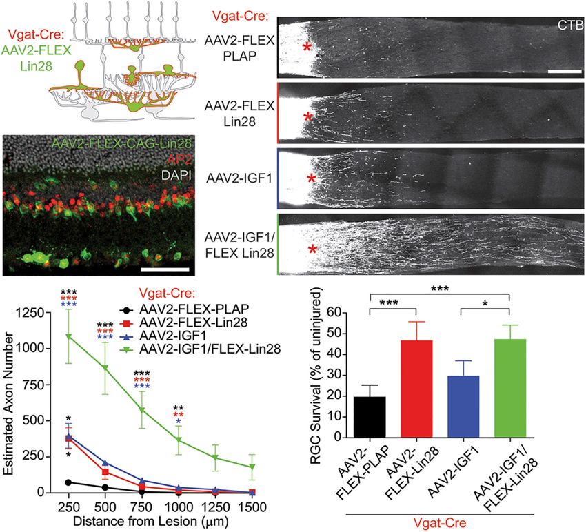
regeneration in the optic nerve. Zhang et al. (2019) showed that nevertheless increase axon regeneration, as we discussed above.
optic nerve injury increases the activity of ACs (Zhang et al.,
2019), which in turn puts a brake on regeneration by inhibiting
RGC activity and reducing these cells’ responsiveness to growth AMACRINE CELLS AND ZINC
factors (Zhang et al., 2019) (Figure 1). When hyperactive SIGNALING IN THE RETINA
ACs were silenced, as confirmed by diminished levels of the
immediate-early gene c-fos in these cells, RGCs showed increased In addition to diminishing RGCs’ activity state, do ACs produce
physiological activity and improved responsiveness to insulin- other signals that suppress RGC survival and regenerative
like growth factor IGF1 (Zhang et al., 2019). This improved ability? Our lab recently reported that one such signal may be
responsiveness was mediated by increased expression of the IGF1 mobile zinc (Zn2+ ) (Li et al., 2017a; Trakhtenberg et al., 2018).
receptor on RGCs’ primary cilia, which serve as the growth factor- Elevation of mobile Zn2+ in AC terminals within the inner
sensing antennae of these cells (Guemez-Gamboa et al., 2014), plexiform layer (IPL) of the retina, as demonstrated by selenite
leading to increased RGC survival and axon regeneration. In autometallography (AMG), is one of the earliest changes seen in
this study, AC activity was suppressed by either overexpressing mouse retina after optic nerve injury (Li et al., 2017a) (Figure 2).
the potassium channel Kir2.1 or by overexpressing an RNA- Normally, zinc is covalently bound to proteins, including
binding insulin-sensitizing protein Lin28 specifically in ACs many transcription factors and enzymes, enabling their folding
and horizontal cells. Importantly, whereas IGF1 overexpression and thus their functionality (McCall et al., 2000; Kochanczyk
or blocking inhibition by either silencing ACs or suppressing et al., 2015). Some neurons, including particular cells in the
neurotransmission with a cocktail of GABA and glycine receptor hippocampus, cerebral cortex, and spinal cord, sequester Zn2+ in
antagonists induced a moderate level of regeneration by itself, synaptic vesicles and co-release it with classical neurotransmitters
the combination of AC silencing plus IGF1 overexpression had (Nakashima and Dyck, 2009; Sensi et al., 2009, 2011; Pan
a strongly synergistic effect via increased RGC activity and IGF1 et al., 2011; Kimura and Kambe, 2016). Intracellular levels of
Frontiers in Cellular Neuroscience | www.frontiersin.org 3 April 2021 | Volume 15 | Article 666798Sergeeva et al. Interneurons Influence Optic Nerve Regeneration
FIGURE 1 | Lin28 expression in inhibitory neurons promotes RGC survival and IGF1-induced axonal regeneration. (A,B) Schematic (A) and example (B) confocal
image stack showing expression of AAV2-FLEX-Lin28 in the intact Vgat-Cre transgenic retina where Lin28 expression is restricted to amacrine and horizontal cells.
(C) Representative confocal image stacks of CTB labeled RGC axons 2 weeks after optic nerve crush with amacrine cell restricted expression of Lin28. Asterisks
indicate crush site. (D) Quantification of the extent of RGC axon regeneration in treatment groups restricted to amacrine cells. Asterisk colors indicate the group that
the p value was significant against. (E) Quantification of RGC survival relative to RGC density observed in intact retinas in treatment groups restricted to amacrine
cells. n = 5 mice per group. Scale bar, 50 µm in (B), and 200 µm in (C). *, **, ***p < 0.05, 0.01, 0.001, respectively. Reprinted from Zhang et al. (2019) with
permission.
mobile Zn2+ can vary depending on many factors, including metallothioneins or engage in oxidative reactions, ultimately
oxidative stress and liberation of Zn2+ from oxidized proteins leading to more Zn2+ release (Krȩżel and Maret, 2017). The
(Aravindakumar et al., 1999; Sensi et al., 1999; Spahl et al., apparent elevation of mobile Zn2+ in AC terminals that synapse
2003; Aras and Aizenman, 2011), redistribution of Zn2+ between onto RGCs that occurs soon after optic nerve injury points to AC
intracellular pools (Sekler et al., 2007; Maret, 2017; Ji et al., and Zn2+ dysregulation as a potential major factor of abnormal
2020), and transcriptional and posttranscriptional regulation of retinal circuit homeostasis after injury (Li et al., 2017a) (Figure 2).
Zn2+ -regulating proteins (Saydam et al., 2002; Jackson et al.,
2008). It is important to maintain Zn2+ concentrations within
a narrow range in different intracellular compartments to
Role of Nitric Oxide and Presumptive
maintain proper Zn2+ availability to numerous Zn2+ -binding Role of Glutamate and Bipolar Cells in
proteins while at the same time preventing mismetallation and Retinal Zn2+ Homeostasis
Zn2+ toxicity (Aras and Aizenman, 2011). For this purpose, a Little is known about the mechanisms underlying the increase
complex homeostatic machinery comprised of metal buffering in AMG signal in the retina following the injury of RGC axons.
proteins – metallothioneins and zinc transporters (ZnTs and A preliminary report used a novel fluorescent nitric oxide (NO)
ZIPs) has evolved (Hidalgo et al., 2001; Cousins et al., 2006; sensor, Cu2 FL2E (Pluth et al., 2011), to provide evidence that
McAllister and Dyck, 2017). the production of NO is rapidly and persistently upregulated
Metallothioneins, glutathione and other metal-containing in the retina after optic nerve injury, and that NO generation
peptides and proteins can liberate Zn2+ and copper ions (Cu+ is upstream of the accumulation of AMG signal in the retinal
or Cu2+ ) when subjected to oxidative stress (Maret, 1995). For IPL (Li et al., 2017b). One possibility is that reactive nitrogen
example, reactive oxygen species and peroxynitrite can oxidize species produced after injury, e.g., peroxynitrite, can liberate
residues on the metal-binding sites of metal-binding proteins and Zn2+ from metallothioneins (Zhang et al., 2004; Nakamura et al.,
release the cations (Sensi et al., 1999; Hidalgo et al., 2001; Spahl 2015; Wolhuter et al., 2018). Alternatively, NO can contribute
et al., 2003; Zhang et al., 2004; Aras and Aizenman, 2011). Cu+ to an increase of intracellular Zn2+ via a cGMP/PKG-dependent
and Cu2+ , as redox-active ions, can directly displace Zn2+ from release of Zn2+ from internal stores (Jang et al., 2007).
Frontiers in Cellular Neuroscience | www.frontiersin.org 4 April 2021 | Volume 15 | Article 666798Sergeeva et al. Interneurons Influence Optic Nerve Regeneration FIGURE 2 | Zn2+ accumulation in the retina and its role in axon regeneration after optic nerve crush (NC). (A) Zinc accumulates in the inner plexiform layer (IPL) of the retina shortly after NC in wild-type mice (slc30a3+/+ ) but not in mice lacking the zinc transporter ZnT3 (slc30a3- / - ): images and quantitation of AMG staining in the IPL (n = 6 retinas per group) of wild-type and slc30a3− / − littermates. Note elevation of AMG signal on day 1 following NC in wild-type mice and decline to near normal level by day 3 (Scale bar, 25 µm; ∗∗ P < 0.01, ∗∗∗ P < 0.001). (B) Tetanus toxin (TeNT) blocks vesicular release of Zn2+ , causing continued Zn2+ build-up in the IPL: images and quantification of AMG staining in the IPL after NC with and without intraocular injection of TeNT (20 nM). Note elevation of AMG staining in the IPL of normal, uninjured mice and in wild-type mice, at 3 days after NC, a time point at which AMG staining in the IPL would normally dissipate. Deletion of the gene encoding ZnT3 eliminates Zn2+ accumulation in the IPL (Scale bar, 50 µm; ∗∗∗ P < 0.001). Adapted from Li et al. (2017a) with permission. NO is synthetized by nitric oxide synthetase, one isoform of the glutamate transporter GLT-1 in BCs, activation of NMDA of which, NOS1, is expressed exclusively in a subset of ACs receptors, and NO elevation may act upstream of Zn2+ liberation (Yamamoto et al., 1993; Oh et al., 1998). Production of NO in and accumulation in AC terminals after optic nerve injury ACs after optic nerve injury points to the existence of an as (Hanovice et al., 2019). yet unidentified retrograde signal linking RGC axon injury and NOS1 activation. NOS1 activation can be triggered by Ca2+ entering ACs upon activation of voltage-gated calcium channels Effect of Presynaptic Zinc on Retinal or through NMDA or AMPA receptors (Christopherson et al., Ganglion Cells 1999). These latter receptors can be activated by glutamate that is In line with previous studies showing that Zn2+ levels in the either synaptically released by BCs or elevated due to a reversal brain (visualized by AMG) are abolished in mice lacking the of glutamate transporters, e.g., GLT-1, EAAC1, GLAST, that zinc transporter protein ZnT3, the accumulation of Zn2+ in AC are expressed on retinal neurons or glia, including astrocytes terminals following ONC is similarly absent in ZnT3 knock-out and Mueller cells. Glutamate transporters normally take up mice (Li et al., 2017a) (Figure 2A). Because ZnT3 enables Zn2+ extracellular glutamate but can reverse the direction of transport to be sequestered in synaptic vesicles (Palmiter et al., 1996), this and release glutamate upon changes in Na+ and K+ gradients finding implies that the Zn2+ that is mobilized in ACs after ONC or membrane potential (Szatkowski et al., 1990; Danbolt, 2001; is stored in synaptic vesicles (Palmiter et al., 1996; Li et al., 2017a). Grewer et al., 2008; Armbruster et al., 2016; Rimmele et al., In conformity with this idea, the Zn2+ that accumulates in the 2017). Our preliminary studies show that BC-specific knockout of retinal IPL after ONC normally dissipates by 48 hour after ONC GLT-1 may prevent mobile Zn2+ accumulation in AC terminals (Li et al., 2017a) but continues to accumulate if exocytosis is after ONC, as does inhibition of NMDA receptors (Hanovice inhibited using Clostridium tetani neurotoxin (TeNT) (Li et al., et al., 2019). Taken together, these results suggest that reversal 2017a; Sergeeva et al., 2019) (Figure 2B). Blockade of synaptic Frontiers in Cellular Neuroscience | www.frontiersin.org 5 April 2021 | Volume 15 | Article 666798
Sergeeva et al.                                                                                        Interneurons Influence Optic Nerve Regeneration

release from AC terminals with TeNT promotes RGC survival                   potentially facilitate transcription of activity-dependent genes
and optic nerve regeneration (Li et al., 2017a; Sergeeva et al.,            and ultimately add to the effects of RGC activation. Along these
2019). These data suggest that Zn2+ packaged into synaptic                  lines, inhibition of HDAC activity alone has been shown to
vesicles and released from AC terminals, or the neurotransmitter            protect RGCs after injury (Gaub et al., 2011; Zhang et al., 2012;
used by these neurons, or both, may negatively affect RGC                   Chindasub et al., 2013; Janssen et al., 2013; Schmitt et al., 2014).
survival and block axon regeneration.                                          An additive effect on RGC survival by metal chelators was
    It should be noted, however, that because the chelators used            observed in combinatorial treatment with deletion of PTEN,
in the aforementioned studies are not entirely specific to Zn2+ ,           producing survival of RGCs that was substantially greater at
it remains possible that other cations, e.g., Cu+ or Cu2+ , could           12 weeks post ONC compared to PTEN deletion itself (Li et al.,
also be involved. Copper is stored in synaptic vesicles and released        2017a). Knockdown of another intrinsic suppressor of axonal
upon depolarization (Kardos et al., 1989). Moreover, the method             growth, Klf-9, also demonstrated enhanced RGC survival when
used to detect Zn2+ , e.g., AMG, although generally regarded as             combined with chelation (Trakhtenberg et al., 2018).
being specific to Zn2+ (Danscher and Stoltenberg, 2005), may
also provide ambiguous results, as selenite may potentially form
complexes with other divalent cations, suggesting that vesicular            WHY DO AMACRINE CELLS BECOME
Cu+ /Cu2+ may potentially contribute to AMG staining. On the                HYPERACTIVE? A HYPOTHESIS
other hand, the observation that the AMG signal in the retinal
IPL is abolished in mice lacking ZnT3 supports the hypothesis               It is largely unknown how or why ACs become hyperactive after
that the AMG signal reflects Zn2+ per se, provided that ZnT3 does           optic nerve injury (Zhang et al., 2019). Activation of NMDA or
not transport other divalent cations, such as copper. At this stage,        AMPA receptors on ACs by glutamate released from BCs leads
we also do not know whether other metals act downstream or                  to AC depolarization, increased firing, and increased release of
upstream of Zn2+ release and accumulation.                                  glycine and GABA onto RGCs, as well as onto BCs and other
    Synaptic release of Zn2+ from ACs could affect RGC                      ACs (Kolb and Famiglietti, 1974). Activation of GABA or glycine
signaling via numerous pathways. Synaptic Zn2+ can modulate                 receptors on ACs by GABA or glycine released from other ACs
the activity of NMDA, GABA and glycine receptors, thereby                   causes these cells to become hyperpolarized to a level closer to
modulating cell excitation and inhibition (Suwa et al., 2001;               the reversal potential for Cl− , which in these cells is normally
Kaneda et al., 2005; Sensi et al., 2011; Vergnano et al., 2014). Zn2+       more negative than the membrane potential, reducing action
modulates glycine receptors in a biphasic manner, potentiating              potential firing, with a net effect of decreasing inhibitory tone
inhibition at low micromolar concentrations while suppressing               projecting onto RGCs. However, under some circumstances, for
glycinergic currents at high concentrations (Kaneda et al., 2005).          example, early in development, the reversal potential for chloride
Potentiation of glycine receptors on RGCs would be expected to              may be depolarized with respect to the membrane potential
decrease RGC activity which, as noted above, would diminish                 due to the electrochemical gradient driven by high intracellular
RGC survival and axon regeneration (Goldberg et al., 2002a;                 Cl− . Such switch in GABA function is mediated by changes
Goldberg, 2012; Zhang et al., 2019). In addition, Zn2+ interacting          in expression or localization of Cl− transporters: the neuron-
with the Zn2+ -sensing receptor ZnR/GPR39 could regulate the                specific K+ - Cl− cotransporter KCC2 and the Na+ - K+ - Cl−
transport of Na+ , K+ , Cl− (Chorin et al., 2011; Saadi et al., 2012)       cotransporter NKCC1 expressed in immature neurons (Kaila
and trigger Gαq -dependent signaling and subsequent release of              et al., 2014). Consequently, the activation state of ACs depends
Ca2+ from endoplasmic reticulum stores, thereby modulating                  not only on the sum of excitatory and inhibitory inputs onto these
ERK/MAPK and PI3K/Akt/mTOR signaling, both of which are                     cells at any moment, but also on the Cl− gradient that determines
important for cell survival and growth (Azriel-Tamir et al., 2004;          the polarity of the GABAergic and glycinergic drive onto these
Hershfinkel, 2018). Potentially, intracellular Zn2+ elevation can           cells. Alteration of the Cl− gradient may be important in retinal
induce cell death by upregulating proapoptotic factors (Jiang               network dysfunction and has been investigated in several studies
et al., 2001; Zhang et al., 2004, 2006, 2007; Cohen et al., 2012),          (Hoffpauir et al., 2006; Krishnan and Gleason, 2015).
mitochondrial impairment (Sensi et al., 1999; Baud et al., 2004;                Cl− gradient alteration induced by decreased KCC2 function
Ji et al., 2020), synthesis of reactive oxygen species (Wang et al.,        or expression is an important cause of disinhibition in cells and
2004; Bishop et al., 2007), activation of MAPK/p38 signaling and            circuits, and has been shown to participate in several neurological
activation of voltage-gated K+ channels, leading to K+ efflux               disorders including epilepsy (Moore et al., 2017; Liu et al., 2019),
(McLaughlin et al., 2001; Bossy-Wetzel et al., 2004; Zhang and              spasticity after spinal cord injury (Boulenguez et al., 2010; Chen
Rosenberg, 2004; Zhang et al., 2004, 2006, 2007; McCord and                 et al., 2018), autism and Rett syndrome (Tang et al., 2016) and
Aizenman, 2013).                                                            chronic pain (Coull et al., 2003; Hasbargen et al., 2010), all of
    Chelation of Zn2+ after ONC can potentially inhibit histone             which are characterized by a failure of inhibition and neural
deacetylases (HDACs), enzymes that deacetylate histone proteins,            hyperactivation (Nabekura et al., 2002; Kaila et al., 2014). KCC2
thereby rendering chromatin more accessible for transcription.              cotransport utilizes a K+ gradient to extrude Cl− (Payne et al.,
The deacetylating activity of HDACs depends on the binding                  2003; Kaila et al., 2014), therefore the Cl− transporter activity
of Zn2+ in the HDAC active site pocket (Pelzel et al., 2010; Li             may decrease with high extracellular K+ following ischemia,
et al., 2019). Prevention of histone deacetylation by inhibition            injury, Na+ /K+ ATP dysfunction and reduced production of ATP
of HDAC activity caused by removal of Zn2+ from HDACs can                   due to mitochondrial compromise (Kleber, 1984; Hughes and

Frontiers in Cellular Neuroscience | www.frontiersin.org                6                                       April 2021 | Volume 15 | Article 666798
Sergeeva et al. Interneurons Influence Optic Nerve Regeneration
Cidlowski, 1999; Kaila et al., 2014; Doyon et al., 2016). KCC2 is Although the non-cell-autonomous regulation of neuronal
highly expressed in the retina (Vardi et al., 2000; Vu et al., 2000). survival and pathological functioning by other neurons is
With increased extracellular K+ after K+ efflux from injured just starting to be recognized as being important after optic
RGCs (Yu et al., 1997; Diem et al., 2001; Zhong et al., 2013) or nerve injury, neuronal circuits have been implicated in various
activated microglia (Fordyce et al., 2005) KCC2-dependent Cl− pathological processes and cell death in other neurodegenerative
extrusion in ACs may be diminished. diseases (Palop et al., 2006; Simon et al., 2016). In amyotrophic
KCC2, like many intracellular proteins, can be regulated lateral sclerosis, hyperexcitability and death of motoneurons
by phosphorylation, trafficking and proteolytic cleavage (Kaila have been attributed to a non-cell autonomous response to a
et al., 2014; Doyon et al., 2016; Kahle and Delpire, 2016). defect in premotor interneurons (Wainger et al., 2014; Held
Extracellular modifiers of KCC2 expression and function include et al., 2019). In Parkinson’s disease, alterations of basal ganglia
BDNF/TrkB, serotonin/5HT2A, glutamate/NMDA, the Zn2+ - circuitry have been shown to precede loss of substantia nigra
sensing receptor GPR39, and noradrenaline signaling (Wake neurons (McGregor and Nelson, 2019), as was shown for striatal
et al., 2007; Hershfinkel et al., 2009; Bos et al., 2013; Watanabe spiny neurons in Huntington’s disease (Creus-Muncunill and
and Fukuda, 2015; Tang et al., 2019). In addition, KCC2 activity Ehrlich, 2019). In Alzheimer’s disease, early circuitry dysfunction
can be suppressed by both NO and intracellular zinc (Yassin et al., may be induced by amyloid beta-mediated suppression of
2014) shown to be elevated in ACs after injury (Li et al., 2017a,b). glutamate reuptake and a consequent vicious cycle of neuronal
KCC2 independent-, NO-mediated elevation of intracellular hyperactivation and cell death (Zott et al., 2018, 2019). In autism
Cl− could be another potential mechanism of AC disinhibition and Alzheimer’s disease, dysfunction of interneurons has been
after optic nerve injury. In chick ACs in vitro, NO transiently implicated (Palop et al., 2006; Palop and Mucke, 2016; Martinez-
reverses GABA- and glycine-gated currents, converting Losa et al., 2018). Disruption of excitatory and inhibitory circuits
inhibition of ACs into excitation, thereby increasing the and excitatory-inhibitory imbalance also seem important in the
firing of these cells and thus enhanced inhibitory drive on their pathogenesis of Rett syndrome and autism (Nelson and Valakh,
synaptic partners (e.g., RGCs). This NO-induced shift in ECl− is 2015; Patrizi et al., 2020). Here we assemble evidence that
likely due to release of Cl− from intracellular stores (Hoffpauir optic nerve injury induces changes in retinal circuitry that is
et al., 2006; Krishnan and Gleason, 2015; Krishnan et al., 2017; initiated by an as-yet unidentified signal from injured RGCs
Maddox and Gleason, 2017; Maddox et al., 2018). In addition, to retinal interneurons that alters the function of amacrine
NO may drive synaptic glutamate release from BCs without cells, in turn influencing the survival and regenerative capacity
membrane depolarization via a TRPC Ca2+ influx-mediated of injured RGCs.
pathway, as shown in the chick retina (Maddox et al., 2018), Despite considerable progress in the areas of RGC protection
further depolarizing ACs. and optic nerve regeneration, there is still a long way to go
In summary, dysregulation of Cl− gradients in the inner retina before we achieve satisfactory levels of functional recovery. One
may be a part of the early pathological process following optic factor that is now coming to be appreciated is the crosstalk
nerve or RGC injury. Reciprocally connected ACs and BCs, in between cell-intrinsic and cell-extrinsic factors, particularly the
the face of Cl− gradient collapse, can form circuits with positive role of neural circuits and the activity of neurons that form
feedback loops that may rapidly lead to hyperactivation of ACs synapses with the affected cells. A greater understanding of the
and thus increased inhibition of their synaptic targets (Marc and role of circuit activity might substantially augment the outcome
Liu, 2000; Marc et al., 2014; Doyon et al., 2015, 2016). Whether achieved by manipulating RGCs’ intrinsic growth potential and
the complex retinal circuitry is particularly susceptible to cell-extrinsic factors.
persistent disinhibition of ACs after injury remains to be studied.
AUTHOR CONTRIBUTIONS
CONCLUSION: FROM RETINAL All authors conceptualized the study and wrote the manuscript.
GANGLION CELLS TO RETINAL
CIRCUITS
FUNDING
Silencing ACs or introducing chelators into the eye to suppress
Zn2+ accumulation in amacrine cell terminals are additive with We are grateful for the support of the NIH (NEI R01EY027881 to
the effects of manipulating RGC-intrinsic factors (PTEN deletion, PR and LB, NEI R01EY024481 to PR and LB, and NINDS IDDRC
Klf-9 suppression, upregulation of osteopontin) on RGC survival HD018655), and the Adelson Medical Research Foundation.
and regeneration (Li et al., 2017a; Trakhtenberg et al., 2018;
Zhang et al., 2019). These findings suggest that dysfunction of the
retinal network, and particularly interneuron (AC) dysfunction, ACKNOWLEDGMENTS
is part of the pathological process following optic nerve injury,
and that the capacity of RGCs to survive and regenerate may We wish to thank Dr. Xin Tang (Boston Children’s
depend in part on the activity of the other retinal neurons with Hospital/Harvard Medical School) for critically reading
which they are connected. parts of the manuscript.
Frontiers in Cellular Neuroscience | www.frontiersin.org 7 April 2021 | Volume 15 | Article 666798Sergeeva et al. Interneurons Influence Optic Nerve Regeneration
REFERENCES Chierzi, S., Strettoi, E., Cenni, M. C., and Maffei, L. (1999). Optic nerve crush:
axonal responses in wild-type and bcl-2 transgenic mice. J. Neurosci. 19,
Aguayo, A. J., Rasminsky, M., Bray, G. M., Carbonetto, S., McKerracher, L., 8367–8376.
Villegas-Perez, M. P., et al. (1991). Degenerative and regenerative responses of Chindasub, P., Lindsey, J. D., Duong-Polk, K., Leung, C. K., and Weinreb, R. N.
injured neurons in the central nervous system of adult mammals. Philos. Trans. (2013). Inhibition of histone deacetylases 1 and 3 protects injured retinal
R. Soc. Lond. B Biol. Sci. 331, 337–343. doi: 10.1098/rstb.1991.0025 ganglion cells. Invest. Ophthalmol. Vis. Sci. 54, 96–102. doi: 10.1167/iovs.12-
Anderson, M. A., Burda, J. E., Ren, Y., Ao, Y., O’Shea, T. M., Kawaguchi, R., et al. 10850
(2016). Astrocyte scar formation aids central nervous system axon regeneration. Chorin, E., Vinograd, O., Fleidervish, I., Gilad, D., Herrmann, S., Sekler, I., et al.
Nature 532, 195–200. doi: 10.1038/nature17623 (2011). Upregulation of KCC2 activity by zinc-mediated neurotransmission
Apara, A., Galvao, J., Wang, Y., Blackmore, M., Trillo, A., Iwao, K., et al. (2017). via the mZnR/GPR39 receptor. J. Neurosci. 31, 12916–12926. doi: 10.1523/
KLF9 and JNK3 interact to suppress axon regeneration in the adult CNS. jneurosci.2205-11.2011
J. Neurosci. 37, 9632–9644. doi: 10.1523/jneurosci.0643-16.2017 Christopherson, K. S., Hillier, B. J., Lim, W. A., and Bredt, D. S. (1999). PSD-95
Apara, A., and Goldberg, J. L. (2014). Molecular mechanisms of the suppression assembles a ternary complex with the N-methyl-D-aspartic acid receptor and a
of axon regeneration by KLF transcription factors. Neural Regen. Res. 9, 1418– bivalent neuronal NO synthase PDZ domain. J. Biol. Chem. 274, 27467–27473.
1421. doi: 10.4103/1673-5374.139454 doi: 10.1074/jbc.274.39.27467
Aras, M. A., and Aizenman, E. (2011). Redox regulation of intracellular zinc: Cohen, L., Azriel-Tamir, H., Arotsker, N., Sekler, I., and Hershfinkel, M. (2012).
molecular signaling in the life and death of neurons. Antioxid. Redox Signal. Zinc sensing receptor signaling, mediated by GPR39, reduces butyrate-induced
15, 2249–2263. doi: 10.1089/ars.2010.3607 cell death in HT29 colonocytes via upregulation of clusterin. PLoS One
Aravindakumar, C. T., Ceulemans, J., and De Ley, M. (1999). Nitric oxide induces 7:e35482. doi: 10.1371/journal.pone.0035482
Zn2+ release from metallothionein by destroying zinc-sulphur clusters without Coull, J. A., Boudreau, D., Bachand, K., Prescott, S. A., Nault, F., Sik, A., et al.
concomitant formation of S-nitrosothiol. Biochem. J. 344 (Pt 1), 253–258. doi: (2003). Trans-synaptic shift in anion gradient in spinal lamina I neurons
10.1042/0264-6021:3440253 as a mechanism of neuropathic pain. Nature 424, 938–942. doi: 10.1038/
Armbruster, M., Hanson, E., and Dulla, C. G. (2016). Glutamate clearance is locally nature01868
modulated by presynaptic neuronal activity in the cerebral cortex. J. Neurosci. Cousins, R. J., Liuzzi, J. P., and Lichten, L. A. (2006). Mammalian zinc transport,
36, 10404–10415. doi: 10.1523/jneurosci.2066-16.2016 trafficking, and signals. J. Biol. Chem. 281, 24085–24089. doi: 10.1074/jbc.
Azriel-Tamir, H., Sharir, H., Schwartz, B., and Hershfinkel, M. (2004). Extracellular R600011200
zinc triggers ERK-dependent activation of Na+/H+ exchange in colonocytes Creus-Muncunill, J., and Ehrlich, M. E. (2019). Cell-autonomous and non-cell-
mediated by the zinc-sensing receptor. J. Biol. Chem. 279, 51804–51816. doi: autonomous pathogenic mechanisms in Huntington’s disease: insights from
10.1074/jbc.M406581200 in vitro and in vivo models. Neurotherapeutics 16, 957–978. doi: 10.1007/
Baud, O., Li, J., Zhang, Y., Neve, R. L., Volpe, J. J., and Rosenberg, P. A. s13311-019-00782-9
(2004). Nitric oxide-induced cell death in developing oligodendrocytes is Danbolt, N. C. (2001). Glutamate uptake. Prog. Neurobiol. 65, 1–105. doi: 10.1016/
associated with mitochondrial dysfunction and apoptosis-inducing factor s0301-0082(00)00067-8
translocation. Eur. J. Neurosci. 20, 1713–1726. doi: 10.1111/j.1460-9568.2004. Danscher, G., and Stoltenberg, M. (2005). Zinc-specific autometallographic in vivo
03616.x selenium methods: tracing of zinc-enriched (ZEN) terminals, ZEN pathways,
Beirowski, B., Nogradi, A., Babetto, E., Garcia-Alias, G., and Coleman, M. P. and pools of zinc ions in a multitude of other ZEN cells. J. Histochem. Cytochem.
(2010). Mechanisms of axonal spheroid formation in central nervous system 53, 141–153. doi: 10.1369/jhc.4R6460.2005
Wallerian degeneration. J. Neuropathol. Exp. Neurol. 69, 455–472. doi: 10.1097/ de Lima, S., Koriyama, Y., Kurimoto, T., Oliveira, J. T., Yin, Y., Li, Y., et al.
NEN.0b013e3181da84db (2012). Full-length axon regeneration in the adult mouse optic nerve and partial
Belin, S., Nawabi, H., Wang, C., Tang, S., Latremoliere, A., Warren, P., et al. recovery of simple visual behaviors. Proc. Natl. Acad. Sci. U.S.A. 109, 9149–9154.
(2015). Injury-induced decline of intrinsic regenerative ability revealed by doi: 10.1073/pnas.1119449109
quantitative proteomics. Neuron 86, 1000–1014. doi: 10.1016/j.neuron.2015. Dickendesher, T. L., Baldwin, K. T., Mironova, Y. A., Koriyama, Y., Raiker, S. J.,
03.060 Askew, K. L., et al. (2012). NgR1 and NgR3 are receptors for chondroitin sulfate
Benowitz, L. I., He, Z., and Goldberg, J. L. (2017). Reaching the brain: advances proteoglycans. Nat. Neurosci. 15, 703–712. doi: 10.1038/nn.3070
in optic nerve regeneration. Exp. Neurol. 287(Pt 3), 365–373. doi: 10.1016/j. Diem, R., Meyer, R., Weishaupt, J. H., and Bahr, M. (2001). Reduction of potassium
expneurol.2015.12.015 currents and phosphatidylinositol 3-kinase-dependent AKT phosphorylation
Bishop, G. M., Dringen, R., and Robinson, S. R. (2007). Zinc stimulates the by tumor necrosis factor-(alpha) rescues axotomized retinal ganglion cells from
production of toxic reactive oxygen species (ROS) and inhibits glutathione retrograde cell death in vivo. J. Neurosci. 21, 2058–2066.
reductase in astrocytes. Free Radic. Biol. Med. 42, 1222–1230. doi: 10.1016/j. Donahue, R. J., Maes, M. E., Grosser, J. A., and Nickells, R. W. (2020).
freeradbiomed.2007.01.022 BAX-depleted retinal ganglion cells survive and become quiescent following
Bos, R., Sadlaoud, K., Boulenguez, P., Buttigieg, D., Liabeuf, S., Brocard, C., optic nerve damage. Mol. Neurobiol. 57, 1070–1084. doi: 10.1007/s12035-019-
et al. (2013). Activation of 5-HT2A receptors upregulates the function of the 01783-7
neuronal K-Cl cotransporter KCC2. Proc. Natl. Acad. Sci. U.S.A. 110, 348–353. Doyon, N., Prescott, S. A., and De Koninck, Y. (2015). Mild KCC2 hypofunction
doi: 10.1073/pnas.1213680110 causes inconspicuous chloride dysregulation that degrades neural coding.
Bossy-Wetzel, E., Talantova, M. V., Lee, W. D., Scholzke, M. N., Harrop, A., Front. Cell. Neurosci. 9:516. doi: 10.3389/fncel.2015.00516
Mathews, E., et al. (2004). Crosstalk between nitric oxide and zinc pathways Doyon, N., Vinay, L., Prescott, S. A., and De Koninck, Y. (2016). Chloride
to neuronal cell death involving mitochondrial dysfunction and p38-activated regulation: a dynamic equilibrium crucial for synaptic inhibition. Neuron 89,
K+ channels. Neuron 41, 351–365. doi: 10.1016/s0896-6273(04)00015-7 1157–1172. doi: 10.1016/j.neuron.2016.02.030
Boulenguez, P., Liabeuf, S., Bos, R., Bras, H., Jean-Xavier, C., Brocard, C., Duan, X., Qiao, M., Bei, F., Kim, I. J., He, Z., and Sanes, J. R. (2015). Subtype-
et al. (2010). Down-regulation of the potassium-chloride cotransporter KCC2 specific regeneration of retinal ganglion cells following axotomy: effects of
contributes to spasticity after spinal cord injury. Nat. Med. 16, 302–307. doi: osteopontin and mTOR signaling. Neuron 85, 1244–1256. doi: 10.1016/j.
10.1038/nm.2107 neuron.2015.02.017
Chen, B., Li, Y., Yu, B., Zhang, Z., Brommer, B., Williams, P. R., et al. (2018). Feldheim, D. A., and O’Leary, D. D. (2010). Visual map development: bidirectional
Reactivation of dormant relay pathways in injured spinal cord by KCC2 signaling, bifunctional guidance molecules, and competition. Cold Spring Harb.
manipulations. Cell 174, 521–535.e13. doi: 10.1016/j.cell.2018.06.005 Perspect. Biol. 2:a001768. doi: 10.1101/cshperspect.a001768
Cheng, Y., Yin, Y., Zhang, A., Bernstein, A. M., Kawaguchi, R., Gao, K., et al. Fernandes, K. A., Harder, J. M., Fornarola, L. B., Freeman, R. S., Clark, A. F.,
(2020). Transcription factor network analysis identifies REST/NRSF as an Pang, I. H., et al. (2012). JNK2 and JNK3 are major regulators of axonal injury-
intrinsic regulator of CNS regeneration. BioRxiv [Preprint] 12.13.413104. induced retinal ganglion cell death. Neurobiol. Dis. 46, 393–401. doi: 10.1016/j.
BioRxiv:12.13.413104 nbd.2012.02.003
Frontiers in Cellular Neuroscience | www.frontiersin.org 8 April 2021 | Volume 15 | Article 666798Sergeeva et al. Interneurons Influence Optic Nerve Regeneration Fischer, D., He, Z., and Benowitz, L. I. (2004a). Counteracting the Nogo receptor Hughes, F. M. Jr., and Cidlowski, J. A. (1999). Potassium is a critical regulator enhances optic nerve regeneration if retinal ganglion cells are in an active of apoptotic enzymes in vitro and in vivo. Adv. Enzyme Regul. 39, 157–171. growth state. J. Neurosci. 24, 1646–1651. doi: 10.1523/jneurosci.5119-03.2004 doi: 10.1016/s0065-2571(98)00010-7 Fischer, D., Petkova, V., Thanos, S., and Benowitz, L. I. (2004b). Switching mature Jackson, K. A., Valentine, R. A., Coneyworth, L. J., Mathers, J. C., and Ford, D. retinal ganglion cells to a robust growth state in vivo: gene expression and (2008). Mechanisms of mammalian zinc-regulated gene expression. Biochem. synergy with RhoA inactivation. J. Neurosci. 24, 8726–8740. doi: 10.1523/ Soc. Trans. 36(Pt 6), 1262–1266. doi: 10.1042/bst0361262 jneurosci.2774-04.2004 Jang, Y., Wang, H., Xi, J., Mueller, R. A., Norfleet, E. A., and Xu, Z. (2007). NO Fordyce, C. B., Jagasia, R., Zhu, X., and Schlichter, L. C. (2005). Microglia Kv1.3 mobilizes intracellular Zn2+ via cGMP/PKG signaling pathway and prevents channels contribute to their ability to kill neurons. J. Neurosci. 25, 7139–7149. mitochondrial oxidant damage in cardiomyocytes. Cardiovasc. Res. 75, 426– doi: 10.1523/jneurosci.1251-05.2005 433. doi: 10.1016/j.cardiores.2007.05.015 Galvao, J., Iwao, K., Apara, A., Wang, Y., Ashouri, M., Shah, T. N., et al. (2018). The Janssen, K. T., Mac Nair, C. E., Dietz, J. A., Schlamp, C. L., and Nickells, R. W. Krüppel-like factor gene target Dusp14 regulates axon growth and regeneration. (2013). Nuclear atrophy of retinal ganglion cells precedes the bax-dependent Invest. Ophthalmol. Vis. Sci. 59, 2736–2747. doi: 10.1167/iovs.17-23319 stage of apoptosis. Invest. Ophthalmol. Vis. Sci. 54, 1805–1815. doi: 10.1167/iovs. Gaub, P., Joshi, Y., Wuttke, A., Naumann, U., Schnichels, S., Heiduschka, P., 11-9310 et al. (2011). The histone acetyltransferase p300 promotes intrinsic axonal Ji, S. G., Medvedeva, Y. V., and Weiss, J. H. (2020). Zn(2+) entry through the regeneration. Brain 134(Pt 7), 2134–2148. doi: 10.1093/brain/awr142 mitochondrial calcium uniporter is a critical contributor to mitochondrial Gerdts, J., Summers, D. W., Milbrandt, J., and DiAntonio, A. (2016). Axon dysfunction and neurodegeneration. Exp. Neurol. 325:113161. doi: 10.1016/j. self-destruction: new links among SARM1, MAPKs, and NAD+ metabolism. expneurol.2019.113161 Neuron 89, 449–460. doi: 10.1016/j.neuron.2015.12.023 Jiang, D., Sullivan, P. G., Sensi, S. L., Steward, O., and Weiss, J. H. (2001). Zn(2+) Goldberg, J. L. (2004). Intrinsic neuronal regulation of axon and dendrite growth. induces permeability transition pore opening and release of pro-apoptotic Curr. Opin. Neurobiol. 14, 551–557. doi: 10.1016/j.conb.2004.08.012 peptides from neuronal mitochondria. J. Biol. Chem. 276, 47524–47529. doi: Goldberg, J. L. (2012). Role of electrical activity in promoting neural repair. 10.1074/jbc.M108834200 Neurosci. Lett. 519, 134–137. doi: 10.1016/j.neulet.2012.02.003 Kahle, K. T., and Delpire, E. (2016). Kinase-KCC2 coupling: Cl- rheostasis, disease Goldberg, J. L., and Barres, B. A. (2000). The relationship between neuronal susceptibility, therapeutic target. J. Neurophysiol. 115, 8–18. doi: 10.1152/jn. survival and regeneration. Annu. Rev. Neurosci. 23, 579–612. doi: 10.1146/ 00865.2015 annurev.neuro.23.1.579 Kaila, K., Price, T. J., Payne, J. A., Puskarjov, M., and Voipio, J. (2014). Cation- Goldberg, J. L., Espinosa, J. S., Xu, Y., Davidson, N., Kovacs, G. T., and Barres, chloride cotransporters in neuronal development, plasticity and disease. Nat. B. A. (2002a). Retinal ganglion cells do not extend axons by default: promotion Rev. Neurosci. 15, 637–654. doi: 10.1038/nrn3819 by neurotrophic signaling and electrical activity. Neuron 33, 689–702. doi: 10. Kaneda, M., Ishii, K., Akagi, T., Tatsukawa, T., and Hashikawa, T. (2005). 1016/s0896-6273(02)00602-5 Endogenous zinc can be a modulator of glycinergic signaling pathway in the Goldberg, J. L., Klassen, M. P., Hua, Y., and Barres, B. A. (2002b). Amacrine- rat retina. J. Mol. Histol. 36, 179–185. doi: 10.1007/s10735-005-1693-4 signaled loss of intrinsic axon growth ability by retinal ganglion cells. Science Kardos, J., Kovács, I., Hajós, F., Kálmán, M., and Simonyi, M. (1989). Nerve 296, 1860–1864. doi: 10.1126/science.1068428 endings from rat brain tissue release copper upon depolarization. A possible Grewer, C., Gameiro, A., Zhang, Z., Tao, Z., Braams, S., and Rauen, T. (2008). role in regulating neuronal excitability. Neurosci. Lett. 103, 139–144. doi: 10. Glutamate forward and reverse transport: from molecular mechanism to 1016/0304-3940(89)90565-x transporter-mediated release after ischemia. IUBMB Life 60, 609–619. doi: 10. Katome, T., Namekata, K., Guo, X., Semba, K., Kittaka, D., Kawamura, K., et al. 1002/iub.98 (2013). Inhibition of ASK1-p38 pathway prevents neural cell death following Guemez-Gamboa, A., Coufal, N. G., and Gleeson, J. G. (2014). Primary cilia in the optic nerve injury. Cell Death Differ. 20, 270–280. doi: 10.1038/cdd.2012.122 developing and mature brain. Neuron 82, 511–521. doi: 10.1016/j.neuron.2014. Kikuchi, M., Tenneti, L., and Lipton, S. A. (2000). Role of p38 mitogen-activated 04.024 protein kinase in axotomy-induced apoptosis of rat retinal ganglion cells. Hanovice, N. J., Li, Y., Danbolt, N. C., Benowitz, L. I., and Rosenberg, P. A. J. Neurosci. 20, 5037–5044. (2019). “Reversal of glutamate transport contributes to retinal zinc elevation Kim, T., Soto, F., and Kerschensteiner, D. (2015). An excitatory amacrine cell and ganglion cell death after optic nerve injury. Program No. 047.14,” in detects object motion and provides feature-selective input to ganglion cells in Proceedings of the Society for Neuroscience, (Chicago, IL: Neuroscience Meeting the mouse retina. Elife 4:e08025. doi: 10.7554/eLife.08025 Planner). Kimura, T., and Kambe, T. (2016). The functions of metallothionein and ZIP Hasbargen, T., Ahmed, M. M., Miranpuri, G., Li, L., Kahle, K. T., Resnick, D., et al. and ZnT transporters: an overview and perspective. Int. J. Mol. Sci. 17:336. (2010). Role of NKCC1 and KCC2 in the development of chronic neuropathic doi: 10.3390/ijms17030336 pain following spinal cord injury. Ann. N. Y. Acad. Sci. 1198, 168–172. doi: Kleber, A. G. (1984). Extracellular potassium accumulation in acute myocardial 10.1111/j.1749-6632.2010.05462.x ischemia. J. Mol. Cell. Cardiol. 16, 389–394. doi: 10.1016/s0022-2828(84) He, Z., and Jin, Y. (2016). Intrinsic control of axon regeneration. Neuron 90, 80610-0 437–451. doi: 10.1016/j.neuron.2016.04.022 Knoferle, J., Koch, J. C., Ostendorf, T., Michel, U., Planchamp, V., Vutova, P., et al. Held, A., Major, P., Sahin, A., Reenan, R. A., Lipscombe, D., and Wharton, (2010). Mechanisms of acute axonal degeneration in the optic nerve in vivo. K. A. (2019). Circuit dysfunction in SOD1-ALS model first detected in sensory Proc. Natl. Acad. Sci. U.S.A. 107, 6064–6069. doi: 10.1073/pnas.0909794107 feedback prior to motor neuron degeneration is alleviated by BMP signaling. Kochanczyk, T., Drozd, A., and Krezel, A. (2015). Relationship between the J. Neurosci. 39, 2347–2364. doi: 10.1523/jneurosci.1771-18.2019 architecture of zinc coordination and zinc binding affinity in proteins–insights Hershfinkel, M. (2018). The zinc sensing receptor, ZnR/GPR39, in health and into zinc regulation. Metallomics 7, 244–257. doi: 10.1039/c4mt00094c disease. Int. J. Mol. Sci. 19:439. doi: 10.3390/ijms19020439 Kolb, H., and Famiglietti, E. V. (1974). Rod and cone pathways in the inner Hershfinkel, M., Kandler, K., Knoch, M. E., Dagan-Rabin, M., Aras, M. A., plexiform layer of cat retina. Science 186, 47–49. doi: 10.1126/science.186. Abramovitch-Dahan, C., et al. (2009). Intracellular zinc inhibits KCC2 4158.47 transporter activity. Nat. Neurosci. 12, 725–727. doi: 10.1038/nn.2316 Komatsu, M., Wang, Q. J., Holstein, G. R., Friedrich, V. L. Jr., Iwata, J., Kominami, Hidalgo, J., Aschner, M., Zatta, P., and Vasak, M. (2001). Roles of the E., et al. (2007). Essential role for autophagy protein Atg7 in the maintenance metallothionein family of proteins in the central nervous system. Brain Res. of axonal homeostasis and the prevention of axonal degeneration. Proc. Natl. Bull. 55, 133–145. doi: 10.1016/s0361-9230(01)00452-x Acad. Sci. U.S.A. 104, 14489–14494. doi: 10.1073/pnas.0701311104 Hoffpauir, B., McMains, E., and Gleason, E. (2006). Nitric oxide transiently Krȩżel, A., and Maret, W. (2017). The functions of metamorphic metallothioneins converts synaptic inhibition to excitation in retinal amacrine cells. in zinc and copper metabolism. Int. J. Mol. Sci. 18:1237. doi: 10.3390/ J. Neurophysiol. 95, 2866–2877. doi: 10.1152/jn.01317.2005 ijms18061237 Frontiers in Cellular Neuroscience | www.frontiersin.org 9 April 2021 | Volume 15 | Article 666798
Sergeeva et al.                                                                                                                    Interneurons Influence Optic Nerve Regeneration

Krishnan, V., and Gleason, E. (2015). Nitric oxide releases Cl(-) from acidic                 McKeon, R. J., Hoke, A., and Silver, J. (1995). Injury-induced proteoglycans
    organelles in retinal amacrine cells. Front. Cell. Neurosci. 9:213. doi: 10.3389/            inhibit the potential for laminin-mediated axon growth on astrocytic scars. Exp.
    fncel.2015.00213                                                                             Neurol. 136, 32–43. doi: 10.1006/exnr.1995.1081
Krishnan, V., Maddox, J. W., Rodriguez, T., and Gleason, E. (2017). A role                    McLaughlin, B., Pal, S., Tran, M. P., Parsons, A. A., Barone, F. C., Erhardt, J. A., et al.
    for the cystic fibrosis transmembrane conductance regulator in the nitric                    (2001). p38 activation is required upstream of potassium current enhancement
    oxide-dependent release of Cl(-) from acidic organelles in amacrine cells.                   and caspase cleavage in thiol oxidant-induced neuronal apoptosis. J. Neurosci.
    J. Neurophysiol. 118, 2842–2852. doi: 10.1152/jn.00511.2017                                  21, 3303–3311. doi: 10.1523/jneurosci.21-10-03303.2001
Kurimoto, T., Yin, Y., Omura, K., Gilbert, H. Y., Kim, D., Cen, L. P., et al. (2010).         Meyer-Franke, A., Wilkinson, G. A., Kruttgen, A., Hu, M., Munro, E., Hanson,
    Long-distance axon regeneration in the mature optic nerve: contributions of                  M. G. Jr., et al. (1998). Depolarization and cAMP elevation rapidly recruit
    oncomodulin, cAMP, and pten gene deletion. J. Neurosci. 30, 15654–15663.                     TrkB to the plasma membrane of CNS neurons. Neuron 21, 681–693. doi:
    doi: 10.1523/jneurosci.4340-10.2010                                                          10.1016/s0896-6273(00)80586-3
Li, S., Yang, C., Zhang, L., Gao, X., Wang, X., Liu, W., et al. (2016). Promoting             Moore, D. L., Apara, A., and Goldberg, J. L. (2011). Kruppel-like transcription
    axon regeneration in the adult CNS by modulation of the melanopsin/GPCR                      factors in the nervous system: novel players in neurite outgrowth and
    signaling. Proc. Natl. Acad. Sci. U.S.A. 113, 1937–1942. doi: 10.1073/pnas.                  axon regeneration. Mol. Cell. Neurosci. 47, 233–243. doi: 10.1016/j.mcn.2011.
    1523645113                                                                                   05.005
Li, Y., Andereggen, L., Yuki, K., Omura, K., Yin, Y., Gilbert, H. Y., et al. (2017a).         Moore, D. L., Blackmore, M. G., Hu, Y., Kaestner, K. H., Bixby, J. L., Lemmon, V. P.,
    Mobile zinc increases rapidly in the retina after optic nerve injury and regulates           et al. (2009). KLF family members regulate intrinsic axon regeneration ability.
    ganglion cell survival and optic nerve regeneration. Proc. Natl. Acad. Sci. U.S.A.           Science 326, 298–301. doi: 10.1126/science.1175737
    114, E209–E218. doi: 10.1073/pnas.1616811114                                              Moore, Y. E., Kelley, M. R., Brandon, N. J., Deeb, T. Z., and Moss, S. J. (2017).
Li, Y., Wang, F., Chen, X., Wang, J., Zhao, Y., Li, Y., et al. (2019). Zinc-dependent            Seizing control of KCC2: a new therapeutic target for epilepsy. Trends Neurosci.
    deacetylase (HDAC) inhibitors with different zinc binding groups. Curr. Top.                 40, 555–571. doi: 10.1016/j.tins.2017.06.008
    Med. Chem. 19, 223–241. doi: 10.2174/1568026619666190122144949                            Nabekura, J., Ueno, T., Okabe, A., Furuta, A., Iwaki, T., Shimizu-Okabe, C.,
Li, Y., Yuki, K., Omura, K., Gilbert, H. Y., Yin, Y., De Lima, S., et al. (2017b).               et al. (2002). Reduction of KCC2 expression and GABAA receptor-mediated
    “Amacrine cells regulate retinal Zn2+ accumulation, ganglion cell survival,                  excitation after in vivo axonal injury. J. Neurosci. 22, 4412–4417.
    and axon regeneration after optic nerve injury via two distinct nitric oxide              Nakamura, T., Prikhodko, O. A., Pirie, E., Nagar, S., Akhtar, M. W., Oh, C. K., et al.
    mechanisms,” in Neuroscience Meeting Planner. Washington, DC: Society for                    (2015). Aberrant protein S-nitrosylation contributes to the pathophysiology of
    Neuroscience.                                                                                neurodegenerative diseases. Neurobiol. Dis. 84, 99–108. doi: 10.1016/j.nbd.2015.
Lim, J. H., Stafford, B. K., Nguyen, P. L., Lien, B. V., Wang, C., Zukor, K., et al.             03.017
    (2016). Neural activity promotes long-distance, target-specific regeneration of           Nakashima, A. S., and Dyck, R. H. (2009). Zinc and cortical plasticity. Brain Res.
    adult retinal axons. Nat. Neurosci. 19, 1073–1084. doi: 10.1038/nn.4340                      Rev. 59, 347–373. doi: 10.1016/j.brainresrev.2008.10.003
Liu, R., Wang, J., Liang, S., Zhang, G., and Yang, X. (2019). Role of NKCC1                   Nelson, S. B., and Valakh, V. (2015). Excitatory/inhibitory balance and circuit
    and KCC2 in epilepsy: from expression to function. Front. Neurol. 10:1407.                   homeostasis in autism spectrum disorders. Neuron 87, 684–698. doi: 10.1016/j.
    doi: 10.3389/fneur.2019.01407                                                                neuron.2015.07.033
Livesey, F. J., and Cepko, C. L. (2001). Vertebrate neural cell-fate determination:           Norsworthy, M. W., Bei, F., Kawaguchi, R., Wang, Q., Tran, N. M., Li, Y., et al.
    lessons from the retina. Nat. Rev. Neurosci. 2, 109–118. doi: 10.1038/35053522               (2017). Sox11 expression promotes regeneration of some retinal ganglion cell
Maddox, J. W., and Gleason, E. (2017). Nitric oxide promotes GABA release by                     types but kills others. Neuron 94, 1112–1120.e4. doi: 10.1016/j.neuron.2017.05.
    activating a voltage-independent Ca(2+) influx pathway in retinal amacrine                   035
    cells. J. Neurophysiol. 117, 1185–1199. doi: 10.1152/jn.00803.2016                        Oh, S. J., Kim, I. B., Lee, M. Y., Chun, M. H., and Chung, J. W. (1998). NOS-like
Maddox, J. W., Khorsandi, N., and Gleason, E. (2018). TRPC5 is required for                      immunoreactive neurons express GABA-like immunoreactivity in rabbit and
    the NO-dependent increase in dendritic Ca(2+) and GABA release from chick                    rat retinae. Exp. Brain Res. 120, 109–113. doi: 10.1007/s002210050383
    retinal amacrine cells. J. Neurophysiol. 119, 262–273. doi: 10.1152/jn.00500.             Palmiter, R. D., Cole, T. B., Quaife, C. J., and Findley, S. D. (1996). ZnT-3, a
    2017                                                                                         putative transporter of zinc into synaptic vesicles. Proc. Natl. Acad. Sci. U.S.A.
Marc, R. E., Anderson, J. R., Jones, B. W., Sigulinsky, C. L., and Lauritzen, J. S.              93, 14934–14939. doi: 10.1073/pnas.93.25.14934
    (2014). The AII amacrine cell connectome: a dense network hub. Front. Neural              Palop, J. J., Chin, J., and Mucke, L. (2006). A network dysfunction perspective on
    Circuits 8:104. doi: 10.3389/fncir.2014.00104                                                neurodegenerative diseases. Nature 443, 768–773. doi: 10.1038/nature05289
Marc, R. E., and Liu, W. (2000). Fundamental GABAergic amacrine cell circuitries              Palop, J. J., and Mucke, L. (2016). Network abnormalities and interneuron
    in the retina: nested feedback, concatenated inhibition, and axosomatic                      dysfunction in Alzheimer disease. Nat. Rev. Neurosci. 17, 777–792. doi: 10.1038/
    synapses. J. Comp. Neurol. 425, 560–582. doi: 10.1002/1096-9861(20001002)                    nrn.2016.141
    425:4
You can also read