Post-translational modification of RNA m6A demethylase ALKBH5 regulates ROS-induced DNA damage response
←
→
Page content transcription
If your browser does not render page correctly, please read the page content below
Published online 28 May 2021                                                              Nucleic Acids Research, 2021, Vol. 49, No. 10 5779–5797
                                                                                                                https://doi.org/10.1093/nar/gkab415

Post-translational modification of RNA m6A
demethylase ALKBH5 regulates ROS-induced DNA
damage response
Fang Yu1,2,† , Jiangbo Wei 3,4,† , Xiaolong Cui3,4,† , Chunjie Yu1 , Wei Ni5 , Jörg Bungert                                                     2
                                                                                                                                                     ,
Lizi Wu 5 , Chuan He3,4,* and Zhijian Qian 1,2,*
1
 Department of Medicine, UF Health Cancer Center, University of Florida, Gainesville, FL 32610, USA, 2 Department
of Biochemistry and Molecular Biology, University of Florida, Gainesville, FL 32610, USA, 3 Department of Chemistry,

                                                                                                                                                              Downloaded from https://academic.oup.com/nar/article/49/10/5779/6287850 by guest on 10 August 2021
Department of Biochemistry and Molecular Biology, and Institute for Biophysical Dynamics, The University of
Chicago, 929 East 57th Street, Chicago, IL 60637, USA, 4 Howard Hughes Medical Institute, The University of
Chicago, 929 East 57th Street, Chicago, IL 60637, USA and 5 Department of Molecular Genetics and Microbiology,
UF Genetic Institute, University of Florida, FL 32610, USA

Received February 22, 2021; Revised April 28, 2021; Editorial Decision April 29, 2021; Accepted April 30, 2021

ABSTRACT                                                                                 oncogene activation and promotes genomic instability (1,2).
                                                                                         Emerging evidence suggest that gene expression is largely
Faithful genome integrity maintenance plays an                                           dependent of the state of RNA epigenetic modifications.
essential role in cell survival. Here, we identify                                       RNA m6 A modification has recently been discovered to reg-
the RNA demethylase ALKBH5 as a key regu-                                                ulate gene expression through regulation of RNA stability
lator that protects cells from DNA damage and                                            and translation (3,4). However, the role of RNA m6 A mod-
apoptosis during reactive oxygen species (ROS)-                                          ification in ROS-induced cellular responses has not been in-
induced stress. We find that ROS significantly in-                                       vestigated in the past.
duces global mRNA N6 -methyladenosine (m6 A) lev-                                           N6 -Methyladenosine (m6 A), the most abundant inter-
els by modulating ALKBH5 post-translational mod-                                         nal chemical modification of eukaryotic mRNAs, is cat-
ifications (PTMs), leading to the rapid and efficient                                    alyzed by the m6 A methyltransferase complex (MTC)
induction of thousands of genes involved in a va-                                        which is composed of METTL3, METTL14, WTAP,
                                                                                         VIRMA (KIAA1429), RBM15/15B, and ZC3H13 and re-
riety of biological processes including DNA dam-
                                                                                         moved by FTO and ALKBH5 (5–14). m6 A-sites are rec-
age repair. Mechanistically, ROS promotes ALKBH5                                         ognized by reader proteins including the YT521-B ho-
SUMOylation through activating ERK/JNK signaling,                                        mology (YTH) domain family of proteins (YTHDF1/2/3
leading to inhibition of ALKBH5 m6 A demethylase ac-                                     and YTHDC1/2) (15–19), the insulin-like growth factor
tivity by blocking substrate accessibility. Moreover,                                    2 mRNA-binding protein IGF2BPs (IGF2BP1/2/3) (20),
ERK/JNK/ALKBH5-PTMs/m6 A axis is activated by                                            heterogeneous nuclear ribonucleoproteins A2/B1 (HN-
ROS in hematopoietic stem/progenitor cells (HSPCs)                                       RNPA2B1) (21), proline-rich and coiled-coil-containing
in vivo in mice, suggesting a physiological role of this                                 protein 2A (PRRC2A) (22), and SND1 (23), which act as
molecular pathway in the maintenance of genome                                           functional mediators of m6 A. m6 A modification regulates
stability in HSPCs. Together, our study uncovers a                                       almost every stage of mRNA metabolism including RNA
molecular mechanism involving ALKBH5 PTMs and                                            folding as well as mRNA maturation processing, stability,
                                                                                         export, and translation (24,25). m6 A methylation plays an
increased mRNA m6 A levels that protect genomic in-
                                                                                         important role in a variety of biological processes by syn-
tegrity of cells in response to ROS.                                                     chronizing expression of hundreds to thousands of mRNAs
                                                                                         which facilitates cellular transitions between distinct states
INTRODUCTION                                                                             during differentiation and development (24). Due to rapid
                                                                                         response kinetics, the regulation of mRNA modifications is
Oxidative DNA damage as a result of exposure to reactive
                                                                                         particularly important under stress conditions (17,26–30).
oxygen species (ROS) is considered as a major driving force
                                                                                         However, it remains unclear which signaling pathways me-
of tumorigenesis that induces chromosomal abnormalities,

* To
   whom correspondence should be addressed. Tel: +1 352 294 8984; Email: z.qian@ufl.edu
Correspondence may also be addressed to Chuan He. Email: chuanhe@uchicago.edu
†
    The authors wish it to be known that, in their opinion, the first three authors should be regarded as Joint First Authors.


C The Author(s) 2021. Published by Oxford University Press on behalf of Nucleic Acids Research.
This is an Open Access article distributed under the terms of the Creative Commons Attribution License (http://creativecommons.org/licenses/by/4.0/), which
permits unrestricted reuse, distribution, and reproduction in any medium, provided the original work is properly cited.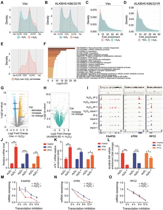
5780 Nucleic Acids Research, 2021, Vol. 49, No. 10
diate stress-induced RNA m6 A modifications and how cel- Comet assay
lular stress regulates m6 A-modifying proteins. Comet assay was performed with the comet kit (R&D SYS-
In this study, we examined the role of RNA demethy- TEMS, Cat# 4250-050-K) according to manufactory in-
lase ALKBH5 in response to ROS stress and observed structions. Briefly, combine cells at 0.5 million per mLwith
that ROS induces a global increase in mRNA m6 A via molten LMA agarose at a ratio of 1: 10 (v/v) and immedi-
inhibition of the ALKBH5. We determined that ROS in- ately pipette 50 l onto comet slice and place it at 4◦ C for
hibits ALKBH5 demethylase activity through ERK/JNK- 30 min in the dark. Immerse slice into 4◦ C lysis buffer for 2
mediated ALKBH5 phosphorylation at serine residues S87 h. Next, immerse slice in alkaline unwinding solution (200
and S325. ALKBH5 phosphorylation facilitates ALKBH5 mM NaOH, 1 mM EDTA, pH > 13) for 20 min at room
SUMOylation by promoting the interaction between temperature. Finally, Electrophoresis was performed in al-
ALKBH5 and SUMO E2 UBC9. Notably, ALKBH5 kaline electrophoresis solution and the comet slices were
is modified by SUMO-1 mainly at lysine residues K86 stained with SYBR Gold dye. And, the tail length was cal-
and K321, which is mediated by the SUMO E3 ligase culated by image J software.
Downloaded from https://academic.oup.com/nar/article/49/10/5779/6287850 by guest on 10 August 2021
PIAS4. Furthermore, we demonstrated that ROS-induced
ERK/JNK/ALKBH5 PTMs/m6 A axis is essential for the Western blot analysis, co-immunoprecipitation and Im-
maintenance of genome integrity and survival of mam- munofluorescence staining
malian cells, and that ERK/JNK/ALKBH5 PTMs/m6 A
axis can be activated in hematopoietic stem/progenitor cells The western blot, co-immunoprecipitation and Immunoflu-
in vivo under physiological condition in response endoge- orescence staining analyses were performed according to
nous ROS. Collectively, our results demonstrate how the standard protocols as described previously (31), using the
mRNA m6 A modification adds another dimension to reg- indicated antibodies. For examining SUMO-modified pro-
ulation of gene expression of DNA damage repair related teins, cells were lysed in denaturing buffer (50 mM Tris–HCl
genes in response to ROS stress. pH7.5, 150 mM NaCl, 4% SDS, 1mM EDTA, 8% glyc-
erol, 50mM NaF, 1 mM DTT, 1mM PMSF and protein
MATERIALS AND METHODS inhibitors) supplemented with 20 mM N-ethylmaleimide
(NEM) and heated at 90◦ C for 10 min. For immunopre-
Plasmids and antibodies cipitation assays, the lysates were further diluted to 0.1%
The pCDH-Strep-ALKBH5 expression plasmid was gen- SDS and immunoprecipitated with antibodies against tar-
erated by cloning the corresponding coding sequence into get proteins at 4◦ C overnight. SUMO-modified proteins
pCDH-Strep vector. All the pCDH-Strep-ALKBH5 K/R were tested by western blotting.
(lysine to arginine) or S/A (serine to alanine) mutants were
derived from pCDH-Strep-ALKBH5 by site-directed mu- Streptavidin pull-down analysis
tagenesis. All expression plasmids for the SUMO systems To determine the effect of ALKBH5 SUMOy-
were kindly provided by Dr. Jiemin Wong’s lab. Antibodies lation on its substrate accessibility, a biotin la-
used in this study were listed as follows: anti-m6 A (Synap- beled RNA oligonucleotide bait was synthesized at
tic Systems# 202003), anti-␥ H2A.X (CST#9719S), Ruimian biotechnology, Shanghai, China (5 -biotin-
anti-␥ H2A.X (Thermo Fisher#MA1-2022), anti- AUGGGCCGUUCAUCUGCUAAAAGG-m6 A-
ALKBH5 (Sigma#HPA007196), anti-ALKBH5 (Thermo CUGCUUUUGGGGCUUGU-3 ). The pull-down
Fisher #703570), anti-FTO (Sigma#SAB2106776), assay was performed according to as described (32). Briefly,
anti-METTL3 (Sigma#SAB2104747), anti-METTL14 transfected HEK293T cells were collected, washed with
(Sigma#HPA038002), anti-SUMO-1 (Thermo Fisher PBS and lysed in lysis buffer (50 mM Tris–HCl pH 7.5,
#33–2400), anti-SUMO-2/3 (CST#4971P), anti- 150 mM NaCl, 0.5% NP-40, 1 mM EDTA, 8% glycerol
ERK (CST#9102S), anti-p-ERK (CST#9106S), anti- supplemented with protease inhibitor mixture, phosphatase
JNK (CST#9252S), anti-p-JNK (CST#4671S), anti- inhibitors and 1 mM DTT). 10% of whole cell lysate was
phophoserine (Sigma#P5747), anti-phophotyrosine used as input and 90% of the whole cell lysate was used
(Sigma#SAB5200015), anti-Strep (Sigma#SAB2702216), for the following Streptavidin sepharose beads pulldown.
anti-Annexin V (Thermo Fisher #17800774), anti-Actin Next, 2 g of biotinylated RNA baits were incubated
(CST#8457S), anti-Tubulin (CST#2146S), anti-Lamin with the above-mentioned whole cell lysate, diluted with
A/C (CST#4777S), anti-HA (CST#2362), anti-Flag binding buffer containing 10 mM Tris–HCl pH 7.5, 150
(Sigma#F1804), anti-IGF2BP2 (CST#14672S), anti- mM NaCl, 1.5 mM MgCl2 , 0.05% NP-40 and subjected to
eIF3A (CST#3411S). rotation at 4◦ C for 2 h. The resulting beads were washed
three times with washing buffer (10 mM Tris–HCl pH
Drug treatment 7.5, 150 mM NaCl, 0.05% NP-40, 1 mM EDTA). The
effect of SUMOylation on ALKBH5 substrate accessibility
For the ROS-induced DNA damage analysis, the indi-
was determined by western blotting using anti-ALKBH5
cated cell lines were treated with or without 100 M hy-
antibodies.
drogen peroxide (H2 O2 ), or 80 M Carbonyl cyanide m-
chlorophenylhydrazone (CCCP) for 6 hours. For the in vivo
shRNA knockdown and quantitative RT-PCR
ROS study, DMSO and 5 mg/kg CCCP was intraperi-
toneally injected in to three pairs of mice. And, all the mice Knockdown of target genes by shRNAs was done as
were sacrificed 12 hours after injection. described previously (31). The vector for shRNAs was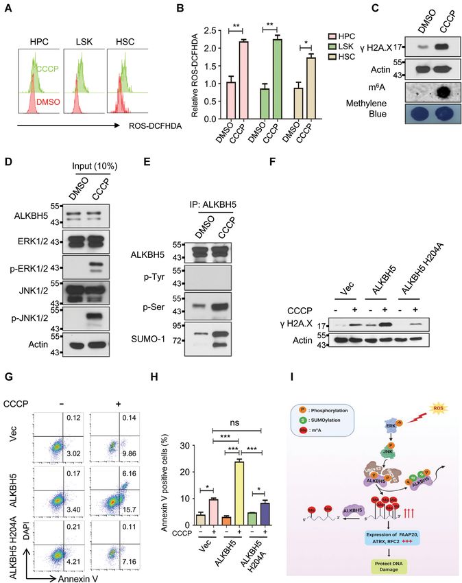
Nucleic Acids Research, 2021, Vol. 49, No. 10 5781
pLKO.1. The sequences for shRNAs are listed in Sup- prepared for both input and IP samples using TruSeq®
plementary Figure Table S1. For qRT-PCR analysis, to- Stranded mRNA Library Prep (Illumina, 20020594) follow-
tal RNA was extracted from various cells as indicated and ing the manufactory protocol. Sequencing was performed
reverses transcribed using kits purchased from Thermo at the University of Chicago Genomics Facility on an Illu-
Fisher. The primer sequences used in the qRT-PCR are mina NextSeq 4000 machine in single-read mode with 50 bp
listed in Supplementary Figure Table S1. per read at around 25–30 M sequencing depth.
Analysis of mRNA m6 A methylation by dot-blot assay m6 A-Seq and RNA-Seq data analysis
6
To analyze mRNA m A methylation, we performed dot- Single-end reads were harvested and trimmed by
blot assays according to a published procedure with minor Trim Galore to remove adaptor sequences and low-
changes (33). Briefly, total RNA was extracted using Trizol quality nucleotides. High-quality reads were then aligned
reagent (Thermo Fisher), and mRNAs were separated us- to UCSC hg19 reference genome by HISAT2 using default
Downloaded from https://academic.oup.com/nar/article/49/10/5779/6287850 by guest on 10 August 2021
ing the dynabeads mRNA purification kit (Thermo Fisher). parameters, and only uniquely mapped reads were retained
The mRNAs were denatured at 95◦ C for 5 min, followed by for all downstream analyses. FeatureCounts software was
chilling on ice directly. Next, 400 ng mRNAs was spotted used to count reads mapped to RefSeq genes, and differen-
to positively charged nylon (GE healthcare), air-dried for 5 tially expressed genes analysis was conducted by Cuffdiff
min, and cross-linked using a UV cross linker. The mem- 2 Software. ExomePeak R package was employed to call
branes were blocked in 5% non-fat milk plus 1% BSA in m6 A peaks on RefSeq transcripts and further generate
PBST for 2 hours and then incubated with anti- m6 A an- differentially methylated m6 A peaks. Peak centers were
tibodies at 4◦ C overnight. After three times washing with then grouped to 3 UTR, CDS and 5 UTR by custom
PBST, the membranes were incubated with Alexa Fluor 680 scripts. Metagene plots were generated by Guitar package,
Goat anti-rabbit IgG secondary antibodies at room temper- and motifs were identified by Homer toolkit. To visualize
ature for 1 h. Membranes were subsequently scanned using sequencing signals at specific genomic regions, we used
image studio. Methylene blue staining was used as a loading Deeptools to normalize all libraries and imported into
control to make sure equal amount of mRNAs was used for IGV.
dot-blot analysis.
Biochemistry assay of ALKBH5 activity in vitro
mRNA m6 A methylation quantification by LC-MS/MS
Similar to a previous report (34), the demethylation ac-
mRNAs from indicated groups were separated by dyn- tivity assay was performed in standard 20 L of reaction
abeads mRNA purification kit two times, followed by the buffer containing KCl (100 mM), MgCl2 (2 mM), SU-
removal of contaminated rRNA with RiboMinus™ Eu- PERNase In (0.2 U/l, life technology), L-ascorbic acid
karyote Kit v2kit (Thermo Fisher). The isolated mRNAs (2 mM), ␣-ketoglutarate (300 M), (NH4 )2 Fe(SO4 )2 ·6H2 O
were subsequently digested into nucleotides with nuclease (150 M), and 50 mM of HEPES buffer (pH 6.5). WT
P1 (Sigma, N8630) in 20 ml of buffer containing 25 mM and SUMOylation-deficient mutant ALKBH5 was purified
NaCl and 2.5 mM ZnCl2 for 1 h at 42◦ C, followed by 1 from HEK293T cells after the treatment of H2 O2 . 100 ng
unit of FastAP Thermosensitive Alkaline Phosphatase (1 polyadenylated RNA purified from HEK293T cells was in-
U/l, Thermofisher Scientific, EF0651) in FastAP buffer cubated with WT or mutant ALKBH5 in the above reac-
were added and the sample was incubated for another 4 h at tion buffer for 1 hour and then quenched by the addition of
37◦ C. The samples were then filtered (0.22 mm, Millipore) 5 mM of EDTA, respectively. Excessive amount of EDTA
and injected into a C18 reverse phase column coupled on- was added to control samples. The RNAs were then iso-
line to Agilent 6460 LC–MS/MS spectrometer in positive lated with 100 L TRIzol® reagents (Thermofisher Scien-
electrospray ionization mode. The nucleosides were quan- tific, #15596018) using standard protocol and subjected to
tified by using retention time and the nucleoside to base RNA digestion prior to LC-MS/MS analysis.
ion mass transitions (268-to-136 for A; 282-to-150 for m6 A.
Quantification was performed by comparing with the stan-
dard curve obtained from pure nucleoside standards run- RIP-RT-qPCR
ning with the same batch of samples. Sixty million cells were collected and re-suspended with
RIPA buffer at 4◦ C for 1 h on a rotator. Then the mRNP
m6 A-Seq and RNA-Seq analysis lysate was centrifuged at 15 000g for 15 min to clear the
lysate. 50 l cell lysate was saved as input, mixed with 1 ml
Total RNA was extracted from indicated cells with or TRIzol. ALKBH5 antibody or igG was added and incu-
without H2 O2 treatment and poly(A)+ RNA was further bate at 4◦ C overnight together with proteinase inhibitor
enriched by dynabeads mRNA purification kit (Thermo and RNase inhibitor. Cell lysate was then mixed with dyn-
Fisher). Particularly, DNase I digestion was performed to abeads protein A/G (1:1 mixture) with continuously rotat-
avoid DNA contamination. mRNA was fragmented by ing at 4 ◦ C for 4 h. The beads were collected, washed and the
Bioruptor® Pico Sonication System and input was saved binding RNA was extracted by TRIzol reagent. Amount of
before m6 A IP. m6 A IP was performed with EpiMark® N6 - target transcripts in both the input and IP RNAs were an-
Methyladenosine Enrichment Kit (NEB, E1610S) follow- alyzed with RT-qPCR, and IP enrichment ratio of a tran-
ing the manufactory protocol. Then, RNA libraries were script was calculated as the ratio of its amount in IP to that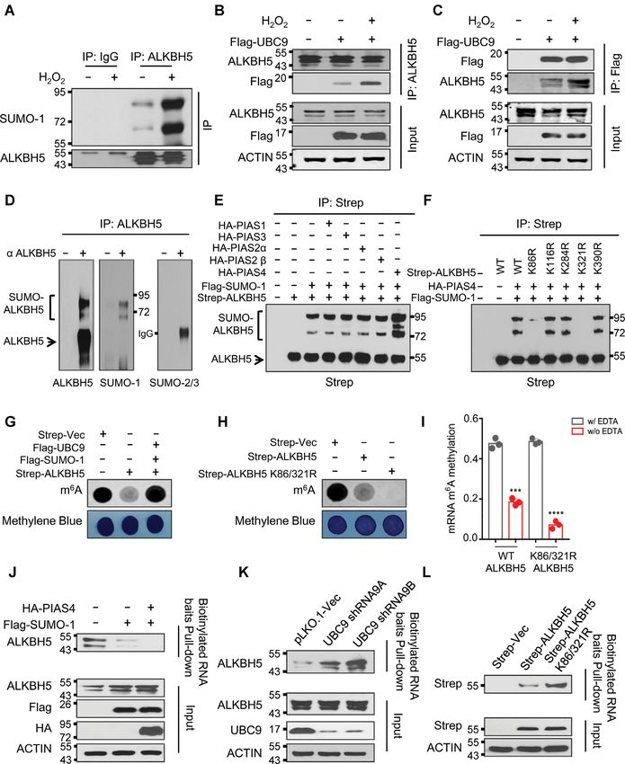
5782 Nucleic Acids Research, 2021, Vol. 49, No. 10
in the input yielded from same amount of cells and normal- after H2 O2 release. Compared with DNA damage, induc-
ized to IgG. The primer sequences used in the qRT-PCR are tion of global mRNA m6 A methylation comes first, which
listed in Supplementary Figure Table S1. occurred at 5 min after H2 O2 treatment and dropped dra-
matically after H2 O2 release (Figure 1J). Together, these
ROS detection by FACS data indicate that exogenous or endogenous ROS-induced
stress significantly up-regulates global mRNA m6 A modifi-
ROS detection assay was performed as previously described cation.
(35). Briefly, bone marrow cells were stained with cell sur-
face markers as we previously described (36), and were incu-
bated with pre-warmed loading buffer containing the probe ROS promotes mRNA m6 A demethylase ALKBH5
DCFHDA (2 -7 -dichlorodihydrofluorescein diacetate) to a SUMOylation
final concentration of 5 M. After 1 h incubation, the in- To determine how H2 O2 induces mRNA m6 A methyla-
tensity of fluorescence was examined by flow cytometry. tion, we examined the expression levels of key mRNA m6 A
Downloaded from https://academic.oup.com/nar/article/49/10/5779/6287850 by guest on 10 August 2021
methylation writers including METTL3 and METTL14,
Statistical analysis and mRNA m6 A erasers FTO and ALKBH5 by RT-qPCR
and western blot analysis. As shown in Supplementary Fig-
Experiments were performed at least three times, and the ure S2A–C, H2 O2 -induced ROS had no effect on transcrip-
representative data were shown. All statistical tests were tion or protein levels of mRNA m6 A erasers, whereas it
performed using the unpaired Student’s test by GraphPad significantly increased both the transcript and protein lev-
Prism 5 software. A value of P < 0.05 was considered statis- els of mRNA m6 A writers (Supplementary Figure S2C–E).
tically significant. In all the results, ‘*’ denotes P < 0.05, ‘**’ This suggests that ROS-induced mRNA m6 A modification
denotes P < 0.01, ‘***’ denotes P < 0.001, and ‘ns’ denotes may occur through up-regulation of m6 A writers METTL3
no significant difference. and METTL14. However, METTL3 or METTL14 knock-
down by specific shRNAs (Supplementary Figure S3A and
RESULTS S3B) only partially blocked ROS-induced up-regulation of
global mRNA m6 A modification (Supplementary Figure
ROS leads to up-regulation of global mRNA m6 A methyla-
S3C and S3D), suggesting the presence of an additional
tion
molecular mechanism that mediates the ROS-induced in-
To examine the role of mRNA m6 A methylation in response crease of global mRNA m6 A modification.
to ROS stress, we treated human cell lines with H2 O2 . Con- Since SUMOylation regulates a variety of cellular pro-
sistent with prior studies (37), H2 O2 induced DNA dam- cesses including cellular response to DNA damage (45), we
age in HEK293T and HeLa cells as evidenced by increased evaluated the possibility that ROS regulates global mRNA
expression of phosphorylated H2 AX (␥ H2 AX), a sensitive m6 A methylation via SUMOylation of mRNA m6 A mod-
marker of DNA damage and genomic instability (38,39) ification writers or erasers. We found that ROS specifically
(Figure 1A–C). Furthermore, H2 O2 significantly increased promotes ALKBH5 SUMOylation (Figure 2A and Supple-
global mRNA m6 A methylation in human HEK293T and mentary Figure S4) but not FTO, METTL3 and METTL14
HeLa cells (Figure 1D and Supplementary Figure S1A). (Supplementary Figure S5A–C). We have also observed
Carbonyl cyanide m-chlorophenylhydrazone (CCCP) was SUMOylation of METTL3 in HEK293T cells (Supplemen-
used to mimic endogenous ROS activation under physiolog- tary Figure S5B), consistent with previously published data
ical conditions (40–43). Similar to H2 O2, CCCP treatment (33). However, ROS did not induce METTL3 SUMOy-
significantly induced phosphorylation of H2 AX as well as lation. Furthermore, ALKBH5 SUMOylation could also
global mRNA m6 A levels in human HEK293T (Supple- be promoted by CCCP-induced endogenous ROS (Supple-
mentary Figure S1B and C) and HeLa cells (Supplemen- mentary Figure S5D). Since E2 conjugation enzyme UBC9
tary Figure S1D and E). More importantly, CCCP-induced is critical for mediating protein SUMOylation (46–48), we
DNA damage and increase in global mRNA m6 A methyla- explored the effects of ALKBH5 SUMOylation on its in-
tion was inhibited by N-acetyl-L-cysteine (NAC), which ab- teraction with UBC9. Co-immunoprecipitation analyses
rogates ROS generated by CCCP treatment, suggesting that showed that ROS strongly increases the interaction between
CCCP-induced DNA damage and global increase in global ALKBH5 and UBC9 (Figure 2B and C). In addition, nu-
mRNA m6 A methylation was mainly attributed to CCCP- merous studies also suggest that Sentrin/SUMO-specific
induced ROS (Figure 1E and F). We next performed quan- proteases 1 and 3 (SENP1 and SENP3) are involved in
titative analysis of the mRNA m6 A/A ratio by LC–QqQ– regulation of protein SUMOylation under oxidative stress
MS/MS using previously described protocol (44). Consis- (49–51). Therefore, we next determined the effect of ROS
tent with the dot blot analysis, both H2 O2 - and CCCP- on the interaction between ALKBH5 and SENP1. Con-
induced ROS significantly increased global mRNA m6 A sistent with previously published results (50,51), SENP3
levels (Figure 1G and H). To determine the effect of ROS was stabilized by H2 O2 treatment in 293T cells. However,
treatment on kinetics of DNA damage and global mRNA we did not observe the interaction between ALKBH5 and
m6 A methylation, we treated 293T cells with H2 O2 at differ- SENP3. (Supplementary Figure S6). Interestingly, the in-
ent time intervals and performed H2 O2 time release analy- teraction between ALKBH5 and SENP1 was significantly
ses. As shown in Figure 1I, phosphorylation of H2A.X was disrupted by H2 O2 treatment (Supplementary Figure S6).
significantly induced 15 min after H2 O2 treatment, peaking Together, these data provide evidence that ROS specif-
at 30 minutes, and diminishing over the following 9 minutes ically promotes ALKBH5 but not FTO, METTL3 and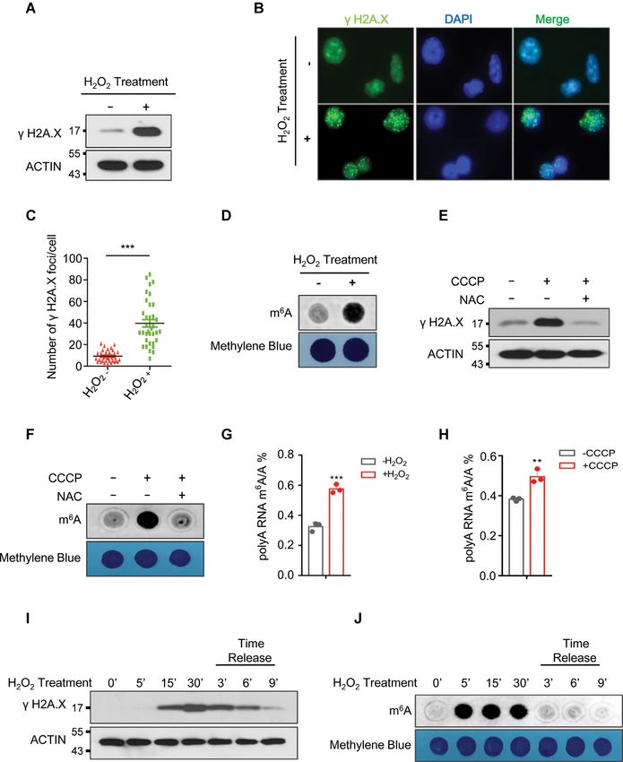
Nucleic Acids Research, 2021, Vol. 49, No. 10 5783
Downloaded from https://academic.oup.com/nar/article/49/10/5779/6287850 by guest on 10 August 2021
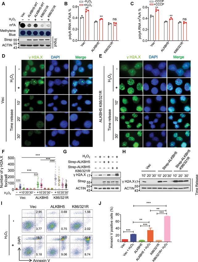
Figure 1. ROS induces up-regulation of global mRNA m6 A methylation. (A, B) Western blot (A), and immunostaining (B) analyses showing phosphory-
lation of H2A.X in HEK293T cells (A), or HeLa cells (B) in the presence or absence of H2 O2 . (C) Number of ␥ H2A.X foci derived from the data shown
in Figure B. (D) Dot-blot assay showing the effect of H2 O2 treatment on global mRNA m6 A levels in HEK293T cells. (E, F) Western blot analyses (E)
showing phosphorylation of H2A.X, and dot-blot assays (F) were conducted to determine the effect of CCCP treatment on global mRNA m6 A levels in
HEK293T cells with or without N-acetyl-L-cysteine (NAC) treatment. (G, H) LC–MS/MS analyses indicating ROS stress induced global mRNA m6 A
modification. (I, J) Western blot analysis (I), and dot-blot analysis (J) showing the kinetics of H2A.X phosphorylation and global mRNA m6 A methylation
in the presence, or absence of H2 O2 treatment respectively.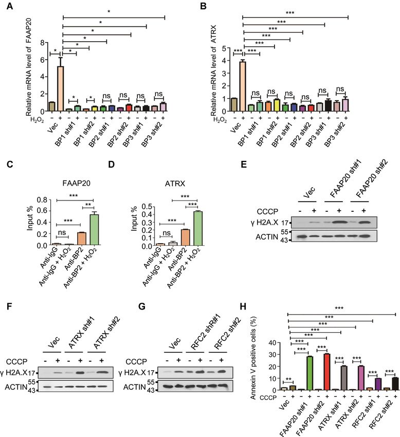
5784 Nucleic Acids Research, 2021, Vol. 49, No. 10

                                                                                                                                                         Downloaded from https://academic.oup.com/nar/article/49/10/5779/6287850 by guest on 10 August 2021
Figure 2. ROS induces mRNA m6 A demethylase ALKBH5 SUMOylation. (A) Denaturing immunoprecipitation (IP) assay indicating H2 O2 -induced ROS
selectively enhances ALKBH5 SUMOylation. (B, C) Reciprocal IP analyses showing ROS facilitates the interaction between ALKBH5 and SUMO E2
conjugation enzyme UBC9. Reciprocal IP assays were performed in cells ectopically expressing Flag-UBC9 with or without H2 O2 treatment. IP antibodies
are shown on the right and western blotting antibodies are shown on the left. (D) Denaturing IP analyses showing that endogenous ALKBH5 is modified by
SUMO-1 but not by SUMO-2/3 in HEK293T cells. (E) Denaturing IP analyses showing that SUMO E3 ligase PIAS4 mediates ALKBH5 SUMOylation.
HEK293T cells were transfected with or without Strep-ALKBH5, HA tagged PIAS family SUMO E3 ligases and Flag-SUMO-1. The cells were collected
for denaturing IP analyses two days after transfection. (F) Denaturing IP analyses showing ALKBH5 SUMOylation mainly occurs at lysine residues
K86 and K321. Strep tagged wild-type ALKBH5 or ALKBH5 lysine (K) to arginine (R) mutants were solely expressed or co-expressed with HA-tagged
PIAS4 and Flag-SUMO-1 in HEK293T cells. (G) Dot-blot analyses showing ALKBH5 mRNA m6 A demethylase activity is inhibited by SUMOylation.
Strep-tagged ALKBH5 was expressed alone or co-expressed with Flag tagged UBC9 and SUMO-1 in HEK293T cells. (H) Dot-blot assays indicating
that blocking ALKBH5 SUMOylation markedly facilitates ALKBH5 mRNA m6 A demethylase activity. Strep tagged vector, or vectors expressing wild-
type ALKBH5 or SUMOylation-deficient mutant ALKBH5 (ALKBH5 K86/321R) were transfected into HEK293T cells. 48 hours after transfection,
dot-blot analyses were conducted to detect global mRNA m6 A levels. (I) In vitro mRNA m6 A demethylase activity analyses indicating that blocking
ALKBH5 SUMOylation dramatically facilitates ALKBH5 mRNA m6 A demethylase activity. Strep tagged wild-type or SD- mutant ALKBH5 (ALKBH5
K86/321R) were expressed in HEK293T cells. (J–L) Substrate pull-down analyses showing that ALKBH5 SUMOylation dramatically blocks its substrate
accessibility. Flag tagged SUMO-1 was expressed alone or co-expressed with HA-tagged PIAS4 in HEK293T cells (J). sh-vector, or two vectors expressing
shRNAs against UBC9 were transfected into HEK293T cells (K). Strep tagged wild-type or SD-mutant ALKBH5 was expressed in HEK293T cells. (L).
Forty-eight hours after transection, whole cell lysates were subjected to biotin labeled m6 A-containing RNA oligo pull-down.
Nucleic Acids Research, 2021, Vol. 49, No. 10 5785
METTL14 SUMOylation by enhancing the interaction of ished ALKBH5 SUMOylation. In addition, we also es-
ALKBH5 and UBC9 and inhibiting the association be- tablished that ALKBH5 K86R/K321R double mutant is
tween ALKBH5 and SENP1. not SUMOylated (Supplementary Figure S10), suggesting
that K86 and K321 are the major ALKBH5 SUMOyla-
tion sites. Notably, ALKBH5 overexpression significantly
ALKBH5 SUMOylation blocks m6 A demethylase activity
reduced the level of mRNA m6 A methylation while co-
by inhibition of substrate accessibility
expression of ALKBH5, UBC9 and SUMO-1, promot-
There are three SUMO proteins including SUMO-1, ing ALKBH5 SUMOylation, did not affect m6 A methy-
SUMO-2 and SUMO-3, which can be covalently con- lation levels (Figure 2G and Supplementary Figure S11).
jugated to the targeted proteins (52). We next deter- Furthermore, the ALKBH5 SUMOylation-deficient mu-
mined whether ectopically expressed ALKBH5 could be tant (ALKBH5 K86R/K321R) inhibited global mRNA
SUMOylated by SUMO-1, SUMO-2 and/or SUMO-3. m6 A methylation more efficiently than wild type ALKBH5
Immunoblotting of the ALKBH5 immunoprecipitates us- (Figure 2H and Supplementary Figure S12). In vitro as-
Downloaded from https://academic.oup.com/nar/article/49/10/5779/6287850 by guest on 10 August 2021
ing strep-tactin beads identified high apparent molec- says revealed that the ALKBH5 K86R/K321R mutant
ular weight ALKBH5 species in HEK293T cells with protein isolated from HEK293T cells had a significantly
co-expression of ALKBH5 and SUMO-1 but not in higher mRNA m6 A demethylation activity than wild-type
cells co-expression of SUMO-2 or SUMO-3 (Supplemen- ALKBH5 (Figure 2I). Taken together, the data demonstrate
tary Figure S7A–C), indicating that ALKBH5 is con- that SUMOylation of ALKBH5 inhibits its m6 A demethy-
jugated with SUMO-1 but not SUMO-2/3. In addi- lase activity in vivo and in vitro.
tion to mono-SUMOylated ALKBH5, higher molecular Next, we analyzed how ALKBH5 SUMOylation reg-
weight ALKBH5 species were also detected, suggesting ulates its m6 A demethylase activity. Western blot anal-
that ALKBH5 is modified with SUMO-1 at multiple ly- ysis showed that increasing ALKBH5 SUMOylation by
sine residues or SUMO-1 is linked to ALKBH5 in polymer PIAS4 or SUMO-1 overexpression or reducing ALKBH5
chain. Consistent with the observation for exogenous Strep- SUMOylation by UBC9 knockdown, does not alter
ALKBH5 in HEK293T cells, immunoprecipitation of the ALKBH5 protein levels (Supplementary Figure S13A and
endogenous ALKBH5 from 293T cells showed the presence B). Additionally, increasing ALKBH5 SUMOylation by
of SUMO-1 high molecular weight species but not SUMO- overexpression of PIAS4 and SUMO-1 did not affect
2/3 modified forms (Figure 2D and Supplementary Figure ALKBH5 subcellular localization (Supplementary Figure
S8B). S13C). Furthermore, wild type and SUMOylation-deficient
In eukaryotes, there are a number of SUMO E3 ligases, mutant ALKBH5 had the same subcellular localization
which increase the efficiency of SUMO conjugation and ac- (Supplementary Figure S13D). Consistently, reducing en-
celerate the rate of SUMO modification (53). The largest dogenous ALKBH5 SUMOylation by UBC9 knockdown
class of SUMO E3 ligase is the PIAS family proteins, which did not affect its subcellular localization (Supplemen-
includes PIAS1, PIAS2 PIAS3, and PIAS4, that all share a tary Figure S13E). Next, we performed a substrate pull-
RING domain (54–58). To examine the mechanism under- down assay using synthesized biotinylated m6 A-containing
lying ALKBH5 SUMOylation, we co-expressed PIAS fam- RNA baits to capture endogenous ALKBH5 from the
ily proteins with ALKBH5 and SUMO-1, and performed HEK293T cells expressing vector, SUMO-1, or SUMO-1
denaturing IP to delineate which SUMO E3 ligase mediates plus PIAS4. The result showed that SUMO-1 overexpres-
ALKBH5 SUMOylation. This analysis showed that among sion markedly inhibits the binding ability of ALKBH5 to
all PIAS family proteins, only co-expression of ALKBH5 its substrate, and that the inhibitory effect of SUMO-1
with PIAS4 and SUMO-1 resulted in extensive SUMOyla- on ALKBH5 substrate binding activity was further aug-
tion of ALKBH5 (Figure 2E, last lane and Supplementary mented by co-expression of PIAS4 and SUMO-1 (Figure
Figure S8C). Additionally, co-immunoprecipitation assay 2J). In contrast, suppression of ALKBH5 SUMOylation by
revealed that ALKBH5 interacts with PIAS4 (Supplemen- knocking down UBC9 enhanced ALKBH5 substrate bind-
tary Figure S8A). ing activity (Figure 2K). In addition, we found that the
SUMOylation of substrates frequently occurs at a ly- ALKBH5 K86R/K321R double mutant significantly en-
sine within the canonical SUMOylation consensus motif hanced ALKBH5 substrate binding activity (Figure 2L).
Kx (D/E), in which represents a large hydrophobic Collectively, our results show that SUMO E3 ligase PIAS4
residue and x represents any amino acid followed by an mediates ALKBH5 SUMOylation at lysine residues K86
acidic residue (59). By using two independent algorithms and K321, and that ALKBH5 SUMOylation markedly in-
GPS-SUMO (60) and JASSA (61), we identified five po- hibits its mRNA m6 A demethylase activity by blocking its
tential SUMOylation sites in ALKBH5, which included ability to bind to m6 A RNA species.
K390, K86, K321, K284 and K116 (Supplementary Figure
S9A). We generated ALKBH5 SUMOylation-deficient mu-
SUMOylation-deficient ALKBH5 overexpression blocks
tants with lysine replaced by arginine, a charged aliphatic
ROS-induced mRNA m6 A methylation, leading to a signif-
acid, within the putative SUMOylation motifs. We per-
icant delay of DNA repair and increase of cell apoptosis
formed denaturing IP analyses to determine which lysine
mutation abolished ALKBH5 SUMOylation. As shown To determine whether ALKBH5 SUMOylation plays a
in Figure 2F and Supplementary Figure S9B, ALKBH5 vital role in ROS-induced global mRNA m6 A modifica-
K86R mutation reduced ALKBH5 SUMOylation dra- tion, we ectopically expressed wild-type or SUMOylation-
matically, and ALKBH5 K321R mutation nearly abol- deficient mutant ALKBH5 in HEK293T cells (Figure 3A)5786 Nucleic Acids Research, 2021, Vol. 49, No. 10
Downloaded from https://academic.oup.com/nar/article/49/10/5779/6287850 by guest on 10 August 2021
Figure 3. ALKBH5 SUMOylation is mainly responsible for ROS-induced global increase in mRNA m6 A methylation. (A) Dot-blot assay showing that
SUMOylation-deficient mutant ALKBH5 overexpression completely blocks ROS-induced global increase in mRNA m6 A methylation. Strep tagged wild-
type or SD-mutant ALKBH5 was transfected into HEK293T cells. Half of cells were used for western blot analysis to confirm the expressions of ectopically
expressed plasmids, and the rest of cells were used for dot-blot analysis. (B, C) LC–MS/MS analyses indicating ROS induces global mRNA m6 A methylation
via ALKBH5 SUMOylation. HEK293T cells stably expressing vector, wild-type, or SUMOylation-deficient mutant ALKBH5 were treated with or without
H2 O2 (B) or CCCP (C) for 6 h, and subjected to LC–MS/MS analysis of mRNA m6 A methylation. (D, E) ␥ H2A.X immunostaining analysis showing
that SUMOylation-deficient mutant ALKBH5 overexpression dramatically delays H2 O2 –induced DNA damage repair. HeLa cells stably expressing Vec
(D), or ALKBH5 K86/321R (E) were treated with H2 O2, which was removed after 6 h. ␥ H2A.X immunostaining analyses were performed at time
intervals between 10 and 30 min. (F) ␥ H2A.X foci quantitative data for D, E and Supplementary Figure S17. (G, H) Western blot analysis showing that
SUMOylation-deficient mutant ALKBH5 overexpression significantly inhibits H2 O2 –induced DNA damage repair. (I) Cell apoptosis analyses showing
ALKBH5 K86/321R overexpression markedly increases ROS-induced cell apoptosis. Apoptosis analyses were performed in HEK293T cells transfected
with Strep tagged vector, or vectors expressing ALKBH5 or Strep tagged ALKBH5 K86/321R in the presence or absence of H2 O2 . (J) Histograms showing
the summary and statistical analysis of Figure 3I.Nucleic Acids Research, 2021, Vol. 49, No. 10 5787
and treated the cells with H2 O2 . As shown in Figure mine whether ROS induced ALKBH5 phosphorylation.
3A, ROS-induced global mRNA m6 A modification was As shown in Figure 4A and Supplementary Figure S18A,
partially reduced by wild-type ALKBH5 overexpression, both H2 O2 and CCCP treatment induced ALKBH5 serine
whereas it was completely blocked by overexpression of but not tyrosine phosphorylation. Threonine phosphory-
ALKBH5 K86R/K321R mutant. Depletion of ALKBH5 lation of ALKBH5 was not induced by H2 O2 either (Fig-
by CRISPR-Cas9-mediated deletion in HEK293T cells was ure 4A). It has been reported that the mitogen-activated
confirmed by western blot (Supplementary Figure S14A), protein kinase (MAPK) signaling pathway, which includes
T7E1 digestion (Supplementary Figure S14B), and DNA the extracellular regulated protein kinase 1/2 (ERK1/2),
sequencing (Supplementary Figure S14C), and ALKBH5 c-Jun N-terminal kinase (JNK) and p38 regulatory path-
knockout remarkably increased global mRNA m6 A modi- ways, is activated by ROS stress (65,66). We thus inves-
fication (Supplementary Figure S14D). We employed LC– tigated whether ERK/JNK signaling pathways mediated
QqQ–MS/MS analysis to establish m6 A/A ratio under per- ROS-induced ALKBH5 phosphorylation and SUMOyla-
oxide and CCCP-induced conditions. We observed that tion. Both H2 O2 and CCCP induced DNA damage and
Downloaded from https://academic.oup.com/nar/article/49/10/5779/6287850 by guest on 10 August 2021
both H2 O2 - and CCCP-induced ROS significantly in- activated the ERK/JNK signaling pathway (Figure 4B
creased mRNA m6 A modification in 293T cells stably ex- and Supplementary Figure S18B). ERK1/2 knockdown by
pressing vector, or wild-type ALKBH5, but did not in- ERK1/2 specific shRNAs suppressed ROS-induced activa-
duce mRNA m6 A methylation levels in ALKBH5 knockout tion of JNK (Figure 4C), suggesting that JNK is a down-
or ALKBH5 K86/K321R mutant-expressing cells (Figure stream mediator of ERK1/2 in response to ROS. Denatur-
3B, C and Supplementary Figure S15A-15B), suggesting ing IP assays revealed that ROS-induced ALKBH5 phos-
that ROS induces an increase in global mRNA m6 A methy- phorylation and SUMOylation were inhibited by either
lation mainly through SUMO modification of ALKBH5. ERK or JNK knockdown (Figure 4C and D), suggesting
To determine the effect of inhibition of ROS-induced that activation of ERK, followed by activation of JNK are
m6 A methylation by SUMOylation-deficient mutant necessary for ROS-induced ALKBH5 phosphorylation and
ALKBH5 on ROS-induced DNA damage, we performed SUMOylation. More importantly, the interaction between
comet analysis (single cell gel electrophoresis assay), ALKBH5 and JNK1/2 was significantly increased by H2 O2
which is used for quantitating DNA damage and repair treatment (Supplementary Figure S19).
with single cell resolution (62,63). As shown in Supple- Previous studies showed that substrate SUMOylation is
mentary Figure S16A and B, ALKBH5 overexpression facilitated by phosphorylation of the upstream or down-
significantly promoted H2 O2 - induced DNA damage, stream serine, tyrosine and threonine sites of the substrate
and SUMOylation-deficient mutant ALKBH5 caused (67,68). We showed that H2 O2 treatment induced serine
more DNA damage than wild-type ALKBH5. We next but not threonine phosphorylation of ALKBH5 (Figure
performed ␥ H2A.X immunostaining analysis in HeLa 4A). To identify the serine sites that are phosphorylated
cells stably expressing Vector, wild-type, or SUMOylation- by ROS-activated JNK signaling, we mutated five serine
deficient mutant ALKBH5. As indicated by ␥ H2A.X foci, residues, S64, S69, S87, S325 and S361, to alanine. These
H2 O2 -induced DNA lesions were repaired within 10 min- serine residues are close to the identified SUMOylation
utes and 20 minutes after removal of H2 O2 in Vector and sites (lysines K86 and K321) of ALKBH5 wild-type. Thus,
wild-type ALKBH5 expressing cells respectively (Figure wild-type or distinct serine to alanine mutants of ALKBH5
3D, F and Supplementary Figure S17), whereas damaged were expressed in HK293T cells for further analysis. As
DNA still persisted in 30 minutes after removal of H2 O2 shown in Figure 4E, ROS-induced ALKBH5 phosphory-
in cells expressing the SUMOylation-deficient mutant lation was significantly reduced by the ALKBH5 S87A mu-
ALKBH5 (Figure 3E and F), indicating that ROS-induced tation but not the S64A, S69A or S361A mutations. Phos-
DNA damage repair can be markedly delayed in the phorylation was almost completely blocked by the S325A
presence of SUMOylation-deficient mutant ALKBH5. mutation, suggesting that S87 and S325 are critical ROS-
Similar results have been observed by Western blot analysis sensitive ALKBH5 phosphorylation sites. Additionally, de-
(Figure 3G and H). In addition, we showed that both naturing IP showed that the ALKBH5 S325A mutation
wild-type ALKBH5 and ALKBH5 K86R/K321R mutant completely abrogates ALKBH5 SUMOylation, while the
overexpression had no effect on cell survival in the absence ALKBH5 S87A, but not the S64A, S69A and S361A mu-
of ROS stress, but significantly sensitized HEK293T tations, significantly inhibit ALKBH5 SUMOylation (Fig-
cells to ROS (Figure 3I and 3J). Of note, cells expressing ure 4F), confirming that ALKBH5 phosphorylation at ser-
ALKBH5 SUMOylation-deficient mutant had a much ine 87 and serine 325 promotes ALKBH5 SUMOylation
higher frequency of apoptosis than the wild-type ALKBH5 in response to ROS stress. Co-IP assay showed that the
overexpressing cells (Figure 3I and J). Thus, the data ALKBH5 S87A mutation inhibited the interaction between
show that ALKBH5 SUMOylation plays a crucial role ALKBH5 and UBC9, and that the ALKBH5 S325A mu-
in ROS-induced global mRNA m6 A modification, DNA tant had a stronger inhibitory effect on the interaction be-
damage repair and cell survival. tween ALKBH5 and UBC9 compared to the ALKBH5
S87A mutant (Figure 4G). Meanwhile, the interaction be-
tween ALKBH5 and SENP1 was significantly enhanced by
ROS induces ALKBH5 phosphorylation and SUMOylation
ALKBH5 S87A and S325A mutants (Supplementary Fig-
by activation of ERK/JNK signaling
ure S20). In addition, we showed that ALKBH5 S325A
Phosphorylation–dependent SUMOylation modifications possessed the strongest mRNA m6 A demethylase activity
were described previously (64). Thus, we aimed to deter- while ALKBH5 S87A had a higher demethylase activity5788 Nucleic Acids Research, 2021, Vol. 49, No. 10
Downloaded from https://academic.oup.com/nar/article/49/10/5779/6287850 by guest on 10 August 2021
Figure 4. ROS induces ALKBH5 phosphorylation and SUMOylation by activating ERK/JNK signaling. (A) Denaturing IP analysis indicating ROS
significantly induces ALKBH5 phosphorylation and SUMOylation in HEK293T cells. (B) Western blot analysis indicating ROS dramatically activates
the ERK/JNK signaling pathway in HEK293T cells. (C) Denaturing IP assay indicating inhibition of ERK dramatically blocks ROS-induced ALKBH5
phosphorylation and SUMOylation. (D) Denaturing IP analysis suggesting inhibition of JNK significantly inhibits ROS-induced ALKBH5 phosphoryla-
tion and SUMOylation. (E) IP analysis showing that ROS induces ALKBH5 phosphorylation at serine residues S87 and S325. (F) Denaturing IP analysis
indicating that blocking ALKBH5 phosphorylation markedly inhibits ALKBH5 SUMOylation. Strep-tagged wild-type ALKBH5 or phosphorylation-
deficient ALKBH5 was overexpressed alone or co-expressed with HA-tagged PIAS4 and Flag-tagged SUMO-1 in HEK293T cells. Denaturing IP assays
were performed to determine the effect of blocking ALKBH5 phosphorylation on ALKBH5 SUMOylation. (G) Co-IP analysis showing that blocking
ALKBH5 phosphorylation inhibits the interaction between ALKBH5 and SUMO E2 UBC9. Flag-tagged UBC9 was overexpressed alone or co-expressed
with Strep tagged wild-type or phosphorylation-deficient ALKBH5 in HEK293T cells and Co-IP assays were performed to determine the effect of block-
ing ALKBH5 phosphorylation on the interaction between ALKBH5 and UBC9. (H) Dot-blot analysis indicating that blocking ALKBH5 phosphoryla-
tion dramatically increases ALKBH5 mRNA m6 A demethylase activity. Strep tagged wild-type or phosphorylation-deficient ALKBH5 was expressed in
HEK293T cells. Two days after transfection, the cells were collected and subjected to dot-blot analysis.Nucleic Acids Research, 2021, Vol. 49, No. 10 5789
than wild-type ALKBH5 (Figure 4H). Collectively, these methylation at specific regions of the FAAP20, ATRX
results suggest that ROS induces ALKBH5 phosphoryla- and RFC2 mRNAs in vector-expressing samples. By con-
tion at serine 87 and serine 325 by activation of ERK/JNK trast, in ALKBH5 K86/321R mutant samples, ROS treat-
signaling, and that ALKBH5 phosphorylation promotes ment led to negligible changes in transcript levels and m6 A
ALKBH5 SUMOylation not only by facilitating the inter- abundance (Figure 5I and Supplementary Figure S21E).
action between ALKBH5 and UBC9, but also by inhibiting Elevated transcription and mRNA m6 A methylation of
the association between ALKBH5 and SENP1 at the same these three genes was further confirmed in control sam-
time. ples by RT-qPCR and methylated RNA immunoprecipi-
tation (MeRIP) followed by RT-PCR analysis (Figure 5J
and K, respectively). FAAP20, ATRX and RFC2 all play
Global gene expression profiling identifies DNA damage re- crucial roles in DNA damage repair (69–71). To further
pair genes as ALKBH5 downstream targets induced by ROS determine whether the altered mRNA m6 A methylation
To further determine how ROS induces stress response and gene expression of these three genes is a consequence
Downloaded from https://academic.oup.com/nar/article/49/10/5779/6287850 by guest on 10 August 2021
pathways through mRNA m6 A methylation-mediated gene of ALKBH5-mediated demethylation, we performed a
expression, we performed RNA sequencing (RNA-Seq) ALKBH5 RNA immunoprecipitation (RIP)-qPCR assay.
and m6 A sequencing (m6 A-Seq) analyses on untreated and As shown in Supplementary Figure S22A-S22C, ALKBH5
H2 O2 -treated HEK293T cells expressing vector (vec sam- can be significantly enriched at mRNAs of FAAP20, ATRX
ples) or ALKBH5 K86/K321R mutant. m6 A-Seq revealed and RFC2. More importantly, H2 O2 treatment remarkably
thousands of differential m6 A peaks mediated by ROS (Fig- reduced ALKBH5 binding ability to FAAP20, ATRX and
ure 5A). As determined by the frequency distribution of dif- RFC2 mRNAs (Figure 5L). Furthermore, ROS-induced
ferential m6 A peaks across the length of mRNA transcripts, up-regulation of these genes related to DNA repair was only
we found that ROS treatment specifically increased mRNA blocked by the SUMOylation-deficient mutant ALKBH5
m6 A in the 3 UTR regions in control samples (Figure 5A). but not wild-type ALKBH5 overexpression, suggesting that
However, overexpression of the ALKBH5 K86R/K321R ALKBH5 SUMOylation plays an important role in ROS-
mutant gene reduced m6 A methylation in the 3 UTR re- induced expression of genes related to DNA damage re-
gion (Supplementary Figure S21A) and completely blocked pair (Supplementary Figure S23A–C). We next determined
ROS-induced mRNA m6 A modification (Figure 5B). Con- whether ROS affected mRNA stability of these three genes.
sistent with m6 A LC–QqQ–MS/MS quantitation experi- By blocking new RNA synthesis with Actinomycin D, we
ments, ROS treatment in vec samples but not in ALKBH5 measured the half-life of FAAP20, ATRX and RFC2 mR-
K86/321R mutant samples induced an obvious increase NAs as determined by qPCR analysis of transcripts of these
in m6 A peaks (Figure 5C and D). We observed that the three genes at different time points in the presence or in
vast majority of increased m6 A peaks were located in the absence of H2 O2 . The results showed that H2 O2 treat-
the 3 UTR, and we also observed a moderate change of ment significantly promoted mRNA stability of FAAP20
these hyper peaks in the 3 UTR compared to other re- and ATRX but not RFC2 mRNA (Figure 5M–O), suggest-
gions, while decreased m6 A peaks were relatively evenly dis- ing that ROS-induced mRNA m6 A methylation of FAAP20
tributed along the mRNAs (Figure 5E and Supplementary and ATRX promotes FAAP20 and ATRX expression pos-
Figure S21B). Notably, while ROS treatment in vec sam- sibly by stabilizing their mRNAs. Taken together, these
ples showed only a minor effect on ALKBH5 protein level, results suggest that ALKBH5 mediates ROS-induced ex-
there was an apparent change in global m6 A abundance in pression of DNA repair genes partially by increasing their
mRNA (Supplementary Figure S21C). We found that genes mRNA stability through reducing mRNA m6 A demethyla-
with significantly increased m6 A modification of their mR- tion of the respective RNAs.
NAs were enriched in multiple cellular processes including Of interest, we also observed that transcription levels of
ribonucleoprotein complex biogenesis, translation, protein both METTL3 and METTL14 can be induced by ROS
processing in the endoplasmic reticulum, DNA repair and in vec samples (Supplementary Figure S24A) but not in
DNA replication (Figure 5F). The analysis of differentially ALKBH5 knockout cells (Supplementary Figure S24B), or
expressed genes in vec expressing cells showed that ROS in cells with expression of ALKBH5 K86R/K321R mu-
treatment results in significant alteration in expression of tant gene (data not shown). Western blot analysis also in-
2157 genes, most of these up-regulated (Figure 5G), while in dicated that expression of ALKBH5 K86R/K321R mu-
ALKBH5 K86/312R mutant samples, ROS treatment only tant gene completely blocked ROS-induced protein levels
leads to two genes with differential expression and none of of METTL3 and METTL14 (Supplementary Figure S24C).
them show differential peaks (Figure 5H). These results in- Thus, these data suggested that ROS promotes transcrip-
dicate that the ROS-induced global increase in mRNA m6 A tion of METTL3 and METTL14 mainly via ALKBH5
methylation and gene expression changes relies on ROS- SUMOylation.
induced ALKBH5 SUMOylation. We found that deletion of ALKBH5 led to up-
Combined with both global transcriptomic and epitran- regulation of FAAP20, ATRX and RFC2 expression but
scriptomic (m6 A methylomes) analysis, we identified 949 not METTL3 and METTL14 (Supplementary Figure S24
genes that have significantly changed levels of m6 A methy- D and S24F), indicating that ALKBH5-mediated METTL3
lation and transcription (Supplementary Figure S21D). and METTL14 expression is dependent on ROS stress while
Among the genes that are involved in DNA damage re- ALKBH5 regulates FAAP20, ATRX and RFC2 in both
pair, ROS stress increased both transcript levels and m6 A homeostasis and stress conditions. In addition, we also ob-5790 Nucleic Acids Research, 2021, Vol. 49, No. 10
Downloaded from https://academic.oup.com/nar/article/49/10/5779/6287850 by guest on 10 August 2021
Figure 5. Global gene expression profiling indicates ALKBH5 downstream targets related to DNA damage repair are induced by ROS. (A, B) The frequency
distribution of m6 A peaks across the length of mRNA transcripts shown by metagene in control cells (A) or in cells expressing ALKBH5 K86/321R (B) with
or without H2 O2 treatment. Each region of the 5 untranslated region (5 UTR), coding region (CDS), and 3 untranslated region (3 UTR) was split into
100 segments, and the percentage of m6 A peaks that fall within each segment was determined. (C, D) The density (line) and frequency (histogram) of m6 A
peaks in control samples (C) or SD-mutant overexpressed samples (D) with or without H2 O2 treatment. (E) The adjusted density (line, top) and distribution
(histogram, bottom) of hyper peaks from (C) across different mRNA regions in control samples with or without H2 O2 treatment. (F) Enrichment analysis
for significantly increased peaks of m6 A modification from (C) in control samples with or without H2 O2 treatment. (G) Differentially expressed genes
shown in volcano figure in control samples with or without H2 O2 treatment. There were 1051 genes withy significantly reduced expression (log2 FC < 0, P
< 0.01), 1106 genes with significantly increased expression (log2 FC > 0, P < 0.01) and 15 102 genes without statistically significant changes in expression.
(H) Differentially expressed genes shown in volcano figure in SD-mutant ALKBH5 overexpressing cells with or without H2 O2 treatment. Almost all the
transcripts show negligible expression changes. (I) m6 A peak visualization of key transcripts in DNA repair in control samples with or without H2 O2
treatment. (J) qRT-PCR analyses showing that ROS markedly up-regulates transcription of the three selected target genes (FAAP20, ATRX and RRC2).
(K) mRNA m6 A methylation validation of the three selected target genes (FAAP20, ATRX and RRC2) by MeRIP analysis. (L) ALKBH5 RIP analyses
showing that H2 O2 -induecd ROS dramatically decreases ALKBH5 enrichment at FAAP20, ATRX and RFC2 mRNAs. (M–O) mRNAs half-life of the
three selected target genes (FAAP20, ATRX and RRC2), with or without ROS treatment.Nucleic Acids Research, 2021, Vol. 49, No. 10 5791
served that ALKBH5 knockout cells become more resis- ALKBH5 but not enzymatic mutant ALKBH5 (ALKBH5
tant to H2 O2 –induced DNA damage (Supplementary Fig- H204A) overexpression in mouse hematopoietic precursor
ure S24E). cell-7 (HPC-7) cells, suggesting that ALKBH5-mediated
ROS response depends on its m6 A demethylase activity
in mouse hematopoietic progenitor cells (Figure 7F–H).
m6 A RNA modification enhances expression of FAAP20,
Taken together, these results suggest that ROS- induced
ATRX by IGF2BPs-mediated mRNA stabilization
ERK/JNK/ALKBH5 PTMs/m6 A methylation axis plays
Despite numerous studies have shown that mRNA m6 A an important role in the maintenance of genome integrity
modification promotes m6 A marked mRNA turnover by of hematopoietic stem/progenitor cell under physiological
YTHDF2-mediated mRNA degradation (16,72–75), sev- condition in vivo in response to ROS.
eral studies also demonstrated that mRNA m6 A modifica-
tion extends mRNA half-life by IGF2BPs-meiated mRNA
DISCUSSION
stabilization (20,76). H2 O2 treatment significantly stabi-
Downloaded from https://academic.oup.com/nar/article/49/10/5779/6287850 by guest on 10 August 2021
lized FAAP20 and ATRX mRNAs but not RFC2 mRNA Reactive oxygen species (ROS)-induced oxidative stress
(Figure 5M-O), thus we further determined the underly- causes extensive cellular damage, and it is one of the major
ing mechanism for m6 A–mediated FAAP20 and ATRX threats to cellular and organismal integrity (77). Here, we
mRNA stabilization. We knocked down IGF2BP1/2/3 provide first evidence showing that mRNA m6 A levels are
in HEK293T cells by gene specific shRNAs (Supple- markedly up-regulated in response to both H2 O2- induced
mentary Figure S25A-C). As shown in Figure 6A and exogenous ROS and CCCP-induced endogenous ROS. We
B, IGF2BP1/2/3 knockdown completely blocked H2 O2 - show that the up-regulation of mRNA m6 A levels play an
induced up-regulation of FAAP20 and ATRX. In addi- essential role in protecting cells from ROS-induced DNA
tion, IGF2BP2 RIP-qPCR analysis revealed that IGF2BP2 damage and cell death. In addition, we uncovered a previ-
bound to FAAP20 and ATRX mRNAs and that H2 O2 ously unrecognized mechanism that lead to up-regulation
treatment significantly increased IGF2BP2 binding ability of mRNA m6 A levels in response to ROS.
to FAAP20 and ATRX mRNAs (Figure 6C and D). To
determine the significance of up-regulation of FAAP20,
Interplay between ERK/JNK signaling pathway and post-
ATRX and RFC2 genes on DNA damage repair and cell
translational modifications of ALKBH5 mediate ROS-
survival in response to ROS, we knocked down all three
induced mRNA m6 A methylation
genes individually by gene specific shRNAs in HEK293T
cells (Supplementary Figure S26A–C). We showed that Emerging data suggest a pivotal role of m6 A methylation
FAAP20, ATRX or RFC2 knockdown markedly increased in response to environmental stressors (17,26,27). Hypoxic
CCCP-induced DNA damage and cell apoptosis (Figure stress induces ALKBH5 expression (28) whereas m6 A RNA
6E-H and Supplementary Figure S27). Collectively, these methylation has been involved in guiding alternative trans-
data suggest that the function of m6 A methylation is medi- lation of mRNA during the integrated stress response (29).
ated by IGF2BP1/2/3 in regulation of FAAP20 and ATRX Earlier study has shown that METTL3 was required for re-
mRNA stabilization and that FAAP20, ATRX and RFC2 localization of DNA polymerase to DNA damage sites
are critical for DNA damage repair and cell survival in re- in response to ultraviolet light induced DNA damage (30).
sponse to ROS. How signaling pathways mediate environmental or endoge-
nous stress-induced alteration of mRNA m6 A modification
remains poorly understood. Numerous studies suggest that
ROS stress response occurs in mouse bone marrow progenitor
global SUMOylation is significantly induced by oxidative
cells in vivo
stress. In addition, a number of SUMO substrates have been
Finally, we examined whether the activation of identified in response to ROS stress (78,79). Oxidative stress
ERK/JNK/ALKBH5-PTMs/m6 A axis occurs in pri- also up-regulates global SUMOylation by cysteine thiol ox-
mary hematopoietic stem/progenitor cells in vivo in idation of the SENP1 and SENP2 catalytic domain, which
response to ROS. As shown in Figure 7A, 7B and Sup- leads to temporal inactivation of both enzymes (49). How-
plementary Figure S28A and S28B, CCCP treatment ever, induction of SUMOylation of ALKBH5 in response
markedly induces ROS in bone marrow hematopoietic to oxidative stress has not been reported.
stem cell enriched population (LSKs, Lin– c-kit+ sca1+ ), We showed that ROS activates the extracellular regu-
hematopoietic progenitor cells (HPCs, Lin– c-kit+ ) as lated protein kinase1/2 (ERK1/2), subsequently leading to
well as lineage negative progenitor cells (Lin– ). Notably, activation of c-Jun N-terminal kinase (JNK). As a con-
CCCP-induced endogenous ROS significantly induced sequence, activated JNK promoted serine but not tyro-
DNA damage in hematopoietic progenitor cells as evi- sine and threonine phosphorylation of ALKBH5. Phos-
denced by increased phosphorylation of H2 AX as well as phorylated ALKBH5 triggered ALKBH5 SUMOylation,
global mRNA m6 A methylation (Figure 7C). Consistent leading to inhibition of its m6 A demethylase activity and
with our observation in human cell lines, CCCP-induced a global increase in mRNA m6 A methylation. Mecha-
ROS activated ERK/JNK phosphorylation as well as nistically, ALKBH5 phosphorylation enhanced the inter-
ALKBH5 phosphorylation and SUMOylation in bone action between ALKBH5 and the SUMO E2 conjugat-
marrow progenitor cells in vivo in mice (Figure 7D and ing enzyme UBC9 and inhibited the interaction between
E). More importantly, CCCP-induced DNA damage and ALKBH5 and desumoylase SENP1, thereby promoting its
cell apoptosis can be significantly facilitated by wild-type SUMOylation in response to ROS stress. More importantly,You can also read