PROCEEDINGS OF THE JOINT INTERNATIONAL SYMPOSIUM "VITAMIN D IN PREVENTION AND THERAPY AND BIOLOGIC EFFECTS OF LIGHT"
←
→
Page content transcription
If your browser does not render page correctly, please read the page content below
ANTICANCER RESEARCH 40: 473-572 (2020)

           PROCEEDINGS OF THE JOINT INTERNATIONAL
             SYMPOSIUM “VITAMIN D IN PREVENTION
         AND THERAPY AND BIOLOGIC EFFECTS OF LIGHT”

                                           5-7 June, 2019
                        Schlossberg Hotel, Homburg/Saar, Germany

                                     Organizing Committee
                    J. Reichrath , M.F. Holick2, M. Friedrich3 and Th. Vogt1
                                  1

                  1Department of Dermatology, Venerology and Allergology,

                  The Saarland University Hospital, Homburg/Saar, Germany;
      2Department of Medicine/Endocrinology, Boston Medical Center, Boston, MA, U.S.A.;

              3Department of Gynecology, Helios Clinic Krefeld, Krefeld, Germany

                                      Local Organizing Committee
                                          M. Preiß, Homburg/Saar
                                       J. Reichrath, Homburg/Saar
                                       R. Saternus, Homburg/Saar
                                       L. Schilling, Homburg/Saar
                                         Th. Vogt, Homburg/Saar

                                             Scientific Board
                                          M.F. Holick, Boston
                                         M. Preiß, Homburg/Saar
                                      J. Reichrath, Homburg/Saar
                                        Th. Vogt, Homburg/Saar

                                          Supported by
                             Deutsche Forschungsgemeinschaft (DFG)
ANTICANCER RESEARCH 40: 473-489 (2020)
doi:10.21873/anticanres.13976
Review
Relevance of Vitamin D in Melanoma Development,
Progression and Therapy
ANNA A. BROŻYNA1, ROBERT M. HOFFMAN2 and ANDRZEJ T. SLOMINSKI3,4,5
1Department of Human Biology, Institute of Biology, Faculty of Biological and Veterinary Sciences,
Nicolaus Copernicus University, Toruń, Poland;
2Anticancer Inc. and Department of Surgery, San Diego, CA, U.S.A.;
3Department of Dermatology, University of Alabama at Birmingham, Birmingham, AL, U.S.A.;
4Comprehensive Cancer Center, Cancer Chemoprevention Program,
University of Alabama at Birmingham, Birmingham, AL, U.S.A.;
5VA Medical Center, Birmingham, AL, U.S.A.
Abstract. Melanoma is one of the most lethal types of skin have anticancer properties and promote differentiation and
cancer, with a poor prognosis once the disease enters metastasis. inhibit proliferation of various cells, including melanoma, the
The efficacy of currently available treatment schemes for most aggressive and lethal type of skin cancer. In this review,
advanced melanomas is low, expensive, and burdened by we provide an overview on the endogenous synthesis and
significant side-effects. Therefore, there is a need to develop new activation of vitamin D via classical and non-classical
treatment options. Skin cells are able to activate vitamin D via pathways. We also present the association of vitamin D and
classical and non-classical pathways. Vitamin D derivatives have melanoma based on epidemiological, experimental and clinical
anticancer properties which promote differentiation and inhibit evidence, showing that defects in vitamin D signaling
proliferation. The role of systemic vitamin D in patients with correlate with progression of melanoma and disease outcome.
melanoma is unclear as epidemiological studies are not Therefore, restoration of the adequate vitamin D signaling can
definitive. In contrast, experimental data have clearly shown that play a role in melanoma therapy.
vitamin D and its derivatives have anti-melanoma properties.
Furthermore, molecular and clinicopathological studies have Introduction to the Ultraviolet B (UVB)
demonstrated a correlation between defects in vitamin D in Skin Biology: A Two-edged Sword
signaling and progression of melanoma and disease outcome.
Therefore, adequate vitamin D signaling can play a role in the Cutaneous synthesis and activation of vitamin D. The main
treatment of melanoma. natural source of vitamin D in the body is its cutaneous
synthesis. Vitamin D3 formation in the skin requires exposure
Skin cells are able to activate vitamin D via classical and non- to ultraviolet B radiation (UVB, λ=290-320 nm) leading to
classical metabolic pathways (1-9). Vitamin D derivatives photolysis of 7-dehydrocholesterol, to form previtamin D3
(precholecalciferol), which is then isomerized to vitamin D3
(cholecalciferol), or phototransformed to tachysterol and
This article is freely accessible online.
lumisterol depending on the UVB dose (1-9). Subsequently,
vitamin D3 is released from keratinocyte membranes to the
Correspondence to: Anna A. Brożyna, Department of Human extracellular space. Vitamin D3 enters the circulating system
Biology, Institute of Biology, Faculty of Biological and Veterinary bound to vitamin D3-binding protein (4) (Figure 1). The serum
Sciences, Lwowska Str. 1, 87-100 Toruń, Poland. Tel: +48 level of hydroxyvitamin D3 is regulated via a negative
566114599, Fax: +48 566114785, e-mail: anna.brozyna@umk.pl; feedback mechanism. Inactivation of both 25(OH)D3 and
Andrzej Slominski, Department of Dermatology, University of
1,25(OH)2D3 is catalyzed by cytochrome P450 family 24
Alabama at Birmingham, AL, U.S.A. Tel: +1 2059345245, Fax: +1
2059960302, e-mail: aslominski@uabmc.edu
subfamily A member 1 (CYP24A1) via hydroxylation (9-13).
Vitamin D3 activation requires a two-step hydroxylation in
Key Words: Melanoma, vitamin D, pigmentation, clinical data, the canonical pathway (Figure 1). The first step includes C25
experimental models, review. hydroxylation catalyzed by cytochrome P450 family 2
473ANTICANCER RESEARCH 40: 473-489 (2020) subfamily R member 1 (CYP2R1) and/or cytochrome P450 nucleotide excision repair whereby they are unable to repair family 27 subfamily B member 1 (CYP27A1), generating 25- UV-induced DNA damage, are more susceptible to both hydroxyvitamin D [calcidiol; 25(OH)D3]. The second melanoma (more than 2,000-fold increased risk in hydroxylation is mediated by cytochrome P450 family 27 comparison to the general population) and non-melanoma subfamily B member 1 (CYP27B1), which generates calcitriol skin cancer (more than a 10,000-fold increased incidence in [1,25(OH)2D3], the biologically active form of vitamin D. The comparison to the general population) (59). Artificial sources systemic levels of active forms of vitamin D3 are regulated by of UV such as solar lamps, tanning beds and UV-based hydroxylation in the liver and kidneys (9, 14-18). In addition, therapies have been reported to be linked to melanoma the above vitamin D3 activation pathways operate in other development (48, 60-67). It is unclear whether UVB or UVA tissues including the skin (9, 10, 19-28). plays a major role in melanomagenesis (58, 68-71). In the non-canonical pathway, vitamin D3 is activated by The mechanism involved in UV-induced carcinogenesis is the action of steroidogenic enzyme CYP11A1 with initial complex and is related to such processes as production 20(OH)D3 and 22(OH)2D3, and further immunosuppression, induction of mutations in a broad range hydroxylation of the side chain by the same enzyme (Figure of genes, stimulation of growth via altered expression of 1) (9, 29, 30). CYP11A1-derived metabolites can be growth factors, cytokines, neuropetides and their receptors, hydroxylated by CYP3A4, CYP27A1, CYP24A1 and and which can affect keratinocytes and melanocytes, and importantly by CYP27B1 producing variety of vitamin D promote melanocyte-fibroblast interactions, and modify hydroxy-derivatives (9, 30, 31). These pathways operate in cadherins, integrins, melanoma inhibitory activity and vivo (32, 33), including in the skin, since CYP11A1 is expression of other genes (Figure 1) (39, 54, 72-83). expressed in skin cells (34). In addition, 7-dehydrocholesterol Although UV fingerprint mutations have been identified in (7DHC) can be metabolized by CYP11A1 to produce genes p53 and cyclin-dependent kinase inhibitor 2A 22(OH)7DHC and 20,22(OH)27DHC, and finally 7- (CDKN2A) in BCC and SCC, the role of p53 in dehydropregnenolone after cleavage of the side chain (35, 36). melanomagenesis is not defined [reviewed in (84)]. After exposure to UVB, these compounds can be transformed to corresponding vitamin D derivatives (37, 38). Melanoma UV and development of melanoma and skin cancer. UVR Epidemiology of cutaneous melanoma. Cutaneous melanoma reaching the Earth’s surface is comprised 95% by UVA is the most common melanoma subtype, with an increasing (λ=320-400 nm) and 5% by UVB (λ=280-320 nm) (39-43). (4-6%) annual incidence rate, mainly in older, fair-skinned UVB is highly mutagenic, generating mostly 6-4 populations of Australia, New Zealand, Northern Europe and photoproducts and pyrimidine or cyclobutane dimers, while North America (53, 85-87). At the same time, a stabilization UVA is less carcinogenic and modifies DNA mostly via in the cutaneous incidence rate in younger populations oxidation of guanine and by generating 8-hydroxyguanine (except USA) has been observed (85). In countries with a [reviewed in (39, 40, 44)]. high incidence rate, such as Australia, New Zeeland and Both artificial and natural UVR are a major risk factors for North America, a preponderance of melanomas among men non-melanoma skin cancer, such as basal cell (BCC) and is observed. On the other hand, an increasing incidence rate squamous cell (SCC) carcinomas, as well as melanomas. has been found in younger (40 years) (85). inducing SCC than is UVA (47, 48). The UV spectrum Surgical removal of melanoma is limited to localized involved in BCC pathogenesis is under the discussion (49- disease (stage I and II) and chemotherapy for melanoma has 52). UVR is the major risk factor for cutaneous melanoma a low response rate. Therefore, there is a need to develop and acts as a full carcinogen (initiator and promoter) (48, 51- new treatment modalities (81, 88-91). The use of molecular- 54). There are several other factors affecting targeted drugs and immune therapies is limited, due to high melanomagenesis such as viruses, chronic inflammation and cost, side-effects and relatively unsatisfactory responses (85). persistent stress, as melanomas can develop on sun-protected A promising treatment option appears to be anti- areas such as mucosa, acral skin and other anatomical sites programmed death receptor 1 (PD1) therapy (92). Vitamin D (55, 56). Intermittent sun exposure and sunburn during represents a new, promising agent, both as chemopreventive childhood and adolescence increase the risk of melanoma, and therapeutic agent. especially in fair-skinned people with blond or red hair and multiple nevi (53, 57). Individuals with genetically Epidemiology of uveal melanoma. Uveal melanoma is the conditioned disease, such as xeroderma pigmentosum, related most frequent primary intraocular cancer, developing mostly to mutations in XP (58) genes, encoding proteins crucial for within the choroid (85-90%), ciliary body (5-8%) and iris (3- 474
Brożyna et al: Vitamin D and Melanoma (Review)
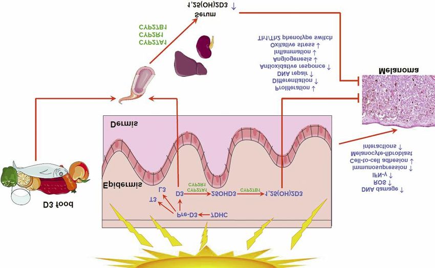
Figure 1. Schematic of vitamin D synthesis, activation and attendant effects on melanoma biology. 7DHC: 7-Dehydrocholesterol; CYP2R1:
cytochrome P450 family 2 subfamily R member 1; CYP27A1: cytochrome P450 family 27 subfamily A member 1; CYP27B1: cytochrome P450
family 27 subfamily B member 1; D3: vitamin D3; INFg: interferon gamma; L3: lumisterol 3; pre-D3: pre-vitamin D3; ROS: reactive oxygen
species; T3: tachysterol 3; Th1: T-helper cell type 1 phenotype; Th2: T-helper cell type 2 phenotype.
5%) (93). It is the second most common melanoma subtype melanoma development is, in part, related to genetic factors,
(93-95). Uveal melanoma affects mostly the Caucasian such as germline mutations, pigmentation, UV-induced
population and people over 50 years of age (93-95). Over the mutations or inability to repair UV-induced DNA damages.
past 40 years, the incidence rate has been stable, with slight, Most melanoma cases are sporadic, but 5-12% of all
but significant increase of incidence for Caucasians (96). melanomas have family history of melanoma (44). Patients
Similarly to cutaneous melanoma, the incidence rate in with multiple nevi are also prone to developing melanoma
Europe increases with latitude, and in the USA a higher (98). About 20% of patients with susceptibility to melanoma
incidence rate was observed in California (94). There is are carriers of a CDKN2A (called also INK4a/ARF) gene
slightly higher incidence rate among men (94, 96). The mutation, coding two structurally distinct proteins, p14ARF and
therapeutic options for uveal melanoma include surgery, p16INK4a, involved in cell-cycle regulation (99). Mutations in
enucleation, radiation, or combination treatment (94, 96). the cyclin-dependent kinase-4 (CDK4) gene confer
The 5-year survival rate (about 75-80%) has been stable for susceptibility to cutaneous melanoma, but mutations in CDK4
the past 40 years (94, 96). The efficacy of immunotherapy are not as frequent as those in CDKN2A (100, 101). In
against uveal melanoma is limited and molecular-targeted addition, germline mutations in the tumor-suppressor BRCA-
therapies are still being investigated (93). Thus, similarly to associated protein-1 (BAP1) gene, ubiquitin C-terminal
cutaneous melanomas, vitamin D-based treatment might hydrolase, encoding the protein interacting with BRCA1, have
serve as novel, adjuvant antitumor therapy (97). been identified in fewer than 1% of cutaneous melanomas.
Melanocortin 1 receptor (MC1R) mutations increase
Risk factors for melanomas. The most important susceptibility to melanoma in general population (102).
environmental risk factor for cutaneous melanoma is natural Atypical cutaneous nevi, light eye color fair skin color,
and artificial UV radiation. The involvement of UV in predisposition to sunburn ocular melanocytosis and iris nevi
475ANTICANCER RESEARCH 40: 473-489 (2020)
are risk factors for uveal and cutaneous melanoma (103). cycle inhibition in cancer can be accompanied by apoptosis,
Chronic exposure to sunlight were not related to the risk of which is also promoted by vitamin D. This is achieved by
uveal melanoma development, but welding was identified as down-regulation of phosphorylated AKT and ERK, leading
a risk factor (103). The majority of uveal melanomas are to apoptosis through activation of forkhead box O 3A
sporadic tumors. However recently mutations in BAP1 were (FOXO3) (148), down-regulation of B-cell lymphoma 2
found to be related to younger age (39-50 years) at (BCL2) and up-regulation of BCL2-associated X protein
diagnosis, and higher risk of second tumors (cutaneous (BAX), BCL2 antagonist/killer 1 (BAK), and BCL2-
melanoma, renal cell carcinoma) has been identified (104). associated agonist of cell death BAD (149) [reviewed in (108,
150)]. Calcitriol induces the expression of adhesion
Classical and Non-classical Vitamin D Derivatives molecules, stimulates cell maturation, and inhibits cancer
progression and metastatic potential (135, 151-153). Vitamin
The main active form of vitamin D, 1,25(OH)2D3 (calcitriol) D inhibits metastasis via the inhibition of vascular endothelial
acts predominantly through binding to the nuclear vitamin D growth factor expression (VEGF) (154, 155).
receptor (VDR). VDR is activated by 1,25(OH)2D3 to form a Vitamin D can prevent cancer by protecting DNA (108,
dimer with the retinoid X receptor (RXR) receptor, is 156-158) as well as inducing the expression of superoxide
translocated to the cell nucleus, and acts as a transcription dismutase, glucose-6-phosphate dehydrogenase, nuclear
factor via binding to VDR-responding element (VDRE) (10, factor erythroid 2 (NF-E2)-related factor 2, proliferating cell
105-108). More than 1,000 target genes, varying broadly in nuclear antigen (PCNA) , BRCA1 and other genes (159-162).
their biological activities, regulated by vitamin D were Vitamin D can modulate immune responses by stimulating
identified depending on cell type (109-112). VDR expression the innate immune response, while inhibiting the adaptive
was identified in many tissues and cells, including epidermal immunity response. Vitamin D attenuates chronic
and dermal skin cells that both synthesize 1,25(OH)2D3 inflammation related to increased cancer risk [reviewed in
(calcitriol) and respond to it (10, 107, 113-115). Non- (163, 164)]. Vitamin D modulates the inflammatory immune
canonical, noncalcemic hydroxylated vitamin D3 forms (9, 30, response by up-regulation of PD-1, as was observed in
33) can also act on VDR (116-119). They can also act as Crohn’s disease (165) and induces the expression of
inverse agonists on retinoic acid-related orphan receptors programmed death-ligand 1 (PD-L1) and PD-L2 via VDR in
(RORs) α and γ (117, 120), which are expressed in normal experimental cell-based models (166). Immune response
and pathological skin cells, including melanoma (120, 121). regulation by vitamin D is linked to the inhibition of type 1
Most recently, it has been shown that vitamin D3 hydroxy- T-helper (Th1) and promotion of Th2 phenotype, including
derivatives can act on arylhydrocarbone receptor (122). These up-regulation of interleukin 10 (IL10) and TGFβ (167, 168).
alternative receptors for vitamin D3 and its metabolites may The role of vitamin D in the immune response in patients
be related to the diverse actions of vitamin D. with cancer appears complex (169).
1,25(OH)2D3 has antitumor properties affecting molecular
Vitamin D and Melanoma: pathways involved in proliferation, apoptosis and
Experimental and Clinical Evidence differentiation, but can also improve effectiveness of classical
anticancer therapies (163, 170). Experimental cell- and animal
Anticancer properties of vitamin D – An overview. Almost 40 model-based data clearly showed that vitamin D and its analogs
years ago the anticancer effects of vitamin D was suggested increased the effectiveness of well-known cancer chemotherapy
by Garland and Garland (123) based on epidemiological drugs (such as doxorubicin, cisplatinum, gemcitabine and
studies and Colston et al. observed the anticancer effects of cyclophosphamide) (171-173). 1,25(OH)2D3 sensitized
vitamin D experimentally (124). Several molecular pathways malignant cells to ionizing radiation (174-179) and proton beam
related to cancer biology, tumor development and progression radiation (180). These data indicate that vitamin D and its
have been proposed to serve as targets for active forms of analogs alone or in combination with standard therapeutic
vitamin D (52, 77, 125-130). Vitamin D and its derivatives schemes can improve the outcome of melanoma therapy.
have been shown to inhibit cancer-cell proliferation. p21
regulates the cell cycle by calcitriol and VDR (131-135). Effects of vitamin D on melanoma cells in vitro. Since anti-
Vitamin D also up-regulates the cell cycle inhibitor, p27 (135- melanoma properties of vitamin D and its analogs were
138). Vitamin D-related mechanisms regulating the cell cycle reviewed recently (38, 130), what follows is only a short
may be related to the growth factor signaling [reviewed in overview. Colston and co-workers showed VDR-expressing
(108, 139)], including up-regulation of insulin-like growth melanoma cells were inhibited by 1,25(OH)2D3 (124). The
factor-binding protein 3 (IGFBP3) and transforming growth anticancer properties of 1,25(OH)2D3 have been shown in
factor-β (TGFβ) expression and signaling pathways (140- various melanoma cell lines. Janjetovic et al. reported the
144), downregulation of hedgehog signaling (145-147). Cell- inhibitory effects of 1,25(OH)2D3 on both pigmented and
476Brożyna et al: Vitamin D and Melanoma (Review)
nonpigmented SkMel-188 melanoma cells (181). A similar of B16 melanoma cells injected into mouse by affecting the
effect was found for 20(OH)D3. Both compounds stimulated extracellular matrix (196), and 1(OH)D2 reduced tumor
VDR translocation into the nucleus, and in nonpigmented growth in Tyr-Tag transgenic mice, which develop
melanoma cells inhibited nuclear factor kappa-light-chain- pigmented ocular tumors, similar to human choroidal
enhancer of activated B cells (NF-ĸB) DNA targeting by melanoma (205). The patient-derived orthotopic xenograph
vitamin D3. Melanin affected melanoma cell susceptibility to (PDOX) model has been developed for melanoma in order
vitamin D3 anticancer activity (181). 1,25(OH)2D3 also to individualize chemotherapy for individual patients with
inhibited colony formation by SkMel-188 cells (182, 183). advanced melanoma. For example, effective therapy was
The antiproliferative activity of 1,25(OH)2D3, calcipotriol and identified for melanoma with/without BRAF-V600 mutation
25(OH)D3 are related to the expression VDR and CYP27B1 (206-212).
(184). The growth inhibition and apoptosis inducing effects of
1,25(OH)2D3 were also observed in other human melanoma Serum vitamin D level in patients with melanoma: Effects on
cell lines, including: A375 (185), ME18 (186), MeWo (187- susceptibility and survival. Garland and Garland (123)
190), RPMI 7951 (191, 192), SM (189), SK Mel 28 (189, suggested that low-sun-exposure-related vitamin D
192-194) and WM1341 (187, 188). The anticancer activity of insufficiency was correlated with higher colon cancer
calcitriol was also demonstrated against mouse B16 and mortality rates. These results were confirmed by other
hamster Bomirski melanoma cells (195). Vitamin D3 inhibited epidemiological reports (213-215) and experiments in animal
invasiveness of malignant cells. Yudoh and co-workers models treated with vitamin D, showing inhibition of tumor
reported inhibition of lysis of IV type collagen and stimulation growth (216, 217) and higher benign and malignant tumor
of basement membrane reconstitution by B16 mouse risk in VDR−/− animals (22, 27, 218, 219).
melanoma cells preincubated with calcitriol for 48 h (196). A recent case–control study showed higher vitamin D levels
Other forms of vitamin D are biologically active and are in serum of healthy controls than in patients at the time of
potential anticancer agents. Vitamin D metabolites melanoma diagnosis. A multivariate model revealed a negative
1,24,25(OH)3D3 and 1,25,26(OH)3D3 inhibited the association between vitamin D sufficiency and melanoma
proliferation of the MM96 cell line, similarly to that found (220). These data confirmed previously published reports on
for 1,25(OH)2D3 (197). It was shown that malignant cells, the correlation of serum vitamin D levels and clinical outcome
including pigmented melanoma cells, possess an active of patients with melanoma, including a relationship between
mechanism of metabolizing of vitamin D (198-200). In the lower Breslow tumor thickness and higher 25(OH)D3 level
addition, several vitamin D derivatives have been developed (221). Subsequent studies confirmed that a lower vitamin D
and identified as non-calcemic or low-calcemic anticancer level was related to greater progression of melanoma [Breslow
agents. 20(OH)D2/3, a non-calcemic vitamin D derivative, thickness, Clark level, the American Joint Committee on
inhibited melanoma cells both in vitro and in vivo (119, 181, Cancer (AJCC) stage], the presence of poor prognostic
183, 201-203). 20,23(OH)2D3, and 1,20(OH)2D3 also markers (ulceration, higher mitotic index), shorter overall
inhibited proliferation and colony formation of melanoma survival and increased risk for melanoma-specific death (222-
cells (183). Metabolites of 20(OH)D3 such as 20,24(OH)2D3 226). However, some investigators (227) did not observe such
and 20,25(OH)2D3, produced by the action of CYP24A1, relationships and found only longer disease-free survival for
inhibited melanoma growth in soft agar more efficiently than patients with higher vitamin D levels. Melanoma risk is
1,25(OH)2D3 and 20(OH)D3 (31). A very recent report related to a higher number of nevi, however Ribero and co-
showed anti-melanoma activity of 21(OH)pD in WM98, workers showed positive correlation of serum vitamin D level
A375 and SK-MEL-188b (VDR−/−CYP27B1−/−) lines. Only and nevi count (228). These authors suggested that melanomas
WM98 and A375 cells were sensitive to calcipotriol (184). associated with a low vitamin D level might be a different
type from those associated with a higher nevi count, thus
Effects of vitamin D and its new analogs on melanoma cells further studies are required to explain the association between
in animal models. The antitumorigenic activity of nevi, melanoma and vitamin D level.
1,25(OH)2D3 in an animal model was reported for the first Correlation between vitamin D intake (supplementary or
time by Eisman and coworkers (204), demonstrating the dietary) and melanoma risk is still incompletely understood
inhibition by 1,25(OH)2D3 of the growth of human (229).
melanoma cells COLO 239F expressing VDR, which were
injected into immunosuppressed mice. Another melanoma Modulation of vitamin D signaling in melanoma – Clinical
cell line, RPMI 7932, with no VDR expression, was experimental data. Our study on clinical material showed
insensitive. The VDR-positive SKMel-188 melanoma cell that the reduction of VDR (both cytoplasmic and nuclear)
line, injected into immunocompromised mice, was inhibited correlated with melanoma progression, being the highest in
by 20(OH)D3 (201). 1,25(OH)2D3 reduced lung metastasis normal skin and benign nevi, and lowest in most advanced
477ANTICANCER RESEARCH 40: 473-489 (2020)
Table I. Changes of vitamin D receptor (VDR, nuclear), cytochrome P450 family 27 subfamily B member 1 (CYP27B1), cytochrome P450 family
24 subfamily A member 1 (CYP24A1), retinoic acid receptor-related orphan receptor alpha (RORα, nuclear), and RORγ (nuclear) in relation to
the expression in normal skin (only statistically significant change are indicated).
Pigmented lesion Immunoexpression
VDR CYP27B1 CYP24A1 RORα RORγ
Nevi =
Melanoma – in situ
↓ ↑ ↓↓ ↓
Melanoma - RGP
↓ ↓ ↑↑↑ ↓↓↓ ↓↓
Melanoma - VGP
↓↓ ↓↓ ↑↑ ↓↓↓ ↓↓
Metastasis =
↓↓↓ ↓↓↓ ↑ ↓↓↓ ↓↓↓
↓↓↓ ↓↓↓ ↓↓↓ ↓↓↓
RGP: Radial growth phase; VGP: vertical growth phase; =: no change; ↑/↑↑/↑↑↑: increase of expression; ↓/↓↓/↓↓↓: decrease of expression.
Table II. Correlation of selected clinico-pathomorphological melanoma features and the expression of vitamin D receptor (VDR, cytoplasmic and
nuclear), cytochrome P450 family 27 subfamily B member 1 (CYP27B1), cytochrome P450 family 24 subfamily A member 1 (CYP24A1), retinoic
acid receptor-related orphan receptor alpha (RORα, cytoplasmic and nuclear), and RORγ (cytoplasmic and nuclear) in primary melanomas. Analysis
was performed with Pearson correlation tests.
Melanoma feature Immunoexpression
VDR CYP27B1 CYP24A1 RORα RORγ
Cytoplasmic Nuclear Cytoplasmic Nuclear Cytoplasmic Nuclear
Breslow thickness r=−0.2860 NSA r=−0.3796 r=−0.4035 r=−0.1924 r=−0.2914 r=−0.4108 r=−0.3109
p=0.0056 p=0.0002 p=0.0008 p=0.0458 p=0.0048 p=0.0003 p=0.0049
Clark level NS r=−0.2659 r=−0.3897 r=−0.3087 r=−0.1879 r=−0.2473 r=−0.4242 r=−0.3254
p=0.0093 p=0.0001 p=0.0087 p=0.0497 p=0.0145 p=0.0002 p=0.0034
pT r=−0.1938 r=−0.3076 r=−0.2870 r=−0.3749 r=−0.2395 r=−0.3284 r=−0.4008 r=−0.3277
p=0.0478 p=0.0036 p=0.0049 p=0.0020 p=0.0193 p=0.0020 p=0.0004 p=0.0034
pN NS NSB r=−0.3159 r=−0.3705 r=−0.2580 r=−0.2802 r=−0.2773 r=−0.2863
p=0.0022 p=0.0023 p=0.0127 p=0.0074 p=0.0115 p=0.0100
pM NS NSC r=−0.2590 NSF NS NS r=−0.2133 NS
p=0.0102 p=0.0415
TILs NSD NS NS NS r=0.2198 r=0.3530 NS NS
p=0.0282 p=0.0009
Overal stage NSE r=−0.3174 r=−0.3826 r=−0.4297 r=−0.2569 r=−0.2953 r=−0.4545 r=−0.4043
p=0.0028 p=0.0002 p=0.0004 p=0.0131 p=0.0051 pBrożyna et al: Vitamin D and Melanoma (Review) In addition, we also observed modulation of the patients after resection of stage II melanoma (n=150), expression of enzymes involved in vitamin D metabolism. treated with 100,000 IU of vitamin D3 every 50 days for 3 CYP27B1 expression, as well VDR, was reduced in years. Disease-free survival has been defined as a primary melanomas, showing the lowest level in most advanced end-point of efficacy. Overall survival, Breslow thickness primary tumors and metastatic melanomas (233). However, and VDR were also measured. A Belgian-Hungarian ViDMe there was a lack of correlation between the presence of an randomized controlled trial (ClinicalTrials.gov Identifier: ulceration or lack of TILs and CYP27B1 expression. NCT01748448) (237) is a multicenter randomized double However, CYP27B1 expression was accompanied by a lower blind placebo-controlled phase III trial, registered in 2012, proliferation index and better overall and disease-free with monthly administration of 100,000 units of vitamin D survival (233). VDR and CYP27B1 expression were also or placebo (Arachidis oleum raffinatum) to 500 patients negatively correlated to pigmentation in melanoma (230, with melanoma. This study examines the relationship 233). The correlation of CYP24A1 and melanoma between disease-free survival, melanoma subtype, anatomic progression is complex. CYP24A1 expression was lowest in site and vitamin D receptor, the vitamin D pathway and metastatic lesions, and highest in benign nevi and localized vitamin D level at the time of diagnosis and at 6 months melanomas (pT1-2, Clark level 1-2, Breslow thickness
ANTICANCER RESEARCH 40: 473-489 (2020)

Conflicts of Interest                                                      pathway of 25-hydroxyvitamin D3 catalyzed by human CYP24.
                                                                           Eur J Biochem 267(20): 6158-6165, 2000. PMID: 11012668.
The Authors have no conflicts of interest to declare.                      DOI: 10.1046/j.1432-1327.2000.01680.x
                                                                      13   Sakaki T, Sawada N, Nonaka Y, Ohyama Y and Inouye K:
Authors’ Contributions                                                     Metabolic studies using recombinant escherichia coli cells
                                                                           producing rat mitochondrial CYP24 CYP24 can convert
                                                                           1alpha,25-dihydroxyvitamin D3 to calcitroic acid. Eur J
AAB conceptualized and wrote the article, RMH wrote the
                                                                           Biochem 262(1): 43-48, 1999. PMID: 10231362. DOI:
subchapter and corrected the article, ATS conceptualized, wrote and
                                                                           10.1046/j.1432-1327.1999.00375.x
corrected the final version of the article.
                                                                      14   Henry HL: Vitamin D hydroxylases. J Cell Biochem 49(1): 4-
                                                                           9, 1992. PMID: 1644853. DOI: 10.1002/jcb.240490103
Acknowledgements                                                      15   Henry HL, Dutta C, Cunningham N, Blanchard R, Penny R,
                                                                           Tang C, Marchetto G and Chou SY: The cellular and molecular
The work was supported in part by grants 2014/15/B/NZ4/00751               regulation of 1,25(OH)2D3 production. J Steroid Biochem Mol
from National Science Centre, Poland to AAB and NIH grants                 Biol 41(3-8): 401-407, 1992. PMID: 1562513. DOI:
1R01AR073004-01A1 and R01 AR071189-01A1 and VA merit                       10.1016/0960-0760(92)90365-p
grant (No. 1I01BX004293-01A1) to ATS.                                 16   Nebert DW and Gonzalez FJ: P450 genes: Structure, evolution,
                                                                           and regulation. Annu Rev Biochem 56: 945-993, 1987. PMID:
References                                                                 3304150. DOI: 10.1146/annurev.bi.56.070187.004501
                                                                      17   Zehnder D and Hewison M: The renal function of 25-
1  Holick MF, Frommer JE, McNeill SC, Richtand NM, Henley                  hydroxyvitamin D3-1alpha-hydroxylase. Mol Cell Endocrinol
   JW and Potts JT Jr.: Photometabolism of 7-dehydrocholesterol            151(1-2): 213-220, 1999. PMID: 10411336. 10.1016/s0303-
   to previtamin D3 in skin. Biochem Biophys Res Commun                    7207(99)00039-8
   76(1): 107-114, 1997. PMID: 194588. DOI: 10.1016/0006-             18   Zhu JG, Ochalek JT, Kaufmann M, Jones G and Deluca HF:
   291x(77)91674-6                                                         CYP2R1 is a major, but not exclusive, contributor to 25-
2 Bikle DD: Vitamin D: An ancient hormone. Exp Dermatol                    hydroxyvitamin D production in vivo. Proc Natl Acad Sci USA
   20(1): 7-13, 2011. PMID: 21197695.                                      110(39): 15650-15655, 2013. PMID: 24019477. DOI:
3 Holick MF: Defects in the synthesis and metabolism of vitamin            10.1073/pnas.1315006110
   D: Exp Clin Endocrinol Diabetes 103(4): 219-227, 1995.             19   Holick MF: Vitamin D: A millenium perspective. J Cell Biochem
   PMID: 7584527. DOI: 10.1055/s-0029-1211354                              88(2): 296-307, 2003. PMID: 12520530. DOI: 10.1002/jcb.10338
4 Holick MF: Photobiology of vitamin D: In: Vitamin D:                20   Bikle DD, Chang S, Crumrine D, Elalieh H, Man MQ, Choi
   Feldman D, Pike JW and Adams JS (eds.). Academic Press, pp.             EH, Dardenne O, Xie Z, Arnaud RS, Feingold K and Elias PM:
   13-22, 2011.                                                            25 Hydroxyvitamin D 1-alpha-hydroxylase is required for
5 Holick MF and Clark MB: The photobiogenesis and                          optimal epidermal differentiation and permeability barrier
   metabolism of vitamin D: Fed Proc 37(12): 2567-2574, 1978.              homeostasis. J Invest Dermatol 122(4): 984-992, 2004. PMID:
   PMID: 212325.                                                           15102089. DOI: 10.1111/j.0022-202X.2004.22424.x
6 Holick MF and DeLuca HF: Vitamin D metabolism. Annu Rev             21   Bikle DD: Vitamin D and the skin: Physiology and
   Med 25: 349-367, 1974. PMID: 4363209. DOI: 10.1146/                     pathophysiology. Rev Endocr Metab Disord 13(1): 3-19, 2012.
   annurev.me.25.020174.002025                                             PMID: 21845365. DOI: 10.1007/s11154-011-9194-0
7 DeLuca HF: Vitamin D: A new look at an old vitamin. Nutr            22   Bikle DD, Oda Y, Tu CL and Jiang Y: Novel mechanisms for
   Rev 29(8): 179-181, 1971. PMID: 4328308. DOI:                           the vitamin D receptor (VDR) in the skin and in skin cancer. J
   10.1111/j.1753-4887.1971.tb07292.x                                      Steroid Biochem Mol Biol 148: 47-51, 2015. PMID: 25445917.
8 Slominski AT, Li W, Kim T, Semak I, Wang J, Zjawiony J and               DOI: 10.1016/j.jsbmb.2014.10.017
   Tuckey RC: Novel activities of CYP11A1 and their potential         23   Lopes N, Sousa B, Martins D, Gomes M, Vieira D, Veronese
   physiological significance. J Steroid Biochem Mol Biol 151:             LA, Milanezi F, Paredes J, Costa JL and Schmitt F: Alterations
   25-37, 2015. PMID: 25448732. DOI: 10.1016/j.jsbmb.2014.                 in vitamin D signalling and metabolic pathways in breast
   11.010                                                                  cancer progression: A study of VDR, CYP27B1 and CYP24A1
9 Tuckey RC, Cheng CYS and Slominski AT: The serum vitamin                 expression in benign and malignant breast lesions. BMC
   D metabolome: What we know and what is still to discover. J             Cancer 10: 483, 2010. PMID: 20831823. DOI: 10.1186/1471-
   Steroid Biochem Mol Biol 186: 4-21, 2019. PMID: 30205156.               2407-10-483
   DOI: 10.1016/j.jsbmb.2018.09.003                                   24   Pillai S, Bikle DD and Elias PM: 1,25-dihydroxyvitamin D
10 Bikle DD: Vitamin D metabolism, mechanism of action, and                production and receptor binding in human keratinocytes varies
   clinical applications. Chem Biol 21(3): 319-329, 2014. PMID:            with differentiation. J Biol Chem 263(11): 5390-5395, 1988.
   24529992. DOI: 10.1016/j.chembiol.2013.12.016                           PMID: 2451669.
11 Jones G, Prosser DE and Kaufmann M: 25-hydroxyvitamin D-           25   Kramer C, Seltmann H, Seifert M, Tilgen W, Zouboulis CC and
   24-hydroxylase (CYP24A1): Its important role in the                     Reichrath J: Characterization of the vitamin D endocrine system in
   degradation of vitamin D: Arch Biochem Biophys 523(1): 9-18,            human sebocytes in vitro. J Steroid Biochem Mol Biol 113(1-2): 9-
   2012. PMID: 22100522. DOI: 10.1016/j.abb.2011.11.003                    16, 2009. PMID: 19027855. DOI: 10.1016/j.jsbmb.2008.10.010
12 Sakaki T, Sawada N, Komai K, Shiozawa S, Yamada S,                 26   Bikle DD, Xie Z, Ng D, Tu CL and Oda Y: Squamous cell
   Yamamoto K, Ohyama Y and Inouye K: Dual metabolic                       carcinomas fail to respond to the prodifferentiating actions of

480
Brożyna et al: Vitamin D and Melanoma (Review)
1,25(OH)2D: Why? Recent results. Cancer Res 164: 111-122, Elmets C, Li W, Hoffman RM and Tuckey RC: On the role of
2003. PMID: 12899516. DOI: 10.1007/978-3-642-55580-0_7 classical and novel forms of vitamin D in melanoma progression
27 Bikle DD, Jiang Y, Nguyen T, Oda Y and Tu CL: Disruption of and management. J Steroid Biochem Mol Biol 177: 159-170,
vitamin D and calcium signaling in keratinocytes predisposes 2018. PMID: 28676457. DOI: 10.1016/j.jsbmb.2017.06.013
to skin cancer. Front Physiol 7: 296, 2016. PMID: 27462278. 39 Slominski AT, Zmijewski MA, Plonka PM, Szaflarski JP and
DOI: 10.3389/fphys.2016.00296 Paus R: How UV light touches the brain and endocrine system
28 Pillai S, Bikle DD and Elias PM: Vitamin D and epidermal through skin, and why. Endocrinology 159(5): 1992-2007,
differentiation: Evidence for a role of endogenously produced 2018. PMID: 29546369. DOI: 10.1210/en.2017-03230
vitamin D metabolites in keratinocyte differentiation. Skin 40 Runger TM: Mechanisms of melanoma promotion by
Pharmacol 1(3): 149-160, 1988. PMID: 3078531. ultraviolet radiation. J Invest Dermatol 136(9): 1751-1752,
29 Slominski A, Semak I, Zjawiony J, Wortsman J, Li W, 2016. PMID: 27542295. DOI: 10.1016/j.jid.2016.04.001
Szczesniewski A and Tuckey RC: The cytochrome p450scc 41 Narayanan DL, Saladi RN and Fox JL: Ultraviolet radiation and
system opens an alternate pathway of vitamin D3 metabolism. skin cancer. Int J Dermatol 49(9): 978-986, 2010. PMID:
FEBS J 272(16): 4080-4090, 2005. PMID: 16098191. DOI: 20883261. DOI: 10.1111/j.1365-4632.2010.04474.x
10.1111/j.1742-4658.2005.04819.x 42 Gasparro FP: Sunscreens, skin photobiology, and skin cancer:
30 Slominski AT, Li W, Kim TK, Semak I, Wang J, Zjawiony JK The need for UVA protection and evaluation of efficacy.
and Tuckey RC: Novel activities of CYP11A1 and their Environ Health Perspect 108 Suppl 1: 71-78, 2000. PMID:
potential physiological significance. J Steroid Biochem Mol 10698724. DOI: 10.1289/ehp.00108s171
Biol 151:25-37, 2015. PMID: 25448732. DOI: 43 Young AR, Claveau J and Rossi AB: Ultraviolet radiation and
10.1016/j.jsbmb.2014.11.010 the skin: Photobiology and sunscreen photoprotection. J Am
31 Tieu EW, Tang EK, Chen J, Li W, Nguyen MN, Janjetovic Z, Acad Dermatol 76(3S1): S100-S109, 2017. PMID: 28038885.
Slominski A and Tuckey RC: Rat CYP24A1 acts on 20- DOI: 10.1016/j.jaad.2016.09.038
hydroxyvitamin D(3) producing hydroxylated products with 44 Jhappan C, Noonan FP and Merlino G: Ultraviolet radiation and
increased biological activity. Biochem Pharmacol 84(12): 1696- cutaneous malignant melanoma. Oncogene 22(20): 3099-3112,
1704, 2012. PMID: 23041230. DOI: 10.1016/j.bcp.2012.09.032 2003. PMID: 12789287. DOI: 10.1038/sj.onc.1206450
32 Slominski AT, Kim TK, Shehabi HZ, Semak I, Tang EK, 45 Rass K and Reichrath J: UV damage and DNA repair in
Nguyen MN, Benson HA, Korik E, Janjetovic Z, Chen J, Yates malignant melanoma and nonmelanoma skin cancer. Adv Exp
CR, Postlethwaite A, Li W and Tuckey RC: In vivo evidence Med Biol 624: 162-178, 2008. PMID: 18348455. DOI:
for a novel pathway of vitamin D(3) metabolism initiated by 10.1007/978-0-387-77574-6_13
p450scc and modified by CYP27B1. FASEB J 26(9): 3901- 46 Leiter U and Garbe C: Epidemiology of melanoma and
3915, 2012. PMID: 22683847. DOI: 10.1096/fj.12-208975 nonmelanoma skin cancer—the role of sunlight. Adv Exp Med
33 Slominski AT, Kim TK, Li W, Postlethwaite A, Tieu EW, Tang EK Biol 624: 89-103, 2008. PMID: 18348450. DOI: 10.1007/978-
and Tuckey RC: Detection of novel CYP11A1-derived secosteroids 0-387-77574-6_8
in the human epidermis and serum and pig adrenal gland. Sci Rep 47 de Gruijl FR: Photocarcinogenesis: UVA vs. UVB radiation.
5: 14875, 2015. PMID: 26445902. DOI: 10.1038/srep14875 Skin Pharmacol Appl Skin Physiol 15(5): 316-320, 2002.
34 Slominski A, Zjawiony J, Wortsman J, Semak I, Stewart J, PMID: 12239425. DOI: 10.1159/000064535
Pisarchik A, Sweatman T, Marcos J, Dunbar C and Tuckey RC: 48 Nilsen LT, Hannevik M and Veierod MB: Ultraviolet exposure from
A novel pathway for sequential transformation of 7- indoor tanning devices: A systematic review. Br J Dermatol 174(4):
dehydrocholesterol and expression of the p450scc system in 730-740, 2016. PMID: 26749382. DOI: 10.1111/bjd.14388
mammalian skin. Eur J Biochem 271(21): 4178-4188, 2004. 49 Rass K: UV damage and DNA repair in basal cell and squamous
PMID: 15511223. DOI: 10.1111/j.1432-1033.2004.04356.x cell carcinomas. In: Molecular Mechanisms of Basal Cell and
35 Slominski AT, Kim TK, Chen J, Nguyen MN, Li W, Yates CR, Squamous Cell Carcinomas. Reichrath J (ed.). Springer:
Sweatman T, Janjetovic Z and Tuckey RC: Cytochrome p450scc- Boston, MA, pp. 18-30, 2018.
dependent metabolism of 7-dehydrocholesterol in placenta and 50 Reichrath J, Lindqvist PG, FR DEG, Pilz S, Kimball SM,
epidermal keratinocytes. Int J Biochem Cell Biol 44(11): 2003- Grant WB and Holick MF: A critical appraisal of the recent
2018, 2012. PMID: 22877869. DOI: 10.1016/j.biocel. reports on sunbeds from the European Commission’s Scientific
2012.07.027 Committee on Health, Environmental and Emerging Risks and
36 Slominski AT, Zmijewski MA, Semak I, Sweatman T, from the World Health Organization. Anticancer Res 38(2):
Janjetovic Z, Li W, Zjawiony JK and Tuckey RC: Sequential 1111-1120, 2018. PMID: 29374748. DOI: 10.21873/
metabolism of 7-dehydrocholesterol to steroidal 5,7-dienes in anticanres.12330
adrenal glands and its biological implication in the skin. PLoS 51 Reichrath J and Reichrath S: Sunlight, vitamin D and malignant
One 4(2): e4309, 2009. PMID: 19190754. DOI: melanoma: An update. Adv Exp Med Biol 810: 390-405, 2014.
10.1371/journal.pone.0004309 PMID: 25207378.
37 Slominski A, Kim TK, Zmijewski MA, Janjetovic Z, Li W, 52 Reichrath J and Rass K: Ultraviolet damage, DNA repair and
Chen J, Kusniatsova EI, Semak I, Postlethwaite A, Miller DD, vitamin D in nonmelanoma skin cancer and in malignant
Zjawiony JK and Tuckey RC: Novel vitamin D photoproducts melanoma: An update. Adv Exp Med Biol 810: 208-233, 2014.
and their precursors in the skin. Dermatoendocrinol 5(1): 7-19, PMID: 25207368.
2013. PMID: 24494038. DOI: 10.4161/derm.23938 53 Ali Z, Yousaf N and Larkin J: Melanoma epidemiology, biology
38 Slominski AT, Brozyna AA, Skobowiat C, Zmijewski MA, Kim and prognosis. EJC Suppl 11(2): 81-91, 2013. PMID:
TK, Janjetovic Z, Oak AS, Jozwicki W, Jetten AM, Mason RS, 26217116. DOI: 10.1016/j.ejcsup.2013.07.012
481ANTICANCER RESEARCH 40: 473-489 (2020) 54 Bocheva G, Slominski RM and Slominski AT: Neuroendocrine 68 Day CP, Marchalik R, Merlino G and Michael H: Mouse aspects of skin aging. Int J Mol Sci 20(11), 2019. PMID: models of UV-induced melanoma: Genetics, pathology, and 31181682. DOI: 10.3390/ijms20112798 clinical relevance. Lab Invest 97(6): 698-705, 2017. PMID: 55 Merkel EA and Gerami P: Malignant melanoma of sun- 28092363. DOI: 10.1038/labinvest.2016.155 protected sites: A review of clinical, histological, and molecular 69 De Fabo EC, Noonan FP, Fears T and Merlino G: Ultraviolet B features. Lab Invest 97(6): 630-635, 2017. PMID: 28092366. but not ultraviolet A radiation initiates melanoma. Cancer Res DOI: 10.1038/labinvest.2016.147 64(18): 6372-6376, 2004. PMID: 15374941. DOI: 56 Rabbie R, Ferguson P, Molina-Aguilar C, Adams DJ and 10.1158/0008-5472.CAN-04-1454 Robles-Espinoza CD: Melanoma subtypes: Genomic profiles, 70 Fernandez AA, Garcia R, Paniker L, Trono D and Mitchell DL: prognostic molecular markers and therapeutic possibilities. J An experimental population study of nucleotide excision repair Pathol 247(5): 539-551, 2019. PMID: 30511391. DOI: as a risk factor for uvb-induced melanoma. Photochem 10.1002/path.5213 Photobiol 87(2): 335-341, 2010. PMID: 21143485. DOI: 57 Oliveria SA, Saraiya M, Geller AC, Heneghan MK and 10.1111/j.1751-1097.2010.00875.x Jorgensen C: Sun exposure and risk of melanoma. Arch Dis 71 Khan AQ, Travers JB and Kemp MG: Roles of UVA radiation Child 91(2): 131-138, 2006. PMID: 16326797. DOI: and DNA damage responses in melanoma pathogenesis. 10.1136/adc.2005.086918 Environ Mol Mutagen 59(5): 438-460, 2018. PMID: 29466611. 58 Noonan FP, Zaidi MR, Wolnicka-Glubisz A, Anver MR, Bahn J, DOI: 10.1002/em.22176 Wielgus A, Cadet J, Douki T, Mouret S, Tucker MA, Popratiloff A, 72 Pastila R and Leszczynski D: Ultraviolet A exposure alters Merlino G and De Fabo EC: Melanoma induction by ultraviolet A but adhesive properties of mouse melanoma cells. Photodermatol not ultraviolet B radiation requires melanin pigment. Nat Commun 3: Photoimmunol Photomed 21(5): 234-241, 2005. PMID: 884, 2012. PMID: 22673911. DOI: 10.1038/ncomms1893 16149935. DOI: 10.1111/j.1600-0781.2005.00166.x 59 Kraemer KH and DiGiovanna JJ: Xeroderma pigmentosum. In: 73 Krengel S, Stark I, Geuchen C, Knoppe B, Scheel G, Schlenke Adam MP, Ardinger HH, Pagon RA et al. (eds.). GeneReviews®, P, Gebert A, Wunsch L, Brinckmann J and Tronnier M: University of Washington, Seattle, WA, USA, 1993-2019. Selective down-regulation of the alpha6-integrin subunit in Available at: melanocytes by UVB light. Exp Dermatol 14(6): 411-419, https://www.ncbi.nlm.nih.gov/books/NBK1397/?report=reader 2005. PMID: 15885076. DOI: 10.1111/j.0906- (Last accessed on 6th June 2019) 6705.2005.00295.x 60 Ghiasvand R, Rueegg CS, Weiderpass E, Green AC, Lund E 74 Marr DG, Poser I, Shellman YG, Bosserhoff AK and Norris and Veierod MB: Indoor tanning and melanoma risk: Long-term DA: Ultraviolet radiation induces release of MIA: A new evidence from a prospective population-based cohort study. Am mechanism for UVR-induced progression of melanoma. Int J J Epidemiol 185(3): 147-156, 2017. PMID: 28077359. DOI: Oncol 25(1): 105-111, 2004. PMID: 15201995. 10.1093/aje/kww148 75 Herlyn M, Berking C, Li G and Satyamoorthy K: Lessons from 61 Stern RS: The risk of melanoma in association with long-term melanocyte development for understanding the biological exposure to PUVA: J Am Acad Dermatol 44(5): 755-761, 2001. events in naevus and melanoma formation. Melanoma Res PMID: 11312420. DOI: 10.1067/mjd.2001.114576 10(4): 303-312, 2000. PMID: 10985664. 62 Roider EM and Fisher DE: Red hair, light skin, and uv- 76 Brenner M, Degitz K, Besch R and Berking C: Differential independent risk for melanoma development in humans. JAMA expression of melanoma-associated growth factors in Dermatol 152(7): 751-753, 2016. PMID: 27050924. DOI: keratinocytes and fibroblasts by ultraviolet A and ultraviolet B 10.1001/jamadermatol.2016.0524 radiation. Br J Dermatol 153(4): 733-739, 2005. PMID: 63 Lo JA and Fisher DE: The melanoma revolution: From UV 16181453. DOI: 10.1111/j.1365-2133.2005.06780.x carcinogenesis to a new era in therapeutics. Science 346(6212): 77 Reichrath J, Reichrath S, Heyne K, Vogt T and Roemer K: 945-949, 2014. PMID: 27050924. DOI: 10.1001/ Tumor suppression in skin and other tissues via cross-talk jamadermatol.2016.0524 between vitamin D- and p53-signaling. Front Physiol 5: 166, 64 Mitra D, Luo X, Morgan A, Wang J, Hoang MP, Lo J, Guerrero 2014. PMID: 24917821. DOI: 10.3389/fphys.2014.00166 CR, Lennerz JK, Mihm MC, Wargo JA, Robinson KC, Devi SP, 78 Slominski A, Wortsman J, Nickoloff B, McClatchey K, Mihm Vanover JC, D’Orazio JA, McMahon M, Bosenberg MW, MC and Ross JS: Molecular pathology of malignant melanoma. Haigis KM, Haber DA, Wang Y and Fisher DE: An ultraviolet- Am J Clin Pathol 110(6): 788-794, 1998. PMID: 9844592. radiation-independent pathway to melanoma carcinogenesis in DOI: 10.1093/ajcp/110.6.788 the red hair/fair skin background. Nature 491(7424): 449-453, 79 Slominski A and Pawelek J: Animals under the sun: Effects of 2012. PMID: 23123854. DOI: 10.1038/nature11624 ultraviolet radiation on mammalian skin. Clin Dermatol 16(4): 65 Fisher DE: UV-tanning behavior: A problem that doesn’t go 503-515, 1998. PMID: 9699062. DOI: https://doi.org/10.1016/ away. Pigment Cell Melanoma Res 24(4): 724, 2011. PMID: s0738-081x(98)00023-6 21535480. DOI: 10.1111/j.1755-148X.2011.00865.x 80 Brozyna A, Zbytek B, Granese J, Carlson AJ, Ross J and 66 Weinstock MA and Fisher DE: Indoor ultraviolet tanning: What Slominski A: Mechanism of UV-related carcinogenesis and its the data do and do not show regarding risk of melanoma and contribution to nevi/melanoma. Expert Rev Dermatol 2(4): 451- keratinocyte malignancies. J Natl Compr Canc Netw 8(8): 867- 469, 2007. PMID: 18846265. 872; quiz 873, 2010. PMID: 20870632. 10.6004/jnccn.2010.0063 81 Slominski AT and Carlson JA: Melanoma resistance: A bright 67 Fisher DE and James WD: Indoor tanning – science, behavior, future for academicians and a challenge for patient advocates. and policy. N Engl J Med 363(10): 901-903, 2010. PMID: Mayo Clin Proc 89(4): 429-433, 2014. PMID: 24684870. DOI: 20818900. DOI: 10.1056/NEJMp1005999 10.1016/j.mayocp.2014.02.009 482
Brożyna et al: Vitamin D and Melanoma (Review)

82 Slominski AT, Zmijewski MA, Zbytek B, Tobin DJ,                   97 Markiewicz A, Brozyna AA, Podgorska E, Elas M, Urbanska
   Theoharides TC and Rivier J: Key role of CRF in the skin              K, Jetten AM, Slominski AT, Jozwicki W, Orlowska-Heitzman
   stress response system. Endocr Rev 34(6): 827-884, 2013.              J, Dyduch G and Romanowska-Dixon B: Vitamin D receptors
   PMID: 23939821. DOI: 10.1210/er.2012-1092                             (VDR), hydroxylases CYP27B1 and CYP24A1 and retinoid-
83 Slominski A, Wortsman J, Luger T, Paus R and Solomon S:               related orphan receptors (ROR) level in human uveal tract and
   Corticotropin releasing hormone and proopiomelanocortin               ocular melanoma with different melanization levels. Sci Rep
   involvement in the cutaneous response to stress. Physiol Rev          9(1): 9142, 2019. PMID: 31235702. DOI: 10.1038/s41598-019-
   80(3): 979-1020, 2000. PMID: 10893429. DOI: 10.1152/                  45161-8
   physrev.2000.80.3.979                                             98 Whiteman DC, Watt P, Purdie DM, Hughes MC, Hayward NK
84 Hocker T and Tsao H: Ultraviolet radiation and melanoma: A            and Green AC: Melanocytic nevi, solar keratoses, and divergent
   systematic review and analysis of reported sequence variants.         pathways to cutaneous melanoma. J Natl Cancer Inst 95(11):
   Hum Mutat 28(6): 578-588, 2007. PMID: 17295241. DOI:                  806-812, 2003. PMID: 12783935. DOI: 10.1093/jnci/95.11.806
   10.1002/humu.20481                                                99 Goldstein AM, Chan M, Harland M, Hayward NK, Demenais F,
85 Nikolaou V and Stratigos AJ: Emerging trends in the                   Bishop DT, Azizi E, Bergman W, Bianchi-Scarra G, Bruno W,
   epidemiology of melanoma. Br J Dermatol 170(1): 11-19, 2014.          Calista D, Albright LA, Chaudru V, Chompret A, Cuellar F, Elder
   PMID: 23815297. DOI: 10.1111/bjd.12492                                DE, Ghiorzo P, Gillanders EM, Gruis NA, Hansson J, Hogg D,
86 Siegel R, Ma J, Zou Z and Jemal A: Cancer statistics, 2014. CA        Holland EA, Kanetsky PA, Kefford RF, Landi MT, Lang J,
   Cancer J Clin 64(1): 9-29, 2014. PMID: 24399786. DOI:                 Leachman SA, MacKie RM, Magnusson V, Mann GJ, Bishop
   10.3322/caac.21208                                                    JN, Palmer JM, Puig S, Puig-Butille JA, Stark M, Tsao H, Tucker
87 Matthews NH, Li WQ, Qureshi AA, Weinstock MA and Cho E:               MA, Whitaker L and Yakobson E: Features associated with
   Epidemiology of melanoma. In: Cutaneous Melanoma: Etiology            germline cdkn2a mutations: A genome study of melanoma-prone
   and Therapy. 2018/02/21 ed. Ward WH and Farma JM (eds.).              families from three continents. J Med Genet 44(2): 99-106, 2007.
   Codon Publications: Brisbane (AU), 2017.                              PMID: 16905682. DOI: 10.1136/jmg.2006.043802
88 Rigel DS: Epidemiology of melanoma. Semin Cutan Med Surg          100 Puntervoll HE, Molven A and Akslen LA: Frequency of
   29(4): 204-209, 2010. PMID: 21277533. DOI: 10.1016/                   somatic braf mutations in melanocytic lesions from patients in
   j.sder.2010.10.005                                                    a cdk4 melanoma family. Pigment Cell Melanoma Res 27(1):
89 Rigel DS: Trends in dermatology: Melanoma incidence. Arch             149-151, 2013. PMID: 24256466. DOI: 10.1111/pcmr.12191
   Dermatol 146(3): 318, 2010. PMID: 20231504. DOI:                  101 Puntervoll HE, Yang XR, Vetti HH, Bachmann IM, Avril MF,
   10.1001/archdermatol.2009.379                                         Benfodda M, Catricala C, Dalle S, Duval-Modeste AB,
90 Rigel DS, Russak J and Friedman R: The evolution of melanoma          Ghiorzo P, Grammatico P, Harland M, Hayward NK, Hu HH,
   diagnosis: 25 years beyond the abcds. CA Cancer J Clin 60(5):         Jouary T, Martin-Denavit T, Ozola A, Palmer JM, Pastorino L,
   301-316, 2010. PMID: 20671054. DOI: 10.3322/caac.20074                Pjanova D, Soufir N, Steine SJ, Stratigos AJ, Thomas L, Tinat
91 Slominski A, Wortsman J, Carlson AJ, Matsuoka LY, Balch CM            J, Tsao H, Veinalde R, Tucker MA, Bressac-de Paillerets B,
   and Mihm MC: Malignant melanoma. Arch Pathol Lab Med                  Newton-Bishop JA, Goldstein AM, Akslen LA and Molven A:
   125(10): 1295-1306, 2001. PMID: 11570904. DOI:                        Melanoma prone families with cdk4 germline mutation:
   10.1043/0003-9985(2001)1252.0.CO;2                           Phenotypic profile and associations with MC1R variants. J
92 Devji T, Levine O, Neupane B, Beyene J and Xie F: Systemic            Med Genet 50(4): 264-270, 2013. PMID: 23384855. DOI:
   therapy for previously untreated advanced BRAF-mutated                10.1136/jmedgenet-2012-101455
   melanoma: A systematic review and network meta-analysis of        102 O’Shea SJ, Robles-Espinoza CD, McLellan L, Harrigan J, Jacq
   randomized clinical trials. JAMA Oncol 3(3): 366-373, 2017.           X, Hewinson J, Iyer V, Merchant W, Elliott F, Harland M,
   PMID: 27787543. DOI: 10.1001/jamaoncol.2016.4877                      Bishop DT, Newton-Bishop JA and Adams DJ: A population-
93 Krantz BA, Dave N, Komatsubara KM, Marr BP and Carvajal               based analysis of germline BAP1 mutations in melanoma. Hum
   RD: Uveal melanoma: Epidemiology, etiology, and treatment             Mol Genet 26(4): 717-728, 2017. PMID: 28062663. DOI:
   of primary disease. Clin Ophthalmol 11: 279-289, 2017. PMID:          10.1093/hmg/ddw403
   28203054. DOI: 10.2147/OPTH.S89591                                103 Nayman T, Bostan C, Logan P and Burnier MN, Jr.: Uveal
94 Mahendraraj K, Lau CS, Lee I and Chamberlain RS: Trends in            melanoma risk factors: A systematic review of meta-analyses.
   incidence, survival, and management of uveal melanoma: A              Curr Eye Res 42(8): 1085-1093, 2017. PMID: 28494168. DOI:
   population-based study of 7,516 patients from the surveillance,       10.1080/02713683.2017.1297997
   epidemiology, and end results database (1973-2012). Clin          104 Helgadottir H and Hoiom V: The genetics of uveal melanoma:
   Ophthalmol 10: 2113-2119, 2016. PMID: 27822007. DOI:                  Current insights. Appl Clin Genet 9: 147-155, 2016. PMID:
   10.2147/OPTH.S113623                                                  27660484. DOI: 10.2147/TACG.S69210
95 Mahendraraj K, Shrestha S, Lau CS and Chamberlain RS: Ocular      105 Silvagno F, Consiglio M, Foglizzo V, Destefanis M and
   melanoma-when you have seen one, you have not seen them all: A        Pescarmona G: Mitochondrial translocation of vitamin D
   clinical outcome study from the Surveillance, Epidemiology and        receptor is mediated by the permeability transition pore in
   End Results (SEER) database (1973-2012). Clin Ophthalmol 11:          human keratinocyte cell line. PLoS One 8(1): e54716, 2013.
   153-160, 2017. PMID: 27822007. DOI: 10.2147/OPTH.S113623              PMID: 23349955. DOI: 10.1371/journal.pone.0054716
96 Aronow ME, Topham AK and Singh AD: Uveal melanoma: 5-             106 Silvagno F and Pescarmona G: Spotlight on vitamin D receptor,
   Year update on incidence, treatment, and survival (SEER 1973-         lipid metabolism and mitochondria: Some preliminary emerging
   2013). Ocul Oncol Pathol 4(3): 145-151, 2018. PMID:                   issues. Mol Cell Endocrinol 450: 24-31, 2017. PMID:
   29765944. DOI: 10.1159/000480640                                      28414049. DOI: 10.1016/j.mce.2017.04.013

                                                                                                                                     483
ANTICANCER RESEARCH 40: 473-489 (2020)
107 Hu L, Bikle DD and Oda Y: Reciprocal role of vitamin D noncalcemic 20-hydroxy- and 20,23-dihydroxyvitamin D. FASEB
receptor on beta-catenin regulated keratinocyte proliferation and J 28(7): 2775-2789, 2014. PMID: 24668754. DOI: 10.1096/fj.13-
differentiation. J Steroid Biochem Mol Biol 144 Pt A: 237-241, 242040
2013. PMID: 24239508. DOI: 10.1016/j.jsbmb.2013.11.002 121 Brozyna AA, Jozwicki W, Skobowiat C, Jetten A and Slominski
108 Jeon SM and Shin EA: Exploring vitamin D metabolism and AT: RORalpha and RORgamma expression inversely correlates
function in cancer. Exp Mol Med 50(4): 20, 2018. PMID: with human melanoma progression. Oncotarget 7(39): 63261-
29657326. DOI: 10.1038/s12276-018-0038-9 63282, 2016. PMID: 27542227. DOI: 10.18632/oncotarget.11211
109 Seuter S, Neme A and Carlberg C: Epigenome-wide effects of 122 Slominski AT, Kim TK, Janjetovic Z, Brozyna AA, Zmijewski
vitamin D and their impact on the transcriptome of human MA, Xu H, Sutter TR, Tuckey RC, Jetten AM and Crossman
monocytes involve ctcf. Nucleic Acids Res 44(9): 4090-4104, DK: Differential and overlapping effects of 20,23(OH)(2)D3
2015. PMID: 26715761. DOI: 10.1093/nar/gkv1519 and 1,25(OH)(2)D3 on gene expression in human epidermal
110 Neme A, Seuter S, Malinen M, Nurmi T, Tuomainen TP, keratinocytes: Identification of ahr as an alternative receptor for
Virtanen JK and Carlberg C: In vivo transcriptome changes of 20,23(OH)(2)D3. Int J Mol Sci 19(10), 2018. PMID: 30297679.
human white blood cells in response to vitamin D: J Steroid DOI: 10.3390/ijms19103072
Biochem Mol Biol pii: S0960-0760(18)30624-1, 2018. PMID: 123 Garland CF and Garland FC: Do sunlight and vitamin D reduce
30537545. DOI: 10.1016/j.jsbmb.2018.11.019 the likelihood of colon cancer? Int J Epidemiol 9(3): 227-231,
111 Nurminen V, Neme A, Seuter S and Carlberg C: The impact of the 1980. PMID: 7440046. DOI: 10.1093/ije/9.3.227
vitamin D-modulated epigenome on VDR target gene regulation. 124 Colston K, Colston MJ and Feldman D: 1,25-dihydroxyvitamin
Biochim Biophys Acta Gene Regul Mech 1861(8): 697-705, 2018. D3 and malignant melanoma: The presence of receptors and
PMID: 30018005. DOI: 10.1016/j.bbagrm.2018.05.006 inhibition of cell growth in culture. Endocrinology 108(3): 1083-
112 Carlberg C: Vitamin D genomics: From in vitro to in vivo. 1086, 1981. PMID: 6257495. DOI: 10.1210/endo-108-3-1083
Front Endocrinol (Lausanne) 9: 250, 2018. PMID: 29875733. 125 Reichrath J, Saternus R and Vogt T: Endocrine actions of vitamin
DOI: 10.3389/fendo.2018.00250 D in skin: Relevance for photocarcinogenesis of non-melanoma
113 Wacker M and Holick MF: Vitamin D - effects on skeletal and skin cancer, and beyond. Mol Cell Endocrinol 453: 96-102, 2017.
extraskeletal health and the need for supplementation. Nutrients PMID: 28526240. DOI: 10.1016/j.mce.2017.05.001
5(1): 111-148, 2013. PMID: 23306192. DOI: 10.3390/nu5010111 126 Reichrath J and Reichrath S: The relevance of the vitamin D
114 Holick MF: Vitamin D deficiency. N Engl J Med 357(3): 266- endocrine system (VDES) for tumorigenesis, prevention, and
281, 2007. PMID: 17634462. DOI: 10.1056/NEJMra070553 treatment of non-melanoma skin cancer (NMSC): Present
115 Bikle DD: Extraskeletal actions of vitamin D: Ann N Y Acad Sci concepts and future perspectives. Dermatoendocrinol 5(1): 38-
1376(1): 29-52, 2016. PMID: 27649525. DOI: 10.1111/nyas.13219 50, 2013. PMID: 24494041. DOI: 10.4161/derm.24156
116 Kim TK, Wang J, Janjetovic Z, Chen J, Tuckey RC, Nguyen 127 Moukayed M and Grant WB: The roles of uvb and vitamin D
MN, Tang EK, Miller D, Li W and Slominski AT: Correlation in reducing risk of cancer incidence and mortality: A review of
between secosteroid-induced vitamin D receptor activity in the epidemiology, clinical trials, and mechanisms. Rev Endocr
melanoma cells and computer-modeled receptor binding Metab Disord 18(2): 167-182, 2017. PMID: 28213657. DOI:
strength. Mol Cell Endocrinol 361(1-2): 143-152, 2012. PMID: 10.1007/s11154-017-9415-2
22546549. DOI: 10.1016/j.mce.2012.04.001 128 Grant WB: Roles of solar uvb and vitamin D in reducing cancer
117 Slominski AT, Kim TK, Hobrath JV, Oak ASW, Tang EKY, Tieu risk and increasing survival. Anticancer Res 36(3): 1357-1370,
EW, Li W, Tuckey RC and Jetten AM: Endogenously produced 2016. PMID: 26977037.
nonclassical vitamin D hydroxy-metabolites act as “Biased” 129 Moukayed M and Grant WB: Molecular link between vitamin
Agonists on VDR and inverse agonists on roralpha and D and cancer prevention. Nutrients 5(10): 3993-4021, 2013.
rorgamma. J Steroid Biochem Mol Biol 173: 42-56, 2017. PMID: 24084056. DOI: 10.3390/nu5103993
PMID: 27693422. DOI: 10.1016/j.jsbmb.2016.09.024 130 Slominski AT, Brozyna AA, Zmijewski MA, Jozwicki W, Jetten
118 Lin Z, Marepally SR, Goh ESY, Cheng CYS, Janjetovic Z, Kim AM, Mason RS, Tuckey RC and Elmets CA: Vitamin D
TK, Miller DD, Postlethwaite AE, Slominski AT, Tuckey RC, signaling and melanoma: Role of vitamin D and its receptors in
Peluso-Iltis C, Rochel N and Li W: Investigation of 20S- melanoma progression and management. Lab Invest 97(6): 706-
hydroxyvitamin D3 analogs and their 1alpha-OH derivatives as 724, 2017. PMID: 28218743. DOI: 10.1038/labinvest.2017.3
potent vitamin D receptor agonists with anti-inflammatory 131 Jiang H, Lin J, Su ZZ, Collart FR, Huberman E and Fisher PB:
activities. Sci Rep 8(1): 1478, 2018. PMID: 29367669. DOI: Induction of differentiation in human promyelocytic HL-60
10.1038/s41598-018-19183-7 leukemia cells activates p21, WAF1/CIP1, expression in the absence
119 Slominski AT, Kim TK, Janjetovic Z, Tuckey RC, Bieniek R, of p53. Oncogene 9(11): 3397-3406, 1994. PMID: 7936668.
Yue J, Li W, Chen J, Nguyen MN, Tang EK, Miller D, Chen 132 Liu M, Lee MH, Cohen M, Bommakanti M and Freedman LP:
TC and Holick M: 20-hydroxyvitamin D2 is a noncalcemic Transcriptional activation of the CDK inhibitor p21 by vitamin
analog of vitamin D with potent antiproliferative and D3 leads to the induced differentiation of the myelomonocytic
prodifferentiation activities in normal and malignant cells. Am cell line U937. Genes Dev 10(2): 142-153, 1996. PMID:
J Physiol Cell Physiol 300(3): C526-541, 2011. PMID: 8566748. DOI: 10.1101/gad.10.2.142
21160030. DOI: 10.1152/ajpcell.00203.2010 133 Saramaki A, Banwell CM, Campbell MJ and Carlberg C:
120 Slominski AT, Kim TK, Takeda Y, Janjetovic Z, Brozyna AA, Regulation of the human p21(WAF1/CIP1) gene promoter via
Skobowiat C, Wang J, Postlethwaite A, Li W, Tuckey RC and multiple binding sites for p53 and the vitamin D3 receptor.
Jetten AM: RORalpha and ROR gamma are expressed in human Nucleic Acids Res 34(2): 543-554, 2006. PMID: 16434701.
skin and serve as receptors for endogenously produced DOI: 10.1093/nar/gkj460
484You can also read