Understanding How Genetic Mutations Collaborate with Genomic Instability in Cancer - ERIBA
←
→
Page content transcription
If your browser does not render page correctly, please read the page content below
cells
Review
Understanding How Genetic Mutations Collaborate with
Genomic Instability in Cancer
Laura J. Jilderda, Lin Zhou                and Floris Foijer *

                                          European Research Institute for the Biology of Ageing (ERIBA), University of Groningen,
                                          University Medical Centre Groningen, 9713 AV Groningen, The Netherlands; l.j.jilderda@umcg.nl (L.J.J.);
                                          l.zhou@umcg.nl (L.Z.)
                                          * Correspondence: f.foijer@umcg.nl

                                          Abstract: Chromosomal instability is the process of mis-segregation for ongoing chromosomes, which
                                          leads to cells with an abnormal number of chromosomes, also known as an aneuploid state. Induced
                                          aneuploidy is detrimental during development and in primary cells but aneuploidy is also a hallmark
                                          of cancer cells. It is therefore believed that premalignant cells need to overcome aneuploidy-imposed
                                          stresses to become tumorigenic. Over the past decade, some aneuploidy-tolerating pathways have
                                          been identified through small-scale screens, which suggest that aneuploidy tolerance pathways can
                                          potentially be therapeutically exploited. However, to better understand the processes that lead to
                                          aneuploidy tolerance in cancer cells, large-scale and unbiased genetic screens are needed, both in
                                          euploid and aneuploid cancer models. In this review, we describe some of the currently known
                                          aneuploidy-tolerating hits, how large-scale genome-wide screens can broaden our knowledge on
                                          aneuploidy specific cancer driver genes, and how we can exploit the outcomes of these screens to
                                          improve future cancer therapy.

                                Keywords: aneuploidy; chromosomal instability; genome wide screens; cancer


Citation: Jilderda, L.J.; Zhou, L.;
Foijer, F. Understanding How Genetic
Mutations Collaborate with Genomic        1. Introduction
Instability in Cancer. Cells 2021, 10,
                                                During each cell division, a cell’s genome is replicated, after which all chromosomes
342. https://doi.org/10.3390/
                                          need to be properly distributed over the two emerging daughter cells. Continuous errors
cells10020342
                                          during chromosome segregation, also known as chromosomal instability (CIN), leads to
                                          cells with chromosome numbers that deviate from the euploid karyotype, a state defined
Academic Editor: Libor Macurek
                                          as aneuploid [1]. Aneuploidy is highly detrimental during development, which is reflected
Received: 20 December 2020
Accepted: 3 February 2021
                                          by the fact that it is the leading cause of spontaneous abortion and mental retardation in
Published: 6 February 2021
                                          humans [2]. When induced experimentally, aneuploidy negatively affects cellular fitness
                                          by reducing cell growth and inducing metabolic and proteotoxic stress [3–6]. However,
Publisher’s Note: MDPI stays neutral
                                          aneuploidy is a hallmark of cancer [7,8], a disease characterized by uncontrolled prolif-
with regard to jurisdictional claims in
                                          eration. This apparent contraction, also known as the aneuploidy paradox [9], suggests
published maps and institutional affil-   that aneuploid cells must activate ‘aneuploidy-coping’ mechanisms in order to adopt a
iations.                                  malignant fate. Therefore, the cellular stresses imposed by aneuploidy are considered to be
                                          attractive targets for therapeutic intervention.
                                                The currently-known aneuploidy-tolerating hits and pathways have mostly been identi-
                                          fied from small scale screens or through educated guesses using model systems for aneuploid
Copyright: © 2021 by the authors.
                                          non-transformed cells or cancer cell lines. While these findings are key for our understanding
Licensee MDPI, Basel, Switzerland.
                                          of the biology of aneuploid cells, they unlikely draw the complete picture of aneuploidy toler-
This article is an open access article
                                          ance pathways. This is partly because the screens and model systems used are biased towards
distributed under the terms and           pathways that we already understand reasonably well. Furthermore, these experiments were
conditions of the Creative Commons        mostly done in cultured cells and thus do not account for the in vivo malignant transforma-
Attribution (CC BY) license (https://     tion process and interactions between tissues. To acquire a more comprehensive overview
creativecommons.org/licenses/by/          of how cells adapt to aneuploidy during malignant transformation, unbiased genome-wide
4.0/).                                    in vivo screens that carefully compare the tumor drivers between aneuploid and euploid

Cells 2021, 10, 342. https://doi.org/10.3390/cells10020342                                                      https://www.mdpi.com/journal/cells
Cells 2021, 10, 342 2 of 16
Cells 2021, 10, x FOR PEER REVIEW 2 of 16
cancers are a next important step forward. In this review, we discuss a selection of the model
systems for stable aneuploidy and briefly touch upon models for ongoing CIN. We discuss
some
drivers of the main
between findings
aneuploid fromcancers
and euploid these are
models and howstep
a next important they haveInbeen
forward. this used to identify
aneuploidy-tolerating pathways.
review, we discuss a selection Finally,
of the model we describe
systems for stableseveral typesand
aneuploidy of briefly
large-scale mutagenesis
screens,
touch upon what their
models for advantages
ongoing CIN. Weanddiscuss
limitations
some ofare, and how
the main they
findings can
from be exploited for the
these
models andidentification
unbiased how they have been used to identify aneuploidy-tolerating
of aneuploidy-tolerating mechanisms. pathways. Finally,
we describe several types of large-scale mutagenesis screens, what their advantages and
limitations are, and how they can be exploited for the unbiased identification of aneu-
2. Model Systems for Stable Aneuploidy
ploidy-tolerating mechanisms.
One way to model the consequences of aneuploidy is by inducing chromosomal
2. Model Systems
instability in thefortarget
Stablecells,
Aneuploidy
which leads to an aneuploid cell population comprising cells
One way to model the consequences
with various karyotypes (Figure 1A). of aneuploidy
While CIN is byand
inducing chromosomal
aneuploidy havein-many overlapping
stability in the target cells, which leads to an aneuploid cell population comprising cells
characteristics, they are different concepts and CIN cells may have their own targetable
with various karyotypes (Figure 1A). While CIN and aneuploidy have many overlapping
vulnerabilities
characteristics, they[1].
are It is therefore
different conceptsimportant tomay
and CIN cells distinguish
have their models for stable aneuploidy
own targetable
and models [1].
vulnerabilities for Itongoing CIN.
is therefore To circumvent
important themodels
to distinguish complications that arise from karyotype
for stable aneuploidy
and models for ongoing
heterogeneity withinCIN. theTo circumvent
cell populationthe complications that arise from
caused by ongoing CIN,karyotype
several stable aneuploid
heterogeneity within the cell population caused by ongoing CIN, several stable aneuploid
cell models were engineered from euploid controls (Figure 1B–D) a selection of which are
cell models were engineered from euploid controls (Figure 1B–D) a selection of which are
summarized hereunder.
summarized hereunder.
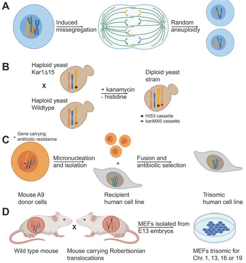
Figure 1. 1.
Figure Examples
Examplesof some
of ofsome
the aneuploidy model systems.
of the aneuploidy (A) Induced
model chromosome
systems. mis-se- chromosome mis-se-
(A) Induced
gregation by weaking the spindle assembly checkpoint using drug treatment or RNAi generates
gregation by weaking
cells with random the(B)
aneuploidies. spindle
Disomicassembly checkpoint
yeast cells can be generatedusing drugwild
by mating treatment
type or RNAi generates
yeast with
cells withyeast carrying
random a KAR1 mutation.
aneuploidies. (B)Lack of KAR1
Disomic interferes
yeast cells with
can nuclear fusion and
be generated byocca-
mating wild type yeast
sionally leads to random chromosome transfer and aneuploidy. Different antibiotic selection
with yeast carrying a KAR1 mutation. Lack of KAR1 interferes with nuclear fusion and occasionally
markers on the chromosomes allow for the selection of disomic cells in a haploid background. (C)
leads to random chromosome
Micronuclei-mediated transfer
chromosome transfer and
can be usedaneuploidy. Different
to generate human antibiotic
aneuploid cells. For selection markers on
this, chromosomes
the mouse donor cellsallow
carryingforanthe
additional human
selection chromosome
of disomic undergo
cells micronucleation
in a haploid background. (C) Micronuclei-
mediated chromosome transfer can be used to generate human aneuploid cells. For this, mouse donor
cells carrying an additional human chromosome undergo micronucleation upon prolonged colcemid
treatment and subsequent cell division. Isolated micronuclei carrying one or more chromosomes
are fused with human acceptor cells. Aneuploid cells are selected for using a selection marker
on the aneuploid chromosome. (D) Mating of wild type mice with mice carrying a Robertsonian
translocation produces embryos with aneuploid karyotypes. Harvesting of cells from E12.5 embryos
from these crosses allowed for the generation of trisomic MEFs.Cells 2021, 10, 342 3 of 16
2.1. Aneuploid Yeast Models
The budding yeast Saccharomyces cerevisiae has been widely used to study aneuploidy
and its cellular effects (Figure 1B). To create aneuploid yeast strains in a haploid back-
ground (i.e., disomes), a KAR1 deficient yeast strain was crossed with wildtype yeast to
prevent nuclear fusion [3]. During such matings, individual chromosomes are occasionally
transferred from one nucleus to the other, leading to aneuploid daughters, which were
selected for using selectable markers. This method yielded 20 yeast strains bearing one
or multiple extra copies for almost all of the yeast chromosomes, which were exploited to
assess the consequences of aneuploidy, as is further discussed below [3].
2.2. Stable Aneuploid Human Cell Lines
To study stable aneuploidy in human cell lines, mouse cells containing an ectopic
human chromosome with a selectable marker were exposed to prolonged colcemid treat-
ment and subsequently released to induce the formation of micronuclei [10–12] (Figure 1C).
Individual micronuclei were isolated and separated by centrifugation after which they
were fused with recipient human cells. Recipient cells with the extra human chromosome
were selected for using a selection cassette. This technique yielded various stable aneu-
ploid human cell lines including defined trisomic and even tetrasomic RPE1, HE35, and
colorectal cancer HCT116 and DLD1 cell lines, which allowed for the studying of effects of
aneuploidy using isogenic aneuploid and euploid cell lines [6,13].
2.3. Stable Aneuploid Mouse Embryonic Fibroblasts
As an alternative strategy to generating stable aneuploid mammalian cell lines, the
Amon lab generated primary mouse embryonic fibroblasts (MEFs) that carry one additional
chromosome (trisomic, Ts) [4] (Figure 1D). These MEF lines were generated by intercrossing
mice homozygous for two different Robertsonian translocations. Male offspring that
carried both translocations were then mated with wild-type mice resulting in a percentage
of the progeny with a trisomy for the chromosome common to the two Robertsonian
translocations. All trisomic embryo’s (except for Ts19 mice, which survived a few days
postnatally) died in utero as expected, but many embryos developed past embryonic day
12.5 allowing for MEF isolation. These efforts yielded MEFs trisomic for chromosome 1, 13,
16, and 19.
3. Models for Ongoing Chromosomal Instability
Most studies that identified aneuploidy-tolerating mechanisms made use of stable
aneuploid cell lines. Therefore, in these lines, the aneuploidy response is expected to mani-
fest homogeneously throughout the population. However, the karyotypes of aneuploid
cancer cells are often much more heterogeneous, a result of ongoing CIN. Therefore, to
understand how the aneuploidy paradox plays out in vivo, ensuing cells with a CIN phe-
notype probably is a more physiologically relevant approach. One way to induce ongoing
CIN is by alleviating the spindle assembly checkpoint (SAC). The SAC acts as a safeguard
mechanism in mitosis that coordinates chromosome attachment to the mitotic spindle. To
prevent CIN, the SAC halts cells in metaphase to prevent sister chromatid separation when
chromosome pairs are unattached or improperly attached [14,15]. Conversely, to provoke
a CIN phenotype, the SAC can be alleviated by the use of drugs that interfere with SAC
proteins such as the MPS1 inhibitors AZ3146 [16] and Reversine [17], or the Mad2 inhibitor
M2I-1 [18] by genetically alleviating SAC proteins such as Mps1, Mad2, or Bub1 [19–21].
Alternatively, CIN can for instance be induced by amplification of centrosomes [22–24], by
interfering with kinetochore structure [25], by disrupting the cohesin complex [26], or by
deregulating microtubule polymerization rates [27].
When comparing the findings between models for stable aneuploidy and ongoing CIN
(the latter extensively discussed elsewhere [28,29]), it appears that similar pathways are
deregulated. However, mapping the consequences for ongoing CIN is complicated by the
intrinsically heterogenous nature of the models. This heterogeneity complicates separatingCells 2021, 10, 342 4 of 16
the stresses triggered by the ongoing CIN and those triggered by the random aneuploidies
provoked by CIN. One solution to this problem is to engineer models for reversible CIN, for
instance, as previously done in Drosophila [30], which allows to uncouple the aneuploidy
and CIN response in vivo.
However, in this review, we mainly focus on the stress pathways identified in stable
aneuploid cell models, as is further discussed below.
4. Early Findings from Model Systems
An important first finding from stable aneuploidy model systems is that aneuploid
cells have a significant proliferative disadvantage compared to their euploid counter-
parts [3,4,6,13]. Furthermore, all aneuploid models showed strong metabolic changes and
induction of stress responses, indicating that aneuploidy decreases cellular fitness, and thus
is expected to suppress tumorigenesis. However, aneuploidy frequently occurs in human
cancer [7,8], a disease characterized by increased cell proliferation. These contradicting
observations are frequently referred to as the ‘aneuploidy paradox’ [9] and suggest that
aneuploid cells need to overcome certain barriers to become malignant. As overcoming
these stresses might be an important step during tumorigenesis, modulators of aneuploidy-
imposed stresses are considered attractive targets for therapeutic intervention [31]. Below,
we discuss some of these stresses and elaborate on how these findings have broadened our
knowledge on how cancer cells deal with aneuploidy.
4.1. Gene Expression Changes
Studies in various aneuploid yeast strains revealed that aneuploidy leads to deregu-
lation of the transcriptome [3], yielding a gene expression signature that is characteristic
of the yeast environmental stress response (ESR). This ESR signature is characterized by
an increased expression of ribosomal biogenesis genes and genes involved in nucleic acid
metabolism. Conversely, genes involved in carbohydrate metabolism displayed decreased
expression. A similar but not identical response was found in human cells when comparing
DNA, mRNA, and protein levels of euploid and aneuploid human cell lines [6]. For this
purpose, multiple human tri-and tetrasomic cell lines were transcriptionally profiled and
compared to aneuploid human cancer cell lines. This led to the identification of a general
transcriptional aneuploid response pattern (ARP) [13]. This ARP signature includes the
upregulation of genes involved in the endoplasmatic reticulum (ER) and Golgi-related
pathways, lysosome/lytic vacuoles, MHC protein complex, antigen processing, and a
downregulation of genes involved in DNA and RNA metabolism and ribosome-related
pathways. The ARP thus might point towards altered growth requirements of aneuploid
cells, which could represent a potential therapeutic target in aneuploid cancer treatment.
While this general ARP pattern seems to hold true for several aneuploid cell strains [13]
and is also observed in vivo (e.g., in aneuploid basal epidermal cells) [32], part of this ane-
uploidy signature is reverted in aneuploid cancers. For instance, while cultured aneuploid
cells appear to have decreased expression of ribosomal genes [13,32], expression these genes
is upregulated in a model for CIN-driven T-ALL [33]. Interestingly, as discussed above,
stable aneuploid yeast strains also exhibit increased expression of ribosome genes [3],
suggesting that yeast cell biology represents the biology of an in vivo cancer cell more than
the biology of mammalian cell lines, at least in its response to aneuploidy.
While aneuploidy clearly triggers expression changes and while there is a remarkable
correlation between gene/chromosome copy number changes and gene expression in stable
aneuploid cells [34], as well as in aneuploid cancers [33,35,36], there is also evidence that the
expression of a small fraction of aneuploid genes is somehow dose-compensated [36–38].
This suggests that some genes need to be epigenetically modulated upon aneuploidization,
potentially uncovering a targetable vulnerability of aneuploid cancer cells.Cells 2021, 10, 342 5 of 16
4.2. Proteotoxicity
As aneuploidy leads to increased gene expression and thus increased protein pro-
duction from the aneuploid chromosomes (in case of chromosome gains) [39], aneuploidy
disrupts the protein complex stoichiometry [6,40]. The resulting excess of ‘aneuploid’ pro-
teins causes proteotoxic stress, such as impaired protein folding [41], unscheduled protein
degradation [42,43] and increased protein aggregation [44]. Together, these processes are
believed to reduce cellular fitness of aneuploid cells [45].
Conversely, reducing proteotoxic stress levels was found to favor the survival of ane-
uploid cells. For instance, fast growing aneuploid yeast strains often harbor inactivating
mutations in UBP6 [46]. UBP6 deubiquitinates substrates at the proteasome, which leads
to recycling of ubiquitin and rescue of proteasome substrates from degradation. Thus,
the absence of UBP6 leads to an increased clearance of proteins, which is beneficial to
aneuploid cells. Together, these findings reveal that proteotoxic stress is an adverse effect
of aneuploidy and, moreover, that UBP6 mutations allow aneuploidy tolerance in yeast by
increasing proteasomal degradation. Conversely, the deletion of UBP3, encoding the deu-
biquitinase UBP3, exerts a significant negative fitness penalty to aneuploid yeast cells [47].
As UBP3 is required for efficient functioning of the ubiquitin-proteasome system, its dele-
tion impairs protein degradation, thereby further increasing proteotoxicity in aneuploid
yeast. This aneuploidy-related liability is conserved in human cells, as depletion of the
human homolog of UBP3 and USP10 also reduces the fitness of chromosomal instable
RPE1 cells [48].
Another cause for the observed proteotoxicity in aneuploid cells is a decrease in heat
shock protein (HSP) (i.e., 90-mediated protein folding), which ultimately leads to protein
aggregation in the cytoplasm [41,44]. Aneuploid yeast and human cells are therefore
more sensitive to HSP90 inhibiters such as 17-AAG [3,41,42]. Conversely, increasing heat
shock protein 1 (HSF1) levels rescues the effects of aneuploidy on HSP90 expression
and resulting proteotoxicity in human cells, thereby identifying HSF1 as an aneuploidy
tolerating gene [41].
Finally, autophagy, a process that removes surplus and damaged proteins and or-
ganelles, is upregulated in aneuploid cells [6]. Stable aneuploid human colon cancer
cells display increased numbers of LC3 foci, an autophagy marker [43], and upregulated
p62-dependent autophagy. Lysosomes play an essential role in autophagy, and protein
aggregates accumulate in the lysosomes of chromosomal instable cells. This triggers a lyso-
somal stress response that can be relieved by activating the transcription factor TFEB, which
increases expression of autophagy genes [49]. Thus, to cope with aneuploidy-imposed
proteotoxicity, aneuploid cells require an upregulation of autophagy, which potentially can
be exploited in the treatment of aneuploid cancers through inhibitors of autophagy such as
chloroquine [42] and bafilomycin A [13,50].
Taken together, aneuploid cells heavily rely on their protein quality control machin-
ery, which renders them more sensitive to compounds interfering in these processes. It
is therefore of the utmost importance to further investigate these potential targetable
vulnerabilities of aneuploid cancer cells.
4.3. Metabolic Stress
Besides proteotoxic stress, aneuploid cells also suffer from metabolic stress [3,4,42].
This is exemplified by the fact that aneuploid cells are much more sensitive to the energy
stress-inducing compound AICAR than their euploid counterparts [42]. Indeed, AICAR
exacerbates aneuploidy-imposed energy stress by activating AMP-activated protein kinase,
which results in the efficient killing of stable trisomic MEFs. Conversely, euploid MEFs
continue to proliferate when exposed to the same concentrations of AICAR.
Similar to AICAR, the glucosylceramide synthase inhibitor, DL-PDMP, also selectively
inhibits the proliferation of Ts13 MEFs [51]. DL-PDMP is a ceramide analogue that in-
hibits glucosylceramide synthase, thereby decreasing ceramide glycosylation. This causes
an accumulation of ceramides, which inhibits proliferation and promotes apoptosis ofCells 2021, 10, 342 6 of 16
aneuploid MEFs in highly aneuploid colorectal cancer cells rather than their euploid coun-
terparts. Aneuploid cells possibly express higher ceramide levels than euploid control cells,
thereby sensitizing them to compounds that further increase these ceramides. However,
inhibition of sphingolipid synthesis was also shown to impair the fitness of aneuploid
yeast [52], suggesting that aneuploid cells critically rely on perfect titration of this pathway
for their survival.
4.4. Inflammatory Response
Finally, while the above-described mechanisms mostly impinge on cell-intrinsic mech-
anisms, accumulating evidence reveals an important role for the immune system in control-
ling the propagation of aneuploid cells. For instance, tri- and tetrasomic RPE1, HCT116, and
DLD1 cell lines display upregulated interferon (IFN) signaling and increased expression
of proteins involved in MHC protein complex and antigen processing [13,53]. Similarly,
aneuploid RPE1 cells exhibit an upregulation of pro-inflammatory cytokines such as IL-6,
IL-8, and CCL2, which could promote immunosurveillance [54].
DNA is normally compartmentalized to the nucleus, but following chromosome
mis-segregation, chromosomes end up outside of the main nucleus in a smaller so-called
micronuclei [55]. When these micronuclei rupture, genomic DNA is released in the cy-
tosol, which activates the cytosolic nucleic acid sensor cyclic guanosine monophosphate
(GMP)-adenosine mononphosphate (AMP) synthase (cGAS) [55,56]. Activated cGAS then
stimulates type I interferon signaling through a stimulator of interferon genes (STING) and
the downstream transcription factor IRF3 ultimately leading to an inflammatory response
of the micro-nucleated cell [57]. The cGAS-STING thus plays a pivotal role in anti-tumor
immunity and STING signaling indeed appears to be altered in a variety of cancers [58,59].
Paradoxically, active STING was found to be crucial for the metastasis of tumors exhibiting
a CIN phenotype. It therefore requires further work to fully understand in which context
cGAS-STING signaling is tumor promoting or suppressive [60].
Most described models that study aneuploidy have looked into what allows aneu-
ploidy tolerance, focusing into what cellular changes or stresses are induced in cells by
aneuploidy. However, the majority of these efforts were educated guesses or small-scale
screens, particularly the studies in mammalian cells. A next important step forward is
therefore to search for the processes that transform aneuploid cells into aneuploid cancer
cells in a completely unbiased fashion, for instance through functional genetic in vivo
screens that systematically compare the drivers between aneuploid and euploid cancers.
Below, we describe which types of genetic screens can be used for cancer gene identifi-
cation and what their main advantages and disadvantages are (Table 1).
Table 1. The advantages and disadvantages of several mutagenesis systems.
Mutagenesis System Advantages Disadvantages
• Labor intensive positional cloning to
• Induces point mutations
identify mutated gene
• Unbiased disease gene discovery based
• Identification of recessive genes in vivo
on phenotyping
Chemical requires back- or inter-crossing; many
• Can be used in forward and reverse
mice required
genetic approaches
• Base pair substitution bias; some genes or
• In vitro and in vivo use
domains more frequently mutated
• Mostly identifies gain of function
• Rapid identification of mutated gene mutations
• Does not require generation of transgenic • (Most) cells must be dividing for
Retrovirus mice for in vivo screens retrovirus integration
• In vitro and in vivo use • Strain-specific effects and limitations
• Limited tissue flexibilityCells 2021, 10, 342 7 of 16
Table 1. Cont.
Mutagenesis System Advantages Disadvantages
• Genome-wide
• Requires generation of transgenic lines
• Loss and gain of function
• Insertion site preference leading to bias
• In vitro and in vivo use
• SB has tendency for local hopping, and
• Allows for the identification of multiple
leaves footprint behind. Note that these
Transposon cooperating mutations
disadvantages are not true for PB
• Can identify the effects of mutations in
transposons
non-coding regions of the genome
• Does not allow for identification of point
• Can be done in vivo in whole organism or
mutations
in tissue specific setup
• Only loss of function
• Genome-wide
• Off target effects
RNA interference • Stable
• Does not identify multiple cooperating
• In vitro and in vivo use
genetic mutations required for phenotype
• Genome-wide
• Can identify loss and gain of function
mutations (CRISPRi/CRISPRa) • Does not identify multiple cooperating
CRISPR-Cas9 • In vitro and in vivo use genetic mutations required for phenotype
• Can be done in vivo in whole organism or
in tissue specific setup
5. Genetic Screens to Identify Cancer-Collaborating Hits
5.1. Chemical Mutagenesis
One way to search for cancer genes is by randomly mutagenizing the genome of
cells and next search for cancer(-like) phenotypes. The alkylating chemical N-ethyl-N-
nitrosourea (ENU) is a potent agent to induce mutations in mice [61], and has been widely
used to find drivers for human disorders in mouse models [62]. For ENU mutagenesis
screens, male mice are treated with ENU and crossed with wild type females to produce
offspring that can be assayed for dominant mutations. Recessive mutations can be identified
by intercrossing or backcrossing pedigree [62]. All offspring are examined for the relevant
disease phenotype by studying their behavior, physiology, or dysmorphology of the mice,
after which the gene responsible for the observed phenotype in mice of interest can be
identified through laborious positional cloning [62].
ENU mutagenesis has been used to find drivers for many human disorders and to
better understand gene function and complex biological systems [62]. For instance, it
was exploited to find mutations that lead to resistance to the EGFR monoclonal antibody
Cetuximab in colorectal cancer patients [63] and provided new leads to treat Cetuximab
resistant tumors. In another example, ENU mutagenesis was used to identify double strand
break (DSB) repair genes in mice when screening for in vivo chromosome damage using
micronuclei as a readout [64]. This screen identified a recessive mutation that leads to
elevated levels of spontaneous and radiation- or mitomycin C-induced micronuclei.
As exemplified above, ENU mutagenesis can be applied to map pathways in bio-
logical systems as well as to better understand cancer susceptibility. However, a major
disadvantage is the large cohort of mice required for such screens and the laborious cloning
needed to identify the causative genes. When retroviral insertional mutagenesis screens
were introduced, these rapidly became an attractive alternative to ENU mutagenesis
screens, as mapping viral integration sites is much less laborious than positional cloning of
ENU-mediated mutations in the genomic DNA [65,66].Cells 2021, 10, 342 8 of 16
5.2. Retroviral Insertional Mutagenesis
Retroviruses consist of an RNA genome that replicates via a provirus (DNA intermedi-
ate). The provirus randomly integrates into the host genome, which results in mutagenesis
at the integration site [67]. Proviral integrations can lead to either activation of a gene, post-
transcriptional dysregulation, gene inactivation, or gene truncation when integrating near
to or in a gene [68]. This gene-disrupting feature was exploited for screening purposes as
the resulting deregulation of affected chromatin could lead to a growth advantage by either
activation of an oncogene or inactivation of a tumor-suppressor gene so that cells carrying
that integration would be enriched in the tumor cell population [69]. Viral integration sites
can easily be identified by cloning the provirus and adjacent cellular DNA from the tumor
DNA [67]. Retroviral insertional mutagenesis has also been extensively used to identify
cancer genes in mouse and human model systems.
Various retroviruses have been used for this purpose. For instance, the Moloney
murine leukemia virus (MoMuLV) was used to identify collaborating drivers in BXH2- and
AKXD-predisposed mice leading to BXH2 myeloid leukemia and AKXD lymphomas [70].
Subsequent mapping of hundreds of proviral insertions revealed several previously-
described collaborating drivers as common insertion sites (CIS) in these lymphomas and
myeloid leukemia’s, providing proof-of-principle, but, importantly, also new cancer driv-
ing genes [70]. Another MoMuLV screen to identify genes that can substitute for Pim1 and
Pim2 in lymphomagenesis in Myc transgenic, Pim1, and Pim2 doubly deficient mice [71],
uncovered several CIS (of which 10 belonged to the Pim complementation group), which
furthered our understanding of Pim1/2-driven lymphomagenesis. Similarly, a MoMuLV
screen performed in Cdkn2a-/- mice identified genes that collaborate with the tumor sup-
pressors p16INK4a and p19ARF encoded by the Cdkn2a locus [72]. Moreover, the CIS included
established cancer drivers and new ones with an enrichment for genes involved in MAPK
signaling. Also other tumor viruses can be exploited for insertional mutagenesis screens,
such as the mouse mammary tumor virus (MMTV), which led to the identification of
various mammary carcinoma genes [73].
In summary, retroviral insertional mutagenesis screens, also known as retroviral tag-
ging screens, can be used to identify genes driving cancer. However, a major disadvantage
is that these viruses display a selective cellular tropism, indicating that these viruses can
only infect certain tissue types [69]. Therefore, retroviral tagging screens were largely
replaced by transposon screens for which it was much easier to target different cell lineages
in vivo. This expedited the development many more specific cancer gene screening models.
5.3. Transposon Mutagenesis Screens
Transposons are mobile genetic elements that can be used to perform unbiased genetic
screens to identify driver genes in human cancers. The most commonly used transpo-
son types in in vivo genetic screens are Sleeping Beauty (SB) [74,75] and Piggyback (PB)
transposons [76–78]. A major advantage of PB compared to SB is that PB transposons do
not leave undesired footprint mutations behind after transposition, which significantly
simplifies the identification of the driver genes [79]. Both SB and PB make use of two
components: (1) a mutagenic gene trap that can either activate or disrupt gene expression
depending on where the transposon integrates [80,81] and (2) the enzyme transposase.
The transposase can be ubiquitously or tissue-specifically expressed, the latter permitting
tissue-specific mutagenesis. Similar to retroviral tagging, the genomic integration sites
are relatively easy to map, for instance using splinkerette-PCR followed by Sanger or
next-generation sequencing.
The first transposon screens used ubiquitously expressed SB transposase and identi-
fied genes that promote sarcomas and hematopoietic malignancies [80,81]. In later screens,
Cre-inducible SB or PB transposase was used to restrict transposon mutagenesis to specific
tissues such as B-cells, liver, and the gastrointestinal tract [82–84]. In addition, transpo-
son screens were employed to find drivers of many other tumor types including breast,
lung, prostate, thyroid, melanoma, and medulloblastoma (reviewed in [85–87]). Together,Cells 2021, 10, 342 9 of 16
these studies showed that transposon mutagenesis can drive tumorigenesis in wild type
or genetically-predisposed backgrounds. Examples include an SB screen performed in
p53-proficient and p53-deficient mice, which identified driver genes of osteosarcoma de-
velopment and metastasis [88]. Another SB screen performed in mice that carried various
colon cancer predisposing mutations [89] identified various new driver genes, which are
highly relevant for early and late cancer stages. Furthermore, an SB screen in Pten mutant
mice identified many new driver genes of triple-negative breast cancer [90]. Similarly, PB
mutagenesis was also successfully used to identify cancer genes in hematopoietic and solid
malignancies [78]. For instance, a PB screen in p19ARF deficient mice identified genes that
cause drug resistance against MDM2-TP53 inhibitors [91].
While we only highlighted a few examples of transposon screens here, it is clear that,
collectively, transposon screens have contributed tremendously to our understanding of
the process of tumorigenesis in vivo. However, it is important to note that transposon
screens randomly mutagenize the genome. There are also more targeted approaches to in-
activate genes, such as RNAi and clustered regularly interspaced short palindromic repeats
(CRISPR), that can also be exploited for genetic screens, as is further discussed below.
5.4. RNA interference Screens
RNA interference (RNAi) is an evolutionarily conserved mechanism among eukary-
otes functioning as a defense mechanism against double stranded-RNA (dsRNA) to target
cellular and viral mRNAs [92,93]. As RNA interference leads to degradation of the tar-
geted RNA molecule [94–97], this mechanism rapidly became exploited as a powerful
technology to silence gene products in vitro and in vivo. For this purpose, short interfering
RNAs (siRNA) are delivered into mammalian cells to induce transient but efficient and
specific down-regulation of target mRNAs [98]. Alternatively, to induce sustained gene-
silencing, short hairpin RNAs (shRNA) can be delivered through retroviral and lentiviral
vectors [99,100].
In addition to targeted downregulation of individual genes, RNAi technology can also
be applied in a high throughput manner. For this purpose, genome-wide shRNA-based
libraries were developed that allowed high-throughput RNAi screens in mammalian cells,
which can be performed as pooled screens or arrayed screens. In the case of arrayed screens,
individual wells with cells are transfected or transduced with individual RNAis/shRNAs
and screened for the assessed phenotype. In pooled screens, large cell populations are
transduced with shRNA libraries (whole genome or targeting specific gene families) at
once. Cells with a phenotype of choice are then selected/enriched for after which the
shRNAs driving the phenotype are identified by sequencing the integrated shRNAs. Such
pooled shRNA screens have been performed both in vitro and in vivo and mostly focused
on genes that contribute to tumorigenesis [100].
For example, one shRNA screen investigating chromatin-modifying enzymes in ag-
gressive castration-resistant prostate cancer showed that knockdown of histone H3K9
demethylase, KDM3B, has an antiproliferative effect in cell lines [101]. Another RNAi
screen revealed that the loss of FBW7 is synthetically lethal with an inactivated SAC in
colorectal cancer cell lines [102]. Importantly, shRNAi screens can also be used in vivo,
exemplified by an shRNA screen that identified over 10 candidate tumor suppressor genes
in a mouse lymphoma model [103].
While most shRNA screens were conducted in mammalian systems, shRNAi screens
are not restricted to mammalian cells. For instance, an RNAi screen in Drosophila used to
identify genes that are required for the viability of CIN cells, revealed that genes involved in
centrosomal and JNK signaling specifically induce apoptosis in cells with a CIN phenotype.
In this screen, Mad2 RNAi was used to alleviate the SAC and provoke CIN, which was
combined with individual RNAi’s targeting a set of kinase and phosphatase genes [104].
Some of the identified hits were, at the time of the screen, already being pursued as
cancer therapy targets, underscoring that such screens can identify drug targets of clinical
significance [105]. Another RNAi screen in cultured Drosophila S2 cells revealed that HSET,Cells 2021, 10, 342 10 of 16
normally a non-essential kinesin motor, is essential for the survival of cells with extra
centrosomes, uncovering HSET as a potentially very selective anti-cancer target for cancers
displaying centrosome amplification [106].
While RNA interference functional screens are still widely-used, they have slowly
begun to lose momentum to CRISPR/ CRISPR associated nuclease 9 (Cas9)-powered
functional genetic screens, as is further discussed below.
5.5. CRISPR-Cas9 Screens
Clustered regularly interspaced short palindromic repeats (CRISPR)/CRISPR associ-
ated nuclease 9 (Cas9) is a powerful genome editing tool that revolutionized the genome
editing field immediately after its discovery. The CRISPR-Cas system originates from
prokaryotic organisms (e.g., bacteria, archaea) in which it provides acquired resistance
against foreign genetic elements such as bacteriophages [107]. Cas9 is a large gene encod-
ing for a single-effector protein that can cleave DNA [108]. The Cas9 protein is guided to
specific DNA sequences through a duplex of a CRISPR RNA (crRNA) and trans-activating
RNA (tracrRNA) [109] and then cleaves the targeted DNA at the so-called protospacer
adjacent motif (PAM) [108]. This prokaryotic immune system was adapted to target DNA
in mammalian cells by combining the crRNA and tracrRNA into one single guide RNA
(sgRNA) construct [110]. This allows for the targeting of Cas9 to any DNA sequence of
choice [110], which then leads to a double-stranded break (DSB) at the targeted DNA
sequence, opening up a wide range of possibilities in the field of genome editing and gene
therapy [111]. Further adaptations to the CRISPR/Cas9 system led to new CRISPR applica-
tions. For instance, a nuclease dead mutant of Cas9 (dCas9) that maintains sgRNA directed
binding of specific DNA sequences, was fused to a transcriptional repressor domain to
yield a Cas9 protein that can inhibit transcription (CRISPR interference or CRISPRi) [112]
or activate transcription when Cas9 is fused to a transcriptional activator domain [113]
(CRISPR activation or CRISPRa). ‘Conventional’ CRISPR, as well as CRISPRi/CRISPRa
technologies, can be exploited for genome-wide or targeted functional genetic screens, for
which many guide RNA libraries (synthetic guide RNA and viral vectors) are available in
both academic and commercial domains.
Indeed, many libraries have been used to identify tumor suppressor genes and onco-
genes [114], as have drug targets and drug resistance mechanisms [115]. For instance, one
recent CRISPR screen in triple negative breast cancer cells showed that inactivation of
genes involved in the anaphase-promoting complex, such as ANAPC4, ANAPC13, and
MAD2L1BP, confers resistance towards Mps1 inhibitors [116]. In another example, a non-
metastatic mouse cancer cell line was transduced with a genome-wide guide RNA library
encompassing 70,000 sgRNAs. The CRISPR polyclonal cell pool was transplanted into
immunocompromised mice and recipient mice were screened for metastatic cancer. Lung
metastases and late-stage primary tumors were enriched for sgRNAs targeting a small
set of genes that included genes involved in pro-apoptotic pathways such as Bid, Pten,
Cdk2na, and Mgmt, suggesting that inactivation of apoptosis drives tumor growth and
metastasis [117].
Together, these screening systems have contributed enormously to our understanding
of organismal and cell biology, as well as the biology of cancer cells. While some screening
systems have lost popularity over newer and more sophisticated screening systems, each
screen type comes with its advantages and disadvantages, as summarized in Table 1.
6. Conclusions and Future Perspectives
In this review we discussed how reverse genetic approaches and small-scale forward
genetics screens in aneuploidy models have been used to determine how cells adapt to
aneuploidy. Aneuploidy is mostly detrimental for cells and initially leads to a proliferative
disadvantage, presumably due to the activation of aneuploidy-imposed stress pathways. It
is therefore likely that aneuploid cells, throughout their malignant transformation process,
need to overcome these stresses. Therefore, the molecular mechanisms underpinning theseCells 2021, 10, 342 11 of 16
aneuploidy-induced stresses are considered to be promising therapeutic targets. The work
of many labs in the last 15 years has significantly improved our understanding of some
of the roadblocks that aneuploid cells need to overcome during tumorigenesis. However,
to our knowledge, no large-scale screens have been reported that systematically compare
the pathways affected in aneuploid cancers to the those affected in euploid cancers. When
performed in an isogenic setting, such screens would surely reveal the differences between
euploid and aneuploid cells on their route to a malignant program.
In this review, we discussed five types of mutagenesis screens that could be suitable for
this goal, each with their own advantages and disadvantages (Table 1). ENU mutagenesis
could be very effective in screening for point mutations that would accelerate the transfor-
mation of aneuploid cells. However, identifying the individual mutations that drive the
phenotype is extremely laborious and many mice would be needed when such a screen
would be performed in vivo. Retroviral mutagenesis allows for rapid identification of the
mutated gene that improves the survival of aneuploid cells. However, these screens only
sample proliferative tissues as the virus will only integrate in dividing cells. Because of this
important limitation, retroviral tagging screens have mostly been surpassed by transposon,
RNAi, and CRISPR screens. Indeed, transposon mutagenesis can be induced in any cell
type within the whole organism, using a ubiquitously expressed transposase or in individ-
ual tissues with a conditional transposase controlled by a tissue-specific Cre-recombinase.
Transposon mutagenesis furthermore allows for the identification of multiple collaborating
driver mutations, which more accurately reflects the complexity of human cancer than a
single mutation. However, transposons do display some insertion site preference, which
yields to some bias in the screened part of the genome. This problem was largely overcome
with the introduction of PiggyBac transposons, which suffer less from ‘local hopping’ and
thus target the whole genome more efficiently [77]. In CRISPR/Cas9 and RNAi interfer-
ence screens, such bias can be eliminated by careful sgRNA/shRNA/RNAi library design.
RNAi have lost some popularity at the benefit of CRISPR screens, as CRISPR screens
completely inactivate the targeted genes instead of (partially) knocking gene expression
down and display fewer off-target effects. Moreover, CRISPR genome engineering offers
many more applications, such as knockdown, knockout, knock-in, activation, and base
editing [118,119], all of which can be exploited in genetic screens.
Altogether, to identify in an unbiased fashion the changes needed to convert an
aneuploid cell into a cancer cell, one would need to setup an in vivo screen that would
compare tumorigenesis in an euploid and aneuploid background. As stable aneuploidy is
probably not sufficient to accelerate cancer in mice, the aneuploid background would need
to be generated by crossing the ‘screening mice’ into a well-characterized CIN-predisposed
background. This would likely work well as in many mouse models for CIN-driven
cancer, CIN alone is not a powerful driver of cancer, but rather an accelerator [120,121].
Given that ongoing CIN is incompatible with early embryonic development [120], the
most suitable CIN predisposition would be a conditional CIN-driving allele that does
not efficiently promote cancer by itself. This could for instance be a Mps1 truncation or
mutation allele [33,122], a Mad2 deletion allele [32], a hypomorphic BubR1 allele [123], or a
Plk4 overexpression allele [24], as well as any other tissue-specific CIN driver. Indeed, in
most of the CIN models, the CIN-driving allele alone leads to aneuploidy but not to rapid
tumorigenesis. However, combining CIN with a single mutation in p53 not only leads
to cancer initiation [32,33,35,122–124] but also to a significant reduction of tumor latency,
which makes this setup very suitable for a mutagenesis screen.
Altogether, we conclude that genome-wide mutagenesis screens in a CIN-predisposed
background will likely yield important steps forward in the identification of more mecha-
nisms of aneuploidy tolerance in vivo.
Author Contributions: This review was written by L.J.J. and F.F. with help of L.Z. Literature research
was performed by L.J.J. and L.Z. All authors have read and agreed to the published version of
the manuscript.Cells 2021, 10, 342 12 of 16
Funding: This work was funded by Dutch Cancer Society Grant 2015-RUG-7833 to F.F. and a Chinese
Scholarship Council (CSC) fellowship to L.Z.
Institutional Review Board Statement: Not applicable.
Informed Consent Statement: Not applicable.
Conflicts of Interest: The authors declare no conflict of interest.
References
1. Schukken, K.M.; Foijer, F. CIN and Aneuploidy: Different Concepts, Different Consequences. BioEssays 2017. [CrossRef] [PubMed]
2. Hassold, T.; Abruzzo, M.; Adkins, K.; Griffin, D.; Merrill, M.; Millie, E.; Saker, D.; Shen, J.; Zaragoza, M. Human aneuploidy:
Incidence, origin and etiology. Environ. Mol. Mutagen. 1996, 28, 167–175. [CrossRef]
3. Torres, E.M.; Sokolsky, T.; Tucker, C.M.; Chan, L.Y.; Boselli, M.; Dunham, M.J.; Amon, A. Effects of aneuploidy on cellular
physiology and cell division in haploid yeast. Science 2007, 317, 916–924. [CrossRef]
4. Williams, B.R.; Prabhu, V.R.; Hunter, K.E.; Glazier, C.M.; Whittaker, C.A.; Housman, D.E.; Amon, A. Aneuploidy affects
proliferation and spontaneous immortalization in mammalian cells. Science 2008, 322, 703–709. [CrossRef]
5. Sheltzer, J.M.; Ko, J.H.; Replogle, J.M.; Passerini, V.; Storchova, Z.; Amon, A. Single-chromosome Gains Commonly Function as
Tumor Suppressors. Cancer Cell 2017, 31, 240–255. [CrossRef] [PubMed]
6. Stingele, S.; Stoehr, G.; Peplowska, K.; Cox, J.; Mann, M.; Storchova, Z. Global analysis of genome, transcriptome and proteome
reveals the response to aneuploidy in human cells. Mol. Syst. Biol. 2012, 8. [CrossRef] [PubMed]
7. Hanahan, D.; Weinberg, R.A. The Hallmarks of Cancer. Cell 2000, 100, 57–70. [CrossRef]
8. Taylor, A.M.; Shih, J.; Ha, G.; Gao, G.F.; Zhang, X.; Berger, A.C.; Schumacher, S.E.; Wang, C.; Hu, H.; Liu, J.; et al. Genomic and
Functional Approaches to Understanding Cancer Aneuploidy. Cancer Cell 2018. [CrossRef]
9. Sheltzer, J.M.; Amon, A. The aneuploidy paradox: Costs and benefits of an incorrect karyotype. Trends Genet. 2011, 27, 446–453.
[CrossRef]
10. Fournier, R.E.K. A general high-efficiency procedure for production of microcell hybrids. Proc. Natl. Acad. Sci. USA 1981, 78,
6349–6353. [CrossRef] [PubMed]
11. Saxon, P.J.; Srivatsan, E.S.; Stanbridge, E.J. Introduction of human chromosome 11 via microcell transfer controls tumorigenic
expression of HeLa cells. EMBO J. 1986, 5, 3461–3466. [CrossRef] [PubMed]
12. Koi, M.; Morita, H.; Yamada, H.; Satoh, H.; Barrett, J.C.; Oshimura, M. Normal human chromosome 11 suppresses tumorigenicity
of human cervical tumor cell line SiHa. Mol. Carcinog. 1989, 2, 12–21. [CrossRef]
13. Dürrbaum, M.; Kuznetsova, A.Y.; Passerini, V.; Stingele, S.; Stoehr, G.; Storchová, Z. Unique features of the transcriptional
response to model aneuploidy in human cells. BMC Genom. 2014, 15, 1–14. [CrossRef]
14. Musacchio, A. The Molecular Biology of Spindle Assembly Checkpoint Signaling Dynamics. Curr. Biol. 2015, 25, R1002–R1018.
[CrossRef] [PubMed]
15. Dou, Z.; Prifti, D.; Gui, P.; Liu, X.; Elowe, S.; Yao, X. Recent Progress on the Localization of the Spindle Assembly Checkpoint
Machinery to Kinetochores. Cells 2019, 8, 278. [CrossRef]
16. Hewitt, L.; Tighe, A.; Santaguida, S.; White, A.M.; Jones, C.D.; Musacchio, A.; Green, S.; Taylor, S.S. Sustained Mps1 activity is
required in mitosis to recruit O-Mad2 to the Mad1-C-Mad2 core complex. J. Cell Biol. 2010, 190, 25–34. [CrossRef]
17. Santaguida, S.; Tighe, A.; D’Alise, A.M.; Taylor, S.S.; Musacchio, A. Dissecting the role of MPS1 in chromosome biorientation and
the spindle checkpoint through the small molecule inhibitor reversine. J. Cell Biol. 2010, 190, 73–87. [CrossRef] [PubMed]
18. Kastl, J.; Braun, J.; Prestel, A.; Möller, H.M.; Huhn, T.; Mayer, T.U. Mad2 Inhibitor-1 (M2I-1): A Small Molecule Protein-Protein
Interaction Inhibitor Targeting the Mitotic Spindle Assembly Checkpoint. ACS Chem. Biol. 2015, 10, 1661–1666. [CrossRef]
19. Resende, L.P.; Monteiro, A.; Brás, R.; Lopes, T.; Sunkel, C.E. Aneuploidy in intestinal stem cells promotes gut dysplasia in
Drosophila. J. Cell Biol. 2018, 217, 3930–3946. [CrossRef] [PubMed]
20. Daniel, J.; Coulter, J.; Woo, J.H.; Wilsbach, K.; Gabrielson, E. High levels of the Mps1 checkpoint protein are protective of
aneuploidy in breast cancer cells. Proc. Natl. Acad. Sci. USA 2011, 108, 5384–5389. [CrossRef]
21. Shi, Q.; Hu, M.; Luo, M.; Liu, Q.; Jiang, F.; Zhang, Y.; Wang, S.; Yan, C.; Weng, Y. Reduced expression of Mad2 and Bub1 proteins
is associated with spontaneous miscarriages. Mol. Hum. Reprod. 2011, 17, 14–21. [CrossRef] [PubMed]
22. Basto, R.; Brunk, K.; Vinadogrova, T.; Peel, N.; Franz, A.; Khodjakov, A.; Raff, J.W. Centrosome Amplification Can Initiate
Tumorigenesis in Flies. Cell 2008, 133, 1032–1042. [CrossRef] [PubMed]
23. Gogendeau, D.; Siudeja, K.; Gambarotto, D.; Pennetier, C.; Bardin, A.J.; Basto, R. Aneuploidy causes premature differentiation of
neural and intestinal stem cells. Nat. Commun. 2015, 6, 1–15. [CrossRef]
24. Levine, M.S.M.S.; Bakker, B.; Boeckx, B.; Moyett, J.; Lu, J.; Vitre, B.; Spierings, D.C.D.C.; Lansdorp, P.M.P.M.; Cleveland, D.W.D.W.;
Lambrechts, D.; et al. Centrosome Amplification Is Sufficient to Promote Spontaneous Tumorigenesis in Mammals. Dev. Cell
2017, 40, 313–322.e5. [CrossRef]
25. Bakhoum, S.F.; Genovese, G.; Compton, D. a Deviant kinetochore microtubule dynamics underlie chromosomal instability. Curr.
Biol. 2009, 19, 1937–1942. [CrossRef]
26. Pezic, D.; Weeks, S.L.; Hadjur, S. More to cohesin than meets the eye: Complex diversity for fine-tuning of function. Curr. Opin.
Genet. Dev. 2017, 43, 93–100. [CrossRef]Cells 2021, 10, 342 13 of 16
27. Schukken, K.M.; Lin, Y.-C.; Bakker, P.; Schubert, M.; Preuss, S.F.; Simon, J.E.; Van den Bos, H.; Storchova, Z.; Colome-Tatche, M.;
Bastians, H.; et al. Altering microtubule dynamics is synergistically toxic with spindle assembly checkpoint inhibition. Life Sci.
Alliance 2020, 3, 1–15. [CrossRef]
28. Tijhuis, A.E.; Johnson, S.C.; McClelland, S.E. The emerging links between chromosomal instability (CIN), metastasis, inflammation
and tumour immunity. Mol. Cytogenet. 2019. [CrossRef] [PubMed]
29. Ye, C.J.; Sharpe, Z.; Heng, H.H. Origins and consequences of chromosomal instability: From cellular adaptation to genome
chaos-mediated system survival. Genes 2020, 1162. [CrossRef] [PubMed]
30. Mirkovic, M.; Guilgur, L.G.; Tavares, A.; Passagem-Santos, D.; Oliveira, R.A. Induced aneuploidy in neural stem cells triggers a
delayed stress response and impairs adult life span in flies. PLoS Biol. 2019, 17, e3000016. [CrossRef]
31. Zhou, L.; Jilderda, L.J.; Foijer, F. Exploiting aneuploidy-imposed stresses and coping mechanisms to battle cancer. Open Biol. 2020,
10. [CrossRef]
32. Foijer, F.; DiTommaso, T.; Donati, G.; Hautaviita, K.; Xie, S.Z.; Heath, E.; Smyth, I.; Watt, F.M.; Sorger, P.K.; Bradley, A. Spindle
checkpoint deficiency is tolerated by murine epidermal cells but not hair follicle stem cells. Proc. Natl. Acad. Sci. USA 2013, 110,
2928–2933. [CrossRef]
33. Foijer, F.; Xie, S.Z.; Simon, J.E.; Bakker, P.L.; Conte, N.; Davis, S.H.; Kregel, E.; Jonkers, J.; Bradley, A.; Sorger, P.K. Chromosome
instability induced by Mps1 and p53 mutation generates aggressive lymphomas exhibiting aneuploidy-induced stress. Proc. Natl.
Acad. Sci. USA 2014, 111, 13427–13432. [CrossRef]
34. Mao, R.; Zielke, C.L.; Ronald Zielke, H.; Pevsner, J. Global up-regulation of chromosome 21 gene expression in the developing
down syndrome brain. Genomics 2003, 81, 457–467. [CrossRef]
35. Foijer, F.; Albacker, L.A.; Bakker, B.; Spierings, D.C.; Yue, Y.; Xie, S.Z.; Davis, S.; Lutum-Jehle, A.; Takemoto, D.; Hare, B.;
et al. Deletion of the MAD2L1 spindle assembly checkpoint gene is tolerated in mouse models of acute T-cell lymphoma and
hepatocellular carcinoma. Elife 2017, 6. [CrossRef]
36. Kahlem, P. Transcript Level Alterations Reflect Gene Dosage Effects Across Multiple Tissues in a Mouse Model of Down Syndrome.
Genome Res. 2004, 14, 1258–1267. [CrossRef]
37. Lyle, R. Gene Expression From the Aneuploid Chromosome in a Trisomy Mouse Model of Down Syndrome. Genome Res. 2004,
14, 1268–1274. [CrossRef] [PubMed]
38. Wang, C.C.; Kazuki, Y.; Oshimura, M.; Ikeo, K.; Gojobori, T. Gene dosage imbalance of human chromosome 21 in mouse
embryonic stem cells differentiating to neurons. Gene 2011, 481, 93–101. [CrossRef] [PubMed]
39. Pavelka, N.; Rancati, G.; Zhu, J.; Bradford, W.D.; Saraf, A.; Florens, L.; Sanderson, B.W.; Hattem, G.L.; Li, R. Aneuploidy confers
quantitative proteome changes and phenotypic variation in budding yeast. Nature 2010, 468, 321–325. [CrossRef] [PubMed]
40. Brennan, C.M.; Vaites, L.P.; Wells, J.N.; Santaguida, S.; Paulo, J.A.; Storchova, Z.; Harper, J.W.; Marsh, J.A.; Amon, A. Protein
aggregation mediates stoichiometry of protein complexes in aneuploid cells. Genes Dev. 2019, 33, 1031–1047. [CrossRef]
41. Donnelly, N.; Passerini, V.; Dürrbaum, M.; Stingele, S.; Storchová, Z. HSF 1 deficiency and impaired HSP 90-dependent protein
folding are hallmarks of aneuploid human cells. EMBO J. 2014. [CrossRef]
42. Tang, Y.-C.; Williams, B.R.; Siegel, J.J.; Amon, A. Identification of aneuploidy-selective antiproliferation compounds. Cell 2011,
144, 499–512. [CrossRef]
43. Stingele, S.; Stoehr, G.; Storchova, Z. Activation of autophagy in cells with abnormal karyotype. Autophagy 2013, 9, 246–248.
[CrossRef]
44. Oromendia, A.B.; Dodgson, S.E.; Amon, A. Aneuploidy causes proteotoxic stress in yeast. Genes Dev. 2012, 26, 2696–2708.
[CrossRef]
45. Oromendia, A.B.; Amon, A. Aneuploidy: Implications for protein homeostasis and disease. DMM Dis. Model. Mech. 2014, 7,
15–20. [CrossRef] [PubMed]
46. Torres, E.M.; Dephoure, N.; Panneerselvam, A.; Tucker, C.M.; Whittaker, C.A.; Gygi, S.P.; Dunham, M.J.; Amon, A. Identification
of aneuploidy-tolerating mutations. Cell 2010. [CrossRef]
47. Dodgson, S.E.; Kim, S.; Costanzo, M.; Baryshnikova, A.; Morse, D.L.; Kaiser, C.A.; Boone, C.; Amon, A. Chromosome-specific and
global effects of aneuploidy in Saccharomyces cerevisiae. Genetics 2016, 202, 1395–1409. [CrossRef] [PubMed]
48. Dodgson, S.E.; Santaguida, S.; Kim, S.; Sheltzer, J.; Amon, A. The pleiotropic deubiquitinase ubp3 confers aneuploidy tolerance.
Genes Dev. 2016. [CrossRef] [PubMed]
49. Santaguida, S.; Vasile, E.; White, E.; Amon, A. Aneuploidy-induced cellular stresses limit autophagic degradation. Genes Dev.
2015, 29, 2010–2021. [CrossRef]
50. Cai, Y.; Crowther, J.; Pastor, T.; Abbasi Asbagh, L.; Baietti, M.F.; De Troyer, M.; Vazquez, I.; Talebi, A.; Renzi, F.; Dehairs, J.; et al.
Loss of Chromosome 8p Governs Tumor Progression and Drug Response by Altering Lipid Metabolism. Cancer Cell 2016, 29,
751–766. [CrossRef]
51. Tang, Y.C.; Yuwen, H.; Wang, K.; Bruno, P.M.; Bullock, K.; Deik, A.; Santaguida, S.; Trakala, M.; Pfau, S.J.; Zhong, N.; et al.
Aneuploid cell survival relies upon sphingolipid homeostasis. Cancer Res. 2017, 77, 5272–5286. [CrossRef] [PubMed]
52. Hwang, S.; Gustafsson, H.T.; O’Sullivan, C.; Bisceglia, G.; Huang, X.; Klose, C.; Schevchenko, A.; Dickson, R.C.; Cavaliere, P.;
Dephoure, N.; et al. Serine-Dependent Sphingolipid Synthesis Is a Metabolic Liability of Aneuploid Cells. Cell Rep. 2017, 21,
3807–3818. [CrossRef]You can also read