Expansion of CD10neg neutrophils and CD14+HLA-DRneg/low monocytes driving proinflammatory responses in patients with acute myocardial infarction ...
←
→
Page content transcription
If your browser does not render page correctly, please read the page content below
RESEARCH ARTICLE

                                     Expansion of CD10neg neutrophils and
                                     CD14+HLA-DRneg/low monocytes driving
                                     proinflammatory responses in patients
                                     with acute myocardial infarction
                                     Daniela Fraccarollo*, Jonas Neuser, Julian Möller, Christian Riehle,
                                     Paolo Galuppo, Johann Bauersachs

                                     Department of Cardiology and Angiology, Hannover Medical School, Hannover,
                                     Germany

                                     Abstract Immature neutrophils and HLA-DRneg/low monocytes expand in cancer, autoimmune
                                     diseases and viral infections, but their appearance and immunoregulatory effects on T-cells after
                                     acute myocardial infarction (AMI) remain underexplored. We found an expansion of circulating
                                     immature CD16+CD66b+CD10neg neutrophils and CD14+HLA-DRneg/low monocytes in AMI patients,
                                     correlating with cardiac damage, function and levels of immune-inflammation markers. Immature
                                     CD10neg neutrophils expressed high amounts of MMP-9 and S100A9, and displayed resistance to
                                     apoptosis. Moreover, we found that increased frequency of CD10neg neutrophils and elevated
                                     circulating IFN-g levels were linked, mainly in patients with expanded CD4+CD28null T-cells.
                                     Notably, the expansion of circulating CD4+CD28null T-cells was associated with cytomegalovirus
                                     (CMV) seropositivity. Using bioinformatic tools, we identified a tight relationship among the
                                     peripheral expansion of immature CD10neg neutrophils, CMV IgG titers, and circulating levels of
                                     IFN-g and IL-12 in patients with AMI. At a mechanistic level, CD10neg neutrophils enhanced IFN-g
                                     production by CD4+ T-cells through a contact-independent mechanism involving IL-12. In vitro
*For correspondence:                 experiments also highlighted that HLA-DRneg/low monocytes do not suppress T-cell proliferation but
fraccarollo.daniela@mh-hannover.     secrete high levels of pro-inflammatory cytokines after differentiation to macrophages and IFN-g
de                                   stimulation. Lastly, using a mouse model of AMI, we showed that immature neutrophils
Competing interests: The             (CD11bposLy6GposCD101neg cells) are recruited to the injured myocardium and migrate to
authors declare that no              mediastinal lymph nodes shortly after reperfusion. In conclusion, immunoregulatory functions of
competing interests exist.           CD10neg neutrophils play a dynamic role in mechanisms linking myeloid cell compartment
Funding: See page 23                 dysregulation, Th1-type immune responses and inflammation after AMI.

Preprinted: 21 September 2020
Received: 22 January 2021
Accepted: 11 July 2021
Published: 22 July 2021              Introduction
                                     Despite advances in interventional therapies patients with large acute myocardial infarction (AMI)
Reviewing editor: Noriaki
Emoto, Kobe Pharmaceutical
                                     are at higher risk of heart failure morbidity and mortality (Heusch and Gersh, 2017). Immunity and
University, Japan                    inflammation play a key role in the pathogenesis of ischemic heart failure, and the complex role of
                                     immune cells during the wound healing process after injury is currently the focus of intensive
   Copyright Fraccarollo et al.
                                     research efforts. Understanding the immune mechanisms operating during AMI could pave the way
This article is distributed under
                                     to develop more effective strategies to prevent progressive dilative cardiac remodeling, functional
the terms of the Creative
Commons Attribution License,         deterioration and heart failure and to reduce adverse cardiovascular events.
which permits unrestricted use           HLA-DRneg/low monocytes and immature neutrophils expand in pathological conditions such as
and redistribution provided that     cancer, infection, and inflammation, (Gabrilovich and Nagaraj, 2009) and have recently been impli-
the original author and source are   cated in the pathogenesis of severe COVID-19 (Silvin et al., 2020; Deutsche COVID-19 OMICS Ini-
credited.                            tiative (DeCOI) et al., 2020). A rapid depression of monocytic HLA-DR expression was observed in

Fraccarollo et al. eLife 2021;10:e66808. DOI: https://doi.org/10.7554/eLife.66808                                                1 of 31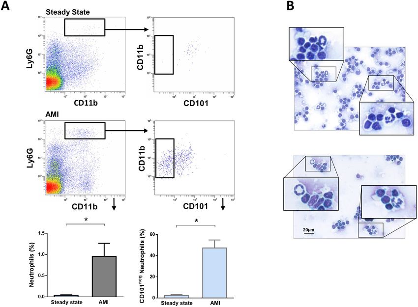
Research article                                                                               Immunology and Inflammation Medicine

                                        patients with AMI (Haeusler et al., 2012). Maréchal et al., 2020 studying neutrophil phenotypes in
                                        acute coronary syndrome found increased percentage of immature low density neutrophils in
                                        patients with ST-elevation MI. However, the role of immature neutrophils as well as HLA-DRneg/low
                                        monocytes in immune mechanisms operating during AMI remains largely unexplored.
                                           By integrating flow cytometric immunophenotyping of monocyte, neutrophil, and lymphocyte
                                        subsets, ex vivo experiments with sorted cells as well as bioinformatic tools this study investigated
                                        the appearance and the functional immune properties of immature neutrophils and HLA-DRneg/low
                                        monocytes in patients with AMI. Moreover, we explored whether increased frequency of immature
                                        neutrophils and HLA-DRneg/low monocytes are linked to circulating levels of immune regulators and
                                        acute inflammation markers such as G-CSF, S100A9/S100A8, MMP-9, NGAL, MPO, IL-6, TNF-a, IL-
                                        1ß and IFN-g.
                                           Since a crucial role for immature neutrophils in the orchestration of adaptive immunity has
                                        recently emerged, we mostly focused on neutrophil-mediated regulation of T-cell response. Environ-
                                        mental factors especially cytomegalovirus (CMV) infection are likely to provide a significant contribu-
                                        tion to functional diversity of T-cells. Therefore, we also investigated the impact of CMV on CD4+
                                        T-cell homeostasis and on the immunoregulatory properties of immature neutrophils derived from
                                        patients with AMI.
                                           In addition, using a mouse model of reperfused AMI we addressed whether immature neutrophils
                                        migrate into the injured myocardium and populate heart-draining lymph nodes in response to acute
                                        ischemic injury.

                                        Methods
                                        Patients and study design
                                        The study protocol is in accordance with the ethical guidelines of the 1975 declaration of Helsinki
                                        and has been approved by the local ethics committee of Hannover Medical School. Patients referred
                                        to our department for acute coronary syndrome (ACS) were included after providing written
                                        informed consent. Patients suffering from active malignant diseases or receiving immunosuppressive
                                        therapy were not included. Seventy-one patients (Table 1) were categorized into unstable angina
                                        (UA, n=11), Non-ST-elevation MI (NSTEMI, n=16), and ST-elevation MI (STEMI, n=44). Left ventricu-
                                        lar (LV) ejection fraction was measured in 2D echocardiographic studies using bi-plane Simpson’s
                                        method. Seventeen healthy volunteers were recruited as control subjects.

Table 1. General traits.
                                                                   UA (N=11)                 NSTEMI (N=16)                    STEMI (N=44)
Age (years)                                                        63.3±2.5                  64.9±3.4                         60.6±1.7
Gender                           Male/Female                       9/2                       14/2                             36/8
BMI (kg/m2)                                                        27.5±0.9                  28.2±0.9                         27.5±0.7
Blood analyses                   LDL (mg/dL)                       92.2±19.2                 95.5±9.5                         138.2±7.3
                                 CK (IU/L)                         120.0 (87.0–444.0)        189.0 (126.8–377.0)              373.5 (110.5–931.2)
                                 CKmax (IU/L)                      120.0 (86.5–440.0)        403.5 (150.5–578.2)              1343.5 (574.8–1917.0)
                                 CK-MB (IU/L)                      19.0 (17.0–22.0)          32.0 (23.5–57.0)                 47.0 (24.5–91.5)
                                 LVEF (%)                          53.3±3.7                  54.1±2.2                         51.0±1.5
                                 Troponin (ng/L)                   12.8 (5.3–22.7)           99.0 (36.7–273.5)                337.0 (84.0–962.0)
                                 Creatinine (mmol/L)               83.0±4.5                  88.6±4.8                         98.5±8.3
                                 CRP (mg/L)                        1.6 (0.7–3.4)             1.8 (1.1–4.3)                    2.5 (1.2–4.7)

Data are presented as mean ± SEM or as median (IQR). LDL, low density lipoprotein; CK, creatine kinase; CKmax, maximum CK; CK-MB, creatine kinase-
myocardial band; LVEF, left ventricular ejection fraction; CRP, C-reactive protein.

Fraccarollo et al. eLife 2021;10:e66808. DOI: https://doi.org/10.7554/eLife.66808                                                             2 of 31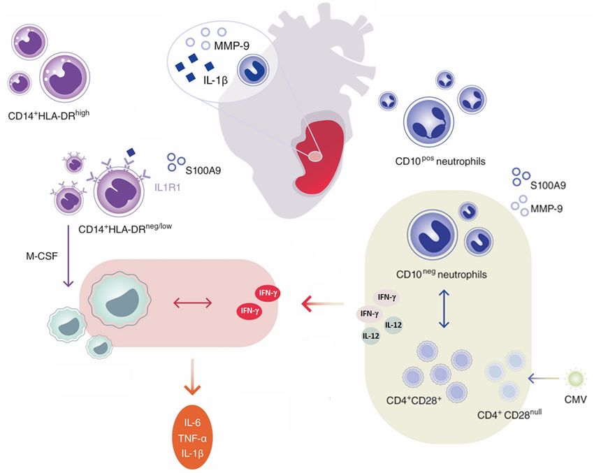
Research article                                                                          Immunology and Inflammation Medicine

                                    Flow cytometry
                                    Venous blood was collected in EDTA tubes, stored at room temperature and processed within 1 hr
                                    of collection. White blood cell count was measured by an automated hematology analyzer (XT
                                    2000i, Sysmex). Serum was separated within 45 min and stored at 80˚C. For multiparameter flow
                                    cytometry whole blood (100 mL) was incubated with fluorochrome-conjugated antibodies for 30 min
                                    at room temperature in the dark, followed by lysis of red blood cells with Versalyse Lysing Solution
                                    (Beckman Coulter) (Haghikia et al., 2018). Finally, the cells were washed twice with Hanks0 Balanced
                                    Salt solution (4 mL). For cell sorting or flow cytometric analysis of monocyte-derived macrophages,
                                    cells were resuspended in ice-cold FACS-staining buffer (PBS, supplemented with 0.5% bovine
                                    serum albumin and 2 mM EDTA) and immunostaining was performed on ice. The following antibod-
                                    ies were used: anti-CD14 (Clone M5E2, 1:50, BD Biosciences; RRID:AB_1645464); anti HLA-DR
                                    (Clone L243, 1:30, BioLegend; RRID:AB_314682, RRID:AB_314684, RRID:AB_893567); anti-CD16
                                    (Clone 3G8, 1:50, BioLegend; RRID:AB_2562990); anti-CX3CR1 (Clone 2A9-1, 1:50, BioLegend;
                                    RRID:AB_1595456); anti-CCR2 (Clone K036C2, 1:50, BioLegend; RRID:AB_2562004); anti-CD66b
                                    (Clone G10F5, 1:30, BioLegend; RRID:AB_314496); anti-CD10 (Clone HI10a, 1:20, BioLegend; RRID:
                                    AB_2561833, RRID:AB_314914); anti-CD3 (Clone SK7, 1:30, BD Biosciences, RRID:AB_1645475);
                                    anti-CD4 (Clone RPA-T4, 1:30, BD Biosciences; RRID:AB_10895807); anti-CD28 (Clone CD28.2, 1:30,
                                    BioLegend; RRID:AB_2561910); anti-CCR7 (Clone G043H7, 1:30, BioLegend; RRID:AB_10916389);
                                    anti-CD45RA (Clone HI 100, 1:30, BioLegend; RRID:AB_314412); anti-CD114 (Clone LMM741, 1:25,
                                    BioLegend; RRID:AB_2083867); anti-CD177 (Clone MEM-166, 1:25, BioLegend; RRID:AB_2072603);
                                    anti-CD11b (Clone ICRF44, 1:25, BioLegend; RRID:AB_10933428); anti-CD101 (Clone BB27, 1:25,
                                    BioLegend; RRID:AB_2716106); anti-MERTK (Clone 590H11G1E3, 1:25, BioLegend; RRID:AB_
                                    2687289); anti-CD163 (Clone GHI/61, 1:25, BD Biosciences; RRID:AB_2738379). Fluorescence minus
                                    one (FMO) controls were included during acquisition for gating analyses to distinguish positive from
                                    negative staining cell populations. FACS data were acquired on a Gallios flow cytometer and ana-
                                    lyzed with Gallios software (Beckman Coulter).

                                    Isolation of blood mononuclear cells and neutrophils
                                    Peripheral blood was collected in EDTA tubes and mononuclear cells (PBMC) were isolated by den-
                                    sity gradient centrifugation using Ficoll-Paque Premium (GE Healthcare Biosciences). CD14+HLA-
                                    DRneg/low/CD14+HLA-DRhigh monocytes were FACS-sorted from PBMC. Granulocytes/neutrophils
                                    were isolated from the erythrocyte fraction by dextran sedimentation or from whole blood by immu-
                                    nomagnetic selection (130-104-434, MACSxpress Whole Blood Neutrophil Isolation Kit; Miltenyi Bio-
                                    tec). CD10neg/CD10pos neutrophils were separated by flow-cytometric sorting. Cells were sorted in
                                    RTL Lysis Buffer plus 1% b-mercaptoethanol (74134, RNeasy Plus Mini Kit; QIAGEN), or in sterile
                                    Sorting Medium [RPMI 1640 supplemented with 10% (v/v) Heat-Inactivated Fetal Bovine Serum (HI-
                                    FCS; A3840001; Gibco) or with 5% autologous serum]. Cell sorting was performed using a FACS
                                    Aria Fusion or FACS Aria IIu (BD Biosciences). Cytospin (Shandon Cytospin 4; Thermo Scientific)
                                    preparations were stained with May-Grünwald Giemsa or Wright-Giemsa (Polysciences Europe
                                    GmbH; Astral Diagnostics).

                                    Macrophage generation and stimulation
                                    For in vitro differentiation of monocytes into macrophages, FACS-sorted cells were suspended at
                                    0.5106 cells/mL in RPMI 1640 medium supplemented with 10% HI-FCS and 1% PenStrep
                                    (10378016; Gibco). CD14+HLA-DRneg/low/CD14+HLA-DRhigh monocytes were cultured in 96 well
                                    plates (200 mL/well) in the presence of 20 ng/mL M-CSF (216-MC-005; R and D Systems) for 4 days
                                    (Murray et al., 2014). Monocyte-derived macrophages [(Mb), in RPMI 1640 medium supplemented
                                    with 2% HI-FCS] were stimulated with IFN-g [M(IFN-g), 20 ng/mL; 285-IF; R and D Systems], IL-4 [M
                                    (IL-4), 20 ng/mL, 130-093-920; Miltenyi Biotec], or dexamethasone [M(dexa), 1 mM; Sigma]
                                    (Xue et al., 2014) for 48 hr.

                                    T-cell proliferation assays in presence of monocytes
                                    Isolation of CD3+ T-cells was performed using Dynabeads Untouched Human T-cells Kit (11344D,
                                    Invitrogen). CD3+ T-cells were stained with CellTrace Violet Cell Proliferation Kit (C34571; Invitrogen)
                                    and resuspended at 1x10⁶/mL in T-Cell Activation Medium (OpTmizer CTS T-Cell Expansion culture

Fraccarollo et al. eLife 2021;10:e66808. DOI: https://doi.org/10.7554/eLife.66808                                                    3 of 31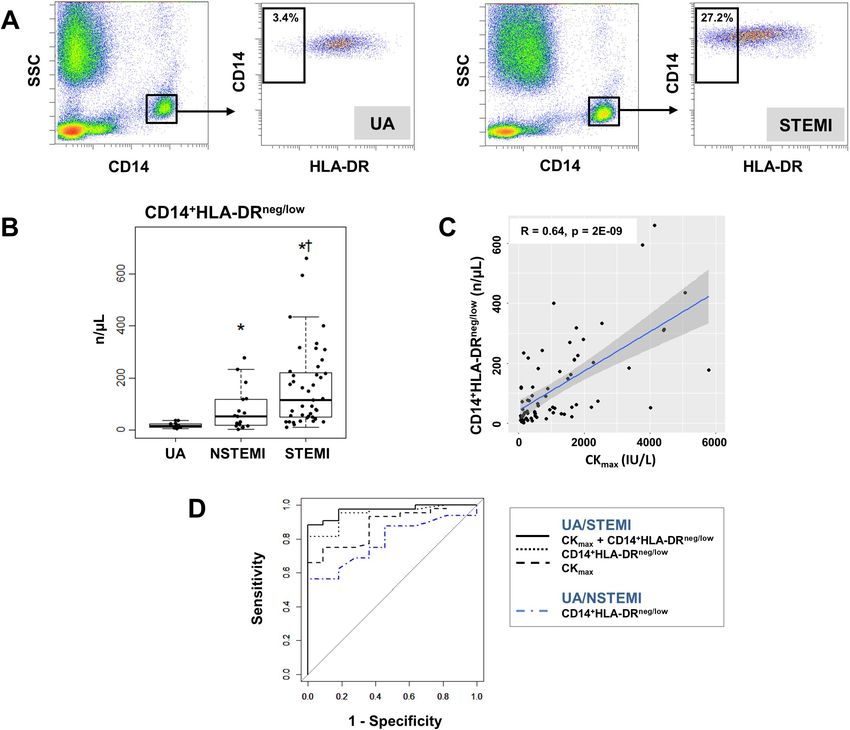
Research article                                                                        Immunology and Inflammation Medicine

                                    medium supplemented with L-glutamine/PenStrep). CD3+ T-cells were co-cultured in 96-well plates
                                    with CD14+HLA-DRneg/low and CD14+HLA-DRhigh monocytes at a ratio of 1 to 1 (T-cells: monocytes).
                                    T-cells were stimulated with Dynabeads Human T-Activator CD3/CD28 (11131D; Gibco) and T-cell
                                    proliferation was assessed 4 days later by CellTrace Violet dilution by flow cytometry.

                                    Chemotaxis assay
                                    Chemotaxis of CD14+HLA-DRneg/low/CD14+HLA-DRhigh monocytes was analyzed with the ChemoTx
                                    Disposable Chemotaxis System (NeuroProbe, NRP-106–8; Hölzel Diagnostika) (Chen et al., 2015).
                                    Human S100A9 (A42590, Invitrogen) was diluted in RPMI 1640 medium and placed in the lower
                                    chamber. Cells were washed with RPMI 1640 medium and preincubated for 30 min at 37˚C before
                                    addition to the upper chamber. Monocytes were allowed to migrate for 3 hr and migration was ana-
                                    lyzed by flow cytometry. The migration index was defined as the number of cells migrating in
                                    response to S100A9 divided by the number of cells migrating in response to medium alone
                                    (Chen et al., 2015).

                                    Assessment of neutrophil apoptosis
                                    CD10neg/CD10pos neutrophils (1x10⁶/mL) were cultured for 24 hr in RPMI 1640 medium supple-
                                    mented with 5% HI-FCS and 1% PenStrep. Apoptosis rate was assessed by flow cytometry using a
                                    Vybrant DyeCycle Violet/SYTOX AADvanced Apoptosis Kit (A35135, Invitrogen) and Apotracker
                                    Green (Barth et al., 2020) (427402; Biolegend), according to the manufacturer’s protocols. Stained
                                    neutrophils were analyzed using a Gallios cytometer and Gallios software (Beckman Coulter).

                                    T-cell activation assays in presence of CD10neg/CD10pos neutrophils
                                    CD4+ T-cells were isolated from PBMC using the MojoSort Human CD4 T Cell Isolation Kit (480009;
                                    BioLegend) or by flow-cytometric sorting. The CD28 MicroBead Kit (130-093-247; Miltenyi Biotec)
                                    was used for isolation of CD4+CD28null T-cells from PBMC. CD4+ T-cells and CD4+CD28null T-cells
                                    were resuspended at 1x10⁶/mL in T-Cell Activation Medium and stimulated with Dynabeads Human
                                    T-Activator CD3/CD28. For transwell experiments CD4+ T-cells and CD10neg/CD10pos neutrophils
                                    were co-cultured in 24 well plates at a ratio of 1 to 2 (T-cells: neutrophils) for 24 hr. CD10neg/
                                    CD10pos neutrophils were cultured in 0.4 mm transwell inserts (140620, Thermo Scientific) and CD4+
                                    T-cells in the well beneath the insert. In some experiments, CD10neg/CD10pos neutrophils were cul-
                                    tured overnight in T-Cell Activation Medium. The cell-free supernatants derived from CD10neg/
                                    CD10pos neutrophils were added to CD4+ T-cells cultured in 96-well plates (8104 cells/well) in the
                                    presence of neutralizing anti-IL-12 antibody (4 mg/mL; MAB219, R and D Systems; RRID:AB_
                                    2123616) or isotype control (4 mg/mL; MAB002, R and D Systems; RRID:AB_357344). CD4+CD28null
                                    T-cells were cultured with cell-free supernatants derived from CD10neg neutrophils. Culture superna-
                                    tants were collected after 24 hr incubation.

                                    LEGENDplex and ELISA assays
                                    Blood levels of G-CSF, MMP9, S100A9/S100A8, NGAL, MPO, TNF-a, IL-6, IL-1ß, and IFN-g were
                                    measured using bead-based multiplex assays (740180; 740589; 740929; LEGENDplex BioLegend).
                                    Serum samples were screened for CMV-specific IgG antibodies with the CMV-IgG-ELISA PKS Medac
                                    enzyme immunoassay (115-Q-PKS; Medac Diagnostika), using a cut-off value of >0.55 AU/mL for
                                    defining seropositivity according to manufacturer’s guidelines. Levels of IFN-g, IL-12, TNF-a, IL-6,
                                    and IL-1ß in the cell-culture supernatants were measured by ELISA (DIF50; R and D Systems) and
                                    using bead-based immunoassay (740929; LEGENDplex BioLegend).

                                    RT-quantitative PCR
                                    RNA was isolated from cells sorted in RTL Lysis Buffer using the RNeasy Plus Mini Kit (QIAGEN)
                                    according to the manufactures’ protocol. RNA quantification and quality testing were assessed by
                                    NanoDrop 2000 (Thermo Fisher Scientific) and Bioanalyzer 2100 (Agilent).
                                       cDNA synthesis was performed using 3 ng (human-neutrophils), 1 ng (mouse-neutrophils), and 10
                                    ng (monocytes) of total RNA and iScript Reverse Transcription Supermix (Bio-Rad). Relative quantita-
                                    tion of mRNA expression levels was determined with CFX96 Touch Real Time PCR using SsoAd-
                                    vanced Universal SYBR Green Supermix and PrimePCR Primers (Bio-Rad). ß-actin (ACTB) was chosen

Fraccarollo et al. eLife 2021;10:e66808. DOI: https://doi.org/10.7554/eLife.66808                                                4 of 31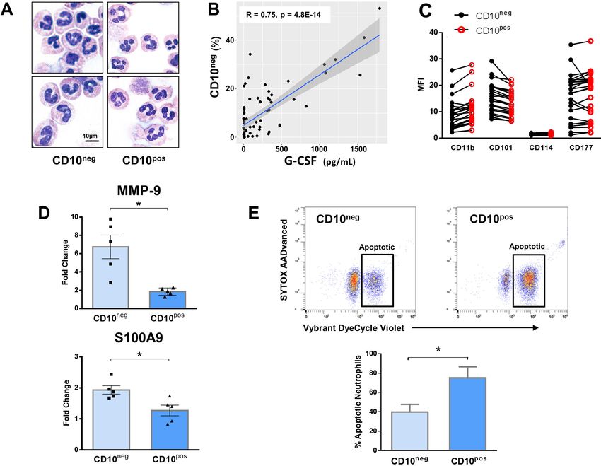
Research article                                                                          Immunology and Inflammation Medicine

                                    as an endogenous control. PCR amplification was performed at initially 95˚C for 30 s followed by 40
                                    cycles at 95˚C for 5 s and terminated by 60˚C for 30 s. The delta-delta Ct method was employed for
                                    data analysis.

                                    Animal experiments
                                    Study protocol
                                    All animal experiments were conducted in accordance with the Guide for the Care and Use of Labo-
                                    ratory Animals published by the National Institutes of Health (Publication No. 85–23, revised 1985).
                                    All procedures were approved by the Regierung von Unterfranken (Würzburg, Germany; permit No.
                                    54–2531.01-15/07) and by the Niedersächsisches Landesamt für Verbraucherschutz und Lebensmit-
                                    telsicherheit (Oldenburg, Germany; permit No. 33.12-42502-04-11/0644; 33.9-42502-04-13/1124
                                    and 33.12-42502-04-17/2702). C57BL/6 mice of both sexes were used in this study
                                    (Fraccarollo et al., 2019; Galuppo et al., 2017; Fraccarollo et al., 2017; Fraccarollo et al., 2011).

                                    Mouse model of reperfused AMI
                                    Myocardial ischemia was induced by transient left coronary artery ligation in age- and gender-
                                    matched mice. Briefly, mice were anesthetized with 2% isoflurane in a 100% oxygen mix, intubated,
                                    and ventilated using a ventilator (MINIVENT mouse ventilator model 845) with the tidal volume
                                    adjusted based on body weight (10 mL/g BW). Buprenorphine (0.1 mg/kg BW) was intraperitoneally
                                    administered for postoperative pain relief. The left coronary artery was ligated with a 6–0 silk suture
                                    just below the left auricular level (Fraccarollo et al., 2019; Galuppo et al., 2017; Fraccarollo et al.,
                                    2017; Fraccarollo et al., 2011). The suture was passed through a segment of PE-10 tubing. One
                                    hour after ischemia the tube was removed to allow for reperfusion. In sham-operated control mice,
                                    the ligature around the left anterior descending coronary artery was not tied.

                                    Isolation of immune cells and fluorescence-activated cell sorting
                                    Mice were anesthetized, intubated and ventilated. Blood samples were drawn from the inferior vena
                                    cava into EDTA-containing tubes. Neutrophil count was measured by an automated hematology
                                    analyzer (XT 2000i, Sysmex). After lysis of red blood cells with RBC Lysis Buffer (420301; BioLegend),
                                    cell suspension was centrifuged at 400 g for 16 min. Single-cell suspensions from heart-draining
                                    lymph nodes were prepared with a 70 mm cell strainer and collected by centrifugation at 400 g for
                                    16 min. Mouse femurs were harvested, bone marrows were flushed with FACS-staining buffer,
                                    passed through a 70 mm cell strainer (BD Biosciences) and the cell suspension was centrifuged at
                                    400 g for 16 min. The pelleted cells were washed and resuspended in ice-cold FACS-staining buffer.
                                    The hearts were perfused for 6 min with the Perfusion Buffer (113 mM NaCl, 4.7 mM KCl, 0.6 mM
                                    KH2PO4, 0.6 mM Na2HPO4), 1.2 mM MgSO4, 12 mM NaHCO3, 10 mM KHCO3, 10 mM HEPES, 30
                                    mM Taurine, 5.5 mM glucose, 10 mM 2,3-Butanedione monoxime, and subsequently digested for 8
                                    min with the Digestion Buffer (0.2 mg/mL Liberase Roche Diagnostics; and 400 mM calcium chloride
                                    in perfusion buffer), using a modified Langendorff perfusion system. The ischemic-reperfused area
                                    and surviving myocardium were separated using a dissecting microscope. Subsequently, the heart
                                    tissue was smoothly pipetted through a sterile low waste syringe several times in order to obtain a
                                    cell suspension in Stop Buffer (perfusion buffer supplemented with 10% (v/v) HI-FCS). The cell sus-
                                    pension was carefully filtered through a 70 mm cell strainer in a 50 mL conical tube, and the cell
                                    strainer was washed with perfusion buffer. Then, the cell suspension was centrifuged at 400 g for 20
                                    min. The pelleted cells were washed and resuspended in ice-cold FACS-staining buffer
                                    (Fraccarollo et al., 2019; Galuppo et al., 2017; Fraccarollo et al., 2017). To prevent capping of
                                    antibodies on the cell surface and non-specific cell labeling all steps were performed on ice and pro-
                                    tected from light. Cells were preincubated with Fc Block (Mouse BD Fc Block; BD Biosciences; RRID:
                                    AB_394657) for 10 min. Subsequently, fluorochrome-conjugated antibodies were added and incu-
                                    bated for 30 min. Finally, the cells were washed twice with ice-cold FACS-staining buffer. After pre-
                                    selection in side scatter (SSC) vs. forward scatter (FSC) dot plot to exclude debris and FSC vs. Time-
                                    of-Flight (ToF) dot plot to discriminate doublets by gating single cells, blood monocytes were identi-
                                    fied as CD45+/CD11b+/Ly6G /CD115+ cells, blood neutrophils as CD45+/CD11b+/Ly6G+ cells,
                                    infarct macrophages as CD45+/CD11b+/Ly6G /F4/80+ cells and infarct neutrophils as CD45+/
                                    CD11b+/F4/80 /Ly6G+ cells (Fraccarollo et al., 2019; Galuppo et al., 2017; Fraccarollo et al.,

Fraccarollo et al. eLife 2021;10:e66808. DOI: https://doi.org/10.7554/eLife.66808                                                    5 of 31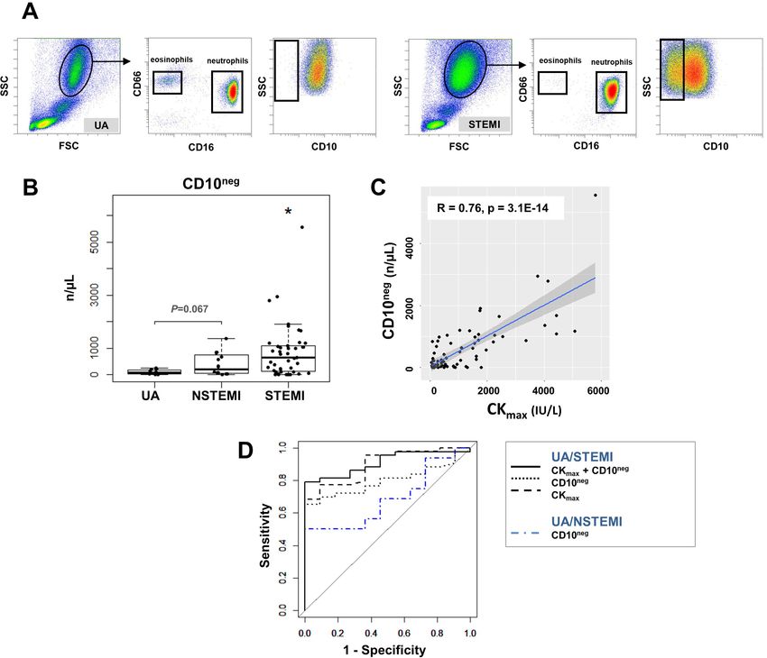
Research article                                                                         Immunology and Inflammation Medicine

                                    2017). The following antibodies were used: anti-CD45 (clone 104, 1:100, BioLegend; RRID:AB_
                                    893350; clone 30-F11, 1:100, BD Biosciences; RRID:AB_394003); anti-F4/80 (clone BM8, 1:100, BioL-
                                    egend, RRID:AB_2734779; clone T45-2342, 1:100, BD Biosciences; RRID:AB_893481); anti-CD11b
                                    (clone M1/70, 1:100, eBioscience, RRID:AB_2637408; BD Biosciences, RRID:AB_394002); anti-CD4
                                    (clone GK1.5, 1:100, BioLegend; RRID:AB_389302); anti-CD115 (clone AFS98, 1:100, BioLegend;
                                    RRID:AB_2562760, RRID:AB_11218983); anti-Ly6G (clone 1A8, 1:100, BioLegend, RRID:AB_1877163;
                                    1:200, BD Biosciences, RRID:AB_394208, RRID:AB_2739207); anti-CXCR2 (clone SA045E1, 1:100,
                                    BioLegend; RRID:AB_2565563, RRID:AB_2565689); anti-CD101 (clone Moushi101, 1:100, eBio-
                                    science, RRID:AB_1210729; clone 307707, 1:100, BD Biosciences, RRID:AB_2738821). For MMP-9
                                    and IL-1ß intracellular staining, the Cytofix/Cytoperm Fixation/Permeabilization Kit was used accord-
                                    ing to the manufacturer’s protocol (BD Biosciences). Antibodies included anti-MMP-9 (AF909, 1:100,
                                    R and D Systems; RRID:AB_355706); anti IL-1ß (ab9722, 1:100, Abcam; RRID:AB_308765); donkey
                                    anti-goat secondary antibody (A-11055, Invitrogen; RRID:AB_2534102) and goat anti-rabbit second-
                                    ary antibody (A-11034, Invitrogen; RRID:AB_2576217). FMO controls were included during acquisi-
                                    tion for gating analyses to distinguish positive from negative staining cell populations. FACS data
                                    were acquired on a Gallios flow cytometer and analyzed with Gallios software (Beckman Coulter).
                                    Cell sorting was performed using a FACS Aria Fusion (BD Biosciences). Cells were sorted in Lysis-
                                    Buffer (PrepEase RNA Spin Kit, Affymetrix; RNeasy Plus Mini Kit; QIAGEN), (Fraccarollo et al.,
                                    2019; Galuppo et al., 2017) or in sterile Sorting Medium. For morphological analysis, CD101neg/
                                    CD101pos neutrophils and mediastinal lymph node cell suspensions were centrifuged onto cytospin
                                    slides and stained with May-Grünwald Giemsa or Wright-Giemsa. For assessment of apoptosis
                                    FACS-sorted CD101neg/CD101pos neutrophils were cultured for 24 hr in RPMI 1640 medium supple-
                                    mented with 5% HI-FCS and 1% PenStrep. Apoptotic rate was determined by flow cytometry using
                                    Vybrant DyeCycle Violet stain.

                                    RNA-Seq
                                    Total RNA was isolated using PrepEase RNA Spin Kit according to the manufacturer’s instructions
                                    (Fraccarollo et al., 2019; Galuppo et al., 2017). Sorted cells were directly collected in lysis buffer
                                    and immediately processed. RNA quantification and quality testing were assessed by NanoDrop
                                    2000 (Thermo Fisher Scientific) and Bioanalyzer 2100 (Agilent). Libraries for RNA sequencing were
                                    prepared from 30 ng total RNA; from each sample, polyA RNA was purified, converted to cDNA
                                    and linked to Illumina adapters using the Illumina TruSeq stranded mRNA Kit according to the manu-
                                    facturer’s instructions. Samples were multiplexed and sequenced on an Illumina NextSeq 500 in a 75
                                    nt single end setting using a high-output run mode. Raw BCL files were demultiplexed and con-
                                    verted to sample-specific FASTQ files using bcl2fastq v1.8.4 (Illumina). Residual adapter sequences
                                    present in the sequencing reads were removed with Cutadapt version 1.12. Reads (~ 40 million per
                                    sample) were aligned to the mouse reference sequence GENCODE vM8 using STAR version 2.5.2b.
                                    RNA sequencing data analysis was undertaken with the statistical programming language, R. The R
                                    package DeSeq2 (v1.14.1) was used to evaluate differential gene expression (Fraccarollo et al.,
                                    2019; Galuppo et al., 2017).

                                    Statistical analysis
                                    Data are presented as mean ± SEM or as median [interquartile range] as indicated. Normality of
                                    data was assessed by Shapiro-Wilk test. Normal data were analyzed by one-way ANOVA with Tukey
                                    post hoc test, otherwise by Kruskal-Wallis test for comparisons of median values, with Mann-Whitney
                                    U test for multiple comparisons. Unpaired t-test or Mann-Whitney U test was used to compare two
                                    independent groups. Linear regression analysis or Spearman’s rank correlation test was used to
                                    determine relationship between variables. Values of p0.05 were considered statistically significant.

                                    Results
                                    Increased circulating levels of CD14+HLA-DRneg/low monocytes in
                                    patients with acute MI
                                    Flow cytometric immunophenotyping was performed in whole blood from patients with unstable
                                    angina (UA) or acute MI within 24–72 hr of symptom onset (median 43.6 hr). A time course analysis

Fraccarollo et al. eLife 2021;10:e66808. DOI: https://doi.org/10.7554/eLife.66808                                                  6 of 31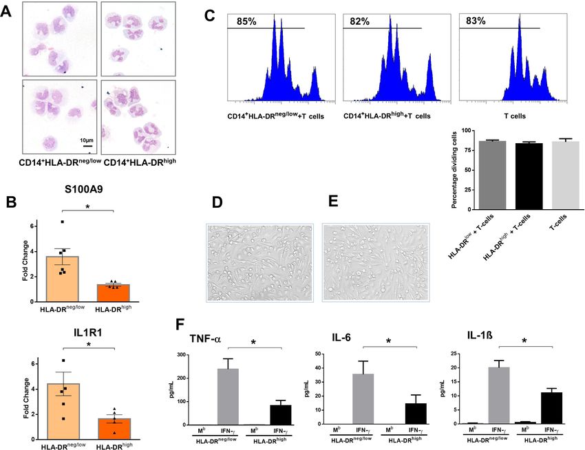
Research article                                                                                     Immunology and Inflammation Medicine

                                     of monocyte subset-frequencies and circulating levels up to day five after MI is shown in Figure 1—
                                     figure supplement 1.
                                        NSTEMI/STEMI patients displayed significantly higher absolute neutrophil and monocyte counts
                                     versus UA patients (Table 2). Based on HLA-DR/CD14/CD16 expression, monocytes can be divided
                                     into different subsets. We detected increased circulating levels of intermediate (HLA-DR++CD14+
                                     +
                                       CD16+CX3CR1+) in ACS patients versus control, and of non-classical (HLA-DR+CD14+CD16+
                                     +
                                       CX3CR1++) in STEMI versus UA patients and controls (Table 2, Figure 1—figure supplement 2).
                                     There were no significant correlations between intermediate/non-classical monocytes and LV ejec-
                                     tion fraction/CKmax.
                                        We found increased percentages and absolute numbers of circulating CD14+HLA-DRneg/low
                                     monocytes in STEMI/NSTEMI patients as compared to UA patients (Figure 1A and B and Figure 1—
                                     figure supplement 2B). Linear regression analysis revealed a positive correlation between circulating
                                     levels of CD14+HLA-DRneg/low monocytes and CKmax (Figure 1C) and a negative correlation with LV
                                     ejection fraction (R=0.44, p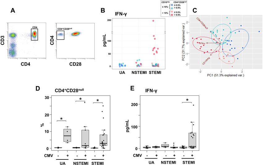
Research article                                                                                 Immunology and Inflammation Medicine

Figure 1. Increased circulating levels of CD14+HLA-DRneg/low monocytes in patients with AMI. (A) Gating strategy to identify CD14+HLA-DRneg/low
monocytes. (B) Circulating levels of CD14+HLA-DRneg/low monocytes in patients with unstable angina (UA; n=11), non-ST-elevation MI (NSTEMI, n=16),
and ST-elevation MI (STEMI, n=44). (C) Linear regression analysis between circulating levels of CD14+HLA-DRneg/low monocytes and maximum CK
(CKmax) in patients with acute coronary syndrome. (D) Receiver operator characteristic (ROC) curve of CD14+HLA-DRneg/low monocytes discriminating
UA/STEMI and UA/NSTEMI patients and the combination of CD14+HLA-DRneg/low monocytes (n/mL) with CKmax. *p

Research article                                                                               Immunology and Inflammation Medicine

Figure 2. CD14+HLA-DRneg/low monocytes from patients with AMI are not immunosuppressive but exhibit an inflammatory phenotype. (A) May-
Grünwald Giemsa stained cytospin preparations of CD14+HLA-DRneg/low and CD14+HLA-DRhigh monocytes. (B) Relative RNA expression of S100A9 and
IL1R1 in CD14+HLA-DRneg/low versus CD14+HLA-DRhigh monocytes. (C) T-cell proliferation in the presence of CD14+HLA-DRneg/low or CD14+HLA-DRhigh
monocytes assessed by CellTrace Violet dilution after 96 hr of co-culture. (D) Macrophages differentiated from CD14+HLA-DRneg/low monocytes and (E)
CD14+HLA-DRhigh cells by 4-day culture with M-CSF. (F) TNF-a, IL-6, and IL-1ß in supernatants of macrophage cultures upon stimulation with IFN-g.
Mb=baseline. CD14+HLA-DRneg/low/CD14+HLA-DRhigh cells were isolated by flow-cytometric sorting from patients with AMI (n=5–6). Data are presented
as mean ± SEM. *p
Research article                                                                               Immunology and Inflammation Medicine

Figure 3. Circulating normal-density CD10neg neutrophils increase in patients with AMI. (A) Gating strategy to identify CD10neg neutrophils.
(B) Circulating levels of CD16+CD66b+CD10neg neutrophils in patients with unstable angina (UA; n=11), non-ST-elevation MI (NSTEMI, n=16), and ST-
elevation MI (STEMI, n=44). (C) Linear regression analysis between circulating levels of CD10neg neutrophils and maximum CK (CKmax). (D) Receiver
operator characteristic (ROC) curve of CD10neg neutrophils (n/mL) discriminating UA/STEMI and UA/NSTEMI patients and the combination of CD10neg
neutrophils with CKmax in patients with acute coronary syndrome. *p
Research article                                                                               Immunology and Inflammation Medicine

                                    immature low-density neutrophils in STEMI patients. However, cancer patients were not excluded
                                    which might have influenced the proportion of low-density CD10neg cells.
                                       Cytospin slides were made after FACS-sorting to examine nuclear morphology (Figure 4A). We
                                    found that the majority of the CD16+CD66b+CD10neg cells has an immature morphology with a lob-
                                    ular nucleus, while CD16+CD66b+CD10pos cells are mature neutrophils with segmented nuclei
                                    (Figure 4A). These findings were obtained when neutrophils were isolated by dextran sedimentation
                                    as well as by negative selection using magnetic beads, indicating that the differences between the

Figure 4. Immature CD10neg neutrophils from patients with AMI express high amounts of MMP-9 and S100A9 and display resistance to apoptosis. (A)
May-Grünwald Giemsa stained cytospin preparations of CD16+CD66b+CD10neg (CD10neg) and CD16+CD66b+CD10pos (CD10pos) neutrophils. (B) Linear
regression analysis between the percentages of CD16+CD66b+CD10neg neutrophils and circulating levels of G-CSF in patients with acute coronary
syndrome (n=71). (C) Mean fluorescence intensity (MFI) of CD11b, CD101, CD114, and CD177 on CD10neg versus CD10pos neutrophils (n=25). (D)
Relative RNA expression of MMP-9 and S100A9 in CD10neg versus CD10pos neutrophils. (E) Percentage of apoptotic neutrophils assessed by flow
cytometry using Vybrant DyeCycle Violet stain and SYTOX AADvanced stain. CD10neg/CD10pos neutrophils were isolated by flow-cytometric sorting
from patients with AMI (n=4–5). *p
Research article Immunology and Inflammation Medicine
neutrophil subpopulations cannot be considered an artifact due to the isolation technique used
(Hardisty et al., 2021). Of note, linear regression analysis revealed a strong positive correlation
between the percentages of CD16+CD66b+CD10neg cells and circulating levels of G-CSF
(Figure 4B). AMI patients with higher systemic concentrations of G-CSF have increased CD10neg
neutrophils levels, suggesting G-CSF-driven immature neutrophil release/expansion.
Immature CD10neg neutrophils from patients with AMI express high
amounts of MMP-9 and S100A9 and display resistance to apoptosis
In an attempt to identify-specific surface antigens that characterize normal-density CD10neg neutro-
phils derived from AMI patients, we next examined the expression of markers associated with neu-
trophil maturation/activation (Hardisty et al., 2021; Silvestre-Roig et al., 2019). Flow cytometry
showed that CD11b, CD101, CD114, and CD177 were expressed at equivalent levels on CD10neg
neutrophils versus CD10pos neutrophils (Figure 4C).
Next, we investigated the expression of inflammatory mediators playing a pathophysiological role
in wound healing after AMI. CD10neg neutrophils sorted from blood of AMI patients express higher
amounts of MMP-9 and S100A9 than CD10pos neutrophils (Figure 4D). No difference was found in
the expression of IRG1 (ACOD1), IL1R1, MMP8, NOS2, and STAT3 (Figure 4—figure supplement
1A), genes regulated in circulating neutrophils as well as in infarct neutrophils in a mouse model of
reperfused AMI (Figure 4—figure supplement 1A–1C).
Mouse neutrophils, unlike human granulocytes, lack CD10 expression (Kalled et al., 1995) Using
next-generation RNA sequencing we identified CD101 among the genes down-regulated by ische-
mia in circulating neutrophils (Figure 4—figure supplement 1B).
Next, we found that CD101 can be used as a surface marker to identify the mature neutrophil
subset among the heterogeneous Ly6GposCXCR2pos neutrophil populations, released into the blood-
stream after ischemia/reperfusion (Figure 4—figure supplement 2A and B). As revealed by mor-
phological analysis circulating CD11bbrightCD101pos neutrophils have a mature morphology, whereas
CD11bdimCD101neg cells are immature neutrophils with ring-shaped nuclei (Figure 4—figure supple-
ment 2B). Recent study in mice showed that CD101 segregates immature neutrophils from mature
neutrophils during G-CSF stimulation and in the tumor setting (Evrard et al., 2018). Notably, the
levels of CD101neg neutrophils in the bone marrow of mice subjected to ischemia followed by 6 hr of
reperfusion were markedly reduced (Figure 4—figure supplement 2C), indicating early release of
immature neutrophils from bone marrow into the circulation after AMI. Moreover, we found that
CD11bposLy6GposCD101neg cells sorted from peripheral blood after AMI express higher amounts of
Irg1 (Acod1), Il1r1, Mmp8, Nos2, and Stat3 than CD101pos neutrophils and showed markedly
reduced CD101 expression (Figure 4—figure supplement 1D and E). Taken together, our results
emphasize interspecies differences in immune responses after ischemic injury, suggesting caution in
applying findings in murine models of myocardial ischemia/reperfusion to patients with AMI.
To further define the functional properties of the immature neutrophil populations derived from
AMI patients we investigated spontaneous apoptosis. The apoptotic rate of CD10neg neutrophils
was substantially reduced compared with CD10pos neutrophils after 24 hr in vitro culture as deter-
mined by flow cytometric analysis using Vybrant DyeCycle Violet/SYTOX AADvanced stain
(Figure 4E) as well as Apotracker Green (Figure 4—figure supplement 3A). The decreased apopto-
sis rate of CD10neg neutrophils was reflected by an increase in the percentage of live cells. Morpho-
logical assessment of cytospins (Figure 4—figure supplement 3B) confirmed the results obtained
by flow cytometry.
Neutrophils may play a crucial role in cardiac healing after AMI by influencing macrophage polari-
zation (Horckmans et al., 2017). Horckmans et al., 2017 found an induction of MerTK expression in
macrophages (differentiated from peripheral blood monocytes of healthy donors) exposed to IL-4 in
the presence of neutrophil supernatants from healthy individuals. Therefore, we next assessed
MerTK expression on macrophages differentiated from CD14+HLA-DRneg/low and CD14+HLA-DRhigh
monocytes and polarized in the presence of supernatants derived from CD10neg neutrophils as well
as CD10pos neutrophils (Figure 4—figure supplement 3C and D). CD14+HLA-DRneg/low/CD14+HLA-
DRhigh cells and CD10neg/CD10pos neutrophils were isolated from patients with AMI. We found a sig-
nificantly higher expression of MerTK on CD14posCD163pos macrophages differentiated from
CD14+HLA-DRneg/low monocytes and stimulated with IFN-g and IL-4 in presence of supernatants
from CD10pos neutrophils (Figure 4—figure supplement 3C). Efficient efferocytosis by human
Fraccarollo et al. eLife 2021;10:e66808. DOI: https://doi.org/10.7554/eLife.66808 12 of 31Research article Immunology and Inflammation Medicine
macrophages requires MerTK induction (Zizzo et al., 2012). Thus, we speculated that factors
secreted by CD10pos neutrophils from patients with AMI may enhance the capacity of HLA-DRneg/low
monocyte-derived macrophages to phagocyte apoptotic cells by regulating MerTK expression.
The alarmin S100A9 have been shown to have chemotactic activity for monocytes (Chen et al.,
2015). Previous experiments have focused on monocytic THP-1 cells and monocytes isolated from
healthy human subjects (Chen et al., 2015). Here, we analyzed S100A9-induced chemotaxis of
CD14+HLA-DRneg/low/CD14+HLA-DRhigh monocytes from patients with AMI. As shown in Figure 4—
figure supplement 3E, S100A9 significantly induced the migration of CD14+HLA-DRhigh as well as
CD14+HLA-DRneg/low monocytes. Therefore, it is also possible that S100A9 regulates CD14+HLA-
DRneg/low/CD14+HLA-DRhigh monocytes recruitment during AMI by functioning as a
chemoattractant.
CD10neg neutrophils and HLA-DRneg/low monocytes are linked to levels
of immune-inflammation markers
In a subgroup of patients we measured serum levels of immune inflammation markers (Table 3).
MMP-9, S100A9/S100A8, NGAL, IL-6, and IL-1ß levels were higher in STEMI patients versus UA
patients.
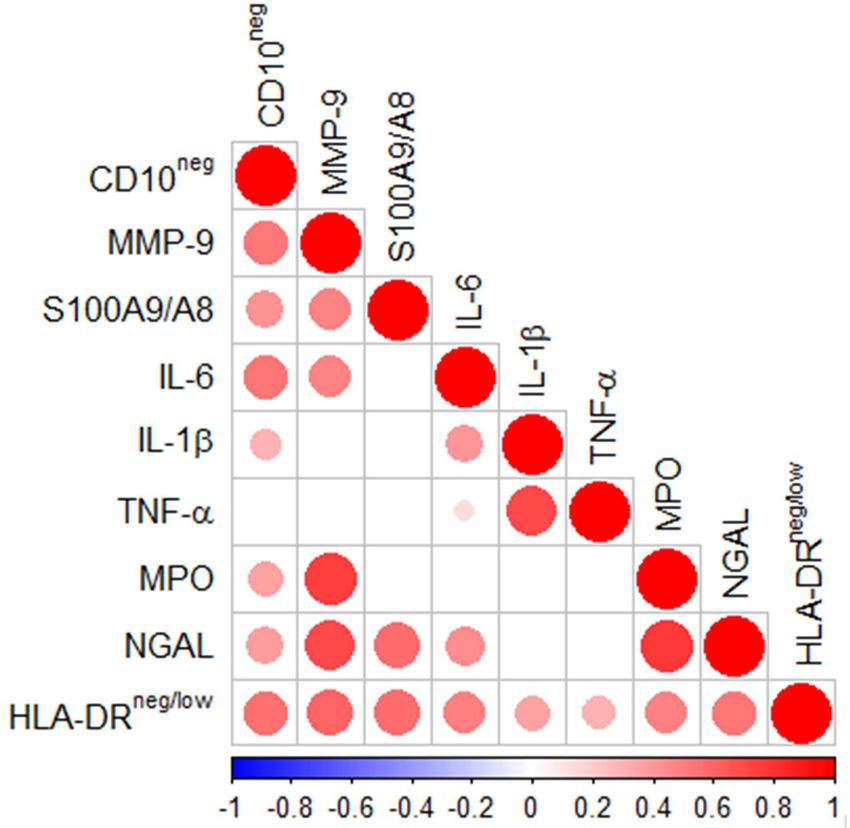
Spearman’s correlation matrix showed multiple intercorrelations among CD10neg neutrophils,
HLA-DRneg/low monocytes and several inflammation markers (Figure 5). The percentages of CD14+-
HLA-DRneg/low cells significantly correlated with circulating levels of MMP-9, S100A9/S100A8, IL-6,
IL-1ß, TNF-a, MPO, and NGAL. Noticeable, CD10neg neutrophils, which expand proportional to the
degree of myocardial injury, significantly correlated with levels of MMP-9, S100A9/S100A8, NGAL,
MPO, IL-6, and IL-1ß (Figure 5).
Immature CD101neg neutrophils are recruited to sites of cardiac injury
and exhibit resistance to apoptosis
We then investigated whether immature CD101neg neutrophils are recruited to the injured myocar-
dium using the mouse model of reperfused AMI. Preliminary experiments showed that current pro-
tocols for tissue dissociation and the recovery of neutrophils from ischemic myocardium involving
long enzymatic digestion times resulted in cell activation/damage and phenotypic changes that can
be easily over-interpreted. Therefore, using a modified Langendorff perfusion system, the infarcted
hearts were perfused for 6 min to remove blood cells and subsequently digested for only 8 min to
preserve cell surface antigens along with expression profiles. Flow cytometry analysis of immune
cells isolated from the ischemic region 3 hr after reperfusion revealed marked infiltration of
CD101neg neutrophils, displaying increased expression of the matrix-degrading protease MMP-9
(Figure 6A). Moreover, as shown in Figure 6B, we found that 24 hr after reperfusion CD101neg neu-
trophils expressed IL-1ß at higher levels compared to CD101pos cells. Morphological analysis of cyto-
spin preparations of sorted neutrophils showed that CD101neg neutrophils isolated from ischemic
myocardium 24 hr after reperfusion still have an immature morphology (Figure 6C). These findings
suggest migration and homing of immature CD101neg neutrophils to ischemic myocardium shortly
after reperfusion.
Table 3. Immune inflammation markers.
UA (N=11) NSTEMI (N=10) STEMI (N=26) p (K-W)
MMP-9 (ng/mL) 429 (320-461) 447 (324-597) 544 (466-758)*Research article Immunology and Inflammation Medicine
Figure 5. Multiple intercorrelations among CD10neg neutrophils, HLA-DRneg/low monocytes and immune-
inflammation markers. Spearman-correlation matrix of CD16+CD66b+CD10neg neutrophils (%), CD14+HLA-DRneg/
low
monocytes (%) and circulating levels of MMP-9, S100A9/S100A8, IL-6, IL-1ß, TNF-a, MPO, and NGAL (levels).
UA (n=11), NSTEMI (n=10), and STEMI (n=26). Each circle illustrates a significant correlation between different
couples of parameters (pResearch article Immunology and Inflammation Medicine Figure 6. Immature CD101neg neutrophils are rapidly recruited to ischemic sites, are a major source of MMP-9 and IL-1ß in the reperfused myocardium and exhibit resistance to apoptosis. (A) Flow cytometric gating strategy to identify neutrophils in the ischemic region, mean fluorescent intensity (MFI) of MMP-9 on CD101neg and CD101pos neutrophils, number of CD101neg and CD101pos neutrophils 3 hr after reperfusion. (B) Flow cytometry identifying infarct neutrophils, mean fluorescent intensity of IL-1ß on CD101neg and CD101pos neutrophils, number of CD101neg and CD101pos neutrophils 24 hr after reperfusion. (C) Wright-Giemsa stained cytospin preparations of CD101neg and CD101pos neutrophils isolated by cell sorting from ischemic myocardium 24 hr after reperfusion. (D) Rate of apoptosis of infarct CD101neg/CD101pos neutrophils determined by flow cytometry using Vybrant DyeCycle Violet stain after 24 hr in vitro culture. CD11bposLy6GposCD101neg (CD101neg) and CD11bposLy6GposCD101pos (CD101pos) cells were isolated by cell sorting from ischemic myocardium 24 hr after reperfusion. Data are presented as mean ± SEM (n=3–4). *p
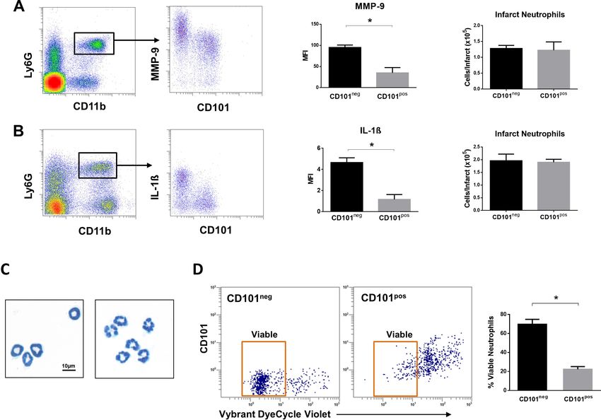
Research article                                                                                Immunology and Inflammation Medicine

Figure 7. Immature CD101neg neutrophils are present in mediastinal lymph nodes draining the heart after acute MI. (A) Flow cytometric analysis of
neutrophil subsets in mediastinal lymph nodes at steady state and 3 hr after reperfusion (AMI). (B) Cytospins of mediastinal lymph node cell
suspensions stained with Wright’s Giemsa. Mice were subjected to 1 hr of coronary occlusion and heart-draining lymph nodes were isolated 3 hr after
reperfusion. Data are presented as mean ± SEM (n=3). *p
Research article                                                                                  Immunology and Inflammation Medicine

Figure 8. Elevated IFN-g levels in patients with expanded CD10neg neutrophils and increased frequency of CD4+CD28null T-cells. (A) Gating strategy
identifying CD4+CD28null T-cells. (B) Scatter plot showing IFN-g levels according to frequency of CD10neg neutrophils and CD4+CD28null T-cells. (C)
Principal component analysis (PCA) showing clustering according to circulating levels of IFN-g, CD10neg neutrophils and CD4+CD28null T-cells. Patients
were stratified based on frequency of CD10neg neutrophils (10% or >10%) and frequency of CD4+CD28null T-cells (0.5% or >0.5%). (D) Frequency of
CD4+CD28null T-cells in patients with acute coronary syndrome stratified according to cytomegalovirus (CMV) serostatus. (E) circulating IFN-g levels
stratified according to CMV serostatus. UA (n=11), NSTEMI (n=13), and STEMI (n=34). *p0.05.
The online version of this article includes the following figure supplement(s) for figure 8:
Figure supplement 1. (A) CD4+CD28null T-cell frequency distribution (log10-transformed CD4+ T-cell fractions) of CMV± (top, n=58), CMV- (middle,
n=23), and CMV+ (bottom, n=35) acute coronary syndrome (ACS) patients. CD4+CD28null T-cells displayed a bimodal distribution related to CMV-
seropositivity. (B) Boxplots show the log10-transformed frequency of CD4+CD28null T-cells in CMV--UA, CMV- (blue) and CMV+ (red) -ACS patients.
Expansion index (dotted line) was calculated as UQ+1.5xIQR of CMV--UA patients chosen as reference group. CD4+CD28null T-cell frequency more than
0.5% was considered as an index of expansion; UQ (upper quantile), IQR (inter-quantile range). (C) CD10neg neutrophils (CD10neg) frequency
distribution (log10-transformed) of CMV± (top, n=71), CMV- (middle, n=31), and CMV+ (bottom, n=40) ACS patients. (D) Boxplots show the log10-
transformed frequency of CD10neg in UA, NSTEMI and STEMI patients. Expansion index (dotted line) was calculated as UQ+1.5xIQR of UA patients.
According, patients with CD10neg frequency more than 10% had expansion. (E) Scaled frequency of CD4+CD28null T-cells and CD10neg neutrophils
stratified by criteria of cell expansion. Hierarchical clustering performed on columns highlights the relationship among CD10neg neutrophils,
CD4+CD28null T-cells, IFN-g production and CMV seropositivity.
Figure supplement 2. Relationship among the peripheral expansion of immature CD10neg neutrophils, CMV IgG titers, CD4+CD28null T-cells, and
circulating levels of IFN-g and IL-12 in patients with AMI.

                                     also performed principal component analysis (PCA) that showed clustering according to elevated cir-
                                     culating levels of IFN-g, high levels of CD10neg neutrophils and peripheral expansion of CD4+-
                                     CD28null T-cells (Figure 8C). The highest IFN-g levels were found in STEMI patients with expanded
                                     CD10neg neutrophils (>10%) and increased frequency of CD4+CD28null T-cells (Figure 8C).
                                         Altered T-cell homeostasis and increased frequency of circulating CD28null T-cells have been
                                     linked to cytomegalovirus (CMV) seropositivity (Pera et al., 2018; Looney et al., 1999; Moro-
                                     Garcı́a et al., 2018). Therefore, we next analyzed the impact of CMV serostatus on CD4+CD28null

Fraccarollo et al. eLife 2021;10:e66808. DOI: https://doi.org/10.7554/eLife.66808                                                              17 of 31
Research article Immunology and Inflammation Medicine
T-cells frequency. We discovered that CD4+ T-cells lacking the costimulatory molecule CD28 showed
expansion across CMV-seropositive (CMV+) patients (Figure 8D).
Frequency distribution of CD4+CD28null T-cells (log10 transformed to improve visualization)
appeared bimodal and analyzing separately in CMV+ and CMV-seronegative (CMV-) patients, the
median was significantly higher by a factor 14.1 in CMV+ patients. (Figure 8—figure supplement
1A). Moreover, CD4+CD28null frequency positively correlated with CMV-IgG antibody levels (R=0.6,
p10%) and frequency of CD4+CD28null T-cells (0.5% or >0.5%) as depicted in
Figure 8C by principal component analysis (PCA). In the derived heatmap IFN-g, CD10neg neutro-
phils, CD4+CD28null, EMRA, and EM CD4+ T-cells were grouped together showing similar patterns
(Figure 8—figure supplement 1E), indicating that persistent CMV infection is associated with
expansion of the effector memory CD4+ T-cell compartment and higher IFN-g levels in patients with
increased frequency of CD10neg neutrophils. Not surprisingly, when stratified according to CMV
serostatus, maximum levels of circulating IFN-g among ACS patients were detected in CMV-seropos-
itive STEMI patients displaying increased levels of CD10neg neutrophils (Figure 8E), indicating a rela-
tion among expansion of immature CD10neg neutrophils, CMV seropositivity and strongly enhanced
levels of IFN-g in patients with large AMI.Of note, PCA performed replacing the percentage of
CD4+CD28null cells with CMV-IgG titer (stratified by a cut-off equal to 0.55) showed a better cluster
grouping (Figure 8—figure supplement 2A), with similar represented total variance (PC1+PC2:
79.5% vs. 83% respectively). The heatmap derived stratifying patients by frequency of CD10neg neu-
trophils and CMV-IgG cut-offs (Figure 8—figure supplement 2B) shows hierarchical subgrouping of
CMV-IgG titers and CD4+CD28null cells. Moreover, Spearman’s correlation matrix revealed a strong
positive correlation between CMV-IgG titers and the percentage of CD4+CD28null cells (Figure 8—
figure supplement 2C). Taken together, these results highlight the complex relationship in vivo
among CMV seropositivity, CD4+CD28null cells, CD10neg neutrophils and IFN-g levels.
CD10neg neutrophils via induction of interleukin-12 enhance priming for
IFN-g production by CD4+ T-cells
Environmental factors such as CMV infection can induce changes in CD4+ T-cell phenotype and func-
tion. Consequently, to provide a mechanistic understanding of the cellular basis for raised IFN-g in
CMV-seropositive patients with expanded CD10neg neutrophils, we investigated IFN-g secretion by
CD4+ T-cells isolated from CMV-/CMV+ patients and its potential link to interleukin 12 (IL-12), potent
inducer of IFN-g (Trinchieri, 2003). In cell-to-cell contact-dependent conditions human neutrophils
can mimic myeloid-derived suppressor cells and suppress T-cell activation through artefactual mech-
anisms (Negorev et al., 2018). Therefore, CD10neg/CD10pos neutrophils were evaluated for their
ability to enhance IFN-g production in cell contact-independent manner. We found that CD10neg
neutrophils strongly enhanced IFN-g and IL-12 production by CD4+ T-cells from CMV+ patients
(Figure 9A and B), when co-cultured using a transwell system where CD4+ T-cells in the lower cham-
ber were separated from neutrophils in the upper chamber. Of note, CD4+ T-cells equally
responded to cell-free supernatants derived from CD10neg neutrophils. IFN-g and IL-12 production
were significantly higher in CD4+ T-cells from CMV+ than CMV- patients. The addition of neutralizing
anti-IL-12 antibody abrogated the IFN-g production by CD4+ T-cells from CMV+ patients in presence
of supernatants derived from CD10neg neutrophils (Figure 9A). Our data indicate that CD10neg
Fraccarollo et al. eLife 2021;10:e66808. DOI: https://doi.org/10.7554/eLife.66808 18 of 31Research article Immunology and Inflammation Medicine
Figure 9. CD10neg neutrophils enhance IFN-g production by CD4+ T-cells via induction of interleukin-12. (A) IFN-g and (B) interleukin-12 production by
CD4+ T-cells stimulated with anti-CD3/CD28 beads and co-cultured for 24 hr in absence (CD4+) or presence of CD10pos neutrophils (CD10pos+CD4+),
CD10neg neutrophils (CD10neg+CD4+) using a transwell system or cultured with cell-free supernatants derived from CD10pos neutrophils (csf-
CD10pos+CD4+), CD10neg neutrophils (csf-CD10neg+CD4+), CD10neg neutrophils in the presence of neutralizing anti-IL-12 antibody (csf-
CD10neg+CD4++IL12Ab). CD4+CD28null T-cells were stimulated with anti-CD3/CD28 beads (CD4+CD28neg) and cultured with cell-free supernatants
derived from CD10neg neutrophils (csf-CD10neg+CD4+CD28neg). CD10neg/CD10pos neutrophils, CD4+ T-cells and CD4+CD28null T-cells were isolated
from CMV-seronegative (CMV-) or CMV-seropositive (CMV+) patients with AMI (n=3–5). Data are represented as fold-change to respective CD3/CD28-
stimulated cells and presented as mean ± SEM. *p0.05.
The online version of this article includes the following figure supplement(s) for figure 9:
Figure supplement 1. IFN-g production by CD4+ T-cells from CMV-seropositive AMI patients stimulated with anti-CD3/CD28 beads and cultured for
24 hr in absence (CD4+) or presence of cell-free supernatants derived from CD10pos neutrophils (csf-CD10pos+CD4+) or CD10neg neutrophils (csf-
CD10neg+CD4+).
neutrophils release soluble factors that efficiently induce a strong Th1 type response. Further studies
aiming at characterizing the neutrophil-secreted immunomodulatory factors are ongoing.
Notably, bioinformatic analysis showed enhanced circulating levels of IL-12 in CMV+ patients with
increased frequency of CD10neg neutrophils (Figure 8—figure supplement 2B). Moreover, Spear-
man’s correlation matrix showed multiple inter-correlations among IL-12 circulating levels, CMV IgG
titers, frequency of CD10neg neutrophils, expanded CD4+CD28null T-cells and particularly circulating
levels of IFN-g (Figure 8—figure supplement 2C). To test whether neutrophils from CMV- or CMV+
patients have different ability to induce IFN-g, CD4+ T-cells from CMV+ patients were cultured in
presence of supernatants derived from CMV-seronegative patients (Figure 9—figure supplement
1). We found that CD10neg neutrophil supernatants markedly enhanced IFN-g secretion (Figure 9—
figure supplement 1), indicating that CD10neg neutrophils from CMV- and CMV+ patients exhibit
the same ability to induce IFN-g production by CD4+ T-cells. CD10neg neutrophils had no effect on
CD3/CD28-stimulated CD4+CD28null T-cells (Figure 9A and B), demonstrating that overproduction
of IFN-g is confined to CD4+ T-cells expressing CD28. Taken together, our findings indicate that
CD4+CD28+ T-cells from CMV+ patients with AMI display a distinct phenotype overproducing IFN-g
in presence of immature neutrophils via induction of interleukin-12.
Fraccarollo et al. eLife 2021;10:e66808. DOI: https://doi.org/10.7554/eLife.66808 19 of 31Research article Immunology and Inflammation Medicine
Discussion
Innate immune mechanisms play a paramount role during AMI and the functional heterogeneity of
monocytes and neutrophils have been the focus of intensive research in recent years. This study
highlights for the first time that immature CD16+CD66b+CD10neg neutrophils and CD14+HLA-DRneg/
low
monocytes promoting proinflammatory immune responses expand in the peripheral blood from
patients with large AMI. We also show that immature neutrophils are recruited to the injured myo-
cardium and migrate to mediastinal lymph nodes shortly after reperfusion, using a mouse model of
AMI. Furthermore, we found a potential link among increased frequency of immature CD10neg neu-
trophils and elevated IFN-g levels, especially in cytomegalovirus-seropositive patients with expanded
CD4+CD28null T-cells. Finally, we could show that CD10neg neutrophils enhance CD4+ T-cells IFN-g
production by a contact-independent mechanism involving IL-12 (Figure 10).
This study uncovered that CD10 can be used as a surface marker to identify the immature neutro-
phil population that expands and promotes proinflammatory effects in patients suffering from AMI.
We believe that immature CD10neg neutrophils derive from MI-induced emergency granulopoiesis.
Both mature (segmented) and immature banded neutrophils are released from the bone marrow
Figure 10. Immature CD10neg neutrophils and HLA-DRneg/low monocytes inducing proinflammatory and adaptive immune responses emerge in patients
with large acute myocardial infarction.
Fraccarollo et al. eLife 2021;10:e66808. DOI: https://doi.org/10.7554/eLife.66808 20 of 31Research article Immunology and Inflammation Medicine
presumably to meet the high demand for more neutrophils, especially in patients with large AMI.
Not surprisingly, in our study higher frequency of circulating CD10neg neutrophils was associated
with increased systemic concentrations of G-CSF, an essential regulator of neutrophil trafficking
from the bone marrow to the blood and important neutrophil survival factor (Costa et al., 2019; Sil-
vestre-Roig et al., 2020). Recently, CD10 has been proposed as a marker that distinguishes mature
from immature neutrophils in healthy volunteers receiving G-CSF for stem cell mobilization
(Marini et al., 2017).
Multiple clinical trials have evaluated the use of G-CSF in patients with AMI after successful revas-
cularization. The majority of these studies found that effective stem cell mobilization with G-CSF
therapy failed to improve left ventricular recovery (Traverse, 2019). Our findings suggest that the
therapeutic benefits of G-CSF therapy after AMI might be compromised due to the release of imma-
ture proinflammatory CD10neg neutrophils.
However, neutrophils may be released from the bone marrow in response to increased damage-
associated molecular patterns such as S100A8/S100A9, secreted from neutrophils as mediators of
sterile inflammation (Sreejit et al., 2020). Of interest, we found that circulating CD10neg neutrophils
express high amounts of S100A9, indicating that immature neutrophils could be an important source
of this alarmin in patients with AMI.
Under inflammatory conditions neutrophils traffic to inflamed tissues as well as to draining lymph
nodes (Costa et al., 2019; Leliefeld et al., 2015) modulating T cell-mediated immune responses.
Our data from the mouse model of AMI are first to provide evidence that immature neutrophils can
populate heart-draining lymph nodes in response to acute ischemic injury. It is intriguing to specu-
late that CD10neg neutrophils migrate into lymph nodes where they could encounter T-cells and
shape adaptive immune responses.
Emerging evidence indicates that immature neutrophils can be T-cell suppressive or do possess
T-cell stimulatory capacities, displaying disease-specific functional plasticity (Costa et al., 2019;
Rahman et al., 2019). Immunostimulatory immature CD10neg neutrophils appear in the circulation of
G-CSF–treated healthy volunteers and contact-dependent mechanisms account for their immunoreg-
ulatory functions (Marini et al., 2017). Here, we provide mechanistic evidence that immature
CD10neg neutrophils from patients with AMI, in a contact-independent way involving IL-12, enhance
priming for IFN-g production in activated CD4+ T-cells. Thus, through diverse mechanisms immature
CD10neg neutrophils may exert immunostimulatory/proinflammatory functions actively participating
in the regulation of adaptive immunity.
Genetic and environmental factors shape the immune system over time. Several studies have
demonstrated that persistent CMV infection is associated with changes in T-cell phenotype and func-
tion (Nikolich-Zugich, 2008; Wertheimer et al., 2014; Davenport et al., 2020). Our results high-
light that CD4+CD28+ T-cells from CMV-seropositive AMI patients are skewed toward a Th1
phenotype, producing large amounts of IFN-g in presence of CD10neg neutrophils. However, results
obtained in vitro cannot be translated directly to the in vivo situation and several cellular and molec-
ular mechanisms could have led to increased circulating levels of the pleiotropic cytokine IFN-g after
AMI. Notably, using bioinformatic tools (PCA and hierarchical clustering) we were able to highlight
the tight relationship among the peripheral expansion of immature CD10neg neutrophils, CMV-
altered CD4+ T-cell homeostasis and high levels of IFN-g in patients with large AMI. Thus, determi-
nation of circulating CD10neg neutrophils levels, particularly in the context of persistent CMV infec-
tion, might help to identify patients at risk for excessive inflammatory immune response.
Although a pathogenetic role of CD4+CD28null T-cells in coronary artery disease and atherogene-
sis have been recognized, important issues have remained unresolved (Dumitriu et al., 2009). A
recent study revealed complex associations between of CD4+CD28null T-cells and cardiovascular dis-
ease (Tomas et al., 2020). CD4+CD28null T-cells are associated with a lower risk for first-time coro-
nary events in a population-based cohort. In contrast, in patients with advanced atherosclerotic
disease an increased frequency of CD4+CD28null T-cells was associated with more frequent major
adverse cardiovascular events (Tomas et al., 2020). Our findings point to a potential link between
CMV induced immune alterations following repeated antigen exposure and the peripheral expansion
of CD4+CD28null T-cells in ACS patients. CMV has been associated with atherosclerosis and
increased risk for cardiovascular diseases. Recent clinical data showed that myocardial ischemia in
CMV-seropositive patients leads to significant changes in the composition of the CD8+ T-cell reper-
toire, accelerating immunosenescence (Hoffmann et al., 2015).
Fraccarollo et al. eLife 2021;10:e66808. DOI: https://doi.org/10.7554/eLife.66808 21 of 31You can also read