Neuropathology of COVID-19 (neuro-COVID): clinicopathological update
←
→
Page content transcription
If your browser does not render page correctly, please read the page content below
Free Neuropathology 2:2 (2021)                                                                                                                                                     Jerry J. Lou et al
doi: https://doi.org/10.17879/freeneuropathology-2021-2993                                                                                                                             page 1 of 14

                                                                                                                                                                                       Review

Neuropathology of COVID-19 (neuro-COVID): clinicopathological
update
Jerry J. Lou1,2*, Mehrnaz Movassaghi1*, Dominique Gordy1, Madeline G. Olson1, Ting Zhang1, Maya S.
Khurana2, Zesheng Chen1, Mari Perez-Rosendahl2, Samasuk Thammachantha3, Elyse J. Singer4, Shino D.
Magaki1, Harry V. Vinters1,4, William H. Yong1,2
1
    Department of Pathology and Laboratory Medicine, David Geffen School of Medicine at UCLA, USA
2
    Department of Pathology and Laboratory Medicine, University of California - Irvine School of Medicine, USA
3
    Department of Pathology, Prasat Neurological Institute, Thailand
4
    Department of Neurology, David Geffen School of Medicine at UCLA, USA

*
    Authors contributed equally and are listed in alphabetical order

Corresponding author:
William H. Yong MD · Department of Pathology and Laboratory Medicine · University of California - Irvine School of Medicine · USA and
Department of Pathology and Laboratory Medicine, David Geffen School of Medicine at UCLA · USA
yongwh@hs.uci.edu

Additional resources and electronic supplementary material: supplementary material

Submitted: 03 September 2020 · Accepted: 14 January 2021 · Copyedited by: Calixto-Hope G Lucas, Jr. · Published: 18. January 2021

Abstract
Coronavirus disease 2019 (COVID-19) is emerging as the greatest public health crisis in the early 21st century. Its
causative agent, Severe Acute Respiratory Syndrome coronavirus 2 (SARS-CoV-2), is an enveloped single-
stranded positive-sense ribonucleic acid virus that enters cells via the angiotensin converting enzyme 2 receptor
or several other receptors. While COVID-19 primarily affects the respiratory system, other organs including the
brain can be involved. In Western clinical studies, relatively mild neurological dysfunction such as anosmia and
dysgeusia is frequent (~70-84%) while severe neurologic disorders such as stroke (~1-6%) and meningoenceph-
alitis are less common. It is unclear how much SARS-CoV-2 infection contributes to the incidence of stroke given
co-morbidities in the affected patient population. Rarely, clinically-defined cases of acute disseminated enceph-
alomyelitis, Guillain-Barré syndrome and acute necrotizing encephalopathy have been reported in COVID-19 pa-
tients. Common neuropathological findings in the 184 patients reviewed include microglial activation (42.9%)
with microglial nodules in a subset (33.3%), lymphoid inflammation (37.5%), acute hypoxic-ischemic changes
(29.9%), astrogliosis (27.7%), acute/subacute brain infarcts (21.2%), spontaneous hemorrhage (15.8%), and mi-
crothrombi (15.2%). In our institutional cases, we also note occasional anterior pituitary infarcts. COVID-19 co-
agulopathy, sepsis, and acute respiratory distress likely contribute to a number of these findings. When present,
central nervous system lymphoid inflammation is often minimal to mild, is detected best by immunohistochem-

Copyright: © 2021 The author(s). This is an open access article distributed under the terms of the Creative Commons Attribution 4.0 International License (https://creativecommons.org/licenses/by/4.0/),
which permits unrestricted use, distribution, and reproduction in any medium, provided the original author and source are credited, a link to the Creative Commons license is provided, and any changes are
indicated. The Creative Commons Public Domain Dedication waiver (https://creativecommons.org/publicdomain/zero/1.0/) applies to the data made available in this article, unless otherwise stated.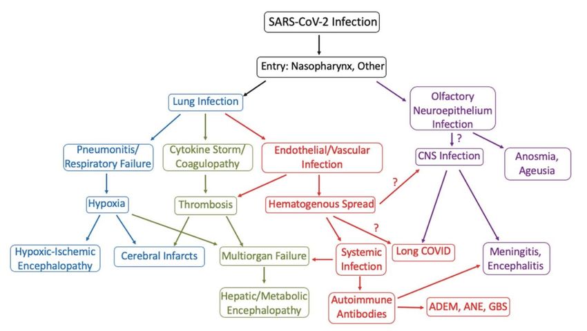
Free Neuropathology 2:2 (2021)                                                                        Jerry J. Lou et al
doi: https://doi.org/10.17879/freeneuropathology-2021-2993                                                page 2 of 14

istry and, in one study, indistinguishable from control sepsis cases. Some cases evince microglial nodules or neu-
ronophagy, strongly supporting viral meningoencephalitis, with a proclivity for involvement of the medulla ob-
longata. The virus is detectable by reverse transcriptase polymerase chain reaction, immunohistochemistry, or
electron microscopy in human cerebrum, cerebellum, cranial nerves, olfactory bulb, as well as in the olfactory
epithelium; neurons and endothelium can also be infected. Review of the extant cases has limitations including
selection bias and limited clinical information in some cases. Much remains to be learned about the effects of
direct viral infection of brain cells and whether SARS-CoV-2 persists long-term contributing to chronic sympto-
matology.

Keywords: CNS, COVID-19, SARS-CoV-2, Brain, Pituitary

                                                             though much remains to be elucidated regarding vi-
1. Introduction                                              ral invasion of the brain, spinal cord and other com-
                                                             ponents of the nervous system.
      In late 2019, a novel infectious disease associ-
ated with pneumonia and acute respiratory distress
emerged in Wuhan, China. The implicated human                2. SARS-CoV-2 virology
coronavirus was genetically related to but distinct
from the underlying viral agent behind the 2003 Se-                SARS-CoV-2 is the latest coronavirus to emerge
vere Acute Respiratory Syndrome (SARS) outbreak,             as a human pathogen. Coronaviruses are single-
SARS coronavirus (SARS-CoV). The International               stranded, positive-sensed ribonucleic acid (RNA) vi-
Committee of Taxonomy of Viruses designated this             ruses subdivided into four genera: alpha-corona-
novel coronavirus as Severe Acute Respiratory Syn-           virus (α-CoV), beta-coronavirus (β-CoV), gamma-
drome coronavirus 2 (SARS-CoV-2)1 and the World              coronavirus (γ-CoV), and delta-coronavirus (δ-CoV).3
Health Organization termed the associated disease            SARS-CoV-2 is a beta-coronavirus, typically 60 to 140
as coronavirus disease 2019 (COVID-19).1 As unsus-           nm4–6 in size, that shares genetic similarities to SARS-
pecting asymptomatic patients readily spread the             CoV (~79% homology) and Middle East Respiratory
disease, the COVID-19 pandemic has swept rapidly             Syndrome coronavirus (MERS-CoV) (~50% homol-
across the world resulting, by the end of December           ogy).7 The SARS-CoV-2 genome encodes 16 non-
2020, in over 79 million confirmed human infections          structural proteins involved in viral replication and
and over 1.7 million deaths.2 These numbers are be-          four structural proteins consisting of the envelope,
lieved to be substantial underestimates of the true          membrane, nucleocapsid, and spike glycoprotein
toll. COVID-19 is best known for its pulmonary in-           (Figure 1).3,8,9 The virus is constantly evolving with
volvement, but other organs are often affected in-           numerous strains of SARS-CoV-2 identified, some
cluding heart, kidney and nervous system. This re-           preferentially localized (at least temporarily) to geo-
view will provide an overview of tissue-based                graphic regions such as Europe or North America.10
COVID-19 neuropathological analyses, almost en-              The mutation rate of SARS-CoV-2 is estimated to be
tirely autopsy derived, as well as a brief discussion        0.84-1.12 x 10⁻³ substitutions per site per year,7,11
of SARS-CoV-2 virology to provide context for under-         which is lower than that of human immunodefi-
standing its pathogenesis, and of diagnostic testing         ciency virus (HIV)12 or influenza A.13 The genetic
limitations that may bias the examined cases. The            spectrum of disparate SARS-CoV-2 strains undergo-
nervous system is affected both secondarily from             ing continued mutation could contribute to the var-
systemic complications such as hypoxia and coag-             iability of neuropathological findings discussed later.
ulopathy, and also likely from primary infection             Antibodies against nucleocapsid and spike proteins
                                                             have been used for immunohistochemical studies.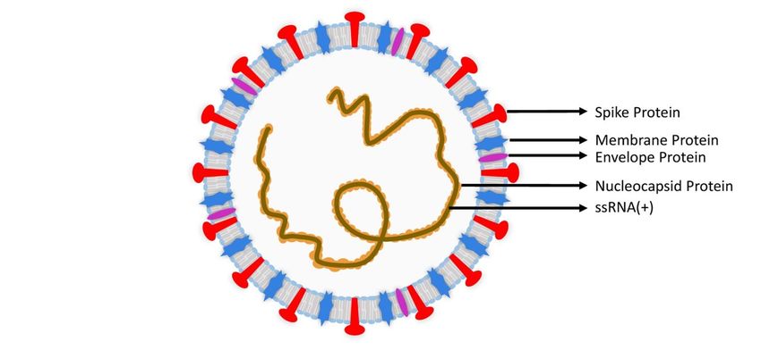
Free Neuropathology 2:2 (2021)                                                                                            Jerry J. Lou et al
doi: https://doi.org/10.17879/freeneuropathology-2021-2993                                                                    page 3 of 14

Figure 1: Severe Acute Respiratory Coronavirus 2 (SARS-CoV-2) structure. There are four structural proteins: spike (S) protein (red), enve-
lope (E) protein (violet), membrane (M) protein (blue), and nucleocapsid (N) protein (orange).

3. ACE2 and other receptors mediate                                       4. Laboratory testing for COVID-19 has
SARS-CoV-2 entry                                                          significant limitations
      The entry of SARS-CoV-2 into human cells is                              In part due to the natural course of disease
commonly thought to be mediated by the interac-                           wherein viral titers rise over time, laboratory testing
tion of the spike protein with the angiotensin con-                       for SARS-CoV-2 does not completely exclude patient
verting enzyme 2 (ACE2) receptor, an important reg-                       infection, complicating the neuropathologist’s man-
ulator of the renin-angiotensin system (RAS).14 Gen-                      agement of surgical or autopsy cases. There may
der, age, lifestyle, smoking, and other patient co-                       also be a bias towards evaluating autopsies of
morbidities are implicated in the modulation of                           COVID-19 patients with severe disease and high viral
ACE2 receptor expression in various tissues.15–21                         loads. The analytical sensitivity of the commonly
ACE2 receptor expression has been reported in the                         used COVID-19 reverse transcriptase polymerase
cerebrum, cerebellum, brainstem, retina, and olfac-                       chain reaction (RT-PCR) assay is excellent with a limit
tory mucosa.22–24 Neurons, vascular pericytes and                         of detection as low as 6.25 copies/µL in a nasopha-
smooth muscle, and glia express the ACE2 recep-                           ryngeal sample.30 In a clinical setting, the sensitivity
tor.22,23,25 In addition to the ACE2 receptor, in vitro                   of RT-PCR may be 83.3% or significantly less, and
studies show that SARS-CoV-2 may gain entry using                         may be affected by infection phase, sample type,
other cell receptors, such as basigin (BSG; CD147),26                     collection procedures, and testing platforms.31,32
neuropilin-1 (NRP1),27 transmembrane serine prote-
ase 2 and 4 (TMPRSS2/4),28,29 and cathepsin L                                  COVID-19 RT-PCR testing may produce false-
(CTSL).9 Expression of ACE2 receptor is highest in ol-                    negative results in the initial phase of infection. In
igodendrocytes, TMPRSS2/4 in neurons, CTSL in mi-                         sequential testing of patients who have symptoms,
croglia, and NRP1 in endothelial cells.25 In principle                    suspicious chest computed tomography (CT) find-
therefore, a broad range of cells in the central nerv-                    ings and an initial negative RT-PCR test, COVID-19
ous system (CNS) have a variety of receptors that                         RT-PCR positivity occurs at a mean of 5.1 ± 1.5 days
may facilitate infection.                                                 after the initial test.32 A patient with a negative RT-
Free Neuropathology 2:2 (2021)                                                                                    Jerry J. Lou et al
doi: https://doi.org/10.17879/freeneuropathology-2021-2993                                                            page 4 of 14

PCR test result in a nasopharyngeal swab but with a                  5. Systemic               pathophysiology                  of
positive SARS-CoV-2 cerebrospinal fluid (CSF) test
has been reported.33 While RT-PCR detection of ge-
                                                                     COVID-19
nomic RNA is not specific for viable virus, identifica-
                                                                           In the first stage of infection, SARS-CoV-2 tar-
tion of sub-genomic RNAs transcribed in infected
                                                                     gets nasal and bronchial epithelial cells as well as
cells have been used in clinical testing to document
                                                                     pneumocytes.28 As infection progresses, SARS-CoV-
the presence of actively replicating virus in lieu of vi-
                                                                     2 infects pulmonary endothelial cells, abrogating the
ral cultures.34 Although chest CT scans may detect
                                                                     epithelial-endothelial barrier.41 A subsequent in-
signs of COVID-19 days before RT-PCR positivity, CT
                                                                     gress of neutrophils and monocytes is followed by
findings overlap with other viral pneumonias.35
                                                                     pulmonary edema and hyaline membrane for-
      Serologic testing has uses for contact tracing,                mation, a component of early acute respiratory dis-
epidemiology, and vaccine studies but is more chal-                  tress syndrome (ARDS).41,42 In severe COVID-19, co-
lenging to use for primary diagnosis given the la-                   agulopathy can occur, reflecting microthrombi for-
tency for development of antibodies.36–40 The most                   mation secondary to endothelial cell inflammation
common serological assays are the rapid lateral flow                 and cytokine storms.41,43 COVID-19 associated hy-
assay, enzyme linked immunosorbent assay (ELISA),                    percoagulability induces venous thromboembolism
and virus neutralization assay. Two-step ELISA as-                   and arterial occlusion.41 The interplay of these sys-
says are quantitative and more reliable than flow as-                temic derangements likely contributes to the patho-
says that are easily scalable but are qualitative.40 The             logic changes seen in the CNS (Figure 2).
virus neutralization assay detects and quantifies an-
tibodies that inhibit viral replication but is techni-
cally complex.37

Figure 2: Flow chart modeling COVID-19 pathogenesis and neurological dysfunction. ADEM= Acute disseminated encephalomyelitis; ANE=
Acute necrotizing encephalopathy; GBS= Guillain-Barré syndrome.
Free Neuropathology 2:2 (2021) Jerry J. Lou et al
doi: https://doi.org/10.17879/freeneuropathology-2021-2993 page 5 of 14
6. COVID-19 neurological manifesta- been documented53,54 though the exact basis for ol-
factory dysfunction and recovery remains to be re-
tions solved.
COVID-19 associated neurological manifesta-
tions range from mild symptoms such as dizziness, Stroke
headache, dysgeusia, or anosmia to severe disorders
such as stroke, Guillain-Barré syndrome (GBS), acute Stroke is the most common debilitating neuro-
hemorrhagic necrotizing encephalopathy, menin- logical disorder associated with COVID-19 and has a
goencephalitis, and cerebral venous thrombosis. predilection for males and the elderly. Two retro-
The frequency of reported neurological signs and spective New York studies reported respectively
symptoms is variable but substantial regardless. In that 1.6% of 1,916 patients with hospitalizations or
an early Chinese retrospective study, 36.4% of 214 emergency department visits for COVID-19 and 0.9%
COVID-19 patients had neurological symptoms of 3,556 hospitalized COVID-19 patients had radio-
which included dizziness (16.8%), headache (13.1%), logically-confirmed ischemic infarcts.55,56 Crypto-
impaired consciousness (7.5%), dysgeusia (5.6%), genic strokes were twice as common in hospitalized
and anosmia (5.1%).44 In Western studies, dysgeusia COVID-19 patients compared to either contempo-
and anosmia are reported in the majority of pa- rary or historical controls.56 A third New York retro-
tients.45,46 A French study reports that 49 out of 58 spective study found 1.1% of 3,218 COVID-19 pa-
(84%) COVID-19 intensive care unit (ICU) patients tients had strokes; in the small subset with acute
had neurological signs which included agitation neuroimaging, 68.5% of strokes were ischemic
(69%), confusion (65%), corticospinal tract signs (44.5% large vessel, 24% lacunar) and 24% were
(67%), and dysexecutive syndrome (33%).47 A study hemorrhagic.57 A retrospective Chinese study of 214
from a British referral center also describes cases of COVID-19 patients reported strokes in six patients
septic or para-infectious encephalopathy, autoim- (2.8%).44 A small American case series reported large
mune encephalitis including acute disseminated en- vessel ischemic strokes in five COVID-19 patients
cephalomyelitis (ADEM), and GBS.48 younger than 50, four of whom had no prior history
of stroke.58 For comparison, the average incidence
of stroke among patients admitted through a U.S.
7. Specific neurological disorders asso- emergency department prior to the pandemic was
ciated with COVID-19 approximately 3.2%.59 Of patients who visited emer-
gency departments or were hospitalized, only 0.2%
Olfactory and gustatory dysfunction of 1486 influenza patients had ischemic infarcts
which was significantly less than COVID-19 patients
In the aforementioned study from China, even after adjusting for age, sex and, race.55
COVID-19 patients had gustatory dysfunction and ol-
factory dysfunction at frequencies of less than 6% In another New York study, 33 out of 755
each.44 In a prospective European study, 88.8% of (4.4%) COVID-19 patients with neuroimaging had
385 COVID-19 patients had gustatory dysfunction evidence of intracranial hemorrhage not associated
and 85.6% of 417 COVID-19 patients had olfactory with trauma, brain metastases, or tumor resec-
dysfunction.45 In a California study, 71% of 59 tion.60 Parenchymal hemorrhages with mass effect
COVID-19 patients recorded ageusia and 68% re- and herniation were present in five patients, punc-
ported anosmia.46 Olfactory and gustatory dysfunc- tate hemorrhages in seven, small to moderate sized
tion in COVID-19 typically resolves after 17 to 30 hemorrhages in 17, and large single site hemor-
days from initial onset.49,50 Although long term fol- rhages without herniation in four.60 Twenty-six of
low-up is generally lacking,51 one study reports res- the 33 intracranial hemorrhages occurred as a trans-
olution rates at day 30 in home-quarantined COVID- formation of an ischemic infarct.60 Of the five pa-
19 patients of 87% for olfactory dysfunction and tients with parenchymal hemorrhages, four had high
82% for gustatory dysfunction.52 Presence of SARS- partial thromboplastin time or anti-factor Xa in the
CoV-2 virions in the olfactory neuroepithelium of 72 hours before the intracranial hemorrhage.60
the nasal mucosa as well as in the olfactory bulb hasFree Neuropathology 2:2 (2021) Jerry J. Lou et al
doi: https://doi.org/10.17879/freeneuropathology-2021-2993 page 6 of 14
Advanced age, cardiovascular disease, cerebro- Japanese male presented with headache, general-
vascular disease, diabetes, chronic respiratory dis- ized fatigue and fever. Nine days after onset of typi-
ease, hypertension, obesity, smoking, and cancer cal COVID-19 symptoms, the patient had altered
are risk factors for severe COVID-19 disease or poor mental status, transient generalized seizures, and
outcome.61–67 In a Canadian study, critically ill neck stiffness. Brain magnetic resonance imaging
COVID-19 patients with blood groups A and AB were (MRI) showed hyperintensities along the lateral ven-
more likely to require ventilation, continuous renal tricle and in the medial temporal lobe including the
replacement therapy, and prolonged intensive care hippocampus. SARS-CoV-2 RNA was not detected by
unit admission.68 In contrast, an earlier American RT-PCR in a nasopharyngeal swab but was in CSF.33
COVID-19 study did not show an association be- Autopsies performed on COVID-19 patients have
tween specific blood groups and either ventilation suggested meningoencephalitis in some cases and
or death.69 Differences in cohorts including mortality these are discussed in section 8.79
rates may account for the contradictory findings.68 It
cannot escape notice that a number of the afore- Other disorders
mentioned COVID-19 prognostic factors are associ-
ated with atherosclerosis and arteriolosclerosis that, A Brazilian study reported central venous
in the setting of COVID-19 respiratory compromise thrombosis (CVT) in three previously healthy COVID-
and coagulopathy, may contribute to strokes. 19 patients younger than 41 years, underscoring the
need for awareness of a hypercoagulable state in
Guillain-Barré syndrome COVID-19 even in this age group.80
GBS is an uncommon, immune-mediated de- Acute hemorrhagic necrotizing encephalopa-
myelinating disease of the peripheral nerves that of- thy, an uncommon complication characterized by
ten follows viral infections. Common presenting multifocal symmetric brain lesions including bilat-
symptoms in COVID-19 patients include symmetrical eral thalamic lesions, has been associated with
flaccid quadriparesis, ataxia, facial weakness, respir- COVID-19, though rarely.81,82 This complication may
atory failure, and lower paresthesia.70–72 RT-PCR of be caused by blood-brain-barrier disruption related
nasopharyngeal swabs tested positive for SARS-CoV- to intracranial cytokine storms.81 COVID-19 associ-
2 but CSF was negative for tested patients.73–75 GBS ated cases of ADEM-like disease and autoimmune
symptoms tend to emerge between day five to 10 encephalitis have been documented.72,83
after COVID-19 symptom onset.71 Over 30 cases of
GBS have been reported.73–76 One proposed mecha- 8. Neuropathological findings and
nism is an autoimmune hyperreaction, triggering re- neuropathogenesis of COVID-19
lease of pro-inflammatory mediators such as inter-
leukin (IL)-6, that cause autoimmune demyelination To examine the spectrum of COVID-19 neuro-
or axonal damage. Alternatively in some cases, pathology, we reviewed 20 papers encompassing
SARS-CoV-2 may induce production of antibodies 184 patients with tissue-based neuropathological
targeting gangliosides, leading to peripheral neurop- analyses including 101 cases analyzed by RT-PCR for
athy.72,77 COVID-19 associated Miller Fisher syn- SARS-CoV-2 and 83 cases by immunohistochemistry
drome (MFS), a variant of GBS with ophthalmople- (IHC) (Table 1).25,53,79,83–99 All cases are autopsies ex-
gia, areflexia, and ataxia, and polyneuritis cranialis cept for four biopsies and one case of unspecified
(PNC), a GBS variant with multiple cranial neuropa- type. The range of histologic findings is broad and in
thies, have been reported.78 part reflects the heterogeneity of neurological find-
ings. The most frequent findings (Table 1) include
Meningitis and encephalitis microglial activation with microglial nodules in a
likely underestimated subset; lymphoid inflamma-
COVID-19 associated meningitis has only infre- tion including perivascular lymphocytosis (Figure
quently been reported but one case highlights the 3a), parenchymal lymphocytic infiltration, and lep-
need for CSF testing if suspected.33 A 24-year-old tomeningeal lymphocytic inflammation; hypoxic-is-Free Neuropathology 2:2 (2021) Jerry J. Lou et al
doi: https://doi.org/10.17879/freeneuropathology-2021-2993 page 7 of 14
chemic changes (Figure 3b); astrogliosis; acute/sub- neuroepithelium (which resides in the mucosa of the
acute brain infarcts; primary hemorrhage; and mi- nasal cavity) and from there to the olfactory bulb
crothrombi. It should be noted that the prevalence and then to other brain regions. This olfactory route
of detected lymphoid inflammation and microglial is used by other coronaviruses, such as SARS-CoV102
nodules is high in papers with immunohistochemical and MERS-CoV.103 COVID-19 patients frequently dis-
studies and low in those without. play MRI hyperintensity in the olfactory cortex.104
The ACE2 receptor, used by SARS-CoV-2 for cellular
Table 1: Neuropathological findings in COVID-19 entry, is expressed in sustentacular cells and stem
brain tissue cells of the nasal olfactory epithelium.23,105 While
one study suggests olfactory neurons themselves do
not express the ACE2 receptor,23 another report
contradicts this finding.106 Besides technical sensitiv-
ity issues, it may be that the expression levels of the
ACE2 receptor and other receptors differ under in-
flammatory conditions as compared to physiologic
conditions. Viral particles have been detected by
electron microscopy and IHC in the olfactory epithe-
lium54 as well as by electron microscopy in olfactory
neurons of the nasal mucosa at autopsy.53 Morbini
and colleagues report ultrastructural evidence of
SARS-CoV-2 particles in the olfactory bulb of a
COVID-19 patient.54 A second theory posits that
SARS-CoV-2 may infect immune cells that cross the
blood-brain barrier and deliver virus into the brain.
This mechanism is well described in HIV.107 White
blood cells including lymphocytes and monocytes
express the ACE2 receptor.108,109 Infection of im-
mune cells by SARS-CoV-2 is an active area of study.
Finally, the third theory extends the well docu-
mented behavior of encephalitic blood-borne coro-
naviruses to enter through the blood-brain barrier
via infection of vessel wall cells.110
Histologically, on routine hematoxylin- and eo-
sin-stained slides, neither viral inclusions nor spe-
Table 1: Some histologic findings are likely to be under-reported
cific cellular changes recognizable as direct viral in-
as reviewed studies are variable in their focus. *Only studies
tabulating the prevalence of microglial nodules are included. fection have been reported. However, SARS-CoV-2
Studies mentioning microglial nodules in their case series but has been detected in the brain by RT-PCR, IHC, and
not enumerating the frequency are excluded. electron microscopy. As many as 54 out of 101
(53.5%) cases were positive for SARS-CoV-2 in the
SARS-CoV-2 is detectable in central nervous sys- brain by RT-PCR and 23 out of 83 (27.7%) cases were
tem tissue positive by IHC (Table 1). In some cases, copies of vi-
rus detected by RT-PCR were low in number and the
At least three routes of CNS infection by SARS- detection of virus in blood or blood cells within in-
CoV-2 have been proposed: retrograde transmission tracerebral vasculature rather than brain cells was a
via olfactory sensory neurons, infiltration of immune possibility. However, IHC has confirmed the pres-
cells, and entry across the blood-brain barrier.100,101 ence of viral antigens in autopsy brain cells. Of note,
The prevalence of anosmia and ageusia in COVID-19 antibodies against SARS-CoV-2 spike protein were
patients led to the theory that SARS-CoV-2 enters more effective in detecting viral antigens than those
the brain via infection of neurons in the olfactoryFree Neuropathology 2:2 (2021) Jerry J. Lou et al
doi: https://doi.org/10.17879/freeneuropathology-2021-2993 page 8 of 14
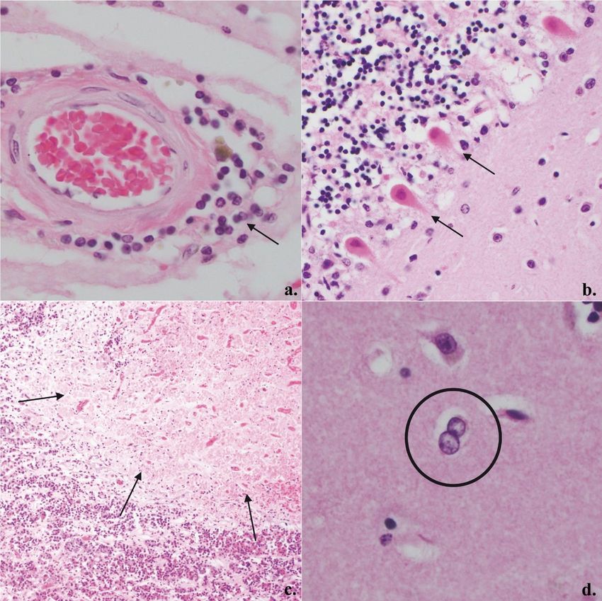
Figure 3: Representative COVID-19 histopathology: a. Mild perivascular lymphoid inflammation, 400X (arrow); b. Eosinophilia in Purkinje
cells compatible with acute hypoxic-ischemic change, 200X (arrows); c. Subacute infarct of anterior pituitary gland, 100X (arrows); d.
Alzheimer Type II astrocytes in basal ganglia, 400X (circle).
targeted against nucleocapsid protein25,87,97 (Supple- related to the quality of the antibodies or the intrin-
mentary Table A). Importantly, staining using both sic accessibility of the relevant epitopes or both is
anti-spike and anti-nucleocapsid antibodies may be unresolved. Staining of virus localizes in scattered
more sensitive than either alone as, in a few cases, cortical neurons and endothelial cells,87 as well as
nucleocapsid protein was detected while spike pro- brainstem cranial nerve roots and isolated cells (cell
tein was not. Whether this finding is a technical issue type unclear) in the medulla oblongata; the images
of the isolated cells had a striking lack of attendantFree Neuropathology 2:2 (2021) Jerry J. Lou et al
doi: https://doi.org/10.17879/freeneuropathology-2021-2993 page 9 of 14
chronic inflammation.25 This disconnect between vi- lymphocytic infiltration is observed to similar de-
ral infection and inflammation raises the question of grees in the control brains of septic (non-COVID-19)
immune evasion. One case exhibited viral staining patients when compared to COVID-19 patients.86
around the edges of subcortical white matter mi-
croinfarcts.87 The immunostaining pattern consists Some cases present findings compatible with
of diffuse cytoplasmic and perinuclear positivity viral meningoencephalitis including parenchymal
with small concentrated foci possibly representing lymphocytic clustering around microglial nodules,
viral inclusion bodies.87 Meinhardt and colleagues, concomitant leptomeningeal lymphocytic inflam-
using in situ hybridization (ISH), identified SARS- mation, and even neuronophagy. In the study by
CoV-2 in the olfactory epithelium and mucus.53 Matschke and colleagues, four out of 16 cases im-
Lastly, viral particles compatible with SARS-CoV-2 munopositive for SARS-CoV-2 had 10 to 49 CD8+ cy-
have been identified by electron microscopy in ol- totoxic T lymphocytes per high power field in at least
factory bulb and frontal lobe tissue.54,96 To date, re- three fields in the medulla oblongata.25 The ob-
gions in which SARS-CoV-2 have been detected by served lymphocytes tended to cluster near activated
IHC, RT-PCR, or electron microscopy relevant to the microglial nodules,25 as commonly seen in viral en-
CNS include cornea, conjunctiva, olfactory epithe- cephalitis. RT-PCR detected SARS-CoV-2 RNA in
lium, olfactory bulb, olfactory tubercle, frontal lobe, three out of the four cases.25 Three studies report
cerebellum, medulla oblongata, cranial nerves and neuronophagy in the medulla associated with histi-
trigeminal ganglion. Most investigations studied lim- ocytic and lymphocytic parenchymal infiltra-
ited areas of brain or did not specify the origin of tion.25,84,89 von Weyhern and colleagues report six
cerebral cortex tissue. Additional studies are needed cases of perivascular and parenchymal lymphocyto-
to better establish the frequency and extent of di- sis with neuronal loss and axon degeneration in the
rect infection in the CNS. As some patients have brainstem, which the authors determined to be ad-
"Long COVID” wherein they have persistent symp- equate to diagnose SARS-CoV-2 viral encephalitis.79
toms for months, whether and how SARS-CoV-2 may In the same study, five of the six cases had concom-
persist in the brain will need to be evaluated in the itant meningitis.79 Respiratory and cardiovascular
future. control centers of the medulla may be affected.53
While the majority of COVID-19 patients with neuro-
Chronic inflammatory and reactive glial changes logical manifestations do not have detectable SARS-
CoV-2 RNA in CSF by RT-PCR,111–113 exceptions to this
Microgliosis and astrocytosis are common in trend have been reported in at least four COVID-19
COVID-19 brains including in the olfactory bulb.25 patients.33,114–116 Anti-SARS-CoV-2 antibodies were
Lymphoid inflammation, which tends to be minimal detected in the CSF of one COVID-19 patient sug-
or mild in many cases, is not uncommon particularly gesting an immune response to viral infection.87 It
in the medulla oblongata and if IHC is used for de- appears therefore that histologically documentable
tection.25 In many studies that did not use IHC, lym- cases of SARS-CoV-2 encephalitis and/or meningitis
phoid inflammation was reported as absent or infre- do occur, with a tendency to involve the brainstem.
quent; this is concordant with our experience and As only a few series report the majority of brainstem
anecdotally with that of colleagues in Asia and in the microglial encephalitis cases, the question arises as
United States (personal communication). A substan- to whether particular viral strains, therapeutic ap-
tial portion of these reactive changes may be sec- proaches, genetic background or detection methods
ondary to systemic issues (e.g. sepsis) or other neu- are responsible for its apparent absence or paucity
ropathology (infarcts, hemorrhages, etc.) rather in the majority of studies.
than a response to direct infection. Microgliosis and Autoimmune mediated inflammation may also
chronic inflammation did not correlate with the se- occur. Autoimmune encephalitis, ADEM, and acute
verity of COVID-19 disease nor were there any dis- necrotizing encephalopathy have also been re-
cernible neuropathological differences in patients ported clinically though histopathologic evaluation
from nursing homes, hospital wards, or intensive has been limited. ADEM-like pathology has been re-
care units.25 Furthermore, microglial activation, ported in one autopsy case.83
perivascular lymphocytosis, and leptomeningealFree Neuropathology 2:2 (2021) Jerry J. Lou et al
doi: https://doi.org/10.17879/freeneuropathology-2021-2993 page 10 of 14
Microthrombi, infarcts, hemorrhages, and “neu- 9. Conclusions
trophilic plugs”
Systemic dysfunction and viral infection of the
SARS-CoV-2 may induce a cytokine storm, a se- CNS can cause a wide range of COVID-19 related
vere hyperimmune reaction characterized by exces- neuropathological changes. The incidence of neuro-
sive and rapid release of cytokines such as IL-6 and pathological findings should be viewed with caution
IL-1β into the blood.117,118 IL-6 activates the coagula- given great variability in what tissues were studied
tion system and increases vascular permeability119 and what types of ancillary studies were (or were
which, in combination with viral endotheliopathy,120 not) performed. Inflammatory changes were re-
may account for the well documented COVID-19 as- ported in a high percentage of cases in some series
sociated coagulopathy.41,121 SARS-CoV-2 has been but not in others, at least in part due to the use of
detected within cerebral endothelial cells by IHC and immunostaining; however, the lack of controls in
electron microscopy.87,96 Hypercoagulability, in turn, most of these studies limit interpretation of these
results in histologic findings of microthrombi, in- findings. Selection or referral bias, the decedent’s
farcts, and hemorrhages. Also, IL-1β plays a major co-morbidities, therapies given, patient’s genetic
role in triggering vascular “neutrophilic plugs” con- background, immune status, immunization status
taining neutrophils and/or platelets as well as neu- and perhaps viral strains of SARS-CoV-2 may also
trophil extracellular traps, a mesh of deoxynucleic contribute to differences and will need to be dis-
acid (DNA)-rich material coated with antimicrobials sected out in the future. Whether or how much of
that entrap and kill microbes.122 These “neutrophilic the inflammatory changes seen are due to autoim-
plugs” have been found in the brain as well as the mune phenomena versus direct viral infection or
lungs, heart, kidneys, and liver of COVID-19 patients other causes remains to be resolved. The presence
who come to autopsy.88 Approximately 1.6% of of microglial nodules and detectable virus are indic-
cases have “neutrophilic plugs” in the brain (Table 1) ative of viral meningoencephalitis in some cases; a
though this percentage is likely an underestimate as predilection for the medulla perhaps with compro-
pathologists do not typically evaluate for these mise of respiratory and cardiovascular control cen-
structures. Pneumonia and ARDS with resultant hy- ters may compound the COVID-19 patient’s often al-
poxemia as well as pre-existing arteriosclerosis are ready tenuous cardiorespiratory function. While
also likely contributory to the cerebral infarcts. In there is evidence for an olfactory route of infection,
addition to cerebral infarcts, we have noted occa- it is unclear whether this is the major mechanism of
sional cases of pituitary infarcts (Figure 3c) in our CNS infection given that the virus is rarely detecta-
COVID-19 autopsies. Numerous extramedullary ble in the olfactory bulb. Cerebral endothelial infec-
megakaryocytes were present in subacute cerebral tion is also present and a hematogenous route of in-
infarcts of one COVID-19 patient.84 fection is therefore plausible perhaps even com-
mon. Data on where the virus is detectable in the
Alzheimer Type II astrocytosis brain is limited as studies have focused on the
frontal lobe and brainstem. It would not be surpris-
Alzheimer Type II astrocytosis (Figure 3d) char- ing to find a greater extent of infection than cur-
acteristic of hepatic encephalopathy has been re- rently confirmed. It should be kept in mind that the
ported.89,97 The reports do not specify whether the sensitivity of virus detection is suboptimal given that
frequency of the Alzheimer Type II astrocytes most samples are derived from autopsies. Lastly, it
reaches a threshold of five or more per 20 high is notable that inflammation does not always coin-
power fields, the cutoff suggested by Agarwal and cide with virus localization, raising the concern that
colleagues for hepatic encephalopathy.123 In a paper the virus may be evading the immune response in
by Solomon and colleagues, four out of four cases the CNS. Whether the CNS is a potential long-term
with Alzheimer Type II astrocytes had chronic liver reservoir of virus and a contributor to “Long COVID”
disease or alcohol use disorder.97 The relative con- remains to be seen. Lastly, increased risk for vascu-
tribution of hepatic encephalopathy to cases cur- lar dementia or neurodegenerative disorders are hy-
rently ascribed as septic or para-infectious encepha- pothetical concerns that may need to be addressed
lopathy remains to be seen. at a later time.Free Neuropathology 2:2 (2021) Jerry J. Lou et al
doi: https://doi.org/10.17879/freeneuropathology-2021-2993 page 11 of 14
Funding Singer, W. Yong) and in part by the UCLA Broad Stem
Cell Research Center COVID 19 Research Award
This work was supported in part by NIMH OCRC #20-70 (H.V. Vinters, S. Magaki, and T. Zhang).
2U24MH10092 and U01 MH083500 (S. Magaki, E.
References
1. Naming the coronavirus disease (COVID-19) and the virus that causes 15. Wang K, Chen W, Zhang Z, et al. CD147-spike protein is a novel route
it. World Health Organization. Accessed October 21, 2020. for SARS-CoV-2 infection to host cells. Signal Transduct Target Ther.
https://www.who.int/emergencies/diseases/novel-coronavirus-2019/ 2020;5(1):283. doi:10.1038/s41392-020-00426-x
technical-guidance/naming-the-coronavirus-disease-(covid-2019)-and-
the-virus-that-causes-it 16. Fernández-Atucha A, Izagirre A, Fraile-Bermúdez AB, et al. Sex
differences in the aging pattern of renin-angiotensin system serum
2. Weekly epidemiological update - 29 December 2020. World Health peptidases. Biol Sex Differ. 2017;8(1). doi:10.1186/s13293-017-0128-8
Organization. Accessed December 30, 2020.
https://www.who.int/publications/m/item/weekly-epidemiological- 17. Hu Y, Li X, Wu N, Wang N, Qui C, Li J. Study on the correlation among
update---29-december-2020 sex, age, and the activity of ACE, ACE2 and the ratio of ACE/ACE2. J
Qiqihar Med Coll. 2018;39(8):884-887. doi:10.3969/j.issn.1002-
3. Ye ZW, Yuan S, Yuen KS, Fung SY, Chan CP, Jin DY. Zoonotic origins of 1256.2018.08.005
human coronaviruses. Int J Biol Sci. 2020;16(10):1686-1697.
doi:10.7150/ijbs.45472 18. Liu Y, Zhao L, Zhang Q, Zhang L, Ren G. Effect of long-term smoking
on expressiong of serum ACE and ACE2 as well as its significance. J
4. Zhu N, Zhang D, Wang W, et al. A Novel Coronavirus from Patients Taishan Med Coll. 2019;40(4):258-260. doi:10.3969/j.issn.1004-
with Pneumonia in China, 2019. N Engl J Med. 2020;382(8):727-733. 7115.2019.04.007
doi:10.1056/NEJMoa2001017
19. Smith JC, Sausville EL, Girish V, et al. Cigarette Smoke Exposure and
5. Caly L, Druce J, Roberts J, et al. Isolation and rapid sharing of the 2019 Inflammatory Signaling Increase the Expression of the SARS-CoV-2
novel coronavirus (SARS-CoV-2) from the first patient diagnosed with Receptor ACE2 in the Respiratory Tract. Dev Cell. 2020;53(5):514-
COVID-19 in Australia. Med J Aust. 2020;212(10):459-462. 529.e3. doi:https://doi.org/10.1016/j.devcel.2020.05.012
doi:10.5694/mja2.50569
20. Bernardi S, Toffoli B, Zennaro C, et al. High-salt diet increases
6. Gulholm T, Basile K, Kok J, Chen S, Rawlinson W. Laboratory diagnosis glomerular ACE/ACE2 ratio leading to oxidative stress and kidney
of severe acute respiratory syndrome coronavirus 2. Pathology. damage. Nephrol Dial Transplant. 2012;27(5):1793-1800.
2020;52(7):745-753. doi:10.1016/j.pathol.2020.09.011 doi:10.1093/ndt/gfr600
7. Koyama T, Platt D, Parida L. Variant analysis of SARS-cov-2 genomes. 21. Lavrentyev EN, Malik KU. High glucose-induced Nox1-derived
Bull World Health Organ. 2020;98(7):495-504. superoxides downregulate PKC-βII, which subsequently decreases ACE2
doi:10.2471/BLT.20.253591 expression and ANG(1-7) formation in rat VSMCs. Am J Physiol - Hear
Circ Physiol. 2009;296(1):106-118. doi:10.1152/ajpheart.00239.2008
8. Naqvi AAT, Fatima K, Mohammad T, et al. Insights into SARS-CoV-2
genome, structure, evolution, pathogenesis and therapies: Structural 22. Xia H, Lazartigues E. Angiotensin-converting enzyme 2 in the brain:
genomics approach. Biochim Biophys acta Mol basis Dis. properties and future directions. J Neurochem. 2008;107(6):1482-1494.
2020;1866(10):165878. doi:10.1016/j.bbadis.2020.165878 doi:10.1111/j.1471-4159.2008.05723.x
9. Ou X, Liu Y, Lei X, et al. Characterization of spike glycoprotein of SARS- 23. Brann D, Tsukahara T, Weinreb C, et al. Non-neuronal expression of
CoV-2 on virus entry and its immune cross-reactivity with SARS-CoV. Nat SARS-CoV-2 entry genes in the olfactory system suggests mechanisms
Commun. 2020;11(1). doi:10.1038/s41467-020-15562-9 underlying COVID-19-associated anosmia. Sci Adv.
2020;6(31):eabc5801. doi:10.1126/sciadv.abc5801
10. Pachetti M, Marini B, Benedetti F, et al. Emerging SARS-CoV-2
mutation hot spots include a novel RNA-dependent-RNA polymerase 24. Chen M, Shen W, Rowan NR, et al. Elevated ACE2 expression in the
variant. J Transl Med. 2020;18(1):1-9. doi:10.1186/s12967-020-02344-6 olfactory neuroepithelium: implications for anosmia and upper
respiratory SARS-CoV-2 entry and replication. Eur Respir J.
11. Day T, Gandon S, Lion S, Otto SP. On the evolutionary epidemiology 2020;56(3):2001948. doi:10.1183/13993003.01948-2020
of SARS-CoV-2. Curr Biol. 2020;30(15):R849-R857. doi:10.1016
/j.cub.2020.06.031 25. Matschke J, Lütgehetmann M, Hagel C, et al. Neuropathology of
patients with COVID-19 in Germany: a post-mortem case series. Lancet
12. Rowland-Jones S, Andrews SM. Recent advances in understanding Neurol. 2020;19(11). doi:10.1016/S1474-4422(20)30308-2
HIV evolution. F1000Research. 2017;6(0):1-7. doi:10.12688
/f1000research.10876.1 26. Wang K, Chen W, Zhou Y Sen, et al. SARS-CoV-2 invades host cells via
a novel route: CD147-spike protein. bioRxiv. Published online January 1,
13. Nobusawa E, Sato K. Comparison of the Mutation Rates of Human 2020:2020.03.14.988345. doi:10.1101/2020.03.14.988345
Influenza A and B Viruses. J Virol. 2006;80(7):3675-3678.
doi:10.1128/jvi.80.7.3675-3678.2006 27. Cantuti-Castelvetri L, Ojha R, Pedro LD, et al. Neuropilin-1 facilitates
SARS-CoV-2 cell entry and provides a possible pathway into the central
14. Gheblawi M, Wang K, Viveiros A, et al. Angiotensin-Converting nervous system. bioRxiv. Published online January 1,
Enzyme 2: SARS-CoV-2 Receptor and Regulator of the Renin-Angiotensin 2020:2020.06.07.137802. doi:10.1101/2020.06.07.137802
System: Celebrating the 20th Anniversary of the Discovery of ACE2. Circ
Res. 2020;126(10):1456-1474. doi:10.1161/CIRCRESAHA.120.317015 28. Hoffmann M, Kleine-Weber H, Schroeder S, et al. SARS-CoV-2 Cell
Entry Depends on ACE2 and TMPRSS2 and Is Blocked by a ClinicallyFree Neuropathology 2:2 (2021) Jerry J. Lou et al
doi: https://doi.org/10.17879/freeneuropathology-2021-2993 page 12 of 14
Proven Protease Inhibitor. Cell. 2020;181(2):271-280.e8. study. Eur Arch Oto-Rhino-Laryngology. 2020;277(8):2251-2261.
doi:10.1016/j.cell.2020.02.052 doi:10.1007/s00405-020-05965-1
29. Zang R, Gomez Castro MF, McCune BT, et al. TMPRSS2 and TMPRSS4 46. Yan CH, Faraji F, Prajapati DP, Boone CE, DeConde AS. Association of
promote SARS-CoV-2 infection of human small intestinal enterocytes. chemosensory dysfunction and COVID-19 in patients presenting with
Sci Immunol. 2020;5(47):eabc3582. doi:10.1126/sciimmunol.abc3582 influenza-like symptoms. Int Forum Allergy Rhinol. 2020;10(7):806-813.
doi:10.1002/alr.22579
30. COVID-19 RT-PCR test - Emergency Use Authorization (EUA)
summary. Laboratory Corporation of America. Accessed September 2, 47. Helms J, Kremer S, Merdji H, et al. Neurologic Features in Severe
2020. https://www.fda.gov/media/136151/download SARS-CoV-2 Infection. N Engl J Med. 2020;382(21):2268-2270.
doi:10.1056/nejmc2008597
31. Long C, Xu H, Shen Q, et al. Diagnosis of the Coronavirus disease
(COVID-19): rRT-PCR or CT? Eur J Radiol. 2020;126:108961. 48. Paterson RW, Brown RL, Benjamin L, et al. The emerging spectrum
doi:10.1016/j.ejrad.2020.108961 of COVID-19 neurology: clinical, radiological and laboratory findings.
Brain. 2020;143(10):3104-3120. doi:10.1093/brain/awaa240
32. Ai T, Yang Z, Hou H, et al. Correlation of Chest CT and RT-PCR Testing
in Coronavirus Disease 2019 (COVID-19) in China: A Report of 1014 49. Paolo G. Does COVID-19 cause permanent damage to olfactory and
Cases. Radiology. 2020;296(2):E32-E40. doi:10.1148 gustatory function? Med Hypotheses. 2020;143:110086.
/radiol.2020200642 doi:10.1016/j.mehy.2020.110086
33. Moriguchi T, Harii N, Goto J, et al. A first Case of 50. Hopkins C, Surda P, Whitehead E, Kumar BN. Early recovery following
Meningitis/Encephalitis associated with SARS-Coronavirus-2. Int J Infect new onset anosmia during the COVID-19 pandemic – an observational
Dis. 2020;94:55-58. doi:10.1016/j.ijid.2020.03.062 cohort study. J Otolaryngol - Head Neck Surg. 2020;49(1):26.
doi:10.1186/s40463-020-00423-8
34. Perera RAPM, Tso E, Tsang OTY, et al. SARS-CoV-2 Virus Culture and
Subgenomic RNA for Respiratory Specimens from Patients with Mild 51. Whitcroft KL, Hummel T. Olfactory Dysfunction in COVID-19:
Coronavirus Disease. Emerg Infect Dis J. 2020;26(11):2701. Diagnosis and Management. JAMA. 2020;323(24):2512-2514.
doi:10.3201/eid2611.203219 doi:10.1001/jama.2020.8391
35. Dai W-C, Zhang HW, Yu J, et al. CT Imaging and Differential Diagnosis 52. Paderno A, Mattavelli D, Rampinelli V, et al. Olfactory and Gustatory
of COVID-19. Can Assoc Radiol J. 2020;71(2):195-200. Outcomes in COVID-19: A Prospective Evaluation in Nonhospitalized
doi:10.1177/0846537120913033 Subjects. Otolaryngol Head Neck Surg. 2020;163(6):1144-1149.
doi:10.1177/0194599820939538
36. Guo L, Ren L, Yang S, et al. Profiling Early Humoral Response to
Diagnose Novel Coronavirus Disease (COVID-19). Clin Infect Dis an Off 53. Meinhardt J, Radke J, Dittmayer C, et al. Olfactory transmucosal
Publ Infect Dis Soc Am. Published online March 2020. SARS-CoV-2 invasion as a port of central nervous system entry in
doi:10.1093/cid/ciaa310 individuals with COVID-19. Nat Neurosci. Published online 2020.
doi:10.1038/s41593-020-00758-5
37. Gauger PC, Vincent AL. Serum Virus Neutralization Assay for
Detection and Quantitation of Serum Neutralizing Antibodies to 54. Morbini P, Benazzo M, Verga L, et al. Ultrastructural Evidence of
Influenza A Virus in Swine. Methods Mol Biol. 2020;2123:321-333. Direct Viral Damage to the Olfactory Complex in Patients Testing
doi:10.1007/978-1-0716-0346-8_23 Positive for SARS-CoV-2. JAMA Otolaryngol Neck Surg.
2020;146(10):972-973. doi:10.1001/jamaoto.2020.2366
38. Okba NMA, Müller MA, Li W, et al. Severe Acute Respiratory
Syndrome Coronavirus 2-Specific Antibody Responses in Coronavirus 55. Merkler AE, Parikh NS, Mir S, et al. Risk of Ischemic Stroke in Patients
Disease 2019 Patients. Emerg Infect Dis. 2020;26(7). with Coronavirus Disease 2019 (COVID-19) vs Patients with Influenza.
doi:10.3201/eid2607.200841 JAMA Neurol. 2020;77(11):1–7. doi:10.1001/jamaneurol.2020.2730
39. Sun B, Feng Y, Mo X, et al. Kinetics of SARS-CoV-2 specific IgM and 56. Yaghi S, Ishida K, Torres J, et al. SARS-CoV-2 and Stroke in a New York
IgG responses in COVID-19 patients. Emerg Microbes Infect. Healthcare System. Stroke. 2020;51(7):2002-2011.
2020;9(1):940-948. doi:10.1080/22221751.2020.1762515 doi:10.1161/STROKEAHA.120.030335
40. Krammer F, Simon V. Serology assays to manage COVID-19. Science. 57. Jain R, Young M, Dogra S, et al. COVID-19 related neuroimaging
2020;368(6495):1060-1061. doi:10.1126/science.abc1227 findings: A signal of thromboembolic complications and a strong
prognostic marker of poor patient outcome. J Neurol Sci.
41. Wiersinga WJ, Rhodes A, Cheng AC, Peacock SJ, Prescott HC. 2020;414:116923. doi:10.1016/j.jns.2020.116923
Pathophysiology, Transmission, Diagnosis, and Treatment of
Coronavirus Disease 2019 (COVID-19): A Review. JAMA. 58. Oxley TJ, Mocco J, Majidi S, et al. Large-Vessel Stroke as a Presenting
2020;324(8):782-793. doi:10.1001/jama.2020.12839 Feature of COVID-19 in the Young. N Engl J Med. 2020;382(20):e60-e60.
doi:10.1056/NEJMc2009787
42. Xu Z, Shi L, Wang Y, et al. Pathological findings of COVID-19
associated with acute respiratory distress syndrome. Lancet Respir Med. 59. National Hospital Ambulatory Medical Care Survey: 2017 Emergency
2020;8(4):420-422. doi:10.1016/S2213-2600(20)30076-X Department Summary Tables. Centers for Disease Control and
Prevention. Accessed January 10, 2021. https://www.cdc.gov
43. Jose RJ, Manuel A. COVID-19 cytokine storm: the interplay between /nchs/data/nhamcs/web_tables/2017_ed_web_tables-508.pdf
inflammation and coagulation. Lancet Respir Med. 2020;8(6):e46-e47.
doi:10.1016/S2213-2600(20)30216-2 60. Dogra S, Jain R, Cao M, et al. Hemorrhagic stroke and anticoagulation
in COVID-19. J Stroke Cerebrovasc Dis. 2020;29(8):104984.
44. Mao L, Jin H, Wang M, et al. Neurologic Manifestations of doi:https://doi.org/10.1016/j.jstrokecerebrovasdis.2020.104984
Hospitalized Patients With Coronavirus Disease 2019 in Wuhan, China.
JAMA Neurol. 2020;77(6):683-690. doi:10.1001/jamaneurol.2020.1127 61. Wu Z, McGoogan JM. Characteristics of and Important Lessons From
the Coronavirus Disease 2019 (COVID-19) Outbreak in China: Summary
45. Lechien JR, Chiesa-Estomba CM, De Siati DR, et al. Olfactory and of a Report of 72 314 Cases From the Chinese Center for Disease Control
gustatory dysfunctions as a clinical presentation of mild-to-moderate
forms of the coronavirus disease (COVID-19): a multicenter EuropeanFree Neuropathology 2:2 (2021) Jerry J. Lou et al
doi: https://doi.org/10.17879/freeneuropathology-2021-2993 page 13 of 14
and Prevention. JAMA. Published online February 2020. 2020;241:10.1212/WNL.0000000000009619.
doi:10.1001/jama.2020.2648 doi:10.1212/wnl.0000000000009619
62. Yang J, Zheng Y, Gou X, et al. Prevalence of comorbidities and its 79. von Weyhern CH, Kaufmann I, Neff F, Kremer M. Early evidence of
effects in patients infected with SARS-CoV-2: a systematic review and pronounced brain involvement in fatal COVID-19 outcomes. Lancet
meta-analysis. Int J Infect Dis IJID Off Publ Int Soc Infect Dis. (London, England). 2020;395(10241):e109. doi:10.1016/S0140-
2020;94:91-95. doi:10.1016/j.ijid.2020.03.017 6736(20)31282-4
63. Chen T, Wu D, Chen H, et al. Clinical characteristics of 113 deceased 80. Cavalcanti D, Raz E, Shapiro M, et al. Cerebral Venous Thrombosis
patients with coronavirus disease 2019: retrospective study. BMJ. associated with COVID-19. Am J Neuroradiol. 2020;41(8):1-7.
2020;368:m1091. doi:10.1136/bmj.m1091 doi:10.3174/ajnr.a6644
64. Jordan RE, Adab P, Cheng KK. COVID-19: risk factors for severe 81. Poyiadji N, Shahin G, Noujaim D, Stone M, Patel S, Griffith B. COVID-
disease and death. BMJ. 2020;368:m1198. doi:10.1136/bmj.m1198 19-associated Acute Hemorrhagic Necrotizing Encephalopathy: Imaging
Features. Radiology. 2020;296(2):E119-E120. doi:10.1148
65. Wang B, Li R, Lu Z, Huang Y. Does comorbidity increase the risk of /radiol.2020201187
patients with COVID-19: evidence from meta-analysis. Aging (Albany
NY). 2020;12(7):6049-6057. doi:10.18632/aging.103000 82. Dixon L, Varley J, Gontsarova A, et al. COVID-19-related acute
necrotizing encephalopathy with brain stem involvement in a patient
66. Simonnet A, Chetboun M, Poissy J, et al. High Prevalence of Obesity with aplastic anemia. Neurol - Neuroimmunol Neuroinflammation.
in Severe Acute Respiratory Syndrome Coronavirus-2 (SARS-CoV-2) 2020;7(5):e789. doi:10.1212/NXI.0000000000000789
Requiring Invasive Mechanical Ventilation. Obesity (Silver Spring).
2020;28(7):1195-1199. doi:10.1002/oby.22831 83. Reichard RR, Kashani KB, Boire NA, Constantopoulos E, Guo Y,
Lucchinetti CF. Neuropathology of COVID-19: a spectrum of vascular and
67. Lighter J, Phillips M, Hochman S, et al. Obesity in patients younger acute disseminated encephalomyelitis (ADEM)-like pathology. Acta
than 60 years is a risk factor for COVID-19 hospital admission. Clin Infect Neuropathol. 2020;140(1):1-6. doi:10.1007/s00401-020-02166-2
Dis. Published online April 2020. doi:10.1093/cid/ciaa415
84. Jensen MP, Le Quesne J, Officer-Jones L, et al. Neuropathological
68. Hoiland RL, Fergusson NA, Mitra AR, et al. The association of ABO findings in two patients with fatal COVID-19. Neuropathol Appl
blood group with indices of disease severity and multiorgan dysfunction Neurobiol. 2020;n/a(n/a). doi:10.1111/nan.12662
in COVID-19. Blood Adv. 2020;4(20):4981-4989. doi:10.1182
/bloodadvances.2020002623 85. Jaunmuktane Z, Mahadeva U, Green A, et al. Microvascular injury
and hypoxic damage: emerging neuropathological signatures in COVID-
69. Latz CA, DeCarlo C, Boitano L, et al. Blood type and outcomes in 19. Acta Neuropathol. 2020;140(3):397-400. doi:10.1007/s00401-020-
patients with COVID-19. Ann Hematol. 2020;99(9):2113-2118. 02190-2
doi:10.1007/s00277-020-04169-1
86. Deigendesch N, Sironi L, Kutza M, et al. Correlates of critical illness-
70. Padroni M, Mastrangelo V, Asioli GM, et al. Guillain-Barré syndrome related encephalopathy predominate postmortem COVID-19
following COVID-19: new infection, old complication? J Neurol. neuropathology. Acta Neuropathol. 2020;140(4):583-586.
2020;(Table 1):1-3. doi:10.1007/s00415-020-09849-6 doi:10.1007/s00401-020-02213-y
71. Toscano G, Palmerini F, Ravaglia S, et al. Guillain–Barré Syndrome 87. Song E, Zhang C, Israelow B, et al. Neuroinvasion of SARS-CoV-2 in
Associated with SARS-CoV-2. N Engl J Med. 2020;382(26):2574-2576. human and mouse brain. bioRxiv. Published online September 8,
doi:10.1056/NEJMc2009191 2020:2020.06.25.169946. doi:10.1101/2020.06.25.169946
72. Dalakas MC. Guillain-Barré syndrome: The first documented COVID- 88. Schurink B, Roos E, Radonic T, et al. Viral presence and
19–triggered autoimmune neurologic disease. Neurol - Neuroimmunol immunopathology in patients with lethal COVID-19: a prospective
Neuroinflammation. 2020;7(5):e781. doi:10.1212 autopsy cohort study. The Lancet Microbe. 2020;1(7):e290-e299.
/NXI.0000000000000781 doi:10.1016/S2666-5247(20)30144-0
73. Caress JB, Castoro RJ, Simmons Z, et al. COVID-19-associated 89. Al-Dalahmah O, Thakur KT, Nordvig AS, et al. Neuronophagia and
Guillain-Barré syndrome: The early pandemic experience. Muscle Nerve. microglial nodules in a SARS-CoV-2 patient with cerebellar hemorrhage.
2020;62(4):485-491. doi:10.1002/mus.27024 Acta Neuropathol Commun. 2020;8(1):147. doi:10.1186/s40478-020-
74. Trujillo Gittermann LM, Valenzuela Feris SN, von Oetinger Giacoman 01024-2
A. Relation between COVID-19 and Guillain-Barré syndrome in adults. 90. Hernández-Fernández F, Valencia HS, Barbella-Aponte RA, et al.
Systematic review TT - Relación entre COVID-19 y síndrome de Guillain- Cerebrovascular disease in patients with COVID-19: neuroimaging,
Barré en adultos. Revisión sistemática. Neurologia. 2020;35(9):646-654. histological and clinical description. Brain. 2020;143(10):3089-3103.
doi:10.1016/j.nrl.2020.07.004 doi:10.1093/brain/awaa239
75. Abu-Rumeileh S, Abdelhak A, Foschi M, Tumani H, Otto M. Guillain- 91. Fabbri VP, Foschini MP, Lazzarotto T, et al. Brain ischemic injury in
Barré syndrome spectrum associated with COVID-19: an up-to-date COVID-19-infected patients: a series of 10 post-mortem cases. Brain
systematic review of 73 cases. J Neurol. Published online August 25, Pathol. Published online October 1, 2020. doi:10.1111/bpa.12901
2020:1-38. doi:10.1007/s00415-020-10124-x
92. Patel SD, Kollar R, Troy P, et al. Malignant Cerebral Ischemia in A
76. Rahimi K. Guillain-Barre syndrome during COVID-19 pandemic: an COVID-19 Infected Patient: Case Review and Histopathological Findings.
overview of the reports. Neurol Sci. 2020;41(11):3149-3156. J Stroke Cerebrovasc Dis. 2020;29(11):105231. doi:10.1016
doi:10.1007/s10072-020-04693-y /j.jstrokecerebrovasdis.2020.105231
77. Ehrenfeld M, Tincani A, Andreoli L, et al. COVID-19 and 93. Patel HN, Syed A, Lobel JS, et al. Cerebellar infarction requiring
autoimmunity. Autoimmun Rev. 2020;19(8):102597. surgical decompression in patient with COVID 19 pathological analysis
doi:10.1016/j.autrev.2020.102597 and brief review. Interdiscip Neurosurg Adv Tech case Manag.
78. Gutiérrez-Ortiz C, Méndez A, Rodrigo-Rey S, et al. Miller Fisher 2020;22:100850. doi:10.1016/j.inat.2020.100850
Syndrome and polyneuritis cranialis in COVID-19. Neurology.Free Neuropathology 2:2 (2021) Jerry J. Lou et al
doi: https://doi.org/10.17879/freeneuropathology-2021-2993 page 14 of 14
94. Bryce C, Grimes Z, Pujadas E, et al. Pathophysiology of SARS-CoV-2: 109. Salamanna F, Maglio M, Landini MP, Fini M. Body Localization of
targeting of endothelial cells renders a complex disease with thrombotic ACE-2: On the Trail of the Keyhole of SARS-CoV-2. Front Med.
microangiopathy and aberrant immune response. The Mount Sinai 2020;7:935.
COVID-19 autopsy experience. medRxiv. Published online January 1, https://www.frontiersin.org/article/10.3389/fmed.2020.594495
2020:2020.05.18.20099960. doi:10.1101/2020.05.18.20099960
110. Bergmann CC, Lane TE, Stohlman SA. Coronavirus infection of the
95. Remmelink M, De Mendoca R, D’Haene N, et al. Unspecific post- central nervous system: host-virus stand-off. Nat Rev Microbiol.
mortem findings despite multiorgan 1 viral spread in COVID-19 patients. 2006;4(2):121-132. doi:10.1038/nrmicro1343
Crit Care. 2020;21(1):495. doi:10.1186/s13054-020-03218-5
111. Neumann B, Schmidbauer ML, Dimitriadis K, et al. Cerebrospinal
96. Paniz-Mondolfi A, Bryce C, Grimes Z, et al. Central nervous system fluid findings in COVID-19 patients with neurological symptoms. J Neurol
involvement by severe acute respiratory syndrome coronavirus-2 (SARS- Sci. 2020;418:117090. doi:10.1016/j.jns.2020.117090
CoV-2). J Med Virol. 2020;92(7):699-702. doi:10.1002/jmv.25915
112. Espíndola O de M, Siqueira M, Soares CN, et al. Patients with
97. Solomon IH, Normandin E, Bhattacharyya S, et al. Neuropathological COVID-19 and neurological manifestations show undetectable SARS-
Features of Covid-19. N Engl J Med. 2020;383(10):989-992. CoV-2 RNA levels in the cerebrospinal fluid. Int J Infect Dis. 2020;96:567-
doi:10.1056/NEJMc2019373 569. doi:10.1016/j.ijid.2020.05.123
98. Hanley B, Naresh KN, Roufosse C, et al. Histopathological findings 113. Bellon M, Schweblin C, Lambeng N, et al. Cerebrospinal fluid
and viral tropism in UK patients with severe fatal COVID-19: a post- features in SARS-CoV-2 RT-PCR positive patients. Clin Infect Dis.
mortem study. The Lancet Microbe. 2020;1(6):e245-e253. Published online August 8, 2020:ciaa1165. doi:10.1093/cid/ciaa1165
doi:https://doi.org/10.1016/S2666-5247(20)30115-4
114. Hung ECW, Chim SSC, Chan PKS, et al. Detection of SARS
99. Kantonen J, Mahzabin S, Mäyränpää MI, et al. Neuropathologic coronavirus RNA in the cerebrospinal fluid of a patient with severe acute
features of four autopsied COVID-19 patients. Brain Pathol. Published respiratory syndrome. Clin Chem. 2003;49(12):2108-2109.
online August 6, 2020. doi:10.1111/bpa.12889 doi:10.1373/clinchem.2003.025437
100. Zubair AS, McAlpine LS, Gardin T, Farhadian S, Kuruvilla DE, Spudich 115. Zhou L, Zhang M, Wang J, Gao J. SARS-Cov-2: Underestimated
S. Neuropathogenesis and Neurologic Manifestations of the damage to nervous system. Travel Med Infect Dis. 2020;36:101642.
Coronaviruses in the Age of Coronavirus Disease 2019: A Review. JAMA doi:10.1016/j.tmaid.2020.101642
Neurol. 2020;77(8):1018-1027. doi:10.1001/jamaneurol.2020.2065
116. Virhammar J, Kumlien E, Fällmar D, et al. Acute necrotizing
101. Iadecola C, Anrather J, Kamel H. Effects of COVID-19 on the Nervous encephalopathy with SARS-CoV-2 RNA confirmed in cerebrospinal fluid.
System. Cell. 2020;183(1):16-27.e1. doi:10.1016/j.cell.2020.08.028 Neurology. 2020;95(10):445-449.
doi:10.1212/WNL.0000000000010250
102. Netland J, Meyerholz DK, Moore S, Cassell M, Perlman S. Severe
acute respiratory syndrome coronavirus infection causes neuronal 117. Chen G, Wu D, Guo W, et al. Clinical and immunological features of
death in the absence of encephalitis in mice transgenic for human ACE2. severe and moderate coronavirus disease 2019. J Clin Invest.
J Virol. 2008;82(15):7264-7275. doi:10.1128/JVI.00737-08 2020;130(5):2620-2629. doi:10.1172/JCI137244
103. Hao XY, Lv Q, Li F Di, Xu YF, Gao H. The characteristics of hDPP4 118. Huang C, Wang Y, Li X, et al. Clinical features of patients infected
transgenic mice subjected to aerosol MERS coronavirus infection via an with 2019 novel coronavirus in Wuhan, China. Lancet.
animal nose-only exposure device. Anim Model Exp Med. 2019;2(4):269- 2020;395(10223):497-506. doi:10.1016/S0140-6736(20)30183-5
281. doi:10.1002/ame2.12088
119. Song P, Li W, Xie J, Hou Y, You C. Cytokine storm induced by SARS-
104. Politi LS, Salsano E, Grimaldi M. Magnetic Resonance Imaging CoV-2. Clin Chim Acta. 2020;509:280-287. doi:https://doi.org
Alteration of the Brain in a Patient With Coronavirus Disease 2019 /10.1016/j.cca.2020.06.017
(COVID-19) and Anosmia. JAMA Neurol. 2020;77(8):1028-1029.
doi:10.1001/jamaneurol.2020.2125 120. Goshua G, Pine AB, Meizlish ML, et al. Endotheliopathy in COVID-
19-associated coagulopathy: evidence from a single-centre, cross-
105. Fodoulian L, Tuberosa J, Rossier D, et al. SARS-CoV-2 Receptors and sectional study. Lancet Haematol. 2020;7(8):e575-e582.
Entry Genes Are Expressed in the Human Olfactory Neuroepithelium doi:10.1016/S2352-3026(20)30216-7
and Brain. iScience. 2020;23(12):101839. doi:10.1016
/j.isci.2020.101839 121. Thachil J, Tang N, Gando S, et al. ISTH interim guidance on
recognition and management of coagulopathy in COVID-19. J Thromb
106. Nampoothiri S, Sauve F, Ternier G, et al. The hypothalamus as a hub Haemost. 2020;18(5):1023-1026. doi:10.1111/jth.14810
for putative SARS-CoV-2 brain infection. bioRxiv. Published online
January 1, 2020:2020.06.08.139329. doi:10.1101/2020.06.08.139329 122. Yaqinuddin A, Kvietys P, Kashir J. COVID-19: Role of neutrophil
extracellular traps in acute lung injury. Respir Investig. 2020;58(5):419-
107. Kim WK, Corey S, Alvarez X, Williams K. Monocyte/macrophage 420. doi:10.1016/j.resinv.2020.06.001
traffic in HIV and SIV encephalitis. J Leukoc Biol. 2003;74(5):650-656.
doi:10.1189/jlb.0503207 123. Agarwal AN, Mais DD. Sensitivity and Specificity of Alzheimer Type
II Astrocytes in Hepatic Encephalopathy. Arch Pathol Lab Med.
108. Trojanowicz B, Ulrich C, Kohler F, et al. Monocytic angiotensin- 2019;143(10):1256-1258. doi:10.5858/arpa.2018-0455-OA
converting enzyme 2 relates to atherosclerosis in patients with chronic
kidney disease. Nephrol Dial Transplant. 2017;32(2):287-298.
doi:10.1093/ndt/gfw206You can also read