Cardiovascular complications of COVID-19 - JCI Insight
←
→
Page content transcription
If your browser does not render page correctly, please read the page content below
Cardiovascular complications of COVID-19
Farnaz Farshidfar, … , Navid Koleini, Hossein Ardehali
JCI Insight. 2021;6(13):e148980. https://doi.org/10.1172/jci.insight.148980.

 Review

The emergence of the novel SARS coronavirus 2 (SARS-CoV-2), the causative agent of coronavirus disease 2019
(COVID-19), has resulted in an unprecedented pandemic that has been accompanied by a global health crisis. Although
the lungs are the main organs involved in COVID-19, systemic disease with a wide range of clinical manifestations also
develops in patients infected with SARS-CoV-2. One of the major systems affected by this virus is the cardiovascular
system. The presence of preexisting cardiovascular disease increases mortality in patients with COVID-19, and
cardiovascular injuries, including myocarditis, cardiac rhythm abnormalities, endothelial cell injury, thrombotic events, and
myocardial interstitial fibrosis, are observed in some patients with COVID-19. The underlying pathophysiology of COVID-
19–associated cardiovascular complications is not fully understood, although direct viral infection of myocardium and
cytokine storm have been suggested as possible mechanisms of myocarditis. In this Review, we summarize available
data on SARS-CoV-2–related cardiac damage and discuss potential mechanisms of cardiovascular implications of this
rapidly spreading virus.

Find the latest version:
https://jci.me/148980/pdf
REVIEW

                                      Cardiovascular complications of COVID-19
                                      Farnaz Farshidfar, Navid Koleini, and Hossein Ardehali
                                      Feinberg Cardiovascular Research Institute, Feinberg School of Medicine, Northwestern University, Chicago, Illinois, USA.

                                              The emergence of the novel SARS coronavirus 2 (SARS-CoV-2), the causative agent of coronavirus
                                              disease 2019 (COVID-19), has resulted in an unprecedented pandemic that has been accompanied
                                              by a global health crisis. Although the lungs are the main organs involved in COVID-19, systemic
                                              disease with a wide range of clinical manifestations also develops in patients infected with SARS-
                                              CoV-2. One of the major systems affected by this virus is the cardiovascular system. The presence of
                                              preexisting cardiovascular disease increases mortality in patients with COVID-19, and cardiovascular
                                              injuries, including myocarditis, cardiac rhythm abnormalities, endothelial cell injury, thrombotic
                                              events, and myocardial interstitial fibrosis, are observed in some patients with COVID-19. The
                                              underlying pathophysiology of COVID-19–associated cardiovascular complications is not fully
                                              understood, although direct viral infection of myocardium and cytokine storm have been suggested
                                              as possible mechanisms of myocarditis. In this Review, we summarize available data on SARS-
                                              CoV-2–related cardiac damage and discuss potential mechanisms of cardiovascular implications of
                                              this rapidly spreading virus.

                                      Introduction
                                      Coronavirus disease 2019 (COVID-19) has resulted in a global pandemic that emerged in 2019 and is the
                                      result of infection with the novel enveloped RNA beta coronavirus SARS coronavirus 2 (SARS-CoV-2).
                                      The first cases of the disease were identified in Wuhan, China, in late 2019, and the disease rapidly spread
                                      throughout the world, infecting more than 168 million individuals and causing 3.5 million deaths world-
                                      wide as of May 28, 2021 (1).
                                          SARS-CoV-2 primarily infects the respiratory system, manifesting a range of clinical presentations,
                                      from asymptomatic subclinical infection to severe acute respiratory distress syndrome (ARDS) that requires
                                      mechanical ventilation and admission to the intensive care unit (ICU). Although respiratory failure is the
                                      primary cause of death, cardiovascular complications, such as acute myocardial injury and myocarditis
                                      (2–4), cardiac fibrosis (5), arrhythmias (6), endothelial dysfunction (7), dysautonomia (8), and thrombotic
                                      events (9), may also contribute to overall morbidity and mortality of COVID-19 patients. The pathophys-
                                      iology of the cardiac manifestations in COVID-19 remains to be fully elucidated. Lack of sufficient histo-
                                      logical evidence to thoroughly assess cardiac pathologies, especially in cases such as myocarditis in which
                                      histological examination is a part of diagnostic criteria, renders our understanding of cardiac manifesta-
                                      tions of SARS-CoV-2 limited. Furthermore, medications currently used to treat COVID-19 may also affect
                                      the cardiovascular system. The occurrence of cardiovascular manifestations may also influence the severity
                                      of COVID-19, and underlying cardiovascular conditions may increase mortality. Thus, understanding the
                                      mechanisms of COVID-19–mediated cardiovascular disease may lead to improvement in the treatment
                                      and management of these patients. In this Review, we summarize recent studies on COVID-19–related
Conflict of interest: HA receives
income through expert witness         cardiovascular injury and the pathophysiology of cardiovascular manifestations.
activities.
                                      SARS-CoV-2 entry into host cells
Copyright: © 2021, Farshidfar et
                                      Some of our understanding of receptor recognition by SARS-CoV-2 originated from coronaviruses responsible
al. This is an open access article
published under the terms of the      for prior epidemics, including SARS and Middle East respiratory syndrome (MERS) (10, 11). Structural studies
Creative Commons Attribution 4.0      have shown that the transmembrane spike protein (S protein) on the viral surface mediates the entry of SARS-
International License.                CoV-2 into cells. The receptor-binding domain on the S protein recognizes the host cell receptor, angiotensin
                                      converting enzyme 2 (ACE2), which acts as a functional receptor for viral entry (12, 13). Immunolocalization
Reference information: JCI Insight.
2021;6(13):e148980.                   of ACE2 in different human tissues revealed that ACE2 is widely distributed in all organs and is especially
https://doi.org/10.1172/jci.          abundant in alveolar epithelial cells and enterocytes of the small intestine; i.e., two organs that are in direct
insight.148980.                       contact with the virus as it enters the body (14). Endothelial cells (ECs), cardiomyocytes, and pericytes in the

                                                                                                                                                                  1
REVIEW
heart also express ACE2, and thus might be direct targets of SARS-CoV-2. S protein binding to ACE2 requires
TMPRSS2-mediated proteolysis (15); therefore, expression of both ACE2 and TMPRSS2 is thought to be
required for SARS-CoV-2 entry (Figure 1A). Multiple bioinformatic and experimental approaches have been
employed to identify cells that coexpress these entry factors. Comprehensive single-cell RNA-Seq (scRNA-Seq)
with subsequent evaluation of protein levels and cellular localization using immunohistochemistry revealed
high expression of both of these receptors in the lung, particularly in alveolar epithelial type II cells, confirm-
ing the lung as the principal target of SARS-CoV-2. Remarkably, cardiomyocytes display the second highest
coexpression of ACE2 and TMPRSS2 (19% of cells) (16). In contrast, another study that evaluated multiple
scRNA-Seq data sets noted a lack of coexpression of these receptors in cardiomyocytes (17). Whether expres-
sion of ACE2 and TMPRSS2 varies between different age groups, sexes, and races needs further investigation.
It has been shown that SARS-CoV-2 viral particles are present within ECs of capillaries. Additionally,
in postmortem tissue analysis of COVID-19 patients, diffuse infiltration of mononuclear cells associated
with endothelium and apoptosis of ECs were detected, and endothelitis and endothelial dysfunction in
cardiac tissue were reported as consequences of SARS-CoV-2 infection (18). These findings suggest that
ECs may also be the direct target of the virus via ACE2 (14, 18). ACE2 expression is also highly enriched in
cardiac pericytes. Crosstalk between pericytes and ECs plays a major role in EC function and maintenance;
therefore, pericyte injury can result in capillary EC dysfunction (Figure 1B). Patients with underlying car-
diovascular disease have a high level of ACE2 expression in pericytes and more severe disease (19). Thus,
a cardiovascular disease–related increase in ACE2 may explain increased SARS-CoV-2–associated cardiac
damage in individuals with baseline cardiovascular disease.
Cardiovascular complications of COVID-19
Myocarditis
Myocarditis is an inflammatory disease of the myocardium that presents with a wide range of symptoms
(20). Established histological, immunological, and immunohistochemical criteria (called the Dallas cri-
teria) are currently used to diagnose this disease (21). Based on the Dallas criteria, acute myocarditis is
defined as “an inflammatory infiltrate of the myocardium with necrosis and/or degeneration of adjacent
myocytes not typical of the ischemic damage associated with coronary artery disease” (22). Different infec-
tious and noninfectious triggers can cause myocarditis, although viral infections by coxsackievirus B, ade-
novirus, parvovirus B19, hepatitis C virus, Epstein-Barr virus, cytomegalovirus, and human herpesvirus
6 are the most commonly identified causes (20). Additionally, postmortem heart biopsies have shown the
presence of myocarditis in some HIV-infected patients (23).
SARS-CoV-2–related coronavirus family members, SARS-CoV and MERS-CoV, have also been report-
ed to cause myocarditis (24–26). Coronavirus-related myocarditis was first reported in 1980 in a 43-year-old
man with an upper respiratory tract infection who was hospitalized in Helsinki because of prolonged fever,
tiredness, and chest pain. The patient was diagnosed with myocarditis, and later, a significant increase in
coronavirus-specific antibody was noted in his blood tests, indicating that in addition to initial upper respi-
ratory infection, coronaviruses can cause subsequent myocarditis (27).
Given that early reports of SARS-CoV-2 infection did not histologically assess myocarditis, the preva-
lence of this complication in COVID-19 patients is not clear. Several studies have demonstrated elevations
in cardiac enzymes and alterations in ECG and echocardiography suggestive of acute myocardial injury in
COVID-19 patients (6, 28). However, only a small number of these studies provided endomyocardial biop-
sy (or in some cases autopsy) results to distinguish between sterile myocardial damage and myocarditis.
The first case of a patient with fulminant COVID-19–related myocarditis was reported in a 63-year-
old male with no history of heart disease or hypertension who initially presented with symptoms con-
sistent with pneumonia. Further blood tests revealed high IL-6 and elevated levels of myocardial inju-
ry markers, including troponin I, myoglobin, and N-terminal brain natriuretic peptide (NT-BNP). On
echocardiography, an enlarged left ventricle, decreased left ventricular ejection fraction, diffuse myocar-
dial dyskinesia, and pulmonary hypertension were observed (29). Since that initial report, additional
cases of COVID-19–related myocarditis have been diagnosed using cardiac MRI (CMR) (Tables 1 and
2) (2, 28, 30–36) and in postmortem analysis of fatal cases and endomyocardial biopsies (Tables 3 and
4) (37–40). In a study of 41 laboratory-confirmed COVID-19 patients who were admitted to a designat-
ed hospital in Wuhan, China, 12% of the patients demonstrated acute cardiac injury, which was defined
JCI Insight 2021;6(13):e148980 https://doi.org/10.1172/jci.insight.148980 2REVIEW
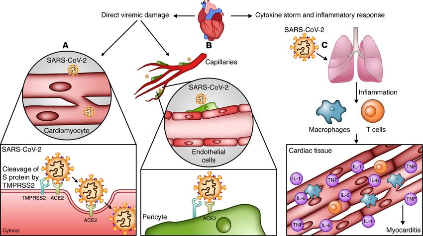
Figure 1. Possible mechanisms of acute myocardial injury and myocarditis in SARS-CoV-2 infection. Two main mechanisms by which SARS-CoV-2 causes myo-
cardial injury are direct virus-induced damage and secondary damage due to cytokine storm and inflammatory responses. (A) There is evidence that cardiomyo-
cytes express the receptors required for SARS-CoV-2 entry to the cell. Transmembrane protease TMPRSS2 cleaves the SARS-CoV-2 spike (S) protein, thus facili-
tating its activation and binding to cell-entry receptor angiotensin converting enzyme 2 (ACE2). (B) In addition, pericytes, which are abundant in cardiac tissue and
required for endothelial cell (EC) function and maintenance, express ACE2. Injury of these cells by SARS-CoV-2 results in EC dysfunction. (C) Furthermore, cytokine
storm and systemic inflammatory responses initiated by the virus can also lead to cardiac tissue damage and myocarditis. Illustrated by Rachel Davidowitz.
as either an increase in cardiac biomarkers or the presence of new abnormalities on electrocardiography
or echocardiography (41). An autopsy study of COVID-19 patients revealed mononuclear infiltrate,
predominantly composed of lymphocytes, that was associated with focal myocyte necrosis (39). Addi-
tionally, a fatal case of fulminant myocarditis, which was confirmed by biopsy, was reported in a 2-year-
old SARS-CoV-2–infected patient (42).
In the majority of patients, myocarditis presents concurrently with SARS-CoV-2–related respiratory
symptoms. Nevertheless, delayed presentation of cardiac complications occurring weeks after initial symp-
tomatic COVID-19 can also occur (43–45). Myocarditis documented by CMR may present as a postacute
sequela of SARS-CoV-2 infection in up to 19% of individuals (46), and isolated myocarditis without con-
comitant respiratory disease has been reported as an atypical presentation of COVID-19 (47, 48). The
subclinical presentation of ongoing or resolving myocarditis is also reported (49–51), and asymptomatic or
mild disease with CMR findings suggestive of cardiac injury have been demonstrated among young com-
petitive athletes with COVID-19 (51).
Myocarditis is now defined as a risk factor for increased mortality in patients with COVID-19 (3).
Although the recognition that COVID-19 may cause acute myocarditis may facilitate early diagnosis
and possible prevention of myocarditis-related mortality, lack of an understanding of the mechanism(s)
by which SARS-CoV-2 contributes to myocarditis and cardiac damage hinders thorough management
of this condition. Direct viral damage of cardiomyocytes, a hyperinflammatory state, and cytokine
storm, which usually occur in severe cases, have been suggested as the main drivers of acute myocardial
injury and myocarditis (Figure 1).
Direct viremic damage. Among coronavirus family members, SARS-CoV and MERS-CoV are less
likely to directly infect the myocardium. In a postmortem analysis of patients who died from SARS-CoV,
the virus was not detected in the heart by immunohistochemistry or in situ hybridization (52). Anoth-
er study that analyzed SARS-CoV RNA in postmortem tissue samples from 7 patients also suggested
JCI Insight 2021;6(13):e148980 https://doi.org/10.1172/jci.insight.148980 3REVIEW
Table 1. Studies reporting myocarditis confirmed by CMR in patients under 40
Author (ref.) Study type, patient(s) Lab findings/ECG/echo CMR findings Patient diagnosis/outcome
PMH/presenting symptoms
Beşler et al. (2) Case report, 20 y M ↑TnI, CK-MB, NT-proBNP, CRP Subepicardial LGE of the Myocarditis combined with
No CVD/febrile sensation and posterolateral wall in the COVID-19/Discharged on day 7
CP midventricle suggestive of
myocarditis
Bonnet et al. (133) 27 y M ↑TnI and NT-proBNP/Echo: Isolated ventricular Severe case of IVNC and acute
No PMH/respiratory distress enlarged LV with impaired noncompaction (IVNC) and myocarditis/Discharged on
LVEF 20% acute myocarditis day 9
Clark et al. (134) 59 COVID+ collegiate athletes, NL TnI/1 patient: new LV LGE in 46%, with 4 (15%) Myocarditis in 2 asymptomatic
19–21 y (n = 37 F, n = 22 M) dysfunction (LVEF 45%) after meeting modified Lake Louise COVID-19+ athletes (3%)
NA ↑dyspnea criteria for myocarditis
Dahl et al. (135) Case report, 37 y M ↑TnT, NT-proBNP, CRP, Diffuse myocardial edema COVID-19 myocarditis as
No PMH/fever, headache, and PCT/ECG: sinus tach with the most likely diagnosis/
unilateral painful neck swelling moderately flattened T/ Discharged on day 11
Echo: initially NL, ↓40% in sys
function on day 2
Kim et al. (31) Case report, 21 y F ↑TnI and NT-proBNP/ECG: Myocardial wall edema, Myocarditis combined with
Febrile sensation, nonspecific ventricular extensive transmural LGE COVID-19
coughing, sputum, diarrhea, conduction delay and PVCs/
and SOB Echo: severe sys dysfunction
Oberweis et al. (136) Case report, 8 y M ↑TnT, NT-proBNP, CRP, IL-6, Biventricular sys dysfunction Myocarditis/Discharged on
NA/fever, coughing, ↓weight, and D-dimer, leukopenia with with small PE, mild day 10
and severe fatigue lymphopenia/ECG: discrete subepicardial LGE of the lateral
ST↑ in V3 consistent with wall, and signs of diffuse
pericarditis/Echo: impaired LV edema
function
Paul et al. (33) Case report, 35 y M ↑TnI/ECG: repolarization in the Subepicardial LGE Acute myocarditis/recovered
Overweight/CP and fatigue precordial leads/Echo: NL predominating in the inferior after 3 weeks
and lateral walls
Sardari et al. (137) Case report, 31 y M NL TnT/ECG: NL/Echo: mild LV Edema/inflammation in Active myocarditis
DOE and low-grade fever dysfunction midinferoseptal and inferior
wall, subepicardial fibrosis in
the midinferior wall on LGE
Starekov et al. (138) Case series, n = 145, 17–23 y, 2 Patient 1: NL BNP, ESR, and Patient 1: predominantly distal Patient 1: myopericarditis
cases of myocarditis CRP, ↑TnI/ECG: ST and T LV inferolateral wall on LGE Patient 2: myocarditis/
NA/Patient 1: initially changes in anterolateral leads/ Patient 2: inferior basal LV Recovered
asymptomatic, mild dyspnea Echo: mild ↓ in global LV strain wall; inferior RV insertion on
reported on 1-month F/U Patient 2: NL BNP, TnI, ESR, LGE
Patient 2: fever, myalgias, and CRP/ECG: NL/Echo: NL
cough, mild dyspnea, sore
throat, congestion, ↓taste
Trogen et al. (34) Case report, 17 y M ↑TnI, BNP, CRP, ferritin, and LV size NL, LVEF 40% and RV Acute myocarditis associated
Spondylolysis and a history D-dimer/ECG: sinus tach and size NL, RVEF 39%, an area with SARS-CoV-2/Discharged
of asthma/fluid-responsive T inversion particularly in the of midwall LGE at the inferior on day 5
septic shock after 7 days of inferior leads/Echo: LVEF LV–RV junction corresponding
fever, GI symptoms, and neck mildly depressed to an area of ↑T2 signal and an
pain area of hypokinesia consistent
with myocarditis
CVD, cardiovascular disease; CP, chest pain; Tn, troponin; CK-MB, creatine kinase-MB; NT-proBNP, N-terminal prohormone B-type natriuretic peptide; CRP,
C-reactive protein; LGE, late gadolinium enhancement; PMH, past medical history; echo, echocardiography; LV, left ventricle; LVEF, left ventricular ejection
fraction; PCT, procalcitonin; T, T wave; NL, normal; sys, systolic; SOB, shortness of breath; PVC, premature ventricular contraction; PE, pericardial effusion;
DOE, dyspnea on exertion; F/U; follow-up; ESR, erythrocyte sedimentation rate; RV, right ventricle; GI, gastrointestinal; tach, tachycardia.
that SARS-CoV has less tropism for the heart than other coronaviruses (53). Additionally, in a 33-year-
old patient who died from MERS-CoV, histopathological analysis of viral particles in different tissues
revealed no remarkable viral particles in heart tissue (54). However, viral RNA was detected in heart
samples collected from a transgenic mouse model expressing the MERS-CoV receptor, human dipepti-
dyl peptidase 4 (DPP4, also known as CD26) (55).
JCI Insight 2021;6(13):e148980 https://doi.org/10.1172/jci.insight.148980 4REVIEW
Table 2. Studies reporting myocarditis confirmed by CMR in patients 40 years old and over
Author (ref.) Study type, patient(s) Lab findings/ECG/echo CMR findings Patient diagnosis/outcome
PMH/presenting symptoms
Coyle et al. (139) Case report, 57 y M ↑TnI, NT-proBNP and A recovery of EF to 82% and Myocarditis as the most likely
HTN/SOB, fevers, cough, inflammatory markers, diffuse biventricular and cause of the acute cardiac
myalgias, ↓appetite, nausea, lymphopenia/ECG: sinus biatrial edema with a small injury/Discharged on day 19
diarrhea tach/Echo: moderate diffuse area of LGE
hypokinesis, LVEF 35%–40%
Doyen et al. (28) Case report, 69 y M ↑TnI/ECG: LVH and diffuse Subepicardial LGE of the apex Myocarditis due to COVID-19/
HTN/cough, fever, dyspnea inverted T/Echo: mild LVH, NL and inferolateral wall Discharged from the ICU after
LVEF 3 wk
Inciardi et al. (30) Case report, 53 y F ↑TnT, NT-proBNP, and CK-MB/ ↑wall thickness, LVEF 35%, Acute myopericarditis/
NA/severe fatigue for 2 days Echo: PE, diffuse hypokinesis, myocardial IE Improvement
LVEF 40%
Luetkens et al. (140) Case report, 79 y M ↑TnT and NT-proBNP/ECG & echo: Diffuse myocardial IE, diffuse Myocarditis/Recovered
Asthma/fatigue, SOB, NL myocardial inflammation
syncope
Nicol et al. (43) Case report, 40 y M ↑TnI, BNP, CRP, Fg, PCT, D-dimer, Myocardial inflammation, Acute myocarditis/
Obesity/fever, odynophagia, and IL-6; pathological analysis: focal lateral subepicardial Discharged on day 9
and left neck pain IE, necrosis, interstitial and enhancement with prolonged
perivascular infiltrates, SARS- T1 relaxation times on LGE
CoV-2 RT-PCR: negative/ECG: imaging
sinus tach/Echo: LVEF 45%,
subtle hypertrophy and akinesia
of LV, small PE
Sala et al. (32) Case report, 43 y F ↑TnT, NT-proBNP, and CRP A recovery of sys function, Acute virus-negative
NA/oppressive CP and EMB: diffuse T cell inflammatory diffuse myocardial edema, lymphocytic myocarditis
dyspnea infiltrates, huge IE, and limited absence of detectable associated with SARS-
necrosis. No virus genome within myocardial scar/necrotic foci CoV-2 respiratory infection/
the myocardium/ECG: low EAR, on LGE Discharged on day 13
mild ST↑ (V1–V2 and aVR),
reciprocal ST↓ (V4–V6)/Echo:
LVEF 43%
Trpkov et al. (141) Case report, 62 y F ↑TnT and NT-proBNP/ECG: sinus Extensive subepicardial LGE COVID-19–related fulminant
Primary progressive tach with diffuse anterolateral in the anterolateral and myocarditis/Discharged
MS/acute altered LOC, inferolateral LV walls, ↑tissue
ST↑/point-of-care ultrasound: several days later
hypoxemia, shock severe LV dysfunction mapping–based markers of
inflammatory injury, ↑ECV,
LVEF 24%
Warchoł et al. (142) Case report, 74 y M ↑TnT, NT-proBNP, LDH, and LA enlargement and global LV Acute myocardial injury
AF, catheter ablation, HTN, D-dimer hypokinesia; EF 20%; large, meeting diagnostic criteria
DM, and hypothyroidism/ patchy, and linear nonischemic for clinically suspected
hemodynamically unstable pattern of subepicardial and myocarditis/Transferred
new-onset VT intramural fibrosis in the to a COVID-19–specialized
inferior and inferolateral wall hospital on day 7
Yokoo et al. (36) Case report, 81 y M ↑TnT/ECG: no signs of ischemia/ LGE areas with an ischemic Myocarditis/Discharged
HTN and ischemic stroke/ Echo: ↓EF (35%) pattern on the LV base
fever, dyspnea, and 91% septum, pronounced diffuse
oxygen saturation hypokinesia, and global sys
function involvement
HTN, hypertension; SOB, shortness of breath; Tn, troponin; NT-proBNP; N-terminal prohormone B-type natriuretic peptide; tach, tachycardia; echo,
echocardiography; LVEF, left ventricular ejection fraction; LGE, late gadolinium enhancement; LVH, left ventricular hypertrophy; T, T wave; NL, normal; CK-MB,
creatine kinase-MB; PE, pericardial effusion; IE, interstitial edema; Fg, fibrinogen; PCT, procalcitonin; LV, left ventricle; CP, chest pain; CRP, C-reactive protein; EAR,
ectopic atrial rhythm; sys, systolic; MS, multiple sclerosis; LOC, level of consciousness; ECV, extracellular volume; AF, atrial fibrillation; DM, diabetes mellitus; VT,
ventricular tachycardia; LDH, lactate dehydrogenase; LA, left atrium.
It remains controversial as to whether SARS-CoV-2 directly infects myocardial cells. Evaluation of SARS-
CoV-2 RNA by quantitative reverse transcriptase PCR (RT-PCR) in 39 autopsy cases indicated viral presence
in the myocardium of 24 cases, although this was not associated with infiltration of mononuclear cells in the
myocardium, as would be seen in myocarditis (56). Another study reported lymphocytic myocarditis and
JCI Insight 2021;6(13):e148980 https://doi.org/10.1172/jci.insight.148980 5REVIEW
Table 3. Case series studies reporting myocarditis confirmed by biopsy or autopsy
Author (ref.) Study type, patient(s) Lab findings/ECG/echo/biopsy findings
PMH/presenting symptoms
Basso et al. (40) International multicenter study, n = 21 autopsies, 3 Multifocal lymphocytic myocarditis, containing
patients with myocarditis (59–86 y) substantial T lymphocytes and a significant proportion of
HTN, DM, prior CVD macrophages, involving LV and RV, with RV predominance
in 1 case; CD4+ lymphocyte predominance in 2 cases and
CD8+ lymphocytes in 1 case
Bois et al. (37) Case series, n = 15 postmortem evaluation, 5 cases with Active lymphocytic myocarditis; one case with extensive
focal myocarditis (71–86 y) myocardial involvement with fibrosis and focal active
1–3 comorbidities per patient myocyte injury (smoldering myocarditis), the remaining
cases with only focal active myocarditis
Bradley et al. (143) Case series, n = 14 postmortem evaluation, 1 case of ↑Troponin
myocarditis (76 y F) Aggregates of lymphocytes surrounding necrotic
HLP, osteoporosis/respiratory distress, hypotension, myocytes, myocardial viral RNA+ by PCR but negative
tachycardia, fever immunohistochemistry and electron microscopy, low RNA
level in the cardiac tissue suggesting the possibility of
contamination by circulating virus
Buja et al. (57) Case series, multicenter study, n = 23 postmortem analysis, SARS-CoV-2 RNA detected in the cardiac tissue,
1 case of myocarditis lymphocytic myocarditis
HTN, obesity, DM
Duarte-Neto et al. (144) Case series, n = 10, 33–83 y, 2 cases of myocarditis Ultrasound-guided minimally invasive autopsy (MIA-US):
HTN, DM, chronic cardiopathy, COPD, chronic renal disease, mild lymphomononuclear myocarditis
neoplasia
Edler et al. (145) Case series, n = 80, 1 case of myocarditis (71 y M) A small lymphocytic infiltrate in RV of the heart in 1 case as
Cardiac insufficiency, DM, lung granuloma a sign of myocarditis
Falasca et al. (39) Case series, n = 22 autopsies, 12 cases of myocarditis Lymphocytic myocarditis characterized by mononuclear,
NA predominantly lymphocytic infiltrate, associated with focal
myocytes necrosis, fibrinous, and hemorrhagic areas with
myofiber disarray
Rapkiewicz et al. (146) Case series, n = 7 autopsies, 1 case of focal acute Minimally ↑troponin on the day of death/No ECG changes/
lymphocytic epimyocarditis (44 y M) Point-of-care ultrasound: ↓EF; a focal acute lymphocytic
HTN, HLP, DM, obesity, RCC/fever, cough, myalgia epimyocarditis, with no virions in cardiomyocytes
Weckbach et al. (147) Case series, n = 18, 1 case of myocarditis Lymphocytic myocarditis, ↑macrophages accompanied by
NA fibrosis; negative SARS-CoV-2 qRT-PCR
HTN, hypertension; DM, diabetes mellitus; CVD, cardiovascular disease; LV, left ventricle; RV, right ventricle; F, female; HLP, hyperlipidemia; COPD, chronic
obstructive pulmonary disease; M, male; RCC, renal cell carcinoma; EF, ejection fraction.
viral RNA detection by RT-PCR in 1 out of 23 postmortem fatal COVID-19 cases (57). Electron microscopy
of endomyocardial biopsy samples of a patient with COVID-19–related cardiogenic shock also revealed the
presence of the virus in the myocardial interstitium (58). Finally, it has been shown that SARS-CoV-2 can
directly infect human pluripotent stem cell–derived cardiomyocytes, which is contingent on ACE2 expression
(59). In contrast to these findings, postmortem analysis of three patients who died from COVID-19 in China
revealed that despite the presence of pathological changes, such as degeneration and necrosis of parenchymal
cells in the heart, SARS-CoV-2 was not detectable by RT-PCR (60). Similarly, in two male patients, aged 36
and 39 years old, myocarditis was noted based on CMR, and endomyocardial biopsy demonstrated myocar-
dial inflammation without necrosis or presence of viral genome by RT-PCR (61).
Cytokine storm and inflammatory response. A number of reports suggest that the hyperinflammatory state
that can occur in COVID-19 patients contributes to myocardial injury and increased mortality (Figure 1C)
(62–64). Severe cases of COVID-19 commonly present with high plasma levels of inflammatory cytokines
and a prolonged proinflammatory response, leading to extensive tissue damage (65, 66). Retrospective
analysis of 191 COVID-19 patients in China revealed increased serum inflammatory markers, including
TNF-α, C-reactive protein (CRP), ferritin, D-dimer, and IL-6, and cardiac damage markers, including tro-
ponin T (67). Comparison of laboratory tests between patients who died from COVID-19 and those who
recovered suggested that CRP and procalcitonin levels were associated with mortality. Poor prognosis was
linked to cytokine storm and a subsequent decrease in helper and suppressor T cells (68–70). Furthermore,
a recent study that included whole-genome sequencing of 2244 patients with COVID-19 from 208 ICUs in
JCI Insight 2021;6(13):e148980 https://doi.org/10.1172/jci.insight.148980 6REVIEW
Table 4. Case report studies reporting myocarditis confirmed by biopsy or autopsy
Author (ref.) Study type, patient(s) Lab findings/ECG/echo/biopsy findings
PMH/presenting symptoms
Craver et al. (38) Case report, postmortem analysis, Enlarged flabby heart with eosinophilic myocarditis on autopsy (diffuse
17 y M inflammatory infiltrates composed of lymphocytes and macrophages, with
No PMH/cardiac arrest after a 2-day prominent eosinophils with multiple foci of myocyte necrosis and minimal if
history of headache, dizziness, N/V any interstitial fibrosis)
Gauchotte et al. (148) Case report, 69 y M ↑TnI and CK-MB/Echo: nondilated LV and severe and diffuse LV hypokinesia,
DM, HTN, and IHD/fever, asthenia, LVEF 30%; fulminant myocarditis: an intense multifocal inflammatory
and abdominal pain infiltration, in both ventricles and septum, composed in its majority of
macrophages and CD8+ cytotoxic T lymphocytes, edematous myocardium
containing dystrophic cardiomyocytes without necrosis and a hypocellular
confluent area of fibrosis in posterior wall of LV; SARS-CoV-2 RNA RT-PCR+ in
heart tissue
Hu et al. (149) Case report, 37 y M ↑TnT, CK-MB, and BNP/ECG: ST↑ (III and AVF)/Echo: LVEF 27%/CT angiography:
CP, dyspnea, diarrhea no coronary stenosis/Biopsy from septum: SARS-CoV-2 PCR+
Kesici et al. (42) Case report, 2 y M NL acute phase reactants/Echo: severe cardiac failure; dilated cardiomyopathy
N/V and poor oral intake secondary to viral myocarditis and SARS-CoV-2 RT-PCR+
Nicol et al. (43) Case report, 40 y M ↑TnI and BNP/ECG: sinus tach/Echo: subtle hypertrophy and akinesia of
Obesity/fever, odynophagia, and left posterolateral LV wall with small PE; LVEF 45%/CMR: compatible with
neck pain acute myocarditis/Interstitial edema, small foci of necrosis, interstitial and
perivascular inflammatory infiltrates composed of plasmocytes, T lymphocytes,
few neutrophils, and a dense and diffuse infiltration by macrophages; negative
SARS-CoV-2 RT-PCR
Sala et al. (32) Case report, 43 y F ↑TnT and NT-proBNP/ECG: low atrial ectopic rhythm, mild ST↑ (V1–V2 and aVR,
Oppressive CP and dyspnea reciprocal), ST↓ (V4–V6)/Echo: mild LV sys dysfunction; LVEF 43%, inferolateral
wall hypokinesis/CMR: diffuse myocardial edema, absence of detectable
myocardial scar/necrotic foci on LGE/Diffuse T lymphocyte inflammatory
infiltrates with huge interstitial edema and limited foci of necrosis, no replacement
fibrosis, and absence of the SARS-CoV-2 genome within the myocardium
PMH, past medical history; N/V, nausea and vomiting; IHD, ischemic heart disease; Tn, troponin; CK-MB, creatine kinase-MB; echo, echocardiography; LV,
left ventricle; LVEF, left ventricular ejection fraction; CP, chest pain; BNP, brain natriuretic peptide; NL, normal; tach, tachycardia; PE, pericardial effusion;
NT-proBNP, N-terminal prohormone B-type natriuretic peptide; sys, systolic; LGE, late gadolinium enhancement.
the United Kingdom showed that critical and life-threatening COVID-19 has a genomic association with
SNPs in mediators of organ inflammation genes, including those encoding tyrosine kinase 2, DPP9, and
monocyte/macrophage chemotactic receptor CCR2 (71). These findings emphasize a critical role of hyper-
inflammation in COVID-19 pathogenesis. It remains to be elucidated whether such phenotypes contribute
to increased risk of myocarditis or cardiovascular events.
A number of studies support a role for hyperinflammation in cardiac manifestations of COVID-19.
Patchy mononuclear infiltration in epicardium, mainly CD4+ T cells, was observed in 15 out of 25
COVID-19 patients in a postmortem analysis of their hearts (72). Blood levels of troponin T, which
correlates with higher mortality, showed a positive linear correlation with CRP, indicating a possi-
ble role for inflammatory response in COVID-19–related cardiac damage (73). Furthermore, a retro-
spective analysis of 353 COVID-19 patients, of whom 79 had myocardial injury, demonstrated that
high neutrophil/lymphocyte ratio, D-dimer, lactate dehydrogenase, and inflammatory cytokines were
positively associated with cardiac troponin I levels. Thus, these markers have the potential to pre-
dict patients at high risk of developing myocardial injury (74). In a case report of a 6-year-old boy,
who presented with persistent fever and was later confirmed to have concurrent parvovirus B19 and
SARS-CoV-2 infections, blood tests exhibited pancytopenia, hypertriglyceridemia, and hypocalcemia
and elevated IL-6, D-dimer, CRP, procalcitonin, and cardiac biomarkers. Echocardiography showed
decreased left ventricle systolic function, and CMR obtained 20 days after symptom onset revealed the
presence of edema with no evidence of cardiomyocyte necrosis. The authors suggested a possible role
of cytokine storm in myocardial injury rather than the direct injury by the virus (75). Finally, in April
2020, eight children from England with either a positive SARS-CoV-2 test or previous exposure to a
COVID-19–infected family member presented with fever, shock, hyperinflammation, and myocardial
JCI Insight 2021;6(13):e148980 https://doi.org/10.1172/jci.insight.148980 7REVIEW
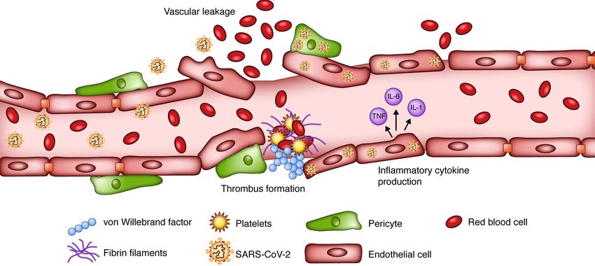
Figure 2. SARS-CoV-2 and endothelial cell dysfunction. Direct damage to endothelial cells (ECs) caused by SARS-CoV-2 disrupts cell integrity, resulting in
EC activation and vascular leakage. Consequent exposure of vWF, which is involved in platelet aggregation and fibrin formation, leads to thrombus forma-
tion. Cytokines secreted by activated ECs can further augment the vascular inflammation, permeability, and leakage. Illustrated by Rachel Davidowitz.
involvement (76). Similar cases were subsequently reported in other countries (77, 78), and in May
2020, this condition, which has been proposed as a potential cause of myocardial injury, was termed
multisystem inflammatory syndrome in children (MIS-C) by the CDC (79).
Myocardial interstitial fibrosis
Diffuse and focal myocardial fibrosis in the hearts of patients with COVID-19 has been reported and
can occur in the absence of cardiac symptoms. A study showed that 7 of 26 patients who recovered
from COVID-19 but later developed cardiac symptoms displayed edema and fibrosis by late gadolin-
ium enhancement in CMR (80). Diffuse interstitial fibrosis was noted on CMR in a case report of a
45-year-old female without a history of myocarditis who presented with palpitation and atypical chest
pain 3 months after contracting COVID-19 (5). Similar findings of diffuse fibrosis were also reported
in a previously healthy 49-year-old male who presented with dyspnea 6 weeks after the initial onset
of COVID-19 symptoms (44). In addition, the autopsy result of 14 COVID-19 patients revealed focal
myocardial fibrosis in 6 cases; however, all had a past history of myocardial infarction (MI) (81). A
postmortem analysis of another 4 patients who died because of SARS-CoV-2 showed mild focal fibrosis
in cardiac tissue in 2 of the patients (82). It is of note that one of these patients had a medical history of
chronic lymphocytic leukemia and the other patient underwent renal transplantation 3 months before
contracting the virus. In another study, analysis of cardiac tissue by endomyocardial biopsy in a patient
with cardiogenic shock revealed low-grade inflammation with focal interstitial fibrosis (58). In contrast
to these findings, a case series of 4 pediatric patients who were admitted to the ICU because of MIS-C
revealed edema but no evidence of necrosis or fibrosis via CMR (83). Edema without fibrosis by CMR
was also reported in 2 patients with myocarditis due to COVID-19 (61). It is important to note that in
all these cases, it is not clear whether fibrosis existed before COVID-19 contraction or developed subse-
quently because of the infection. Moreover, the absence of a history of underlying heart disease does
not exclude the possibility of past fibrosis in heart tissue.
Replacement of necrotic cardiomyocytes (as a result of myocarditis, vasculitis, and microinfarc-
tions) by fibroblasts appears to be the main mechanism of fibrosis in COVID-19 patients. In addition,
cytokine storm and infiltration of the myocardium with immune cells, which can potentially initiate
fibroblast to myofibroblast conversion and subsequent matrix remodeling, are among other possible
mechanisms of fibrosis in these patients (84).
JCI Insight 2021;6(13):e148980 https://doi.org/10.1172/jci.insight.148980 8REVIEW
EC dysfunction and vasculitis
ECs play a role in the regulation of immune response, inflammatory reactions, coagulation, and plate-
let function. As a result, these cells are key players in various pathologies associated with COVID-19
(85, 86). EC dysfunction and vasculitis, although currently considered as one of the main cardiovas-
cular complications of COVID-19, are also believed to be among other mechanisms that may underlie
COVID-19–induced myocarditis.
A postmortem analysis of patients with COVID-19 demonstrated the presence of SARS-CoV-2 in the
ECs of multiple organs (18) Direct viral infection of ECs, via SARS-CoV-2 receptors ACE2 and TMPRSS2
that are expressed on ECs (87), can lead to endothelial dysfunction and disruption of vascular integrity,
causing subsequent leakage (Figure 2) (88). Hyperinflammation and hypercoagulability have also been
reported as complications of EC dysfunction in COVID-19 patients (88). Postmortem studies demonstrat-
ed higher ACE2 expression in infected patients, which was associated with altered endothelial morphology,
disruption of cell junctions, detachment of cells from the basement membrane, and cell swelling (89).
scRNA-Seq studies have demonstrated that genes associated with immunomodulation, leukocyte acti-
vation, cytokine production, and antigen presentation are expressed in ECs. Specifically, these transcripts
are highly enriched in lung ECs compared with other organs (90). These results suggest that ECs may play
a critical role in initiation and maintenance of inflammation. Furthermore, binding of SARS-CoV-2 to
ACE2 impairs its enzymatic function, leading to bradykinin accumulation (87, 91), which is associated
with increased vascular permeability (88). Thrombotic events are also commonly observed in hospitalized
COVID-19 patients, especially those admitted to the ICU (92). Disruption of EC integrity exposes base-
ment membrane to circulatory platelets, initiating platelet aggregation and thrombosis (88). Additionally,
ECs express P-selectin, vWF, and fibrinogen in response to IL-1β and TNF-α, causing platelets to directly
bind to ECs and become activated (93). Activated ECs are hypercontractile, which can lead to disruption
of cell-cell junctions and vascular leakage.
Finally, a recent study showed that patients who contracted SARS-CoV-2 demonstrated EC dysfunc-
tion as shown by a 6% reduction in flow-mediated dilation (FMD) (94). A 6% reduction in FMD is clinical-
ly significant because every 1% reduction is associated with a 13% greater risk of a cardiovascular event (7).
Thrombotic events
Thrombotic events are also commonly observed in hospitalized COVID-19 patients, especially those
admitted to the ICU (92). Autopsy of 4 patients with COVID-19 revealed the presence of large emboli
in the lungs and multiple microthrombi in other organs, including the brain (9). The presence of emboli
has been associated with increased disease severity and mortality (67, 94–98). The underlying etiology
of the prothrombotic state observed in patients with COVID-19 is multifactorial. As mentioned earlier,
EC damage results in subsequent exposure of collagen within the extracellular matrix, leading to acti-
vation and recruitment of platelets (88). Additionally, activated ECs express a number of surface pro-
teins, including P-selectin, that function as cell adhesion molecules to recruit platelets and leukocytes
(93). An exaggerated inflammatory response, with elevated proinflammatory cytokines, also results in a
predisposition to coagulopathy (9, 81, 99). Other factors that play significant roles in creating a hyper-
coagulable state and subsequent thrombus formation include disseminated intravascular coagulation,
which commonly occurs in critically ill patients, and hyperferritinemia, which is associated with mac-
rophage activation syndrome (100, 101). Additionally, ICU admission of patients requiring mechanical
ventilation prolongs immobilization and venous stasis, which aggravates coagulopathy and frequently
complicates disease course (96). Treatment with anticoagulants may lower mortality in hospitalized
COVID-19 patients, as suggested by some studies (102).
Acute coronary events in COVID-19 patients have been reported since the emergence of the pandem-
ic. In a systematic review of 1527 patients with COVID-19, 8% presented with acute MI, and MI risk was
13 times higher in patients with severe clinical symptoms (103). However, in a case series of 28 patients
with COVID-19 who either presented with ST-elevation MI (STEMI) or developed STEMI during hospi-
talization, a lesion could not be identified in 40% of the patients who underwent coronary angiography
(97). Similarly, out of 9 patients with COVID-19 who had STEMI, 3 patients displayed no obstructive
disease (4). These observations suggest that these patients may have had acute thrombus formation that
led to their MI. Other case reports also provide evidence that culprit lesions were not necessarily present
in coronary arteries (19, 98); thus, this diagnostic challenge remains to be further elucidated.
JCI Insight 2021;6(13):e148980 https://doi.org/10.1172/jci.insight.148980 9REVIEW
Cardiac arrhythmias
COVID-19 patients can also present with arrhythmia. Heart palpitations were reported as a presenting
symptom in 7% of 137 individuals infected with SARS-CoV-2 hospitalized in Hubei province in China
(104). In another report from China, the rate of arrhythmias in 138 confirmed COVID-19 cases was even
higher at 16.7% (6). The most common arrhythmia observed in COVID-19 patients is sinus tachycardia.
It is not clear whether sinus tachycardia is due to increased cardiac output secondary to fever, hypoxia,
inflammatory stress, and medications or to myocardial structural changes (105).
A study of 700 patients admitted for COVID-19 infection reported 25 incidents of atrial fibrillation
(AF), 9 bradyarrhythmias, and 10 nonsustained ventricular tachycardias (NSVTs). In addition, ICU admis-
sion was associated with incidents of AF and NSVT (106). Similarly, atrial arrhythmias were recorded on
the ECGs of 27.5% of the patients admitted to the ICU compared with none of those who were treated in
a non-ICU setting (107). Ventricular arrhythmias also occur in COVID-19 patients with critical conditions
(108), making these patients vulnerable to cardiogenic shock, which requires further assessment to deter-
mine the need for extracorporeal membrane oxygenation (109). Medication side effects, inflammation of
the myocardium, edema of the interstitial tissue, fibrosis, and myocarditis, leading to structural changes,
conduction abnormalities, and dysregulation of ion channels (Na+ and K+), are among the underlying
mechanisms by which cardiac arrhythmias happen in COVID-19 patients (110). Nonetheless, new occur-
rence of tachyarrhythmia accompanied by an elevation in serum cardiac biomarkers in a patient can be
suggestive of myocarditis (63, 105).
Dysautonomia
Dysautonomia is a medical condition caused by malfunction of the autonomic nervous system (ANS),
generally due to the failure or overactivity of the sympathetic or parasympathetic components of the ANS.
This condition has been reported in patients with COVID-19 and may occur as a severe acute manifesta-
tion of COVID-19 or as part of the chronic sequelae of extended disease referred to as “long COVID”
(111–113). Recent studies suggest that some patients with long COVID may experience symptoms of auto-
nomic dysfunction, especially postural orthostatic hypotension (POTS), which is defined by symptoms of
orthostatic intolerance, including palpitation, headache, lightheadedness, fatigue, presyncope, shortness
of breath, chest pain, sleep disturbances, and gastrointestinal symptoms upon upright position (114–116).
Although the pathophysiology of POTS in COVID-19 remains to be elucidated, a number of mechanisms,
including hypovolemia, invasion of the sympathetic nervous system and/or medullary centers in the brain-
stem, and autoimmunity, are among the potential underlying causes (114, 117).
Medications
A wide range of compounds have been under investigation to treat COVID-19, but there currently is no
specific treatment available for this rapidly spreading disease. A number of agents, including chloroquine
and hydroxychloroquine, initially received emergency use authorization (EUA) from the FDA for treat-
ment and/or prophylaxis of COVID-19. However, because of lack of clinical efficacy and side effects,
especially cardiac adverse events, the FDA revoked EUA (118).
Among proposed treatment options, remdesivir is currently approved for use in hospital-
ized COVID-19 patients regardless of disease severity (119). Remdesivir is an antiviral agent with
broad-spectrum activity against several viruses, including SARS-CoV and MERS-CoV. It is an ade-
nosine analogue and a prodrug activated inside the cells via conversion to its pharmacologically active
form, adenosine nucleoside triphosphate. The active form of remdesivir inhibits RNA-dependent RNA
polymerase, thus resulting in RNA synthesis arrest (120, 121). Compassionate use of remdesivir has
been tried in critically ill COVID-19 patients and has shown modest improvement in condition and
shortened recovery times. However, because of insufficient information regarding its efficacy, trials are
currently being conducted to assess the clinical impact of remdesivir (122). Remdesivir demonstrated
cardiovascular side effects in 2 patients with COVID-19 who developed bradycardia, with QT interval
prolongation and T wave abnormality in 1 patient (123). In addition, in a randomized controlled trial
(RCT) conducted in China, 1 case of cardiac arrest was reported in a patient receiving remdesivir
(120). Nevertheless, the data are insufficient to conclude whether or not remdesivir is safe, particularly
in those with underlying cardiovascular disease. Thus, ongoing surveillance with an emphasis on car-
diovascular aspects is needed in patients with COVID-19.
JCI Insight 2021;6(13):e148980 https://doi.org/10.1172/jci.insight.148980 10REVIEW
In contrast to the therapies with cardiovascular adverse effects that were initially intended to combat
COVID-19, a number of agents have been studied for their favorable cardiovascular profiles in COVID-19
patients. Given the risk of thrombotic events in COVID-19 patients, use of anticoagulants has been recommend-
ed in all hospitalized patients with COVID-19, especially those with critical conditions who have no contrain-
dication for anticoagulation (124). Colchicine is among other therapies that, because of its antiinflammatory
properties, has been under investigation as a cardiovascular therapy in COVID-19 patients (125). A recently
published meta-analysis of studies on colchicine demonstrated a significantly lower mortality rate with a pos-
sible lower risk of mechanical ventilation in patients with COVID-19 (126). The findings of the GRECCO-19
randomized clinical trial on the effect of colchicine on cardiac and inflammatory markers revealed a decrease in
D-dimer and an improved clinical condition in patients who received colchicine (127). The results of ongoing
randomized clinical trials will determine whether colchicine is effective in reducing cardiac injury in COVID-19.
In addition to aforementioned medications, ACE inhibitors, angiotensin receptor blockers (ARBs), and
sodium glucose cotransporter-2 (SGLT-2) inhibitors are some of the drug classes that have been studied to be
repurposed for COVID-19 treatment (128). The association of ACE inhibitors/ARBs with decreased mortality
in cohorts of COVID-19 patients (129), along with their proposed mechanism in reducing viral entry in vitro
(130), has prompted a number of RCTs on the effect of these therapeutics in COVID-19 patients (131). Never-
theless, there is not yet clear evidence regarding the clinical impact of ACE inhibitors/ARBs in COVID-19. In
regard to SGLT-2 inhibitors, evidence suggests that they may have potential renoprotective and cardioprotec-
tive effects (132). To investigate the organ protection benefits of SGLT-2 inhibitors in COVID-19, an ongoing
international, multicenter, randomized clinical study is evaluating dapagliflozin compared with placebo in pre-
vention of COVID-19 complications or death and improvement of clinical recovery (Dapagliflozin in Respira-
tory Failure in Patients with COVID-19, DARE-19; https://clinicaltrials.gov NCT04350593).
Conclusions
Since the emergence of COVID-19, multiple groups have reported cardiovascular complications associ-
ated with SARS-CoV-2 infection. A hyperinflammatory state and cytokine storm can lead to fulminant
myocarditis. In addition, SARS-CoV-2 can potentially infect cardiomyocytes, ECs, and pericytes in
the myocardium, leading to acute myocardial injury. Importantly, cardiac arrhythmias, the most com-
mon being tachyarrhythmias, and diffuse and focal fibrosis have been observed in COVID-19 patients.
COVID-19 is also associated with a hypercoagulable state. Therefore, patients with COVID-19 should
be monitored for cardiovascular events, especially patients with a past medical history of cardiovascular
disease. Further investigations are required to screen and treat patients at risk of cardiovascular compli-
cations and to elucidate the mechanisms by which SARS-CoV-2 complicates the cardiovascular system.
Despite tremendous research on the cardiovascular complications of COVID-19 and its mechanisms, a
number of unanswered questions remain to be addressed. Although high expression of ACE2 in endothelial
cells and SNPs in certain inflammatory loci are associated with more severe disease and higher incidence of car-
diac complications, a causal effect is yet to be determined. In addition, short- and long-term effects of COVID-19
in competitive athletes in whom the resumption of physical activity is important should be studied, and screen-
ing protocols to identify patients at risk of myocarditis should be developed. Because postacute sequela of
SARS-CoV-2 infection is one of the presentations of COVID-19–associated myocarditis, future studies should
also determine the target group, timing of the screening after initial recovery from COVID-19, and the tests that
would help in distinguishing individuals at risk. Finally, given that COVID-19–associated cardiovascular com-
plications can potentially be debilitating and sometimes life-threatening, routine screening protocols (including
ECG, cardiac markers, echocardiography, or CMR and timing of each test based on each patient’s past medical
history and predisposing factors) should be developed to identify patients at risk for myocarditis.
Acknowledgments
HA is supported by NIH grants National Heart, Lung, and Blood Institute HL140973, HL138982, and
HL140927 and Leducq Foundation.
Address correspondence to: Hossein Ardehali, SQBRC 8-521, 303 E Superior Ave, Chicago, Illinois 60611,
USA. Phone: 312.503.2342; Email: h-ardehali@northwestern.edu.
JCI Insight 2021;6(13):e148980 https://doi.org/10.1172/jci.insight.148980 11REVIEW
1. Dong E, et al. An interactive web-based dashboard to track COVID-19 in real time. Lancet Infect Dis. 2020;20(5):533–534.
2. Beşler MS, Arslan H. Acute myocarditis associated with COVID-19 infection. Am J Emerg Med. 2020;38(11):2489.e1–2489.e2.
3. Ruan Q, et al. Clinical predictors of mortality due to COVID-19 based on an analysis of data of 150 patients from Wuhan,
China. Intensive Care Med. 2020;46(5):846–848.
4. Bangalore S, et al. ST-segment elevation in patients with Covid-19 — a case series. N Engl J Med. 2020;382(25):2478–2480.
5. Jagia P, et al. Myocardial fibrosis detected by cardiovascular magnetic resonance in absence of myocardial oedema in a patient
recovered from COVID-19. BMJ Case Rep. 2020;13(12):e240193.
6. Wang D, et al. Clinical characteristics of 138 hospitalized patients with 2019 novel coronavirus-infected pneumonia in Wuhan,
China. JAMA. 2020;323(11):1061–1069.
7. Ratchford SM, et al. Vascular alterations among young adults with SARS-CoV-2. Am J Physiol Heart Circ Physiol.
2021;320(1):H404–H410.
8. Dani M, et al. Autonomic dysfunction in ‘long COVID’: rationale, physiology and management strategies. Clin Med (Lond).
2021;21(1):e63–e67.
9. Bryce C, et al. Pathophysiology of SARS-CoV-2: targeting of endothelial cells renders a complex disease with thrombotic
microangiopathy and aberrant immune response. The Mount Sinai COVID-19 autopsy experience [preprint]. https://doi.org/1
0.1101/2020.05.18.20099960. Posted on medRxiv May 22, 2020.
10. Li W, et al. Angiotensin-converting enzyme 2 is a functional receptor for the SARS coronavirus. Nature. 2003;426(6965):450–454.
11. de Wit E, et al. SARS and MERS: recent insights into emerging coronaviruses. Nat Rev Microbiol. 2016;14(8):523–534.
12. Zhou P, et al. A pneumonia outbreak associated with a new coronavirus of probable bat origin. Nature. 2020;579(7798):270–273.
13. Kuba K, et al. A crucial role of angiotensin converting enzyme 2 (ACE2) in SARS coronavirus-induced lung injury. Nat Med.
2005;11(8):875–879.
14. Hamming I, et al. Tissue distribution of ACE2 protein, the functional receptor for SARS coronavirus. A first step in understand-
ing SARS pathogenesis. J Pathol. 2004;203(2):631–637.
15. Ragia G, Manolopoulos VG. Inhibition of SARS-CoV-2 entry through the ACE2/TMPRSS2 pathway: a promising approach
for uncovering early COVID-19 drug therapies. Eur J Clin Pharmacol. 2020;76(12):1623–1630.
16. Zhou L, et al. SARS-CoV-2 targets by the pscRNA profiling of ACE2, TMPRSS2 and furin proteases. iScience.
2020;23(11):101744.
17. Sungnak W, et al. SARS-CoV-2 entry factors are highly expressed in nasal epithelial cells together with innate immune genes.
Nat Med. 2020;26(5):681–687.
18. Varga Z, et al. Endothelial cell infection and endotheliitis in COVID-19. Lancet. 2020;395(10234):1417–1418.
19. Chen L, et al. The ACE2 expression in human heart indicates new potential mechanism of heart injury among patients infected
with SARS-CoV-2. Cardiovasc Res. 2020;116(6):1097–1100.
20. Caforio AL, et al. Current state of knowledge on aetiology, diagnosis, management, and therapy of myocarditis: a position
statement of the European Society of Cardiology Working Group on Myocardial and Pericardial Diseases. Eur Heart J.
2013;34(33):2636–2648.
21. Richardson P, et al. Report of the 1995 World Health Organization/International Society and Federation of Cardiology task
force on the definition and classification of cardiomyopathies. Circulation. 1996;93(5):841–842.
22. Aretz HT, et al. Myocarditis. A histopathologic definition and classification. Am J Cardiovasc Pathol. 1987;1(1):3–14.
23. Cooper LT. Myocarditis. N Engl J Med. 2009;360(15):1526–1538.
24. Peiris JSM, et al. Clinical progression and viral load in a community outbreak of coronavirus-associated SARS pneumonia: a
prospective study. Lancet. 2003;361(9371):1767–1772.
25. Chong PY, et al. Analysis of deaths during the severe acute respiratory syndrome (SARS) epidemic in Singapore: challenges in
determining a SARS diagnosis. Arch Pathol Lab Med. 2004;128(2):195–204.
26. Alhogbani T. Acute myocarditis associated with novel Middle East respiratory syndrome coronavirus. Ann Saudi Med.
2016;36(1):78–80.
27. Riski H, et al. Carditis associated with coronavirus infection. Lancet. 1980;2(8185):100–101.
28. Doyen D, et al. Myocarditis in a patient with COVID-19: a cause of raised troponin and ECG changes. Lancet.
2020;395(10235):1516.
29. Zeng J-H, et al. First case of COVID-19 complicated with fulminant myocarditis: a case report and insights. Infection.
2020;48(5):773–777.
30. Inciardi RM, et al. Cardiac involvement in a patient with coronavirus disease 2019 (COVID-19). JAMA Cardiol. 2020;5(7):819–824.
31. Kim I-C, et al. COVID-19-related myocarditis in a 21-year-old female patient. Eur Heart J. 2020;41(19):1859.
32. Sala S, et al. Acute myocarditis presenting as a reverse Tako-Tsubo syndrome in a patient with SARS-CoV-2 respiratory infec-
tion. Eur Heart J. 2020;41(19):1861–1862.
33. Paul JF, et al. Myocarditis revealing COVID-19 infection in a young patient. Eur Heart J Cardiovasc Imaging. 2020;21(7):776.
34. Trogen B, et al. COVID-19-associated myocarditis in an adolescent. Pediatr Infect Dis J. 2020;39(8):e204–e205.
35. Irabien-Ortiz Á, et al. Fulminant myocarditis due to COVID-19. Rev Esp Cardiol (Engl Ed). 2020;73(6):503–504.
36. Yokoo P, et al. COVID-19 myocarditis: a case report. Einstein (Sao Paulo). 2020;18:eRC5876.
37. Bois MC, et al. COVID-19-associated nonocclusive fibrin microthrombi in the heart. Circulation. 2021;143(3):230–243.
38. Craver R, et al. Fatal eosinophilic myocarditis in a healthy 17-year-old male with severe acute respiratory syndrome coronavirus
2 (SARS-CoV-2c). Fetal Pediatr Pathol. 2020;39(3):263–268.
39. Falasca L, et al. Postmortem findings in Italian patients with COVID-19: a descriptive full autopsy study of cases with and with-
out comorbidities. J Infect Dis. 2020;222(11):1807–1815.
40. Basso C, et al. Pathological features of COVID-19-associated myocardial injury: a multicentre cardiovascular pathology study.
Eur Heart J. 2020;41(39):3827–3835.
41. Huang C, et al. Clinical features of patients infected with 2019 novel coronavirus in Wuhan, China. Lancet.
2020;395(10223):497–506.
42. Kesici S, et al. Fulminant COVID-19-related myocarditis in an infant. Eur Heart J. 2020;41(31):3021.
JCI Insight 2021;6(13):e148980 https://doi.org/10.1172/jci.insight.148980 12You can also read