Workshop on Cardiac Modeling - KIT
←
→
Page content transcription
If your browser does not render page correctly, please read the page content below
Workshop on Cardiac Modeling

     Towards an integrated numerical heart model,
      Coupling the relevant physics the right way

                April 15-17 2019, Bad Herrenalb

Organizers:
 Prof. Dr. Christian Wieners
 Prof. Dr. Olaf Dössel, Dr. Axel Loewe
 Prof. Dr. Bettina Frohnapfel
 Prof. Dr. Vincent Heuveline
Workshop Schedule

              Monday                               Tuesday                      Wednesday
              15.04.19                                 16.04.19                     17.04.19

                                     9:00-10:00             Peter Kohl            Rolf Krause

10:00-10:15   Welcoming / Opening
                                    10:00-10:30             Coffee break           Coffee break

10:15-11:15       Luca Dedè                                                       Stefan Frei
                                    10:30-11:30          Steven Niederer
                                                                                 Henry Sutanto

11:15-12:15       Gernot Plank
                                    11:30-12:30   The integrated heart model    Alfonso Santiago

12:15-13:30        Lunch break
                                    12:30-13:30             Lunch break               Lunch

13:30-14:00      Jan Christoph

14:00-14:30     Stefano Pagani

14:30-15:00   Ekaterina Kovacheva   13:30-16:00           Social program

15:00-15:30        Coffee break

15:30-16:30      Poster session
                                    16:00-16:30             Coffee break

16:30-17:00   Nagaiah Chamakuri
                                    16:30-17:30 Electro-Mechanical benchmark
17:00-17:30     Simone Pezzuto

17:30-18:30      Gary Mirams        17:30-18:30         Maxime Sermesant

                                    20:00-21:30 Poster session / get-together

                                                   i
Abstracts
Monday, April 15
Cardiac Electromechanics: Multiscale Modeling, Coupling Schemes,
and Numerical Simulation Invited
speaker
Luca Dedèa , A. Gerbi, F. Regazzonia , A. Quarteronia
a MOX, Politecnico di Milano, Milano, Italy
We consider the mathematical and numerical modeling of cardiac electromechanics with ap-
plication to the left ventricle of the human heart. We proceed by integrating state-of-the art
models for the electrophysiology of the tissue, mechanical activation at the cellular level, and
the passive mechanical response of the muscle, thus yielding a coupled electromechanical pro-
blem within the active strain paradigm. We consider the spatial approximation of the Partial
Differential Equations therein involved by means of the Finite Element method and the time
discretization by Backward Differentiation Formulas. We numerically solve the coupled elec-
tromechanics problem by exploiting both monolithic and staggered approaches, for which we
verify, compare, and critically discuss their accuracy properties and computational efficiency
in simulating the whole cardiac cycle. In addition, we develop a multiscale model for cardiac
electromechanics that accounts for miscroscopic active force generation at the cellular level
within the active stress paradigm; with this aim, we exploit model order reduction techniques
based on Machine Learning algorithms to enable efficient numerical simulations of multiscale
electromechanics. Finally, we present several numerical results of the electromechanics problem
in the human left ventricle obtained in the high performance computing framework.
This project has received funding from the European Research Council (ERC) under the
European Union’s Horizon 2020 research and innovation programme: grant agreement No
740132, iHEART - “An integrated Heart Model tor the Simulation of the Cardiac Function”,
2017–2022.
Multiphysics modeling of total heart function Invited
speaker
Gernot Plank
Medical University Graz, Graz, Austria
Advances in numerical techniques and the ever increasing computational power have rendered
the execution of forward models of total heart function feasible. Using such models based on
clinical images and parameterized to reflect a given patient’s cardiac anatomy and physiology,
is considered a highly promising approach to comprehensively and quantitatively characterize
cardiovascular function in a given patient. Assuming that models correctly capture all funda-
mental mechanisms relevant to a given clinical problem, it is anticipated that modeling and
simulation of cardiovascular function will play a pivotal role in future precision medicine as a
1method for stratifying diseases, optimizing therapeutic procedures, predicting outcomes and
thus for better informing clinical decision making.
However, to translate modeling into a clinically applicable modality a number of key chal-
lenges hast to be addressed. In particular, expensive computational models must be made
efficient enough to be compatible with clinical time frames. This can be addressed either with
hierarchical models of varying complexity which are cheaper to evaluate, by using computa-
tional efficient techniques such as spatio-temporal adaptivity, or by exploiting the power of
new HPC hardware through massive parallelization or the use of accelerators. Further, the
etiology of most cardiac pathologies comprises Multiphysics aspects, requiring the coupling
of various physics, which may be characterized by very different space and time scales, ren-
dering their coupling a challenging endeavor. Finally, to be of clinical utility generic models
must be specialized based on clinical data, which requires complex parameterization and data
assimilation procedures to match model behavior with clinical observations.
Electromechanical Vortex Filaments and Vortex-Substrate
Interactions during Cardiac Fibrillation talk
Jan Christoph
University Medical Center Göttingen, Department of Cardiology and Pneumology, Göttingen,
Germany
The visualization of the highly dynamic electrical wave phenomena evolving within the heart
muscle during cardiac fibrillation is a major scientific challenge. In recent work, we demons-
trated that simultaneous imaging of both electrical and mechanical dynamics of the heart can
provide novel insights into the spatio-temporal organization of cardiac fibrillation within the
heart muscle. Using high-resolution 4D ultrasound, we showed that it is possible to identify
mechanical filament-like phase singularities within the contracting, fibrillating heart wall. The
mechanical filaments appear to evolve like fingerprints of electrical vortex filaments through
the ventricular muscle, indicating the core regions of three-dimensional electrical scroll waves.
On the deforming ventricular surface, it can be observed that electrical spiral vortices create
vortex-like mechanical deformation patterns, which similarly rotate and whose core regions
co-exist and co-localize with the core regions or phase singularities of the electrical vortices.
Furthermore, it is possible to observe interactions of electrical and mechanical vortices with
heterogeneities such as scar tissue, as both electrical and mechanical phase singularities equal-
ly attach to or co-localize with the heterogeneities. Lastly, the integration of the data into
computer models could be used to infer in 3D the electrical wave patterns that had caused
the deformations, but can not be measured yet directly.
2Uncertainty quantification in cardiac electrophysiology disease
modeling talk
Stefano Pagania , Andrea Manzonia , Alfio Quarteronia,b
a MOX, Politecnico di Milano, Milano, Italy
b EPFL, Lausanne, Switzerland
We develop a computationally efficient framework to perform uncertainty quantification (UQ)
in cardiac electrophysiology in order to improve the ability of cardiac models to reproduce both
physiological and pathological patient-specific behaviors. Electrophysiology numerical models,
obtained from the discretization of nonlinear parametrized coupled system of ordinary and
partial differential equations (PDEs), are inevitably affected by uncertainty, e.g., in (i) the
computational domain, (ii) physical coefficients and (iii) boundary conditions.
We address a complete UQ pipeline, including: (i) a variance-based sensitivity analysis
for the selection of the most relevant input parameters; (ii) forward UQ (or uncertainty pro-
pagation) to investigate the impact of intra-subject variability on clinically relevant outputs
related to the cardiac action potential; (iii) backward UQ (or parameter and state estimation
and data assimilation) in view of both model calibration and personalization.
In this context, numerical strategies involve the approximation of PDEs for several (usually,
order of thousands) input parameter values, thus making high-fidelity, or full-order, techniques
(e.g. the finite element method) ill-suited.
To mitigate this computational burden, we replace the high-fidelity model with compu-
tationally less expensive projection-based local reduced-order models aimed at reducing the
state-space dimensionality. Numerical experiments dealing with both physiological and patho-
logical cases illustrate the ability of the UQ pipeline based on reduced-order models to realize
a cost-effective-but still accurate-methodology.
This project has received funding from the European Research Council (ERC) under
the European Union’s Horizon 2020 research and innovation programme (grant agreement
n. 740132).
A Bidirectionally Coupled Model of Electrophysiology and
Elastomechanics of the Human Heart talk
Tobias Geracha , Ekaterina Kovachevaa , Larissa Hüttera , Olaf Dössela , Axel Loewea
a Institute of Biomedical Engineering, Karlsruhe Institute for Technology (KIT), Karlsruhe, Germany
The contraction of the heart is a complex process involving the interaction of different me-
chanisms. On the one hand, electrical activation propagates through the tissue and leads to
tension development and thus to mechanical contraction of the myocytes. On the other hand,
the deformation of the myocardium and stretch activated channels (SAC) influence the electri-
cal excitation. Each of these mechanisms is modelled by a different mathematical description
and can be reproduced in-silico. Nevertheless, to estimate how these mechanisms influence
each other, a coupling method is needed to interconnect the electrical propagation in the
tissue and the heart mechanical contraction. In this study we investigate whether a bidirec-
tional coupling of the electrophysiology and the elastomechanics is necessary during normal
sinus rhythm. The electromechanical propagation was simulated on an idealized left ventricle
geometry for three different scenarios: first, we simulated the elastomechanics self-contained,
second the elastomechanics strongly coupled with the electrophysiology, and finally we added
SACs to the electrophysiology model. We compared the global indicator ejection fraction as
3well as measurements on a local scale such as the action potential and tension development for
a single cell. This will help us to decide which complexity of the coupled model is necessary to
simulate certain scenarios with sufficient accuracy. In the future this study will be extended
to include a cardiomyopathy case, where the mutual influence of both electrophysiology and
the elastomechanics will also be evaluated and compared to the healthy heart beat. This will
provide important insights in terms of developing new diagnostic tools and therapeutic options
for the treatment of a cardiomyopathy.
An efficient and accurate numerical methods for the solution of
bidomain model. talk
Nagaiah Chamakuri
University of Hohenheim, Stuttgart, Germany
The most complete description of cardiac bioelectrical activity at the cardiac tissue is given
by the bidomain model which consists of a system of a non-linear partial differential equati-
ons (PDEs). The evolution equation is coupled through the non-linear reaction term with a
stiff system of ordinary differential equations (ODEs) describing the ionic currents through
the cellular membrane. Many attempts to made to increase the bidomain solver efficiency by
using decoupled strategies and operator splitting schemes. More importantly, the monodomain
equations are often decoupled into one parabolic equation that is computationally cheap to
solve and other set of ODEs which are even very cheap to solve by using implicit-explicit
(IMEX) time stepping schemes. Thus, it is not clear if commonly used splitting methods can
outperform a coupled approach by maintaining the good accuracy. Moreover, the splitting
methods constrain the maximum time step that may be used for stability as well as accuracy
considerations. In this talk, we present the numerical results for the coupled solver approach as
compared with commonly used splitting methods by considering more sophisticated physiolo-
gical models. Our numerical results demonstrate that the coupled method is computationally
slower than the conventional uncoupled methods but it produces more accurate results. In
this regard, the novel memory efficient computational technique will be demonstrated to solve
such coupled system of PDEs.
Enabling high-dimensional uncertainty quantification for cardiac
electrophysiology via multifidelity techniques talk
Simone Pezzutoa , Alessio Quaglinoa , Rolf Krausea
a Università della Svizzera italiana, Lugano, Switzerland
Mathematical modeling of the heart, as many other models in biomedical sciences, involves a
large number of parameters and simplifying approximations. Uncertainties for cardiac models
are ubiquitous, including anatomy, fiber direction, and electric and mechanical properties of
the tissue. Hence, both UQ and parameter sensitivity naturally arise during modeling, and
they shall become fundamental in view of clinical applications. Despite the relevance of UQ
for cardiac models has been acknowledged multiple times in the last 5 years [1], the literature
on this topic is still very limited.
For high-dimensional input uncertainties, e.g., substrate heterogeneity or cardiac fibers
orientation, the method of choice for UQ is the classic Monte Carlo (MC) method. MC con-
vergence rate does not suffer from the curse of dimensionality, but it is notoriously slow. While
4sampling a random field can be done very efficiently via the pivoted Cholesky decomposition,
computing the cardiac activation from the bidomain equation is a computational demanding
task. A single patient-tailored simulation can take several CPU-hours even on a large cluster.
This makes uncertainty quantification (UQ) unfeasible, unless modeling reduction strategies
are employed.
One such strategy is represented by multifidelity methods [2]. A key ingredient of the
multifidelity approach is the choice of low-fidelity models. Typical strategies are projection-
based or data-fit surrogates, which however need to be trained anew for each patient and may
become inefficient for a large dimensionality of the input, as in the case under considerati-
on. Instead, a more physics- based approach is to take advantage of the natural hierarchy of
available models. These include different cellular models for the monodomain equation, the
time-independent eikonal equation, and the 1D geodesic point activation [3]. By exploiting the
statistical correlations in this hierarchy, we observed a reduction of the computational cost by
at least two orders of magnitude, enabling to perform a full analysis within a reasonable time
frame. Moreover, we incorporate Bayesian techniques, which provide confidence intervals and
full probability distributions at selected points, thus augmenting the information provided by
standard frequentist approaches.
References:
[1] Pathmanathan, P. & Gray, R. A. (2018). Validation and trustworthiness of multiscale
models of cardiac electrophysiology. Frontiers in Physiology, vol. 9, no. FEB, pp. 1-19.
[2] Peherstorfer, B., Willcox, K., & Gunzburger, M. (2018). Survey of multifidelity methods
in uncertainty propagation, inference, and optimization. SIAM Review, 60(3), 550-591.
[3] Quaglino, A., Pezzuto, S., Koutsourelakis, P.S., Auricchio, A., Krause, R. (2018). Fast
uncertainty quantification of activation sequences in patient-specific cardiac electrophysiology
meeting clinical time constraints. Int J Numer Meth Biomed Engng, e2985.
Uncertainty Quantification in Cardiac Electrophysiology Modelling invited
speaker
Gary Mirams
Centre for Mathematical Medicine & Biology, School of Mathematical Sciences, University of
Nottingham, Nottingham, England
The Uncertainty Quantification (UQ) field typically focusses on the challenging process of
propagating uncertainties in model inputs and parameter values through to model predicti-
ons. But perhaps more important in cardiac electrophysiology modelling is our uncertainty in
model structure - the right set of equations to use. Unpicking whether variability in experimen-
tal data is biologically-relevant or due to experimental artefacts is also a challenge. Here, I’ll
discuss sources of uncertainty that are important to consider when doing cardiac electrophy-
siology modelling. We will examine a case study selecting and parameterising models for the
hERG/IKr potassium current. There are over seven different structures and thirty paramete-
risations in the literature for this one current. I’ll show how designing more information-rich
experiments helps us to minimise uncertainty in parameter values with short training experi-
ments, whilst leaving time to do independent validation experiments which help with model
selection. The project raises questions about how best to deal with discrepancy between the
models and reality when using model predictions to make real world decisions that are incre-
asingly safety-critical.
5Tuesday, April 16
The Non-Textbook Heart: Structure, Electrics, Mechanics invited
speaker
Peter Kohl
Institute for Experimental Cardiovascular Medicine, University Heart Centre Freiburg / Bad
Krozingen, Faculty of Medicine, University of Freiburg, Germany
The heart is an amazing organ. It beats once per second, about 2 billion times by the time
we retire, and if it stops – so does life. The volume it pumps in a year is equivalent to that of
an Olympic-sized swimming pool. Pumping itself involves intra-cardiac volume redistribution
between atria and ventricles, without a discernible change in the overall external volume occu-
pied by the blood-filled heart. This mechanical activity results from electrically-orchestrated
contractions of billions of individual heart muscle cells. Each of them displays slightly diffe-
rent stress-strain behaviour, depending on the local mechanical environment which differs as
a function of basico-apical and transmural position. The mechanical environment furthermore
changes differentially with any alteration in pre- (volume) or after- (pressure) load, such as on
every breath we take, when we change posture, or during exercise. The matching of local me-
chanical activity to global demand requires finely tuned auto-regulatory abilities, and all that
in the absence of the kind of neuro-muscular junctions that tune skeletal myofibre activity. In
addition, the cross-talk between electrics and mechanics is far from uni-directional, as electrical
excitation and conduction, as well as the mechanisms underlying electro-mechanical coupling,
are exquisitely mechano-sensitive. Add to this the observation that the heart contains more
non-myocytes than muscle cells, combined with recent insight into electrical coupling between
those different cell populations, and it becomes clear that we need to take a fresh look at
the intriguing aspects of cardiac structure and function that extend beyond current textbook
knowledge. This lecture will address some of those aspects that may be of relevance on the
path towards an integrated numerical heart model.
Applying Cardiac Modelling to Study Drugs, Devices and Diagnosis invited
speaker
Steven Niederer
King’s College London, London, England
The ability to measure the heart, its shape, its structure and its function across multiple
spatial and temporal scales continues to grow. Interpreting this data remains challenging.
Computational biophysical models of the heart allow us to quantitatively link and interpret
these large disparate data sets within the context of known cardiac physiology and invariable
physical constraints. Within these models, we can infer unobservable states, propose and test
new hypothesis and predict how systems will respond to challenges increasing our ability to
interrogate and understand biological systems. We are increasingly applying this approach to
modelling human hearts to investigate clinical applications.
In this presentation, I will give an overview on our modelling work simulating anthracycline-
induced heart failure, how we are using models of individual patients to study cardiac re-
synchronisation therapy and how we are using simulations to characterise the anatomy and
pathophysiology of atrial fibrillation patients. Finally, I will present some of our preliminary
results on simulating the four-chamber heart to begin simulating the interactions between
atrial and ventricular function.
6Biophysics & AI for Computational Cardiology: Learning by Heart invited
speaker
Maxime Sermesant
Inria, Sophia Antipolis, France
Electromechanical models of the heart have made important progress over the last decades,
and personalised models can now be envisaged for clinical applications. However there are still
challenges in translating such models to the clinics. The recent progress in computing power
and available data makes it possible to develop accurate data-driven approaches for health-
care but such artificial intelligence approaches often lack of robustness. Machine learning and
biophysical modelling are very complementary approaches, with biophysical models offering a
principled way to introduce physiological constraints. In this talk I will present results on per-
sonalised electromechanical models of the heart and research where we combined biophysics
and AI in different ways in order to leverage their strengths. Different clinical applications in
computational cardiology will be presented.
Wednesday, April 17
Coupling Scales, Coupling Physics - Multi-physics in Cardiac
Simulation invited
speaker
Rolf Krausea , P. Zulian, S. Pozzi, M. Favino, M. Nestola, P. Benedusi
a Università della Svizzera italiana, Lugano, Switzerland
In this talk we present and discuss discretization and solution methods in space, time, and
space-time for cardiac simulation. Starting from pure electrophysiology, we discuss the coup-
ling of electrophysiology and mechanics, and eventually comment on fluid structure interaction
and contact in the heart valves.
As it turns out, either on the side of the solution method (multigrid) or on the side of
the coupling of different discretizations (mortar methods), discrete L2 projections turn out to
be a versatile ingredient - may it be for the construction of multi-level approximation spaces
(electrophysiology) , for the discretization of contact constraints (heart valves) , or for the
transfer of discrete quantities (FSI). We will describe our discretization and solution methods
and will comment on how to handle efficiently the arising discrete constrained and coupled
systems, with particular focus on their parallel solution.
Eventually, we will comment on how to deal with uncertain data and in which way the
created multi-level hierarchies can be exploited for uncertainty quantification.
7Modelling and simulation of mechano-chemical fluid-structure
interaction with application to atherosclerotic plaque growth talk
Stefan Freia , Thomas Richterb , Thomas Wickc
a University College London, England
b University of Magdeburg, Gerrmany
c University of Hannover, Germany
In this talk, we present a numerical framework for mechano-chemical fluid-structure interac-
tions with long-term effects. In particular, we investigate a model for atherosclerotic plaque
growth in arteries including the interaction of the growing solid with the flow in the vessel.
The mechano-chemical interaction is modelled by a multiplicative splitting of the deformation
gradient.
This application includes two particular difficulties: First, growth may lead to very large
deformations, up to full clogging of the fluid domain. Therefore, we use a Fully Eulerian
approach, that is able to handle very large deformations up to contact. The second difficulty
stems from the different time scales: while the dynamics of the fluid demand to resolve a
scale of milliseconds to seconds, growth typically takes place in a range of months. To include
both long-scale and short-scale effects in an efficient and accurate way, we derive a temporal
two-scale approach.
The numerical methodology is substantiated with several numerical tests that include
comparisons of the Eulerian approach to an ALE method, the performance of the temporal
two-scale algorithm as well as numerical convergence studies.
Understanding species-, atrial-, disease- and rate-specific effects of
clinically used antiarrhythmic drugs using computational models talk
Henry Sutantoa , Lian Laudya , Michael Clerxb , Dobromir Dobrevc , Harry J.G.M. Crijnsa ,
Jordi Heijmana
a Department of Cardiology, CARIM School for Cardiovascular Diseases, Maastricht University,
Maastricht, The Netherlands
b Department of Computer Science, University of Oxford, Oxford, United Kingdom
c Institute of Pharmacology, West German Heart and Vascular Center, University Duisburg-Essen,
Essen, Germany
Background: Cardiac arrhythmias remain a major cause of death and disability. Despite
the improved understanding of arrhythmia mechanisms, progress in the development of new
antiarrhythmic drugs (AADs) has been limited and clinical application of currently available
AADs often remains suboptimal, likely in large part due to the incomplete understanding of the
complex mechanisms-of-action of AADs. Here, we present a novel user-friendly computational
tool designed to facilitate a better understanding of AADs (the Maastricht Antiarrhythmic
Drug Evaluator; MANTA).
Methods: MANTA integrates published computational cardiomyocyte models of different
species (mouse, guinea-pig, rabbit, dog, human), regions (atrial, ventricular, purkinje) and
disease conditions (atrial fibrillation- and heart failure-related remodeling). It enables simu-
lations of the effects of clinically available AADs (Vaughan-William Classes I, III, IV and
multi-channel blockers) on action potential (AP) properties and the occurrence of proarrhyth-
mic effects such as early-afterdepolarizations. AAD effects were simulated based on published
IC50 values for each cardiac ion channel and by integrating state-dependent block of IN a by
8Class I AADs using a Markov-model approach in all cardiomyocyte models.
Results: Markov model parameters were optimized to replicate published INa characteri-
stics (voltage-dependent activation, inactivation, recovery from inactivation) and AP upstroke
velocity in all cardiomyocyte models and reproduced experimental use-dependent onset and
recovery of IN a inhibition by flecainide and lidocaine. MANTA provides a graphical user in-
terface allowing users to select different AADs, concentrations, and experimental conditions
(rate, electrolyte concentrations). Using MANTA, we demonstrated and characterized import-
ant species-, rate-, cell-type-, and disease-state-specific AAD effects, including 1) a stronger
effect of Class III AADs in large mammals than in rodents; 2) a frequency-dependent decrease
in upstroke velocity with Class I AADs and reverse use-dependence of Class III AADs; 3)
ventricular-predominant effects of pure IKr blockers and preferential reduction in atrial AP
upstroke velocity with vernakalant; and 4) excessive AP prolongation with Class III AADs
other than amiodarone in heart failure.
Conclusion: The effects of AADs are complex and highly dependent on the experimental or
clinical conditions. MANTA is a powerful, freely available tool able to reproduce a wide range of
AAD characteristics that enables analyses of the underlying ionic mechanisms. Use of MANTA
is expected to improve understanding of AAD effects on cellular electrophysiology under a
wide range of conditions, which may provide clinically-relevant information on the safety and
efficacy of AAD treatment, thereby potentially improving cardiac arrhythmia management.
A tightly coupled three-physics model of the human heart invited
speaker
Alfonso Santiagoa , Jazmin Aguado-Sierraa , Federica Saccob , Constantine Butakoffb , Mariano
Vázqueza
a BSC, ELEM Biotech, Barcelona, Spain
b BSC, Universitat Pompeu Fabra, Barcelona, Spain
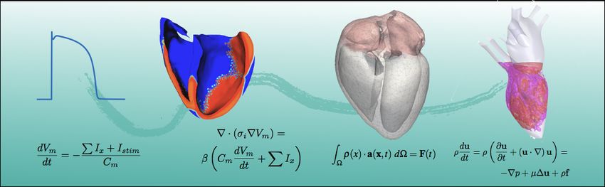
The heart is a complex system. From the engineer’s standpoint, it gathers electrical depolariza-
tion, mechanical deformation and fluid dynamics, tightly coupled in a very intricate geometry.
Although simplified and reduced order models are crucial nowadays to transfer science to
industry and clinic, comprehensive and integrative models of the heart are also required to
understand the complex connections between the different physics. This detailed modelling
strategy leads to an expensive computational cost, requiring an efficient use of the resources
following a clear parallel programming strategy, from the algorithms down to the implemen-
tation issues. In this talk we see the heartbeat from an engineer’s point of view, decomposing
it in each one of the independent physical problems. After, the way the problems are tightly
coupled is explained. On the one hand, the Land model is used for the excitation-contraction
coupling and the mechano-electric feedback. On the other hand the bidirectional fluid-structure
Interaction problem is tackled with a staggered quasi-Newton approach, with a specific focus
on the parallel implementation. The FSI problem in the heart is particularly complex due
to the similar densities of both domains, a fact that makes the system prone to added mass
instability in a partitioned scheme. Together with this challenge, two FSI interfaces should be
simultaneously coupled and converged: the right and left ventricles. Finally, we show two use
cases for this three-physics model.
9Poster session
Development of a simulation environment to understand the
charge-based mapping of cardiac physiology from single
measurement points
M. E. Hesara , R. Vitushinskyb and S. Ingebrandta
a Institute for Materials in Electrical Engineering 1, RWTH Aachen University, Aachen, Germany
b RAM Group DE GmbH, Zweibrücken, Germany
In many research projects as well as in clinical tests the cardiac physiology has been investiga-
ted noninvasively. To date, the differential measurement of the body surface potential (BSP)
is well-known and state-of-the-art in clinical routine (Electrocardiography – ECG). Only after
differential BSP measurements, solving the ill-posed inverse problem [1] of the heart is possi-
ble. Typically, numerical or analytical modeling of the heart as an electronic signal generator
(e.g. 2D or 3D dipole [3], mono-domain [2], bi-domain [3] etc.) is utilized to understand and
simulate the signal propagation from the heart as a signal source towards the micro- and
nanoelectrodes used for low ohmic skin-electrode contacts.
In our project, in contrast to ECG, we are utilizing an ultra-low noise, charge based sensing
technique from single measurement points on the skin to acquire cardiac signals representing
mechanical and electrical features of the heart activity. We coin this technique single point
cardiography (SPC) as a complementary technique to routine ECG. Our signals contain peri-
odic features, which show signal components of standard ECG overlaid by components, which
we attribute to the mechanical action of the heart. We are aiming to develop a simulation
environment and solve the inverse problem of the heart action including electrical activity and
mechanical movement of the tissue using the classical simulation techniques.
Moreover, similar to other biological signal recordings, noise reduction and signal averaging
techniques are required to extract the prominent signal features of the SPC recordings. Here,
the sensor front ends, which are highly sensitive to the tiny skin charge changes, are posing
high demands for electromagnetic shielding.
To approach this problem from a fundamental science aspect, we develop a 3D upper body
geometrical model similar to the human anatomy to verify the numerical and mathematical
models for signal generation and propagation. This will allow us to verify our simulation re-
sults with standard signal sources.
References:
[1] Nejib Zemzemi. Theoretical and Numerical study of the electric activity of the heart. Mo-
deling and Numerical simulation of electrocardiograms. Mathematics [math]. Université Paris
Sud - Paris XI, 2009. English.
[2] Coudière, Y., Rioux, M.: Virtual electrodes mechanisms predictions with a current-lifted
monodomain model. Comput. Cardiol. 39, 837–840 , 2012.
[3] Gerardo-Giorda, L. An Introduction to Mathematical and Numerical Modeling of Heart
Electrophysiology. In Nonlinear Dynamics in Biological Systems (pp. 83-111). Springer, Cham.
, (2016).
Acknowledgements:
Funding of the German Ministry of education and Research (BMBF) under the project SIN-
Dynamik (13GW0180B) is acknowledged.
10Initiation and maintenance of re-entrant cardiac propagation: a
computational vulnerability study
Luca Azzolin
Institute of Biomedical Engineering, Karlsruhe Institute for Technology (KIT), Karlsruhe, Germany
Nowadays, a large share of the global population is affected by heart rhythm disorders. Com-
putational modelling is a useful tool for understanding the dynamics of cardiac arrhythmias.
Several recent clinical and experimental studies suggest that atrial fibrillation is maintained
by re-entrant drivers (e.g. rotors). As a consequence, numerous works have addressed atrial
arrhythmogenicity of a given electrophysiological model using different methods to simulate
the perpetuation of re-entrant activity. However, no common procedure to test atrial fibril-
lation vulnerability has yet been defined. Here, we systematically evaluate and compare two
state-of-the-art methods. The first one is rapid extra-stimulus pacing from rim of the four
pulmonary veins. The second consists of placing phase singularities in the atria, estimating an
activation time map by solving the Eikonal equation and finally using this as initial condition
for the electrical cardiac propagation simulation. In this way, we are forcing the wavefronts to
follow re-entrant circuits with low computational cost thus less simulation time. We aim to
identify a methodology to quantify arrhythmia vulnerability on patient-specific atrial geome-
tries and substrates. We will proceed with in-silico experiments, comparing the results of these
two methods to initiate re-entrant activity, checking the influence of the different parameters
on the dynamics on the re-entrant drivers and finally extracting a valid set of parameters allo-
wing to reliably assess re-entry vulnerability. The final objective is to come up with an easily
reproducible minimal set of simulations to assess vulnerability of a particular atrial substrate
(cellular and tissue model) or of distinct anatomical atrial geometries to arrhythmic episodes.
Given the great need of exploring susceptibility to atrial arrhythmias, i.e. after a first ablation
procedure, this study can provide a useful tool to test new treatment strategies and to learn
how to prevent the onset and progression of atrial fibrillation.
Optimization of Cardiac Resynchronization Therapy in 3D
Electrophysiological Ventricular Models
Edison Carpioa , Juan F. Gomeza , Rafael Sebastianb , Alejandro Lopez-Pereza , Eduardo
Castellanosc , Jesus Almenralc , Jose M. Ferreroa , Beatriz Trénora
a Universitat
Politècnica de València, València, Spain
b Universitat
de València, València, Spain
c HM Hospitales, Universidad CEU-San Pablo
Cardiac Resynchronization therapy (CRT) is an assigned treatment for cardiac diseases, such
as heart failure (HF) with left bundle branch block (LBBB), which reduces electrical ventricu-
lar dyssynchrony. In CRT, an electrical stimulus is usually applied to both the right ventricle
(RV) and the left ventricle (LV). An optimal location of the LV pacing lead is fundamental to
an effective CRT. The LV posterior-lateral wall is the usual location recommended for CRT
application. Nevertheless, several optimization criteria such as the latest electrical activated
region in the LV or the LV site leading to the shortest QRS have also been proposed to guide
the location for the LV pacing lead.
The aim of the present study was to systematically analyze the optimal location LV pacing
lead based on these three different optimization criteria during CRT procedure, using compu-
ter simulations. A human ventricular anatomical model including cardiac conduction system
was used to reproduce the electrical behavior of a tissue under non-pathological and HF and
11LBBB conditions. Precordial leads signals were computed upon human torso geometry. Com-
putational simulations were performed using a modified version of the O’Hara et al. action
potential model.
Simulation results showed that the greatest reduction in electrical ventricular dyssynchrony
during CRT application, measured as the shortest total ventricular activation time (TAT), was
reached when the LV lead was located in the mid-posterior wall. This LV site is in accordance
with the region leading to the shortest QRS. On the other hand, the latest electrically activated
area of the LV did not lead to the shortest TAT.
In conclusion, an optimal location of the LV lead is important to achieve a higher degree
of electrical synchrony in a heart with HF and LBBB. A criterion based on the shortest QRS
could be used to determine the optimal location of the LV lead.
ASIC - a hybrid modeling system for a prediction of critical states in
the ICU
Konstantin Sharafutdinova , Richard Polzina , Andreas Schupperta
a Joint Research Center for Computation Biomedicine RWTH Aachen, Aachen, Germany
The demand for intensive care medicine will strongly increase over the next years facing unmet
medical needs, such as early diagnosis of the acute respiratory distress syndrome (ARDS).
Within the ASIC project we are developing a system for continuous analysis of data obtained
from the hospital patient data management system (PDMS) in order to enable model-based
‘algorithmic surveillance’ of the state of critically ill patients. We are developing and integrating
a hybrid modeling system which consists of two components: the Virtual Patient (VP) and
the Diagnostic Expert Advisor (DEA).
The VP is a model-based system which relies on the physiological models of respiratory
and cardiac system and allows personalized modeling of a patient physiology. In contrast to
the VP the DEA is a data-driven component which utilizes ML tools and will support the VP
model in stratification of patients and parameters estimation for individual patients. These
two components together build up a hybrid modeling system which will enable individual
prognosis for a particular patient.
The importance of the atrial heterogeneous wall thickness and fibre
orientation transmurality in fibrillatory patterns
Sara Rochera , Alejandro Lopeza , Ana Ferrera , Laura Martineza , Damián Sanchez-Quintanab ,
Javier Saiza
a Centro de Investigación e Innovación en Bioingeniería, Universitat Politècnica de València,
Valencia, Spain
b Departamento de Anatomía, Universidad de Extremadura, Badajoz, Spain
Atrial fibrillation (AF) is the most prevalent cardiac arrythmia and is considered a major
cause of morbidity and mortality. However, despite the progress in health technology, AF
treatment is still suboptimal because the physiopathology of the disease remains incompletely
understood. Multiscale cardiac modelling has been increasingly used since it provides a pro-
mising framework to advance in AF diagnosis and treatment. Atrial wall thickness and fibre
orientation have been suggested to have a significant role in arrhythmogenic dynamics. Thus,
in this study we have developed a highly detailed 3D model of the human atria.
12We improved our previous atrial model by adding an anatomical and heterogeneous de-
scription of the wall thickness (from 0,5 to 7 mm; mean value of 3 mm) and realistic and
transmural fibre orientation. The new 3D model has a spatial resolution of 300 µm and is
comprised of 1.945.101 hexahedral elements and 2.174.034 nodes. The Courtemanche model
was used to solve the electrical activity and tissue propagation was described by the mon-
odomain formalism. To reproduce the heterogeneity in action potential morphology and in
tissue conduction of the different atrial regions, nine cellular models and ten tissue materials
were defined. Additionally, we added the electrical remodelling characteristic of a chronic AF
substrate.
The electrical behaviour of the new model in control conditions was validated by comparing
the propagation sequence in sinus rhythm with respect to the experimental local activation
times. Then, we compared the fibrillatory activity of the new model with two models with
the same electrophysiological properties but less anatomically detailed: i) a model with ho-
mogeneous wall thickness of 600-900 µm and ii) a model with genuine heterogeneous wall
thickness, both including detailed fibre orientation without transmurality. We observed how
the three models reproduced different fibrillatory patterns with the appearance of rotors at
different areas. Therefore, our results suggest that the anatomical and functional definition
of the model affects atrial activation, especially in abnormal heart rhythms, highlighting the
importance of using realistic models to obtain reliable outcomes.
In conclusion, our new highly detailed model is an excellent tool for gaining insight into AF,
allowing to consider new variables of study like effects of transmurality in fibrosis or ablation.
Impact of ventricular deformation on the body surface potential
map during repolarization
Robin Mossa,b , Eike M. Wülfersa,b , Gunnar Seemanna,b
a Facultyof Medicine, University of Freiburg, Freiburg, Germany,
b Institute for Experimental Cardiovascular Medicine, University Heart Center Freiburg Bad
Krozingen, Germany
Computational cardiac modelling of electrophysiology is frequently used to understand how
and where signals measured on the body surface originate. Yet, one aspect impacting cardiac
signals is rarely considered or stated as negligible – the effect of deformation. As deformation
of the heart occurs mainly during ventricular contraction, this simplification might be valid
when looking at the P-wave or QRS complex. But when considering the movement of the
heart one has to consider how this shift of field origins might affect the surface signal.
To asses these effects, we combined two previous existent frameworks of mechanics (Cardio-
Mechanics) and electrophysiology (acCELLerate). We calculated excitation propagation using
the monodomain equation and the resulting electrical field within the torso using the Poisson
equation. The consequential deformation of the heart was then determined by the governing
equation for balance of linear momentum. Therefore, we considered the active stress origina-
ting from cellular contraction, the passive elastic properties of the tissue, stress resulting from
interaction with the blood within the heart, as well as the interaction with the surrounding
tissue.
The overall differences between simulated signals (deforming vs. non-deforming heart) on
the body surface map decrease with distance from the originating sources. Thus, when looking
at the Einthoven I-III leads which are measured relatively far away from the heart, the change
13in T-wave amplitude is rather small (∼8-25%). When looking at the Wilson I-VI leads, which
are located towards the apex of the heart, this change increases to up to 35%. Overall, our
results show that there is a distinct impact of deformation on the repolarization signals on the
body surface. It has yet to be determined how this change compares to the overall naturally
occurring variability in measured signals.
The SuLMaSS project: Development of a sustainable open source
software package for cardiac electrophysiology
Gunnar Seemanna , Gernot Plankb , Edward Vigmondc , Yung-Lin Huanga , Eike M. Wülfersa ,
Jorge Sánchezd , Mark Nothsteind , Felix Bachd , Robert Ulrichd , Michael Selzerd , Axel Loewed
a University of Freiburg, Freiburg, Germany,
b Medical University of Graz, Graz, Austria,
c LIRYC and University Bordeaux, Bordeaux, France,
d Karlsruhe Institute of Technology, Karlsruhe, Germany
In the SuLMaSS project, we will advance, develop, build, evaluate, and test infrastructure
for sustainable lifecycle management of scientific software. The infrastructure will be tested
and evaluated by the successor of the cardiac electrophysiology simulator CARPentry which
will be advanced towards optimal usability and a large and active user community. First, we
are going to provide a high quality, user-friendly cardiac electrophysiology simulation software
package that accommodates attestable needs of the scientific community. Second, we will
deliver infrastructure components for testing, safe-keeping, referencing, and versioning, which
has been advanced, evaluated, and thoroughly tested during SuLMaSS. Third, the way software
lifecycle management will be performed will be documented and disseminated and will serve as
a best practice example for sustainable scientific software also for other communities. Scientific
software development in Germany and beyond will benefit through the best practice role model
and the advanced infrastructure that will, in part, also be available for external projects.
Particularly, we are going to advance the cardiac simulator based on a detailed user needs
analysis and a target performance comparison. We will extend the unique proposition of the
software and add value for the wider scientific cardiac electrophysiology community. By provi-
ding the user-friendly software under an open source license, we will offer the optimal solution
for a large share of research groups potentially leveraging computational cardiac modeling
methods. SuLMaSS will drive and showcase the infrastructure formation, thus serving as a
lighthouse project.
SuLMaSS aims to support the full research lifecycle from exploration through conclusive
analysis and publication, to archival, and sharing of data and source code, thus increasing
the quality of research results. Moreover, we will support the full lifecycle of research software
from requirements management and architecture design through community-based collabo-
rative development and advancement, to testing, archival, change management, continuous
integration, dissemination, and user documentation, thus improving sustainability of research
software.
14Influence of the fibrotic tissue arrangement during persistent atrial
fibrillation
Jorge Sáncheza,b , Luca Azzolina , Axel Loewea , Olaf Dössela , Javier Saizb , Beatriz Trénorb
a Instituteof Biomedical Engineering, Karlsruhe Institute for Technology (KIT), Karlsruhe,
Germany,
b Centro de Investigación e Innovación en Bioingeniería (Ci2B), Universitat Politècnica de València,
Valencia, Spain.
The mechanisms that initiate, maintain and terminate atrial fibrillation (AF) are still unclear.
Inflammation is associated with structural remodeling of the atrial substrate and recent stu-
dies also suggest that cytokines like the transforming growth factor β1 (TGF-β1) may alter
the myocyte’s electrophysiology. The aim of this work is to analyze the contribution of AF
myocyte’s electrical remodeling, coupling of myofibroblasts and myocytes, collagen deposition
and inflammatory cytokines during persistent atrial fibrillation (peAF).
Human atrial myocyte and myofibroblast electrophysiology was simulated using mathe-
matical models proposed by Koivumäki et al. under persistent atrial fibrillation remodeled
myocytes of 3 different regions of the atria: right atrium posterior wall (RA), left atrium pos-
terior wall (LA) and pulmonary vein (PV). We simulated 2D patches of 5 × 5cm with a spatial
discretization of 100µm and created a circular fibrotic region of 2cm diameter. In this region,
we introduced random uniformly distributed fibrosis with three different densities: 10%, 20%
and 40%. Additionally, we considered different ratios of elements that represent collagen depo-
sition (non-conductive elements) and myofibroblast coupling (0%-100%, 25%-75%, 50%-50%,
75%-25% and 100%-0% correspondingly). Furthermore, myocytes within this region were elec-
trically remodeled due to peAF and the TGF-β1.
The results of our simulations show that reentry dynamics change depending on the cha-
racteristics of the fibrotic region. Different ratios of myofibroblasts – collagen changes the
dynamic of the reentry with low fibrosis density (10%). For high fibrosis density (20% and
40%) due to a block of conduction the reentry meanders around the fibrotic region.
Our results suggest that structural remodeling and the presence of cytokines like TGF-β1
due to an inflammatory process may alter the dynamics of the arrhythmia.
Stretching Strands of Neonatal Murine Cardiac Myocytes
Co-Cultured with Myofibroblasts Causes Prominent but only
Transient Conduction Slowing
Andrea Buccarelloa , Frédéric Michoudb , Stéphanie P. Lacourb , Jan P. Kuceraa
a Institut für Physiologie, Universität Bern, Bern, Switzerland
b Bertarelli Foundation Chair in Neuroprosthetic Technology, Laboratory for Soft Bioelectronic
Interfaces, Institute of Microengineering, Institute of Bioengineering, Centre for Neuroprosthetics,
École Polytechnique Fédérale de Lausanne (EPFL), Geneva, Switzerland
15Author Index
Azzolin Loewe
Luca, 11, 15 Axel, 3, 14, 15
Buccarello Moss
Andrea, 15 Robin, 13
Carpio Niederer
Edison, 11 Steven, 6
Chamakuri
Nagaiah, 4 Pagani
Christoph Stefano, 3
Jan, 2 Pezzuto
Dössel Simone, 4
Olaf, 3, 15 Plank
Dedè Gernot, 1, 14
Luca, 1
Sánchez
Eyvazi Hesar Jorge, 14, 15
Milad, 10 Saiz
Javier, 12, 15
Frei Santiago
Stefan, 8 Alfonso, 9
Seemann
Gary
Gunnar, 13, 14
Mirams, 5
Sermesant
Gerach
Maxime, 7
Tobias, 3
Sharafutdinov
Hütter Konstantin, 12
Larissa, 3 Sutanto
Henry, 8
Kohl
Peter, 6 Trénor
Kovacheva Beatriz, 11, 15
Ekaterina, 3
Krause Vigmond
Rolf, 4, 7 Edward, 14
16You can also read