Revisiting hemochromatosis: genetic vs. phenotypic manifestations - Annals of Translational Medicine
←
→
Page content transcription
If your browser does not render page correctly, please read the page content below
Review Article on Unresolved Basis Issues in Hepatology
                                                                                                                                       Page 1 of 16

Revisiting hemochromatosis: genetic vs. phenotypic
manifestations
Gregory J. Anderson1^, Edouard Bardou-Jacquet2
1
    Iron Metabolism Laboratory, QIMR Berghofer Medical Research Institute and School of Chemistry and Molecular Bioscience, University of
Queensland, Brisbane, Queensland, Australia; 2Liver Disease Department, University of Rennes and French Reference Center for Hemochromatosis
and Iron Metabolism Disease, Rennes, France
Contributions: (I) Conception and design: Both authors; (II) Administrative support: None; (III) Provision of study materials or patients: None; (IV)
Collection and assembly of data: None; (V) Data analysis and interpretation: None; (VI) Manuscript writing: Both authors; (VII) Final approval of
manuscript: Both authors.
Correspondence to: Gregory J. Anderson. Iron Metabolism Laboratory, QIMR Berghofer Medical Research Institute, 300 Herston Road, Brisbane,
Queensland 4006, Australia. Email: greg.anderson@qimrberghofer.edu.au.

                  Abstract: Iron overload disorders represent an important class of human diseases. Of the primary iron
                  overload conditions, by far the most common and best studied is HFE-related hemochromatosis, which
                  results from homozygosity for a mutation leading to the C282Y substitution in the HFE protein. This
                  disease is characterized by reduced expression of the iron-regulatory hormone hepcidin, leading to increased
                  dietary iron absorption and iron deposition in multiple tissues including the liver, pancreas, joints, heart
                  and pituitary. The phenotype of HFE-related hemochromatosis is quite variable, with some individuals
                  showing little or no evidence of increased body iron, yet others showing severe iron loading, tissue damage
                  and clinical sequelae. The majority of genetically predisposed individuals show at least some evidence of
                  iron loading (increased transferrin saturation and serum ferritin), but a minority show clinical symptoms
                  and severe consequences are rare. Thus, the disorder has a high biochemical penetrance, but a low clinical
                  prevalence. Nevertheless, it is such a common condition in Caucasian populations (1:100–200) that it remains
                  an important clinical entity. The phenotypic variability can largely be explained by a range of environmental,
                  genetic and physiological factors. Men are far more likely to manifest significant disease than women, with
                  the latter losing iron through menstrual blood loss and childbirth. Other forms of blood loss, immune system
                  influences, the amount of bioavailable iron in the diet and lifestyle factors such as high alcohol intake can
                  also contribute to iron loading and disease expression. Polymorphisms in a range of genes have been linked
                  to variations in body iron levels, both in the general population and in hemochromatosis. Some of the genes
                  identified play well known roles in iron homeostasis, yet others are novel. Other factors, including both co-
                  morbidities and genetic polymorphisms, do not affect iron levels per se, but determine the propensity for
                  tissue pathology.

                  Keywords: Hemochromatosis; HFE; iron overload; environmental modifiers; genetic modifiers

                  Submitted Jul 25, 2020. Accepted for publication Nov 26, 2020.
                  doi: 10.21037/atm-20-5512
                  View this article at: http://dx.doi.org/ 10.21037/atm-20-5512

^ ORCID: 0000-0002-8814-5866.

© Annals of Translational Medicine. All rights reserved.                   Ann Transl Med 2021;9(8):731 | http://dx.doi.org/ 10.21037/atm-20-5512
Page 2 of 16 Anderson and Bardou-Jacquet. Genetic and phenotypic presentation of hemochromatosis
Introduction can take decades for the body to accumulate sufficient iron
to lead to clinical manifestations. Indeed, many individuals
Haemochromatosis can be defined in a number of ways, but,
do not progress to clinical disease at all. Iron demands
in its broadest sense, it refers to a disease associated with
are particularly high during the growing years, so young
excess iron in the body (1,2). Where that iron comes from
people with HFE mutations rarely show overt symptoms.
is an important consideration. Iron loading syndromes that
Nevertheless, even in childhood, evidence of iron overload
result from defects in proteins directly involved with body
may be apparent in the form of increased transferrin
iron acquisition (dietary iron absorption) or its regulation
saturation (TSAT) in genetically predisposed individuals,
are known as primary iron loading disorders. Secondary
and this correlates with the development of iron overload
iron loading usually results from blood transfusions,
in young adult life (13). With the onset of menstruation,
which are frequently used to treat haemoglobinopathies or
females also regularly lose iron through menstrual blood
hemolytic syndromes, but excess iron can also be associated
loss which slows their rate of iron accretion. Symptomatic
with various chronic liver diseases, including alcoholic liver
HH normally appears between 40 and 60 years in men,
disease. Conditions that require transfusion also frequently and not until after menopause in women (3,4). Some
exhibit ineffective erythropoiesis, and this also drives an forms of hemochromatosis lead to early iron loading and
increase in dietary iron absorption. Because humans have a tissue damage, and such juvenile hemochromatosis will be
very limited capacity to excrete iron, excess iron can remain considered briefly below.
in the body for a considerable time. The most useful markers of body iron status are TSAT,
This review will concern itself largely with primary which reflects the amount of recently absorbed iron
iron overload and particularly hemochromatosis resulting and iron being trafficked around the body, and serum
from mutations in the homeostatic iron regulator (HFE) ferritin (SF), which represents body iron stores (14). A
gene (3,4). HFE-related hemochromatosis (HH) is one of raised TSAT is the first indication of excess iron, and SF
a family of mechanistically related diseases characterized rises later after iron has accumulated in the tissues (15).
by reduced expression of the iron regulatory hormone Newly absorbed iron first binds to transferrin (TF) in the
hepcidin (see below), but it is by far the most prevalent of blood and is transported to sites of iron utilization and
these disorders and the one encountered most frequently storage (16). Under normal circumstances, TF is
clinically. HH is almost exclusively a Caucasian disease of approximately 30% saturated with iron, giving it a
northwestern European origin, and a single mutation in the considerable buffering capacity to deal with excess iron. In
HFE gene is responsible for most disease (5). Homozygosity HH, the TSAT is typically greater than 60% in men or 50%
for this mutation is found at highest frequency in Ireland in women, but can become fully saturated with high iron
[1:83], but is also prevalent (usually around 1:200) in other loading (17). In this situation, the level of non-transferrin-
parts of Europe and globally where there are northern bound iron (NTBI) in the blood increases. NTBI is
European-derived populations (6-9). normally only present in the circulation at extremely
In hemochromatosis, intestinal iron absorption is low concentrations, but in HH it can reach much higher
inappropriately high for a given body iron load (10), and levels (18,19). This form of iron is very readily taken up
this characteristic, combined with the limited capacity of by tissues and can be particularly toxic to cells (17,20). It
humans to excrete iron (11), leads to iron accumulation seems likely that NTBI is the major form in which iron is
and its deposition in many body tissues. Although iron delivered to the tissues in HH (17).
is essential for normal cellular function, it is also toxic Once within cells, any iron not immediately used for
when present in excess (12), and it is iron-related free metabolic purposes is stored in ferritin, a large, multi-
radical-mediated tissue damage that underlies the clinical subunit protein that acts as a nanocage to sequester iron in
consequences of the condition. a non-damaging form (21). It is synthesized on demand as
the cellular iron content increases, and intracellular ferritin
levels can become extremely high, particularly in specialized
The hemochromatosis phenotype
iron storage cells, such as hepatocytes. Ferritin-bound iron
A typical person genetically predisposed to HH will can be mobilized if the systemic demand for iron rises, and
accumulate extra iron from their diet from birth (3,4). The this is why phlebotomy is such as effective treatment for
extra iron amounts to only several milligrams each day, so it HH. Small amounts of ferritin can also be released into the
© Annals of Translational Medicine. All rights reserved. Ann Transl Med 2021;9(8):731 | http://dx.doi.org/ 10.21037/atm-20-5512Annals of Translational Medicine, Vol 9, No 8 April 2021 Page 3 of 16
circulation, and the levels of this SF are proportional to method is also useful to assess body iron levels when the SF
body iron stores. This makes SF an extremely useful clinical may be unreliable due to concomitant disease.
marker for diagnosing and monitoring HH. The upper Arthropathy is another common feature of HH, and affects
limit of the SF normal range is approximately 300 μg/L 25–50% of symptomatic patients (31,32). It particularly
in men and 200 μg/L in women (14). In HH, SF usually, involves the second and third metacarpophalangeal joints of
but not always, exceeds the normal range and can even the hands initially, but can progress to involve other joints
reach several thousand. Levels of SF above 1,000 μg/L are including the hips, wrists, ankles and knees. HH can also be
strongly correlated with increased risk of tissue damage, and associated with various bone abnormalities including reduced
even death (22-24), but lower iron loads, giving SF values bone mass, altered bone microarchitecture, osteoporosis,
between the upper limit of normal and 1,000 μg/L, have osteopenia and fractures (33,34).
also been shown to be clinically significant (25,26). Since The pancreas, heart and anterior pituitary are also
SF can also be increased by inflammation (21), or released important sites of iron accumulation (3,4). Increased iron
from damaged tissues, an elevated value is not specific for in the pancreatic Islets can reduce insulin production,
iron loading, although in the absence of inflammation or and diabetes mellitus is a frequent complication of HH.
significant infection (e.g., metabolic syndrome, hepatitis), In the heart, iron accumulates in the cardiomyocytes and
SF provides a reliable index of the body iron load. this can lead to congestive heart failure and arrhythmias
Many tissues can become iron loaded in HH, with the when iron loading is severe (35,36). Interestingly, despite
liver, heart, pancreas, anterior pituitary, and joints being these negative effects on cardiac function, HH is associated
particularly severely affected (3). Consequently, clinical with a lower risk of coronary artery disease (37). Using
signs can include liver dysfunction, arthropathy, increased a combination of human data and murine models to
skin pigmentation, cardiomyopathy and diabetes mellitus. investigate the mechanisms underlying this phenomenon,
Iron loading is usually progressive, as is the development of it was demonstrated that HFE represses hepatocyte
symptoms (15), but the clinical picture can be quite variable, low-density lipoprotein (LDL) receptor expression,
with different individuals manifesting different symptoms and consequently dysfunctional HFE is associated with
and at different times. Early symptoms can include lethargy, increased receptor levels and enhanced plasma LDL-
weakness, abdominal pain and weight loss. Since the liver cholesterol clearance (38). Iron accumulation in the anterior
is the first major organ that is exposed to newly absorbed pituitary can affect the production of a range of hormones
iron, it is not surprising that liver iron levels can rise early and this characteristic underlies the hypogonadism that
in HH and become quite high (27). In a typical adult male, often accompanies the disease (39).
the amount of storage iron, i.e., iron in excess of immediate Treatment of most cases of hemochromatosis involves
metabolic needs, is approximately 1 g, and in women it is therapeutic phlebotomy (40). The removal of each 100
approximately 300 mg (28). Much of this storage iron is in mL of blood removes approximately 40–50 mg of iron, so
the liver. In HH, the amount of iron in the liver can reach aggressive phlebotomy can rapidly deplete body iron levels.
40 g or more (3). It is a testimony to the storage capacity of Most of the adverse consequences of HH resolve following
intracellular ferritin that it can sequester a large amount of iron removal, with the extent of resolution being dependent
iron for a long time before tissue damage becomes apparent. on the level of tissue damage prior to the commencement
Advanced HH is associated with fibrosis and cirrhosis of of phlebotomy (41-43). If the disease is treated early and
the liver, and affected individuals are at a much higher risk before severe tissue pathology, an essentially complete
of developing hepatocellular carcinoma (HCC), portal return to normality can be achieved. However, if tissue
hypertension and end-stage liver disease (29). A large North damage is severe, e.g., advanced cirrhosis of the liver,
American HH screening study (HEIRS) showed that men recovery may be more limited. Individuals diagnosed and
who were homozygous for the major HFE mutation had treated early have a normal life expectancy (44,45), while
an odds ratio of 3.3 for liver disease (9). While in the past, those diagnosed after the development of cirrhosis have
a liver biopsy followed by the chemical measurement of a decreased life expectancy, even if they are de-ironed by
its iron content was required to accurately assess the body phlebotomy (9). Some clinical features of HH may not
iron load, the contemporary approach is to use magnetic resolve following phlebotomy, including the arthropathy
resonance imaging (MRI) following by post-scan analysis and increased risk of liver cancer, suggesting that early and
of the data to generate an iron concentration (30). This irreversible changes can occur in some tissues (46,47).
© Annals of Translational Medicine. All rights reserved. Ann Transl Med 2021;9(8):731 | http://dx.doi.org/ 10.21037/atm-20-5512Page 4 of 16 Anderson and Bardou-Jacquet. Genetic and phenotypic presentation of hemochromatosis
Hemochromatosis genetics high, hepcidin levels are low, more iron is released from
stores, and more iron is absorbed from the diet. When iron
Although hemochromatosis was named as a disease over
requirements are low, such as in an iron loading situation,
130 years ago (48), for most of that time its precise origin
hepcidin levels are high and iron entry into the plasma is
was unclear. Marcel Simon in 1976 recognized that HH
reduced. One might expect that in HH, hepcidin levels
was an inherited disease that was linked to the human
would be high because of increased body iron, but in fact,
leukocyte antigen (HLA) region on human chromosome
hepcidin expression is considerably reduced in HH (62).
6 (49). However, it was not until 1996 that the affected
This explains why people with HH continue to absorb iron
gene was identified as HFE (5), and thereafter the genetics
even though they are already carrying a considerable iron
of HH became well established. HH is the most common
load, as they are not able to increase their hepcidin levels to
recessive, autosomally inherited genetic disease in humans.
restrict iron intake. These studies demonstrate that HFE
Almost all clinical HH is associated with homozygosity
acts as an upstream regulator of hepcidin (63). Interestingly,
for a mutation which leads to the substitution of a tyrosine
hepcidin expression can be increased by inflammation by a
residue for a cysteine at position 282 of the HFE protein. pathway that is independent of HFE (64), and thus chronic
This mutation is usually simply referred to as C282Y (or inflammation has the potential to suppress the iron loading
pCys282Tyr). The high prevalence of this mutation indicates phenotype (65).
a very strong founder effect, and potentially there is some Not all forms of inherited iron loading can be explained
selective advantage for the mutation as it has been retained by mutations in the HFE gene (type 1 HH), and genetic
in the gene pool for at least 4,000 years (50). The only other analyses of these disorders have led to the identification
prevalent mutation in HFE leads to the H63D substitution. of several other key players in body iron homeostasis
This mutation on its own is rarely associated with significant (Table 1). Patients with mutations in the gene which encodes
iron loading (9,51,52), but when present in compound transferrin receptor 2 (TFR2) develop hemochromatosis
heterozygosity with C282Y, moderate iron loading has been (type 3) that is phenotypically very similar to HFE-related
observed in some individuals. However, they are at no or disease, although perhaps a little more severe (66). There
only slightly increased risk of clinical sequelae (9,37,53), and are also very severe forms of iron loading known as juvenile
often in these cases there are comorbidities or contributing hemochromatosis (type 2) where iron accumulates rapidly in
environmental factors. Multiple other mutations in HFE the body in early postnatal life, with consequent early clinical
have been described, but these are rare (54-56). Interestingly, presentation. Mutations in two genes have been found to
deletion of the HFE gene is the most common cause of HH lead to juvenile hemochromatosis, HFE2, the gene encoding
in Sardinia (57). The identification of the C282Y variant hemojuvelin (HJV) (type 2A) and HAMP, which encodes
as the cause of the great majority of HH greatly simplified hepcidin itself (type 2B) (67,68). Types 1–3 hemochromatosis
diagnosis, and today, testing for this mutation is routine share the common feature of reduced hepcidin expression,
when patients present with consistently raised SF and which provides the mechanistic basis for the iron loading
TSAT. In some cases, a typical clinical picture of HH may (1,2,63). In the case of HFE- and TFR2-related HH, the
be seen, but the C282Y variant may not be present. In such reduction in hepcidin is relatively small and the iron loading
cases more comprehensive testing of the genome may be is gradual. In the case of HJV- and HAMP-related disease,
warranted to search for other variants in HFE or other iron- negligible hepcidin is produced and iron loading is rapid and
related genes (46,58). severe. Since the hepcidin regulatory pathway plays such
Within a few years of the identification of HFE, studies an important role in determining body iron levels, it might
into the basic regulation of iron homeostasis, mainly in mice be expected that mutations in FPN, the molecular target of
initially, led to the identification of the liver-derived peptide hepcidin, could also result in iron loading. This is indeed
hormone hepcidin as the “master” regulator of body iron the case and many mutations in FPN have been described
homeostasis (59,60). Hepcidin is secreted predominantly (69,70). Some of these affect the binding of hepcidin to
by the liver and regulates iron entry into the plasma by FPN, meaning that hepcidin is unable to effectively reduce
binding to the iron export protein ferroportin (FPN) and FPN levels (61,71) and thus regulate iron absorption. These
causing its internalisation and subsequent degradation (61). patients (HH type 4B) have an elevated SF and TSAT,
The expression of hepcidin itself is inversely related to like patients with HFE-associated HH. Other mutations
body iron requirements (59,60). When requirements are restrict the ability of FPN to transport iron (69,70) (HH
© Annals of Translational Medicine. All rights reserved. Ann Transl Med 2021;9(8):731 | http://dx.doi.org/ 10.21037/atm-20-5512Annals of Translational Medicine, Vol 9, No 8 April 2021 Page 5 of 16
Table 1 Forms of hemochromatosis and some other iron loading disorders
Disorder Gene symbol OMIM Type of disorder Hepcidin level Iron phenotype and clinical features
HFE-related hemochromatosis HFE 235200 Primary iron Low Late onset (4th–5th decade of life)
(type 1) overload parenchymal iron overload; liver disease;
arthropathy; hypogonadism; diabetes;
cardiomyopathy
Juvenile hemochromatosis HFE2 (HJV) 602390 Primary iron Very low Severe early onset (childhood) parenchymal
(HJV-related) (type 2A) overload iron overload; cardiac disease;
Juvenile hemochromatosis HAMP 613313 Primary iron Very low to liver cirrhosis; hypogonadism; diabetes;
(hepcidin-related) (type 2B) overload absent arthropathy
TFR2-related hemochromatosis TFR2 604250 Primary iron Low Parenchymal iron overload; liver disease;
(type 3) overload arthropathy; hypogonadism; diabetes;
cardiomyopathy; “intermediate” age of
onset
FPN-related hemochromatosis SLC40A1 606069 Primary iron Low to normal† Reticuloendothelial iron overload (spleen
(transport defective) (type 4A) (FPN1) overload and liver); raised SF but normal or
(ferroportin disease) reduced TSAT; fatigue, arthralgia
FPN-related hemochromatosis SLC40A1 606069 Primary iron High Parenchymal iron overload; increased
(hepcidin resistance) (type 4B) (FPN1) overload TSAT and SF; similar clinical features to
HFE-related hemochromatosis
β-thalassemia HBB 613985 Hemoglobinopathy Low to normal Parenchymal and reticuloendothelial iron
overload; anemia; reticulocytosis; cardiac
Sickle cell anemia HBB 603903 Hemoglobinopathy Low to normal
diseases; liver disease; transfusion
X-linked sideroblastic anemia ALAS2 300751 Hemoglobinopathy Low† worsens the phenotype
Pyruvate kinase deficiency PKLR 266200 Hemolytic anemia Low Parenchymal iron overload; anemia
Hereditary spherocytosis Heterogenous 182900 Hemolytic anemia Low Parenchymal iron loading; anemia;
jaundice; splenomegaly
Some other iron-loading disorders
Friedreich ataxia FXN 229300 Mitochondrial iron Unknown† Mitochondrial iron overload; neurological
overload and heart disease
Hereditary atransferrinemia TF 209300 Plasma protein Low Severe hemosiderosis of the heart and
deficiency liver; microcytic anemia
Hereditary CP 604290 Plasma protein Low Iron loading in the brain, retina, liver and
aceruloplasminemia deficiency pancreas; increased SF but low TSAT;
movement disorders and cognitive
impairment; retinal degeneration; cirrhosis;
diabetes mellitus; microcytic anemia
†
, predicted levels based on current knowledge of hepcidin regulation. ALAS2, delta aminolevulinate synthase 2; CP, ceruloplasmin; FPN,
ferroportin; FXN, frataxin; HAMP, hepcidin anti-microbial peptide; HBB, hemoglobin beta; HJV, hemojuvelin; OMIM, Online Mendelian
Inheritance in Man; PKLR, pyruvate kinase, liver and red blood cell; SLC40A1, solute carrier family 40 (iron-regulated transporter), member
1; TF, transferrin; TFR2, transferrin receptor 2; TSAT, transferrin saturation.
type 4A or FPN disease). Affected individuals accumulate Some knowledge of the hepcidin regulatory pathway
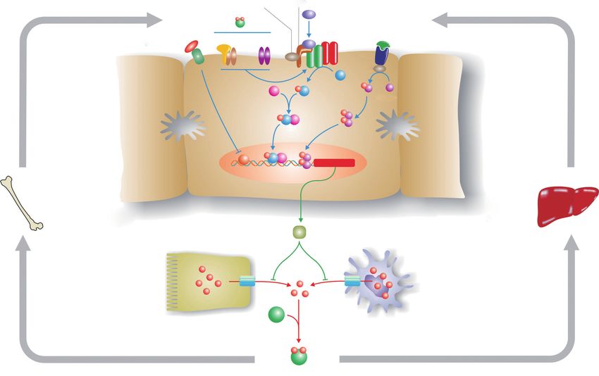
iron in various organs, but they do not absorb dietary iron (Figure 1) is helpful in understanding disease penetrance
efficiently, so SF is increased, but not TSAT. Interestingly, in hemochromatosis (59,60). To modulate body iron
such patients develop relatively few clinical symptoms homeostasis, hepcidin must respond to multiple signals,
despite high tissue iron (70). including body iron load, changes in erythropoiesis,
© Annals of Translational Medicine. All rights reserved. Ann Transl Med 2021;9(8):731 | http://dx.doi.org/ 10.21037/atm-20-5512Page 6 of 16 Anderson and Bardou-Jacquet. Genetic and phenotypic presentation of hemochromatosis
TMPRSS6 HJV
BMP2/
Diferric BMP6
transferrin
HFE/TFR1 TFR2 BMPR-Ⅰ/Ⅱ IL-6
Erythroferrone
IL-6R
? JAK1
P
SMAD P
SMAD4 1/5/8 STAT3
P
? P P
P P
P HAMP
Iron
Erythropoiesis Hepatocyte stores
Hepcidin
FPN FPN
Iron Iron
Enterocyte Macrophage
Diferric transferrin
Figure 1 The hepcidin pathway and the regulation of body iron homeostasis. Hepcidin is the master regulator of body iron homeostasis.
It is synthesized predominantly by hepatocytes and, after secretion, binds to the iron export protein ferroportin (FPN) and removes it from
the surface of target cells. This in turn decreases iron export from these cells. Macrophages and intestinal enterocytes are prime targets, but
most cells express FPN on their surface. The HAMP gene, which encodes hepcidin, is regulated by a complex series of upstream signalling
pathways. The bone morphogenetic protein (BMP)/SMAD pathway is the core regulatory pathway that responds to body iron requirements.
Mutations in various proteins that modulate signalling through this pathway lead to hemochromatosis by reducing hepcidin expression.
These include hemojuvelin (HJV) which acts as a BMP co-receptor, and homeostatic iron regulator (HFE) and transferrin receptor 2 (TFR2),
which modulate signalling through the BMP/SMAD pathway via mechanisms that are not yet fully understood. Increased body iron levels
normally stimulate hepcidin expression via the BMP/SMAD pathway, while proinflammatory cytokines increase hepcidin by signalling
through the JAK/STAT pathway. The suppression of hepcidin by enhanced erythropoiesis is, at least in part, mediated by erythroferrone.
inflammation, hypoxia and other stimuli. The bone SMAD signalling pathway is necessary, but not sufficient.
morphogenetic protein (BMP)/SMAD signalling pathway is Pro-inflammatory cytokines, notably IL-6, will stimulate
the central pathway modulating hepcidin expression. BMPs HAMP expression via the JAK/STAT3 signalling pathway.
act through their cell surface receptors to phosphorylate Several signals appear to be involved in communicating the
SMAD1/5/8 which binds to SMAD4. The resulting iron requirements of the erythroid marrow to hepcidin,
complex enters the nucleus where it binds to and activates with erythroferrone being the best studied (72). This
the HAMP promoter. Several BMPs have been shown to remains an area of active investigation.
stimulate this pathway, including BMP2 and BMP6, and While the roles of HJV, hepcidin and FPN are now
at least BMP6 is responsive to cellular iron levels. For well-defined, precisely how HFE and TFR2 influence
hepcidin to be regulated by inflammation, an intact BMP/ HAMP expression is not fully understood. However,
© Annals of Translational Medicine. All rights reserved. Ann Transl Med 2021;9(8):731 | http://dx.doi.org/ 10.21037/atm-20-5512Annals of Translational Medicine, Vol 9, No 8 April 2021 Page 7 of 16
there is compelling evidence that both proteins are (300–1,000 μg/L) to grossly (>1,000 μg/L) elevated. In two
required for optimal signalling through the BMP/SMAD large population-based studies of HH, up to 84% of male
pathway (73). The proteins may form part of a large multi- and 73% of female C282Y homozygotes had a raised TSAT,
protein complex with BMP receptors and co-receptors that and 88% of male and 57% of female homozygotes had a
enhances HJV-mediated BMP-SMAD signalling (74), but raised SF (9,52). As would be expected from their limited
this has yet to be proven in vivo. Interestingly, mice lacking physiological loss of iron, biochemical penetrance of HH
both HFE and TFR2 have a more severe phenotype than is much higher in men than it is in women (9,52). Even
mice lacking either gene alone (75), suggesting that the though HH is a progressive iron loading disorder, the rate
roles of the two proteins are not fully interchangeable. A of body iron acquisition can vary. It is usually greatest in the
similar situation has been observed in humans (76). first few decades of life, but thereafter diminishes and the
SF level may remain steady or even decline (9,52,78). This
is not unduly surprising. In HFE-related HH, hepcidin
Penetrance of HFE-associated hemochromatosis
levels are reduced, but not absent, so the body retains some
The phenotypic presentation of individuals who are capacity to regulate its iron intake, and it will decline as the
homozygous for the C282Y variant can be quite variable. iron load increases (10).
Some individuals never accumulate significant amounts Only a relatively small number of studies have objectively
of iron nor show clinical manifestations, others show looked at clinical penetrance in HH. Allen et al. (52)
biochemical evidence of iron loading (increased SF and genotyped 31,192 Australians and identified 203 C282Y
TSAT) but no clinical consequences, whereas others show homozygotes. All homozygotes, and a selection of other
iron loading in multiple tissues and clinical sequelae (3,4). genotypes were examined by clinicians who were unaware
The majority of C282Y homozygotes will not develop of the genotype. This study showed that 28.4% of male,
significant disease, even if they have raised iron indices, but only 1.2% of female, C282Y homozygotes developed
so HH is a disorder with low clinical penetrance. In a iron overload-related disease, with disease being defined as
study published by Beutler and colleagues in 2002 that biochemical evidence of iron overload in the presence of
stimulated discussion in this area, only 1 of 152 C282Y at least one clinical symptom (e.g., biopsy-proven fibrosis,
homozygotes met their definition of “frank clinical HCC, arthritis) (52). Nevertheless, in the same study 82%
hemochromatosis” (77). However, many considered this of men and 54% of women had a raised SF (and 73% and
figure to be unrealistically low. The reconciliation of these 69% respectively had a raised TSAT), consistent with the
two views comes with the understanding of how penetrance observation that a predisposing genotype or raised body
is defined. If a biochemical definition is used, the penetrance iron does not lead to overt disease in most individuals.
of C282Y homozygosity is quite high (in the Beutler study, An earlier study by Whitlock et al. (79) analyzed several
SF was raised in 76% of men and 54% of women), but if a longitudinal, population-based studies and found that
clinical definition of phenotype is used, penetrance is much 38–50% of C282Y homozygotes had a raised SF, but
lower. The clinical phenotype itself can vary from very only 10–33% developed HH-related symptoms. These
subtle changes (e.g., fatigue, reduced quality of life) to the analyses appear representative of the true clinical picture.
more overt consequences of pathological damage to organs. Consistent with their higher iron load, men are by far the
If mortality is used as an end point, then penetrance is very most likely to develop end-organ damage in HH (9,52,80).
low indeed. Most clinicians would agree that severe iron- In a more recent study, Pilling et al. examined the large UK
related disease is very infrequent in HH, but many C282Y Biobank population (451,243 participants; 2,890 C282Y
homozygotes will develop some clinical signs, even if subtle. homozygotes) and found that liver disease, diabetes mellitus
There is certainly evidence that iron loading does not need and rheumatoid arthritis and osteoarthritis were over-
to be at a level that precipitates severe disease and organ represented (with odds ratios ranging from 1.5 to 4.3) in
damage to lead to adverse health outcomes (25,26). participants homozygous for the C282Y variant (37).
Biochemical penetrance in HH is simple to ascertain as A SF of greater than 1,000 μg/L confers a clearly
it is based on objective measurements such as SF, TSAT or increased risk of tissue damage and clinical sequelae (23),
body iron load. In most HH patients, the TSAT is often but are individuals with more modest iron accumulation
well above the normal range, and the SF concentration is at increased risk of developing the complications of iron
also usually increased. The latter can range from mildly overload? Recent evidence suggests this is the case, with
© Annals of Translational Medicine. All rights reserved. Ann Transl Med 2021;9(8):731 | http://dx.doi.org/ 10.21037/atm-20-5512Page 8 of 16 Anderson and Bardou-Jacquet. Genetic and phenotypic presentation of hemochromatosis
C282Y homozygotes with a SF value between the upper consequences of that iron may differ. For example,
limit of normal and 1,000 μg/L having a lower mortality one individual may be particularly susceptible to the
than the general population following phlebotomy development of hepatic fibrosis. It is the net effect of these
therapy (25). Another study showed that there was a influences that determines whether someone genetically
beneficial effect of phlebotomy in C282Y homozygotes with predisposed to HH will develop disease and how severe that
SF vales less than 1,000 μg/L, and in this case the effect was will be.
observable after only a few months of iron depletion (26). Environmental and physiological factors play an
These studies demonstrate that there is a benefit in treating important role in determining the severity of HH. This
HH patients with mildly elevated iron indices as it will is perhaps best illustrated by the sex difference in the
reduce their risk of subsequent clinical problems. prevalence of iron loading and HH-related disease (9,37,52).
C282Y homozygosity is responsible for the vast majority Women and men do not fundamentally differ in their iron
of HH, but do other genotypes (particularly C282Y/+, absorption mechanism, but women lose much more iron
C282Y/H63D, H63D/H63D and H63D/+) confer an than men during their lifetime through both menstrual
increased risk of iron loading? In general, these other bleeding and childbirth (85,86), and testosterone suppresses
genotypes have only a very limited effect. Some C282Y or hepcidin, favouring higher iron intake in men (87). Thus, it
H63D heterozygotes have increased TSAT and SF, but they takes women much longer to accumulate iron to clinically
do not develop the complications of iron overload (9,51,81). significant levels, and this never happens at all in many
For example, Pedersen and Milman (82) demonstrated women. After the menopause, women and men accumulate
that TSAT was elevated in 9% of C282Y heterozygotes, iron at similar rates (88). Other forms of blood loss, usually
and 8% of H63D heterozygotes, with corresponding SF pathological, can also ameliorate the HH phenotype, and
values of 9% and 12%. Very few in either group had both regular blood donation can limit body iron accumulation
indices raised. Similarly, H63D homozygotes show very low and hence disease expression, although this is not always the
biochemical penetrance. A small number may have raised case (89-91).
iron indices, but clinical consequences are rare (83). The Another physiological modulator to iron loading is the
situation is a little different for C282Y/H63D compound immune system. Interest in this area was piqued soon after
heterozygotes. These individuals are more likely to have the cloning of the HFE gene with the recognition that HFE
raised iron indices than simple heterozygotes (9,52), and in was a non-classical major histocompatibility complex (MHC)
one study, 0.5–2.0% of compound heterozygotes developed class I-like protein (5), and is consistent with the earlier
clinical evidence of iron overload (53). In the HEIRS association of HH with the HLA system (49). However,
study, male compound heterozygotes were also more likely even before HFE was identified, it was recognized that HH
to report a history of liver disease (9), but a more recent patients with more severe iron loading had abnormally high
meta-analysis did not show a link between compound CD4/CD8 lymphocyte ratios (92) and that this reflected
heterozygosity and liver cirrhosis (84). Many compound constitutively low CD8+ lymphocyte numbers (93-96).
heterozygotes who do show clinical sequelae will present Although the mechanisms are not fully understood, HFE
with a co-morbidity (e.g., fatty liver, viral hepatitis), so it is may act as a suppressor of CD8+ T cell activation and
often difficult to attribute their disease symptoms solely to differentiation (97,98). In addition, primary defects in the
increased iron (15). immune system per se can lead to iron loading, and this
could influence the hemochromatosis phenotype. Not only
do mice lacking both HFE and β2-microglobulin have more
Environmental, physiological and genetic modifiers
severe iron overload than mice lacking only HFE (99), but
of the HFE-hemochromatosis phenotype
mice lacking classical MHC class I proteins also accumulate
A range of genetic, physiological and environmental factors excess iron (100,101). Indeed, extended HLA haplotypes
can contribute to the variable penetrance in HH, and these have been associated with variations in iron loading in
can also operate at different levels. There may be variation HH (95,96,102). The mechanisms linking both the adaptive
in components of the iron homeostatic machinery such that and innate immune systems to iron homeostasis are only
different individuals take up and/or store different amounts partly understood and this represents a fruitful area for
of iron. Alternatively, the amount of iron accumulated further investigation.
by two individuals may be similar, but the pathological In HFE-related HH, age is an important consideration
© Annals of Translational Medicine. All rights reserved. Ann Transl Med 2021;9(8):731 | http://dx.doi.org/ 10.21037/atm-20-5512Annals of Translational Medicine, Vol 9, No 8 April 2021 Page 9 of 16
as iron levels progressively increase over time in many, but the penetrance of HH, genetic modifiers also play a
not all, individuals. The older a person is, the more likely role. Perhaps the clearest demonstration that genetics
they are to have accumulated a sufficiently large amount of contributes to variations in body iron status comes from
iron to show the clinical manifestations of the disease (103). studies with inbred mouse strains which vary widely in
Symptoms are rarely observed in younger patients and it is their capacity to accumulate and store iron (119,120).
usually the 4th or 5th decade of life before significant health Any mutations/polymorphisms that affect the activity of
problems arise (3,4). proteins involved in iron homeostasis, and notably the
In most cases, the iron content of the diet is unlikely hepcidin regulatory pathway, have the potential to alter
to exert a major influence on iron loading in HH, but it body iron levels in HH.
can be a contributing factor (104,105). Vegetarians and A number of studies have specifically looked for
vegans will be relatively protected from iron loading and genetic modifiers of HFE-related HH, while others
may take longer to manifest signs of HH, whereas C282Y have sought genetic explanations for variations in iron
homozygotes who eat a large amount of red meat may status in the general population that could also influence
load relatively quickly (91). In addition, factors which the HH phenotype. Some of these are summarized in
influence the efficiency of iron absorption, either positively Table 2. Some of the modifying variants are in genes
or negatively, could contribute in a small way to body encoding proteins of iron metabolism, so their involvement
iron load. For example, proton pump inhibitors may is not surprising. These include the hepcidin regulators
reduce dietary iron absorption, and individuals taking iron BMP2 (134) and TMPRSS6 (133), and the iron reductase
supplements or substances that increase iron absorption CYBRD1 (122,123), which is predicted to be involved
(such as large doses of vitamin C) may take up relatively in iron absorption, and the iron transport protein
more iron (106,107). C282Y homozygotes do not generally TF (132), but others are novel (121). One of these is
need to be too restrictive about their diet, but they should GNPAT which encodes an enzyme involved in peroxisomal
limit their intake of foods containing large amounts of iron lipid metabolism (124,125). Precisely how GNPAT
or substances that may stimulate its absorption (89). contributes to iron homeostasis is unclear, but in vitro
Other clinical conditions can also influence disease studies have shown it to be a potential regulator of hepcidin
expression in HH. For example, excess alcohol consumption expression (124). The examination of GNPAT variants in
is frequently associated with an increased body iron load and other populations has shown mixed results, some supporting
this reflects a reduction in hepcidin expression (108,109). its involvement in modulating HH risk, but others
Also, C282Y homozygotes who consume excess alcohol have not (126-128). Polymorphisms in yet other genes (such
more severe liver disease and are more likely to progress to as PCSK7 and PNPLA3) have been associated with liver
cirrhosis (110,111). The incidence of non-alcoholic fatty liver disease risk (129-131), and these also may modulate the
disease (NAFLD) is rising globally and it too can contribute HH phenotype. The number of genetic modifiers of HH
to disease expression in HH (112,113). Even in the absence identified will undoubtedly increase in time, but it is most
of HFE mutations, many patients with NAFLD develop mild likely we are dealing with multiple polymorphisms having
iron overload (dysmetabolic iron overload syndrome) (114). small effects rather than a few modifiers with large effects.
Hepcidin levels are inappropriately high in NAFLD, likely
reflecting increased inflammation, but why this does not
Summary and conclusions
limit iron absorption is unclear, and suggests the possibility
of hepcidin resistance (115). Increased iron indices have also There is extensive clinical experience with HH and, in most
been demonstrated in autoimmune and viral hepatitis, and cases, the disorder can be easily diagnosed, particularly
these correlate with inappropriately low hepcidin expression since the advent of genetic testing, and readily treated by
and potentially increased iron absorption, but hepcidin phlebotomy. Biochemical penetrance in C282Y homozygotes
regulation in these conditions appears to be complex (113). is relatively high, but clinical penetrance is relatively low,
Experimental studies have shown that the combination of and clinical sequelae are much more likely to be seen in men
iron and other hepatic toxins increases the severity of liver than in women. Variations in penetrance between individuals
pathology (116-118). reflect a combination of physiological, environmental and
Although environmental factors or co-morbidities genetic factors, and co-morbidities. The physiological and
appear to account for the majority of variation in environmental factors are broadly appreciated, but studies
© Annals of Translational Medicine. All rights reserved. Ann Transl Med 2021;9(8):731 | http://dx.doi.org/ 10.21037/atm-20-5512Page 10 of 16 Anderson and Bardou-Jacquet. Genetic and phenotypic presentation of hemochromatosis
Table 2 Some genes containing polymorphisms/mutations that may potentially modify the HFE-related hemochromatosis phenotype
Gene symbol Gene product Function Reference
ARNTL Aryl hydrocarbon receptor nuclear Linked to TF expression; involved in circadian rhythm generation (121)
translocator-like
BMP2 Bone morphogenetic protein 2 Upstream positive regulator of hepcidin (121)
CYBRD1 Duodenal cytochrome B Iron reductase that may be involved in dietary iron absorption (122,123)
FADS2 Fatty acid desaturase 2 Linked to TF expression; involved in lipid metabolism; changes in (121)
lipid and iron homeostasis are frequently associated
GNPAT Glyceronephosphate O-acyltransferase Peroxisomal protein involved in the production of plasmalogens, a (124-128)
type of lipid
NAT2 N-acetyltransferase 2 Linked to TF expression; involved in xenobiotic metabolism; link to (121)
iron homeostasis unclear.
PCSK7 Proprotein convertase subtilisin/kexin Serine protease involved in processing proproteins in the (129,130)
type 7 constitutive secretory pathway
PNPLA3 Patatin like phospholipase A multifunctional enzyme with both triacylglycerol lipase and (131)
domain-containing protein 3 acylglycerol O-acyltransferase activity; involved in lipid metabolism
(or 1-acylglycerol-3-phosphate in adipocytes
O-acyltransferase or adiponutrin)
TF Transferrin The major plasma iron transport protein (121,132)
TMPRSS6 Transmembrane serine protease 6 Upstream negative regulator of hepcidin (133)
of genetic modifiers are in their relative infancy. In time we Hepatology” published in Annals of Translational Medicine.
will learn more about polymorphisms that affect iron loading The article has undergone external peer review.
and/or disease outcome, but we are likely looking at a genetic
landscape where multiple loci each contribute a small effect. Peer Review File: Available at http://dx.doi.org/10.21037/
In the great majority of cases, HH patients will continue atm-20-5512
to be monitored using conventional blood iron status
parameters, with the use of non-invasive imaging to monitor Conflicts of Interest: Both authors have completed the
organ iron load and tissue pathology, and phlebotomy to ICMJE uniform disclosure form (available at http://dx.doi.
deplete accumulated iron. However, with an increasingly org/10.21037/atm-20-5512). The series “Unresolved Basic
advanced understanding of iron homeostasis mechanisms and Issues in Hepatology” was commissioned by the editorial
disease modifiers, and bespoke therapeutic options on our office without any funding or sponsorship. Dr. GJA reports
doorstep, we now have the tools available to diagnose and personal fees from Protagonist Therapeutics, other from
treat unusual iron overload cases when they present. PharmaNutra, outside the submitted work. EBJ has no
other conflicts of interest to declare.
Acknowledgments
Ethical Statement: The authors are accountable for all
Funding: None. aspects of the work in ensuring that questions related
to the accuracy or integrity of any part of the work are
appropriately investigated and resolved.
Footnote
Provenance and Peer Review: This article was commissioned Open Access Statement: This is an Open Access article
by the Guest Editors (Ralf Weiskirchen and Wolfgang distributed in accordance with the Creative Commons
Stremmel) for the series “Unresolved Basic Issues in Attribution-NonCommercial-NoDerivs 4.0 International
© Annals of Translational Medicine. All rights reserved. Ann Transl Med 2021;9(8):731 | http://dx.doi.org/ 10.21037/atm-20-5512Annals of Translational Medicine, Vol 9, No 8 April 2021 Page 11 of 16
License (CC BY-NC-ND 4.0), which permits the non- Pharmaceuticals (Basel) 2019;12:122.
commercial replication and distribution of the article with 14. Lynch S, Pfeiffer CM, Georgieff MK, et al. Biomarkers of
the strict proviso that no changes or edits are made and the Nutrition for Development (BOND)-Iron Review. J Nutr
original work is properly cited (including links to both the 2018;148:1001S-1067S.
formal publication through the relevant DOI and the license). 15. Powell LW, Seckington RC, Deugnier Y.
See: https://creativecommons.org/licenses/by-nc-nd/4.0/. Haemochromatosis. Lancet 2016;388:706-16.
16. Anderson GJ, Vulpe CD. Mammalian iron transport. Cell
Mol Life Sci 2009;66:3241-61.
References
17. Brissot P, Ropert M, Le Lan C, et al. Non-transferrin
1. Brissot P, Troadec MB, Loréal O, Brissot E. bound iron: a key role in iron overload and iron toxicity.
Pathophysiology and classification of iron overload Biochim Biophys Acta 2012;1820:403-10.
diseases; update 2018. Transfus Clin Biol 2019;26:80-8. 18. Loréal O, Gosriwatana I, Guyader D, et al.
2. Piperno A, Pelucchi S, Mariani R. Inherited iron overload Determination of non-transferrin-bound iron in genetic
disorders. Transl Gastroenterol Hepatol 2020;5:25. hemochromatosis using a new HPLC-based method. J
3. Brissot P, Pietrangelo A, Adams PC, et al. Hepatol 2000;32:727-33.
Haemochromatosis. Nat Rev Dis Primers 2018;4:18016. 19. Le Lan C, Loréal O, Cohen T, et al. Redox active
4. Kowdley KV, Brown KE, Ahn J, Sundaram V. ACG plasma iron in C282Y/C282Y hemochromatosis. Blood
Clinical Guideline: Hereditary hemochromatosis. Am J 2005;105:4527-31.
Gastroenterol 2019;114:1202-18. 20. Cabantchik ZI, Breuer W, Zanninelli G, et al. LPI-
5. Feder JN, Gnirke A, Thomas W, et al. A novel MHC labile plasma iron in iron overload. Best Pract Res Clin
class I-like gene is mutated in patients with hereditary Haematol 2005;18:277-87.
haemochromatosis. Nat Genet 1996;13:399-408. 21. Theil EC. Ferritin: the protein nanocage and iron
6. Byrnes V, Ryan E, Mayne P, et al. Neonatal screening biomineral in health and in disease. Inorg Chem
for the HFE mutations in the Irish population. Gut 2013;52:12223-33.
1999;1:527A. 22. Guyader D, Jacquelinet C, Moirand R, et al.
7. Milman N, Pedersen P, Steig T, et al. Frequencies of the Noninvasive prediction of fibrosis in C282Y homozygous
hereditary hemochromatosis allele in different populations. hemochromatosis. Gastroenterology 1998;115:929-36.
Comparison of previous phenotypic methods and novel 23. Allen KJ, Bertalli NA, Osborne NJ, et al. HFE Cys282Tyr
genotypic methods. Int J Hematol 2003;77:48-54. homozygotes with serum ferritin concentrations below
8. Olynyk JK, Cullen DJ, Aquilia S, et al. A population-based 1000 μg/L are at low risk of hemochromatosis. Hepatology
study of the clinical expression of the hemochromatosis 2010;52:925-33.
gene. N Engl J Med 1999;341:718-24. 24. Barton JC, Barton JC, Acton RT, et al. Increased risk of
9. Adams PC, Reboussin DM, Barton JC, et al. death from iron overload among 422 treated probands
Hemochromatosis and iron-overload screening in a racially with HFE hemochromatosis and serum levels of ferritin
diverse population. N Engl J Med 2005;352:1769-78. greater than 1000 μg/L at diagnosis. Clin Gastroenterol
10. McLaren GD, Nathanson NH, Jacobs A, et al. Regulation Hepatol 2012;10:412-6.
of intestinal iron absorption and mucosal iron kinetics 25. Bardou-Jacquet E, Morcet J, Manet G, et al. Decreased
in hereditary hemochromatosis. J Lab Clin Med cardiovascular and extrahepatic cancer-related mortality
1991;117:390-401. in treated patients with mild HFE hemochromatosis. J
11. Widdowson EM, McCance RA. The absorption and Hepatol 2015;62:682-9.
excretion of iron before, during and after a period of very 26. Ong SY, Gurrin LC, Dolling L, et al. Reduction of body
high intake. Biochem J 1937;31:2029-34. iron in HFE-related haemochromatosis and moderate
12. Galaris D, Barbouti A, Pantopoulos K. Iron homeostasis iron overload (Mi-Iron): a multicentre, participant-
and oxidative stress: An intimate relationship. Biochim blinded, randomised controlled trial. Lancet Haematol
Biophys Acta Mol Cell Res 2019;1866:118535. 2017;4:e607-14.
13. Porto G, Cruz E, Teles MJ, et al. HFE Related 27. Mehta KJ, Farnaud SJ, Sharp PA. Iron and liver fibrosis:
Hemochromatosis: Uncovering the Inextricable Link Mechanistic and clinical aspects. World J Gastroenterol
between Iron Homeostasis and the Immunological System. 2019;25:521-38.
© Annals of Translational Medicine. All rights reserved. Ann Transl Med 2021;9(8):731 | http://dx.doi.org/ 10.21037/atm-20-5512Page 12 of 16 Anderson and Bardou-Jacquet. Genetic and phenotypic presentation of hemochromatosis
28. Cook JD, Skikne BS, Lynch SR, et al. Estimates of iron with or without a family history. Arch Intern Med
sufficiency in the US population. Blood 1986;68:726-31. 2006;166:294-301.
29. Kowdley KV, Brandhagen DJ, Gish RG, et al. Survival 43. Bardou-Jacquet E, Morandeau E, Anderson GJ, et al.
after liver transplantation in patients with hepatic iron Regression of fibrosis stage with treatment reduces long-
overload: the national hemochromatosis transplant term risk of liver cancer in patients with hemochromatosis
registry. Gastroenterology 2005;129:494-503. caused by mutation in HFE. Clin Gastroenterol Hepatol
30. Paisant A, d'Assignies G, Bannier E, et al. MRI for the 2020;18:1851-7.
measurement of liver iron content, and for the diagnosis 44. Adams PC, Speechley M, Kertesz AE. Long-term survival
and follow-up of iron overload disorders. Presse Med analysis in hereditary hemochromatosis. Gastroenterology
2017;46:e279-87. 1991;101:368.
31. Carroll GJ, Breidahl WH, Olynyk JK. Characteristics of 45. Niederau C, Fischer R, Pürschel A, et al. Long-term
the arthropathy described in hereditary hemochromatosis. survival in patients with hereditary hemochromatosis.
Arthritis Care Res (Hoboken) 2012;64:9-14. Gastroenterology 1996;110:1107-19.
32. Kiely PD. Haemochromatosis arthropathy - a 46. Barton JC, Edwards CQ. HFE Hemochromatosis. 2000
conundrum of the Celtic curse. J R Coll Physicians Edinb Apr 3 [updated 2018 Dec 6]. In: Adam MP, Ardinger HH,
2018;48:233-8. Pagon RA, et al. editors. GeneReviews® [Internet]. Seattle
33. Guggenbuhl P, Deugnier Y, Boisdet JF, et al. Bone mineral (WA): University of Washington, Seattle, 1993–2020.
density in men with genetic hemochromatosis and HFE 47. Prabhu A, Cargill T, Roberts N, et al. Systematic review
gene mutation. Osteoporos Int 2005;16:1809-14. of the clinical outcomes of iron reduction in hereditary
34. Jeney V. Clinical impact and cellular mechanisms of hemochromatosis. Hepatology 2020;72:1469-82.
iron overload-associated bone loss. Front Pharmacol 48. Ulvik RJ. Hereditary haemochromatosis through 150
2017;8:77. years. Tidsskr Nor Laegeforen 2016;136:2017-21.
35. Díez-López C, Comín-Colet J, González-Costello J. Iron 49. Simon M, Bourel M, Fauchet R, et al. Association
overload cardiomyopathy: from diagnosis to management. of HLA-A3 and HLA-B14 antigens with idiopathic
Curr Opin Cardiol 2018;33:334-40. haemochromatosis. Gut 1976;17:332-4.
36. Gulati V, Harikrishnan P, Palaniswamy C, et al. 50. Cassidy LM, Martiniano R, Murphy EM, et al. Neolithic
Cardiac involvement in hemochromatosis. Cardiol Rev and Bronze Age migration to Ireland and establishment
2014;22:56-68. of the insular Atlantic genome. Proc Natl Acad Sci USA
37. Pilling LC, Tamosauskaite J, Jones G, et al. Common 2016;113:368-73.
conditions associated with hereditary haemochromatosis 51. Zaloumis SG, Allen KJ, Bertalli NA, et al. Natural history
genetic variants: cohort study in UK Biobank. BMJ of HFE simple heterozygosity for C282Y and H63D:
2019;364:k5222. Erratum in: BMJ. 2019 Oct 23;367:l6157. a prospective 12-year study. J Gastroenterol Hepatol
doi: 10.1136/bmj.l6157. 2015;30:719-25.
38. Demetz E, Tymoszuk P, Hilbe R, et al. The 52. Allen KJ, Gurrin LC, Constantine CC, et al.
haemochromatosis gene Hfe and Kupffer cells control Iron-overload-related disease in HFE hereditary
LDL cholesterol homeostasis and impact on atherosclerosis hemochromatosis. N Engl J Med 2008;358:221-30.
development. Eur Heart J 2020;41:3949-59. 53. Gurrin LC, Bertalli NA, Dalton GW, et al. HFE
39. McDermott JH, Walsh CH. Hypogonadism in C282Y/H63D compound heterozygotes are at low risk
hereditary hemochromatosis. J Clin Endocrinol Metab of hemochromatosis-related morbidity. Hepatology
2005;90:2451-5. 2009;50:94-101.
40. Loréal O, Cavey T, Robin F, et al. Iron as a Therapeutic 54. Barton JC, Edwards CQ, Acton RT. HFE gene: Structure,
Target in HFE-Related Hemochromatosis: Usual and function, mutations, and associated iron abnormalities.
Novel Aspects. Pharmaceuticals (Basel) 2018;11:131. Gene 2015;574:179-92.
41. Falize L, Guillygomarc'h A, Perrin M, et al. Reversibility 55. Piperno A, Arosio C, Fossati L, et al. Two novel nonsense
of hepatic fibrosis in treated genetic hemochromatosis: a mutations of HFE gene in five unrelated Italian patients
study of 36 cases. Hepatology 2006;44:472-7. with hemochromatosis. Gastroenterology 2000;119:441-5.
42. Powell LW, Dixon JL, Ramm GA, et al. Screening 56. Hamdi-Rozé H, Beaumont-Epinette MP, Ben Ali Z, et
for hemochromatosis in asymptomatic subjects al. Rare HFE variants are the most frequent cause of
© Annals of Translational Medicine. All rights reserved. Ann Transl Med 2021;9(8):731 | http://dx.doi.org/ 10.21037/atm-20-5512Annals of Translational Medicine, Vol 9, No 8 April 2021 Page 13 of 16
hemochromatosis in non-c282y homozygous patients with features. Pharmaceuticals (Basel) 2019;12:132.
hemochromatosis. Am J Hematol 2016;91:1202-5. 71. Aschemeyer S, Qiao B, Stefanova D, et al. Structure
57. Le Gac G, Congiu R, Gourlaouen I, et al. Homozygous function analysis of ferroportin defines the binding site
deletion of HFE is the common cause of hemochromatosis and an alternative mechanism of action of hepcidin. Blood
in Sardinia. Haematologica 2010;95:685-7. 2018;131:899-910.
58. McDonald CJ, Ostini L, Wallace DF, et al. Next- 72. Kautz L, Jung G, Valore EV, et al. Identification
generation sequencing: Application of a novel of erythroferrone as an erythroid regulator of iron
platform to analyze atypical iron disorders. J Hepatol metabolism. Nat Genet 2014;46:678-84.
2015;63:1288-93. 73. Xiao X, Alfaro-Magallanes VM, Babitt JL. Bone
59. Ganz T, Nemeth E. Hepcidin and iron homeostasis. morphogenic proteins in iron homeostasis. Bone
Biochim Biophys Acta 2012;1823:1434-43. 2020;138:115495.
60. Silvestri L, Nai A, Dulja A, et al. Hepcidin and the BMP- 74. D’Alessio F, Hentze MW, Muckenthaler MU. The
SMAD pathway: An unexpected liaison. Vitam Horm hemochromatosis proteins HFE, TfR2, and HJV form
2019;110:71-99. a membrane-associated protein complex for hepcidin
61. Nemeth E, Tuttle MS, Powelson J, et al. Hepcidin regulation. J Hepatol 2012;57:1052-60.
regulates cellular iron efflux by binding to ferroportin and 75. Wallace DF, Summerville L, Crampton EM, et al.
inducing its internalization. Science 2004;306:2090-3. Combined deletion of Hfe and transferrin receptor 2 in
62. Bridle KR, Frazer DM, Wilkins SJ, et al. Disrupted mice leads to marked dysregulation of hepcidin and iron
hepcidin regulation in HFE-associated haemochromatosis overload, Hepatology 2009;50:1992-2000.
and the liver as a regulator of body iron homeostasis. 76. Pietrangelo A, Caleffi A, Henrion J, et al. Juvenile
Lancet 2003;361:669-73. hemochromatosis associated with pathogenic mutations
63. Darshan D, Frazer DM, Anderson GJ. Molecular basis of of adult hemochromatosis genes. Gastroenterology
iron-loading disorders. Expert Rev Mol Med 2010;12:e36. 2005;128:470-9.
64. Frazer DM, Wilkins SJ, Millard KN, et al. Increased 77. Beutler E, Felitti VJ, Koziol JA, et al. Penetrance of
hepcidin expression and hypoferraemia associated with an 845G-->A (C282Y) HFE hereditary haemochromatosis
acute phase response are not affected by inactivation of mutation in the USA. Lancet 2002;359:211-8.
HFE. Br J Haematol 2004;126:434-6. 78. Gurrin LC, Osborne NJ, Constantine CC, et al. The
65. van Deuren M, Kroot JJ, Swinkels DW. Time-course natural history of serum iron indices for HFE C282Y
analysis of serum hepcidin, iron and cytokines in a homozygosity associated with hereditary hemochromatosis.
C282Y homozygous patient with Schnitzler's syndrome Gastroenterology 2008;135:1945-52.
treated with IL-1 receptor antagonist. Haematologica 79. Whitlock EP, Garlitz BA, Harris EL, et al. Screening for
2009;94:1297-300. hereditary hemochromatosis: a systematic review for the
66. Camaschella C, Roetto A, Calì A, et al. The gene TFR2 is U.S. Preventive Services Task Force. Ann Intern Med
mutated in a new type of haemochromatosis mapping to 2006;145:209-23.
7q22. Nat Genet 2000;25:14-5. 80. Gan EK, Powell LW, Olynyk JK. Natural history and
67. Papanikolaou G, Samuels ME, Ludwig EH, et al. management of HFE-hemochromatosis. Semin Liver Dis
Mutations in HFE2 cause iron overload in chromosome 2011;31:293-301.
1q-linked juvenile hemochromatosis. Nat Genet 81. Bulaj ZJ, Griffen LM, Jorde LB, et al. Clinical and
2004;36:77-82. biochemical abnormalities in people heterozygous for
68. Roetto A, Papanikolaou G, Politou M, et al. Mutant hemochromatosis. N Engl J Med 1996;335:1799-805.
antimicrobial peptide hepcidin is associated with severe 82. Pedersen P, Milman N. Genetic screening for HFE
juvenile hemochromatosis. Nat Genet 2003;33:21-2. hemochromatosis in 6,020 Danish men: penetrance
69. Détivaud L, Island ML, Jouanolle AM, et al. Ferroportin of C282Y, H63D, and S65C variants. Ann Hematol
diseases: functional studies, a link between genetic and 2009;88:775-84.
clinical phenotype. Hum Mutat 2013;34:1529-36. 83. Gochee PA, Powell LW, Cullen DJ, et al. A population-
70. Vlasveld LT, Janssen R, Bardou-Jacquet E, et al. Twenty based study of the biochemical and clinical expression of
years of ferroportin disease: a review or an update of the H63D hemochromatosis mutation. Gastroenterology
published clinical, biochemical, molecular, and functional 2002;122:646-51.
© Annals of Translational Medicine. All rights reserved. Ann Transl Med 2021;9(8):731 | http://dx.doi.org/ 10.21037/atm-20-5512You can also read