Laboratory Values Interpretation Resource
←
→
Page content transcription
If your browser does not render page correctly, please read the page content below
Updated 2017

Laboratory Values Interpretation Resource
 Academy of Acute Care Physical Therapy – APTA Task Force on Lab Values
                                                                             2017 Members
                                                      James Tompkins, PT, DPT | Co-Chair
                                                     Traci Norris, PT, DPT, GCS | Co-Chair
                                                Kim Levenhagen, PT, DPT, WCC | Co-Chair
                                                               Kate Adeletti, PT, DPT, NCS
                                                                         Courtney Bryan, PT
                                                    Malinda Brown-Crowell, PT, DPT, GCS
                                                                      Jamie Dyson, PT, DPT
                                                                Komal Shah, PT, DPT, NCS
                                                            Kathy Swanick, PT, DPT, OCS
                                                                           Julie Terrell, DPT
                                                                   Risa Maruyama, PT, NCS
                                                                                 Caitlin Price

                                                                         2012 Members
                                                                 Roya Ghazinouri | Chair
                                                                     Samidha Deshmukh
                                                  Sharon Gorman, PT, DPTSc, FNAP, GCS
                                                                          Angela Hauber
                                                                             Mary Kroohs
                                                                         Elizabeth Moritz
                                                   Babette Sanders, PT, DPT, MS, FAPTA
                                                             Darin Trees, PT, DPT, CWS

                                                                            2008 Members
                                                                   Holly McKenzie, PT, DPT
                                                                      Dawn Piech, PT, MPT
                                                                    Jim Smith, PT, DPT, MA

                              Approved by Academy of Acute Care Physical Therapy – APTA
                                               Board of Directors: 8/2008, 12/2011, 1/2017

                                                                                            1
Evolution of the 2017 Edition of the Laboratory Values Interpretation
Resource by the Academy of Acute Care Physical Therapy
As emerging research regarding early mobilization and advancements in medical practice is evolving, the
Academy of Acute Care Physical Therapy – American Physical Therapy Association Task Force on Lab
Values took on the mission of updating the Laboratory Values Interpretation Resource to better accommodate
practitioners’ needs. The task force consisted of physical therapists from across the country in various acute
care settings. Based on practitioners’ feedback, this document was adapted to improve usability in the busy
acute care setting.
The task force set out to use current literature from the past five years. Original sources were captured and
referenced for each item. The task force collaborated with university librarians to ensure the
comprehensiveness of the literature search. After consulting with clinical lab scientists, the task force was
unable to identify a gold standard in regard to a laboratory guide listing reference values (see disclaimer). For
the purpose of consistency, the task force decided to use the reference values from one reputable laboratory
values textbook, unless there was a clinical practice guideline related to that laboratory value. Each laboratory
test captured in this 2017 version has a brief explanation of the test or laboratory panel, reference values,
clinical presentation, and clinical implications. In response to unmet clinical decision-making needs from
membership thus far, updates have been made to the content from the previous version, and a new point-of-
care document listing key laboratory tests has been created for this version. As the task force closes its current
work on this project, it does so in the understanding that this living document needs continuous updating to
ensure that the needs of clinicians will be appropriately accommodated.
Disclaimer
The reference ranges and recommendations in this resource are based on the current, best-available
evidence. Considering the absence of a universal reference range for any of the more than 5,000 lab tests in
existence, accredited laboratories are required to establish and validate their reference values at least
annually. Thus, any given result should be interpreted based on the reference value of the laboratory in which
the test was performed. Reference values must be updated each time a new reagent kit or diagnostic
instrument is added. In addition, differences in patient populations (ethnicity, age, gender, behaviors, and
culture) might result in variability of reference ranges. Abnormal values are defined as those results that are
outside a specific range obtained from a cohort of healthy individuals.1
Physical therapists have the professional responsibility to provide excellent care, adhere to high standards,
and collaborate with other healthcare providers to achieve optimal health outcomes for their patients. Acute
care physical therapists work in an environment that is quickly evolving and therefore should be knowledgeable
regarding critical laboratory values and safe mobility recommendations. Lundberg (1972) defined a critical
value as a “physiological state at such variance with normal as to be life threatening unless something is done
promptly and for which some corrective action can be taken.”2 As critical values might evolve quickly in the
acute care setting, physical therapists should be vigilant in reevaluating safe and effective patient
management. Although the recommendations made in this document are evidence-based, the final judgment
regarding the appropriateness of particular physical therapy interventions should be made by the clinician. The
goal of clinical standardization is not to produce rigid guidelines; it is to establish an evidence- and consensus-
founded treatment approach that could change and evolve based on the patient’s clinical presentation and
individual values, as well as expectations and preferences.
Today’s electronic health record environment allows for fast retrieval of laboratory results. Test names and
specific value ranges are easily visualized with high-priority findings (i.e. critical alerts), having predetermined
indicators or color highlights to bring attention to medical team.
2Table of Contents
1. Understanding Lab Values
a. Trends
b. Risk vs. Benefit of the Therapeutic Intervention
c. Acute vs. Chronic Considerations of the Therapeutic Intervention
d. Gender, Race, and Culture Considerations
e. Age Considerations
2. Complete Blood Count (CBC)
a. White Blood Cells
b. Platelets
c. Hemoglobin
d. Hematocrit
3. Electrolyte Panel
a. Sodium (Na)
b. Potassium (K)
c. Calcium (Ca)
d. Chloride (Cl)
e. Phosphate (PO4)
f. Magnesium (Mg)
4. Kidney Function
a. Blood Urea Nitrogen (BUN)
b. Serum Creatinine
5. Endocrine
a. Glucose/Criteria for Diagnosis of Diabetes
b. Hgb A1C
c. Thyroid Function Tests
6. Acid-Base Disorders
a. Respiratory Alkalosis
b. Respiratory Acidosis
c. Metabolic Alkalosis
d. Metabolic Acidosis
7. Liver Function/Hepatic Panel
a. Serum Albumin/Pre-Albumin
b. Serum Bilirubin
c. Ammonia
d. Model for End-Stage Liver Disease (MELD)
e. FK Trough (Tacrolimus/Prograf Test)
8. Lipid Panel
a. High-Density Lipoprotein (HDL)
b. Low-Density Lipoprotein (LDL)
c. Triglycerides
d. Total Cholesterol
39. Bleeding Ratio/Viscosity
a. International Normalized Ratio (INR)
b. Activated Partial Thromboplastin Time (aPTT)
c. Prothrombin Time (PT)
d. Anti-Factor Xa Assay
e. D-Dimer
f. Algorithm for Mobilizing Patients with Known Lower-Extremity Deep Vein Thrombosis
10. Cardiovascular-Specific Labs
a. Troponin
b. B-Type Natriuretic Peptide (BNP)
c. Creatinine Kinase (CK)
11. References
Appendix A: Point-of-Care Document
41. Understanding Lab Values
a. Trends
Physical therapists should not rely exclusively on a single laboratory finding; instead, they should also consider
a variety of other clinical factors. For instance, clinicians should be aware of the time the laboratory specimen
was drawn, potential drug interactions, or the patient’s recent meals. Likewise, it is important to understand the
significance of trends in the values over time. Electrolyte panels might change with intravenous infusions,
medications, and diet. Patients with chronic medical conditions, such as anemia, might be asymptomatic
during exercise, while a patient with a precipitous drop in hemoglobin and hematocrit might require urgent
medical attention.
When a patient presents with symptoms of a suspected myocardial infarction (MI), cardiac biomarker
laboratory tests are ordered to assist with a differential diagnosis. Cardiac biomarkers are materials released
into the bloodstream when the heart is under stress. Typically, under normal circumstances, these substances
do not appear in circulation; however, when there is insufficient blood flow to the heart, markers associated
with myocardial injury increase in a predictable fashion. Up to 80% of patients with an acute MI will present
with an elevation of troponin within 3 hours of onset of chest pain.3
However, not all patients with cardiac impairments present with obvious symptoms, and they might not have
undergone diagnostic testing. It is not uncommon for patients with complex comorbidities and non-specific and
subtle symptoms, including unexplained fatigue and weakness, to be referred to acute care physical therapy. It
is, therefore, prudent for therapists to be aware of the presence of cardiac biomarkers and potential delays in
the diagnosing of cardiac ischemia.
b. Risk vs. Benefit Considerations of the Therapeutic Intervention
The fundamental consideration when reviewing patient laboratory findings is toward determining an appropriate plan
of care and weighing the anticipated benefit of a therapy intervention against the potential risk to the patient.
Physical therapists should carefully anticipate the physiological changes that might have occurred whenever a
laboratory value is out of range. They should also be aware of the heightened risk level if a value should fall
into the critical range. It is critical to understand pertinent lab values and the subsequent potential of adverse
events when practicing in this kind of practice setting. In weighing risks and benefits, physical therapists should
also consider the potential benefits from a therapeutic plan that increases the patient’s activity. Immediate risks
and benefits, as well as the longer-term consequences over the episode of care, should be assessed. To fully
explore the potential effects of physical therapy intervention, collaboration with other members of the
interprofessional medical team is often necessary. It is prudent and congruent with standards of
professionalism for physical therapists to assist with the development of facility policies, procedures, and
protocols to aid in the clinical decision-making process regarding the use of lab values in determining the
intensity level of therapeutic interventions.
c. Acute vs. Chronic Considerations of the Therapeutic Intervention
In addition to comparing a patient’s specific laboratory values to known reference ranges for a population,
clinical decisions require understanding of the patient’s symptoms and the dynamic physiological changes
indicated by the laboratory tests. As an example, acute laboratory value changes, such as those associated
5with blood loss due to trauma or surgery, might require the physical therapist to select a more conservative
plan of care. At the same time, such acute changes might also suggest the potential for more serious adverse
events contributable to the limited amount of time to physiologically compensate for this acute change. Patients
with chronic medical conditions often have more chronic changes in lab values, commonly associated with
these conditions (e.g., congestive heart failure [CHF], chronic obstructive pulmonary disease [COPD], anemia)
or longer-term medical interventions (e.g., chemotherapy, radiation therapy). Under these circumstances, it is
prudent for the physical therapist to allow the patient a period of time for his or her body to adapt to the
changes in lab values. In turn, this interim period might allow patients to have more resources toward dealing
with potential adverse events caused by increasing cardiorespiratory demand, mobility, and exercise.
d. Gender, Race, and Culture Considerations
Census 2010 indicated increased minority demographic shifts in the United States.4 McClatchey noted that
“genetic heterogeneity within a population leads to person-to-person phenotypic differences that can contribute
to the variability in laboratory test results.”4 Some diseases are more prevalent in specific races and ethnicities.
For example, sickle cell anemia is more prevalent in populations with sub-Saharan African ancestry than with
Caucasians.5 That being said, it is not possible to determine whether racial differences in laboratory values are
genetic or related to lifestyle alone, due to culture and food preferences (e.g., cholesterol).6 Therefore, physical
therapists should be mindful of potential racial differences in laboratory values.
Genetic heterogeneity at the molecular level can lead to differences in the reactivity of an individual’s DNA,
proteins, or cells toward the nucleic acid probes and antibodies that are used as reagents in many diagnostic
tests.4 This type of genetic heterogeneity might result in false-negative findings. As the field of clinical
laboratory medicine progresses, genetic variability will become an increasingly more important consideration in
the development of new tests and in analyzing results from the current test.
In the United States, African Americans tend to have increased muscle mass and skeletal structures compared
to their Caucasian counterparts. Therefore, racial differences in serum levels of creatinine kinase and lactate
dehydrogenase in adults, and in serum alkaline phosphatase in children, are noted. African Americans also
tend to have higher serum total protein levels and higher serum levels of alpha, beta, and gamma globulins,
IgG, and IgA, than Caucasians.4
African Americans tend to have lower hemoglobin (Hgb) values compared to Caucasians.6 In addition, HgbA1c
(A1c) lab values can be altered in patients with sickle hemoglobin, which is present in 8% of the African
American population.7 Other studies have noted racial differences in mean hematocrit (Hct) readings that
decreased over time due to quality of care rendered during the onset of end-stage renal disease, regardless of
socioeconomic status.8 Cultural competence is a non-negotiable skill, subject to rigorous testing similar to any
other core component of the physical therapy profession.9 Leavitt posited “future research stands to provide a
wealth of knowledge on the link between genetics and disparities in health, but the differences remain to be
seen.”10 For those reasons, physical therapists must consider racial variations in laboratory values in order for
culturally competence care.
Sex and Gender Considerations:11 Many lab results will have reference ranges reported as age-specific or
sex-specific values. With regard to interpretation of these reference ranges regarding sex-specific norms, the
therapist needs to consider the patient’s biological sex, gender, and gender identity to avoid referencing the
incorrect “normal” value. A review of the differences of these terms is provided in Table 1.
6Table 1: Definitions pertaining to sex and gender roles.12
Term Definition
Categorical differentiation between men and women, assigned at birth based on brief
Sex
visual examination of external genitalia.
Gender Binary social construct involving characteristics distinguishing men from women.
Gender Identity Person’s sense of being male or female.
Transsexual Outdated term for person who feels they were assigned the incorrect sex.
Overarching term for persons with various identities and expressions that are
Transgender
associated with assignment of incorrect sex.
Legal, medical, and surgical processes that a transsexual person might experience to
Transition
correct the incongruence of incorrect sexual assignment.
Transwoman A person who identifies as female but was assigned the male sex.
Transman A person who identifies as male but was assigned the female sex.
Individual patients might be in the process of transitioning to their preferred gender through medical (i.e.,
hormone replacement therapy), surgical (i.e., gender reassignment surgery), and/or legal (i.e., amending legal
documents to reflect gender identity) means to correct incongruence of sex. Physical therapists should
determine if patients in transition are currently under medical treatment for this transition, which could occur
prior to or in conjunction with surgical transition, and will be continued after surgical transition. If the patient is
on hormone replacement therapy, physical therapists should use the transitioned gender to determine the
reference value. If the patient is not receiving hormone therapy, physical therapists should use the patient’s
biological sex to determine the reference value. For example, a transwomen on estrogen replacement therapy
should have her lab values compared to normal values of females due to the effects of estrogen on her
physiology, whereas a transman on testosterone should have his lab values compared to those of males due
to the effects of testosterone on his physiology. The key factor is not whether the medical record assigns a
patient a particular sex or whether the patient has undergone sexual reassignment surgery, but whether
patients are taking hormone therapy that will affect their physiology and lab chemistry. Knowing the medical
transition status of a transsexual person reduces the risk of misinterpretation of lab values and ensure correct
application of normal reference values consistently.12
e. Age Considerations
This outline was created to assist the clinician with lab value considerations for the general population. The
clinician should be aware that “norms” are created for the healthy adult, and each patient’s lab values should
be interpreted within the context of the patient’s current medical status. That is to say, when reading the value
ranges in this section, be aware that considerations for mobility might vary based on the patient’s age and
current medical condition. For example, an 18-year-old boy with a below-normal hematocrit might tolerate this
lower level better than a 90-year-old male with the same low hematocrit. Thus, a clinician might be more willing
to mobilize a patient with a below-normal value who is younger and has overall more reserve. Conversely,
patients being treated for certain blood cancers can more safely participate in mobility with lower platelet levels
vs. the general population, the latter likely being at an increased risk of bleeding.
We have not included lab ranges for the pediatric population. Please refer to the Academy of Pediatric Physical
Therapy for more information, as normative values might differ from the adult populations.
72. Complete Blood Count (CBC)
Complete Blood Count (CBC)
Provides results regarding the concentration of red blood Causes Presentation Clinical Implications
cells, white blood cells, and platelets in a blood sample.1
Infection
Leukemia
Neoplasm Fever
Trauma Malaise
Symptoms-based approach when
Surgery Lethargy
determining appropriateness for activity,
Sickle-cell disease Dizziness
Trending Upward especially in the presence of fever.
Stress/pain Bleeding
(leukocytosis)13
Medication-induced Bruising
> 11.0 109/L Consider timing of therapy session due
Smoking Weight loss
to early-morning low level and late-
Obesity (unintentional)
afternoon high peak.14
Congenital Lymphadenopathy
Chronic inflammation Painful inflamed joints
White Blood Cells Connective tissue
disease
Routine test to identify Anemia
the presence of Viral infections
Trending Weakness Symptoms-based approach when
infection, inflammation, Chemotherapy
Downward Fatigue determining appropriateness for activity,
allergens. Aplastic anemia
(leukopenia)13 Fever especially in the presence of fever.14
Autoimmune disease
< 4.0 109/L Headache
REFERENCE Hepatitis
Shortness of breath
VALUES13
5.0-10.0 109/L Trending
Downward
(neutropenia)13
Neutropenic precautions (dependent on
< 1.5 109/L Low-grade fever
Stem cell disorder facility guidelines).14
Skin abscesses
Bacterial infection
0.5-1.0 109/L = Sore mouth
Viral infection Symptoms-based approach when
moderate Symptoms of
Radiation determining appropriateness for activity,
neutropenia pneumonia
especially in the presence of fever.14
< 0.5 109/L=
severe neutropenia
8Complete Blood Count (CBC) Causes Presentation Clinical Implications
Splenectomy
Inflammation
Neoplasm/cancer Symptoms-based approach when
Stress Weakness determining appropriateness for activity;
Trending Upward Iron deficiency Headache monitor symptoms; collaborate with
(thrombocytosis) Infection Dizziness interprofessional team.13-15
> 450 k/uL Hemorrhage Chest pain
Hemolysis Tingling in hands/feet Elevated levels can lead to venous
Platelets High altitudes thromboembolism.
Strenuous exercise
Trauma
REFERENCE
In presence of severe thrombocytopenia
VALUES Viral infection
(< 20 k/uL): Symptoms-based approach
Nutrition deficiency
140-400 k/uL13 when determining appropriateness for
Leukemia Petechiae
activity; collaborate with
Trending Radiation Ecchymosis
interprofessional team (regarding
Downward Chemotherapy Fatigue
possible need for/timing of transfusion
(thrombocytopenia) Malignant cancer Jaundice
prior to mobilization)14
< 150 k/uL Liver disease Splenomegaly
Aplastic anemia Risk for bleeding
Fall risk awareness (risk of spontaneous
Premenstrual and
hemorrhage).16,17
postpartum
Hemoglobin
Congenital heart
Assess anemia, blood Low critical values (< 5-7 g/dL) can lead
disease Orthostasis
loss, bone marrow to heart failure or death.13
Severe dehydration Presyncope
suppression
(or Dizziness
High critical values (> 20 g/dL) can lead
hemoconcentration) Arrhythmias
REFERENCE to clogging of capillaries as a result of
Trending Upwards Chronic obstructive CHF onset/exacerbation
VALUES hemoconcentration.13
(polycythemia) pulmonary disease Seizure
Male: 14-17.4 g/dL13 (COPD) Symptoms of transient
Symptoms-based approach when
Female: 12-16 g/dL13 Congestive heart failure ischemic attack (TIA)
determining appropriateness for activity,
(CHF) Symptoms of MI
monitor symptoms, collaborate with
Severe burns Angina
interprofessional team.14
Note: Values are slightly High altitude
decreased in elderly.13
9Complete Blood Count (CBC) Causes Presentation Clinical Implications
Monitor vitals including SpO2 to predict tissue
perfusion. May present with tachycardia and/or
orthostatic hypotension.
Medical team might monitor patients with pre-existing
cerebrovascular, cardiac, or renal conditions for
ineffective tissue perfusion related to decreased
hemoglobin.18
Hemoglobin Hemorrhage
(cont.) Nutritional IfComplete Blood Count (CBC) Causes Presentation Clinical Implications
Burns Low critical value (60%) spontaneous
Dizziness
Trending Upward Tend to be elevated blood clotting.13-15
Weakness
(polycythemia) with those living in
Fatigue
higher altitude Symptoms-based approach when
Easy bruising or
Hypoxia due to chronic determining appropriateness for activity;
bleeding
pulmonary conditions monitor symptoms; collaborate with
Hematocrit (COPD, CHF) interprofessional team13-15
Assess blood loss and
fluid balance.
Patient might have impaired endurance;
REFERENCE
progress slowly with activity.
VALUES
Male: 42-52%13 Monitor vitals including SpO2 to predict
Female: 37-47%13 tissue perfusion. Might present with
Leukemia tachycardia and/or orthostatic
Note: Values are Bone marrow failure hypotension.
Pale skin
slightly decreased in Multiple myeloma
Headache
the elderly. 13 Dietary deficiency Medical team might monitor patients
Trending Dizziness
Pregnancy with pre-existing cerebrovascular,
Downward Cold hands/feet
Hyperthyroidism cardiac, or renal conditions for
(anemia) Chest pain
Cirrhosis ineffective tissue perfusion related to
Arrhythmia
Rheumatoid arthritis decreased hematocrit.18
Shortness of breath
Hemorrhage
High altitude If < 25%: Symptoms-based approach
when determining appropriateness for
activity; collaborate with
interprofessional team (regarding
possible need for/timing of transfusion
prior to mobilization)13-15,18
113. Electrolyte Panel
Electrolyte Reference Values Causes Presentation Clinical Implications
The Basic Metabolic Panel (BMP) is a group of specific tests for electrolyte level, blood sugar, kidney status and acid-base balance. Significant changes in
electrolytes, acid-base balance, renal function and blood sugar may indicate kidney failure, respiratory distress, and impaired cognitive status. Changes in
sodium, potassium and calcium alter the excitability of neurons, cardiac, and skeletal muscles that can produce arrhythmias, weakness, and spasms/tremors.1
Increased sodium Irritability
Sodium (Na) Hypernatremia intake Agitation
Impaired cognitive status.
(sodium level Severe vomiting Seizure
> 145 mEg/L) CHF Coma21
Primary determinant of Seizure precautions for patients
Trending Upward Renal insufficiency Hypotension
extracellular fluid with past medical history.21
Cushing's syndrome Tachycardia
volume.
Diabetes20 Decreased urinary output22
REFERENCE Headache
VALUES Lethargic
Diuretic use
Hyponatremia Decreased reflexes
134-142 mEq/L13 Gastrointestinal Impaired cognitive status.
(sodium level Nausea and vomiting (N/V)
impairment
< 130mEq/L) Diarrhea
Burns/wounds Monitor vitals secondary to risk for
Trending Downward Seizure
Hypotonic IV use orthostatic hypotension.23
Coma
Cirrhosis20
Orthostatic hypotension
Pitting edema21
Renal failure Patient at risk for cardiac issues
Metabolic acidosis Muscle weakness/paralysis > 5 mEq/L: Use symptoms-based
Potassium (K) Hyperkalemia
Diabetic ketoacidosis Paresthesia approach when determining
(serum potassium
(DKA) Bradycardia appropriateness for activity 1,20,21
levels > 5.5 mEq/L)
Important for function Addison's disease Heart block
Trending Upward
of excitable cells such Excess potassium Ventricular fibrillation Might exhibit muscle weakness
as nerves, muscles, supplements Cardiac arrest21 during intervention.
and heart. Blood transfusion20
Diarrhea/vomiting Extremity weakness
Symptoms-based approach when
REFERENCE Hypokalemia Gastrointestinal Decreased reflexes
determining appropriateness for
VALUES (serum potassium impairment Paresthesia
activity.1,20,21
levels < 3.5 mEq/L) Diuretics Leg cramps
3.7-5.1 mEq/L13
Trending Downward Cushing's syndrome EKG changes
Severe hypokalemia < 2.5 mEq/L:
Malnutrition Cardiac arrest
collaborate with interprofessional
Restrictive diet Hypotension
team.
ETOH abuse20 Constipation21
12Electrolyte Reference Values Causes Presentation Clinical Implications
Excessive calcium Ventricular dysrhythmias
supplements/antacids Heart block
Bone destruction – Asystole
Hypercalcemia
Calcium (Ca) tumor Coma Symptoms-based approach when
(high levels of
Immobilization Lethargy determining appropriateness for
calcium in blood)
Fracture Muscle weakness activity.1,20,21
Important for bone Trending Upward
Excessive vitamin D Decreased reflexes
formation, cell division
Cancer Constipation
and growth, blood
Renal failure20 Nausea/vomiting21
coagulation, muscle
contraction, and
release of
Anxiety
neurotransmitters.
Confusion
Hypocalcemia Might have impaired cognitive
ETOH abuse Agitation
REFERENCE (low levels of calcium abilities.
Poor dietary intake Seizure
VALUES in blood)
Limited GI absorption EKG changes
Trending Symptoms-based approach when
8.6-10.3 mg/dL13 Pancreatitis Fatigue
Downward determining appropriateness for
Laxative use21 Numbness/tingling
activity.1,20,21
Increased reflexes
Muscle cramps21
Lethargy
Decreased level of
Hyperchloremia
High-salt, low-water diet consciousness
Chloride (Cl) (high levels of
Hypertonic IV Weakness
Determine if appropriate for
chloride in blood) treatment if exhibiting decreased
Metabolic Acidosis Edema
Important for fluid Trending Upward level of consciousness.21
Renal failure21 Tachypnea
balance and acid base Hypertension (HTN)
status. Tachycardia21
Agitation
REFERENCE Hypochloremia Low salt diet
Irritability
VALUES (low levels of chloride Water intoxication
Hypertonicity Monitor level of consciousness and
in blood) Diuresis
98-108 mEq/L13 Increased reflexes motor function.1,20,21
Trending Downward Excessive vomiting
Cramping
and/or diarrhea21
Twitching21
13Electrolyte Reference Values Causes Presentation Clinical Implications
Ventricular dysrhythmia
Bone destruction – Heart block
Hyperphosphatemia tumor Asystole
Phosphate (high level of Immobilization Coma Symptoms-based approach when
(PO4) phosphate in blood) Fracture Lethargy determining appropriateness
Trending Upward Excessive vitamin D Muscle weakness of activity.1,20,21
Necessary for bone Cancer Decreased reflexes
formation, acid-base Renal failure21 Constipation
balance, and storage Nausea/vomting21
and transfer of energy. Anxiety
Confusion
Might have impaired cognitive
REFERENCE Hypophosphatemia ETOH abuse Agitation
abilities.
VALUES (low level of Poor dietary Intake Seizure
phosphate in blood) Limited GI absorption EKG changes
2.3-4.1 mg/dL13 Symptoms-based approach when
Trending Downward Pancreatitis Fatigue
determining appropriateness
Laxative Use21 Numbness/tingling
for activity.1,20,21
Increased reflexes
Muscle cramps21
Diaphoresis
Increased intake of N/V
Magnesium Hypermagnesemia antacids/magnesium Drowsiness
Symptoms-based approach when
(Mg) (high level of citrate Lethargy
determining appropriateness
magnesium in blood) Renal failure Weakness flaccidity
for activity.1,20,21
Concentrated in bone Trending Upward Leukemia Decreased reflexes
and muscle; Dehydration21 Hypotension
concentration primarily Heart block21
regulated by kidneys Increased reflexes
(ordered separately Tremors
from BMP). ETOH abuse Spasticity
Hypomagnesemia
Eating disorders Seizures Symptoms-based approach when
(low level of
REFERENCE Diuresis Nystagmus determining appropriateness
magnesium in blood)
VALUES DKA EKG changes (premature for activity.1,20,21
Trending Downward
Medications21 ventricular contraction
1.2-1.9 mEq/L13 (PVC) → v-tach →v-fib )
Emotional lability21
144. Kidney Function
Kidney Function Reference Values Causes Presentation Clinical Implications
HTN
High-protein diet
Fluid retention
Renal failure Decreased tolerance to
Blood Urea Nitrogen Decreasing volume
Fatigue
activity.21
Poor appetite
(BUN) Trending CHF
N/V
Upward GI Bleed Symptoms-based approach
Itchy/dry skin
Evaluates kidney function. Fever when determining
Decreased cognition
Increased protein appropriateness for activity.1,20,21
Dyspnea
REFERENCE VALUES Catabolism21
Bone pain21
6-25 mg/dL13 Symptoms-based approach
Trending Hepatic disease Uncommon; usually not a
when determining
Downward Malnutrition21 concern21
appropriateness for activity.1,20,21
Reduced urine output
Dark-colored urine
Edema Decreased tolerance to
Renal disease Back pain activity.19
Trending Muscular dystrophy Fatigue
Serum Creatinine Upward Rhabdomyolysis Low fever Symptoms-based approach
Dehydration21 Loss of appetite when determining
Evaluates kidney function. Headache appropriateness for activity.1,20,21
Confusion
REFERENCE VALUES Dyspnea21
Male: 0.7-1.3 mg/dL13
Female: 0.4-1.1 mg/dL13 Age
Pregnancy Fatigue (this is uncommon; can Symptoms-based approach
Trending
Low muscle mass be precursor to autoimmune when determining
Downward
Liver disease disease)21 appropriateness for activity.1,20,21
Low-protein diet21
155. Endocrine
Glucose Reference Values Causes Presentation Clinical Implications
Diabetes mellitus21
Decreased tolerance to
Glucose24 Hyperglycemic
Sepsis
Brain Tumors Diabetic
activity. 21
Trending Upward Certain medications ketoacidosis
Measures blood glucose at the time Symptoms-based approach to
(> 200 mg/dL) IV glucose Severe fatigue21
sample obtained. appropriateness of
After a meal
activity.1,20,21
Pancreatitis
REFERENCE VALUES
70-100 mg/dL
FASTING PLASMA GLUCOSE (FPG) May not tolerate therapy until
Lethargy
glucose level increased.21
90-130 mg/dL Excess insulin21 Irritability
Hypoglycemic Brain injury Shaking
A glucose target between
Trending Downward Pituitary deficiency Extremity
Criteria for the Diagnosis (< 70 mg/dL) Malignancy Weakness
140-180 mg/dL is
recommended for most
of Diabetes24 Addison's disease Loss of
patients in noncritical care
consciousness21
units while hospitalized.24
FPG > 126 mg/dL OR
2-hour Plasma Glucose > 200 mg/dL
Hgb A1C Reference Values Causes Presentation Clinical Implications
Hgb A1C24
Eye disease Monitor vitals if poorly controlled
Shows the average level of blood glucose Heart disease diabetes.
control over the previous 3 months. Kidney disease
Nerve damage Educate importance of exercise for
Diabetes mellitus
REFERENCE VALUES Stroke blood sugar control.
Gum disease
Normal: < 5.7% Non-traumatic Consider for wound care
Pre-diabetes mellitus: 5.7 - 6.4% amputations24 management.24
With diabetes mellitus: > 6.5%
(poor glucose control)
16Thyroid Function Reference Values1 Presentation Clinical Implications
Tremors
Nervousness/lability Decreased exercise tolerance – both
Weakness/muscular atrophy strength and capacity.
Thyroxine (T4) Increased reflexes
Fatigue Monitor heart rate and blood
REFERENCE VALUES Tachycardia – increased cardiac pressure.
Total 4.5-11.5 µg/dL Hyperthyroidism output
Increased T3 and/or T4 Arrhythmias (atrial fibrillation) Patient at risk for dysrhythmias
Hypotension during exercise.
Triiodothyronine (T3) Chronic periarthritis
Proximal weakness Patient in a hypermetabolic state will
Also affects: integumentary, deplete nutrients quickly with
REFERENCE VALUES
gastrointestinal and genitourinary exercise.1
80-200 ng/dL systems
Slow Speech/Hoarseness
Hypothyroidism – frequently
Slow Mental Function
accompanied by myalgia and CK
Thyroid – Stimulating Ataxia
elevation.
Proximal muscle weakness
Hormone (TSH) Carpel tunnel syndrome
Prolonged reflexes More prone to skin tears.
REFERENCE VALUES Hypothyroidism Paresthesia
Increased TSH Muscular/joint edema Activity intolerance; should improve
0.3-3.0 U/mL Back pain
Decreased T3 and or with treatment of hypothyroidism.
Bradycardia
Note: Increased TSH and T4 CHF
decreased T4 = thyroid disease; Poor peripheral circulation Rhabdomyolysis, although rare, can
decreased TSH = pituitary Hyperlipidemia appear in the presence of heavy
disease HTN exercise, alcohol, or medications.
Also affects: integumentary,
gastrointestinal and genitourinary Monitor heart rate – bradycardia.1
systems
176. Acid-Base Disorders1,13,25-27
Normal Values: pH 7.35-7.45 PaO2 80-95 mmHg PaCO2 37-43 mmHg HCO3 20-30 mmol/L13
Cause Symptoms Implications
Respiratory Anxiety sedatives
CHF May need to coordinate
Alkalosis Chronic obstructive Dizziness
CVA Confusion treatments around ventilation.
pulmonary disease Paresthesia
PE meningitis Seizure
(COPD) Chest pain
pH ≥ 7.45 Psychosis Expect somnolence and fatigue.20
Pain
PaCO2 ≤ 35 mmHg
Fever
Respiratory Decreasing
ventilation Neuromuscular disease May need to coordinate
Acidosis Confusion
Depression of central (ALS, GBS, MD) SOB treatments around ventilation.
Fatigue and/or
respiratory center Asthma/chronic Somnolence
lethargy
pH ≤ 7.35 (drugs vs. cerebral obstructive pulmonary Expect somnolence and fatigue.20
PaCO2 ≥ 45 mmHg disease) disease (COPD)
Metabolic Severe vomiting
Decreasing ventilation
Diarrhea May need to coordinate
Alkalosis Causing increasing
Severe dehydration treatments around ventilation.
Hypercapnia CO2 retention
(diuretics)
Cystic fibrosis
pH ≥ 7.45 Retention of Expect somnolence and fatigue.20
Chloride-resistant
HCO3 ≥ 30 mmol/L bicarbonate
Lactic acidosis May need to coordinate mobility
Metabolic Diarrhea or
Ketoacidosis around dialysis (CVVHD vs. HD).
Increased acid other
Acidosis Laxative abuse Kidney disease
production intestinal
Thiazide diuretics Cardiac Expect somnolence and fatigue.20
Decreased renal acid losses
Massive diuresis arrhythmia
pH ≤ 7.35 Excretion Anxiety related
w/ pH Consider risk of arrhythmias with
HCO3 < 24 mmol/L to hypoxia
< 7.120,28-30 mobility.20
18Anion Gap13
The difference between free cations and free anions. The major free cations are Sodium (Na+) and Potassium (K+). The major anions are Chloride
(Cl−) and Bicarbonate (HCO3−).
The anion gap (AG) it is calculated from the equation AG= [(Na+) + (K+)] − [(Cl−) + (HCO − 3 )]- note- K+ may or may not be included- refer to your
specific lab to know if K+ is included in Anion Gap
REFERENCE VALUE
8 to 16 mEq without K+
12 to 20 mEq with K+
Clinical Considerations – Elevated Anion Gap
ETOH Ketoacidosis
Uncontrolled diabetes-Increased ketoacids
Methanol intoxication- Increased formic acid
Tissue hypoxia-Increased lactic acid
Ketogenic diet
Fasting
Poisoning- salicylate, ethynol, methanol
Clinical Decisions
Use a systems-based approach based on the cause of the elevated AG level, not the value itself.
197. Liver Function/Hepatic Panel
Liver Function/Hepatic Panel Reference
Ranges Causes Presentation Clinical Implications
Assesses the liver’s ability to clear bilirubin, total
protein, and albumin.
Severe infections
Congenital disorders
Serum Albumin Severe dehydration
Hepatitis
Clinical features are Assess integumentary daily
Half-life of 21 days. Chronic inflammation
Trending dependent on the
Tuberculosis
Upward cause (i.e. renal, Collaborate with the interprofessional
3.5-5.2 g/dL13 Overdose of cortisone
cardiac, TB, etc.)21 team regarding nutrition31
medications
CHF
Serum Renal Disease
Prealbumin Cancer21
Half-life 2 days; detects
current nutritional status Assess integumentary daily.
within a patient's body.13
Collaborate with the interprofessional
19-39 mg/dL13 team regarding nutrition.
Infection
0-5 mg/dL = severe Low levels occur with prolonged
Nutritional compromise
protein depletion hospital stay.13
Inflammation
Trending Peripheral edema
Liver disease
5-10 mg/dL = moderate Downward Non-healing wound Serum Albumin: < 3.0 g/dL
Crohn's disease
protein depletion Hypotension21 nutritionally compromised; < 2.8 g/dL
Burns
generalized symmetrical peripheral
Malnutrition
10-15 mg/dL (mild edema, poor wound healing, potential
Thyroid disease21
protein depletion)13 drug toxicity
Serum Pre-Albumin: < 10 g/dL
significant nutritional risk, poor wound
healing, generalized edema
20Liver Function/Hepatic Panel Reference
Ranges Causes Presentation Clinical Implications
Assesses the liver’s ability to clear bilirubin, total
protein, and albumin.
Symptoms-based approach when
determining appropriateness for
Serum Bilirubin Cirrhosis Patients with severe
activity. 1, 18, 19
Hepatitis disease might have
Total bilirubin Trending Hemolytic anemia fatigue, anorexia,
Adapt education if decreased
Upward Jaundice nausea, fever, and,
cognition.
0.3-1.0 mg/dL13 Transfusion reaction occasionally, vomiting.
Critical value: > 12 Bile duct occlusion Might have loose, fatty
Patients with advanced disease are at
mg/dL13 Chemotherapy stools.
risk for osteoporosis and bleeding due
to deficiencies of fat soluble vitamins.
Ammonia (NH3) Hepatic encephalopathy
Confusion
Cirrhosis Lethargy
Severe hepatitis Dementia
15-60 µg/dL13
Reye’s syndrome Daytime sleepiness Might need to alter communication
Trending Severe heart disease Tremors and education, and designate patient
Evaluates liver function
Upward Kidney failure Breakdown of fine as an increased fall risk, if
and metabolism. The liver
Severe bleeding of stomach motor skills encephalopathy present.1
converts ammonia from
or intestines (GI system) Numbness and tingling
blood to urea. If the liver
(peripheral nerve
is damaged, then
impair)
increased ammonia levels
Speech impairment
are noted.
21Model for End-Stage Liver Disease (MELD) and MELD-Na32-35
Serum bilirubin, serum creatinine, and INR are laboratory measurements that are utilized to determine a score on the traditional Model for End-
Stage Liver Disease (MELD) equation. The MELD score accurately predicts the survival for adult patients with advanced liver disease. MELD
scores are one of the considerations for allocation of liver transplants.
Recently, the Organ Procurement and Transplantation Network (OPTN) made a change to incorporate serum sodium as another variable for those
individuals with a MELD score of greater than 11 (known as the MELD-Na score). Therapists should be aware of the altered values implications for
each of these laboratory measures when devising a plan of care with patients who are being considered for, or are on, the list for liver
transplantation.
FK Trough (Tacrolimus/Prograf Test)
Avila, J., Zivkovic, S. (2015). The Neurology of Solid Organ Transplantation. Current neurology and neuroscience reports, 15(7), 1-10.
Physical therapists should review FK trough (Tacrolimus/Prograf test) to assess for trends (spikes) when evaluating patients for safe exercise
prescription. The drug is essentially fully metabolized in the liver and intestinal wall, with multiple factors affecting the pharmokinetic and metabolic
profile (age, sex, other organ impairment, diet, and concomitant medications). Tacrolimus is a highly effective immunosuppressant for lowering the
risk of organ transplantation. While dosing is being established by the physician, patients might show tremors, seizures, elevated heartrate, HTN,
blurred vision, nausea and vomiting, and ataxia with increasing trends. Therapeutic range: 6-15 ng/mL
228. Lipid Panel 36
High-Density Lipoprotein
(HDL)
Males ≥ 40 mg/dl
“Good” cholesterol: It helps to remove Females ≥ 50 mg/dl
excess cholesterol deposits from the
arterial lining. Higher levels can reduce the
incidence of coronary heart disease.
Low-Density Lipoprotein
(LDL) Desired Level
Borderline high: High: Very high:
130-159 mg/dl 160-189 mg/dl ≥ 190 mg/dl
< 100 mg/dl
“Bad” cholesterol: It deposits in the arterial
lining and compromises blood flow.
Normal
Borderline high: High: Very high:
Triglycerides 150-199 mg/dl 200-499 mg/dl ≥ 500 mg/dl
< 150 mg/dl
Desired Level
Borderline high: High:
Total Cholesterol 200-239 mg/dl ≥ 240 mg/dl
< 200 mg/dl
Clinical Implications: Cardiovascular disease is the No. 1 cause of death in the United States, with an estimated 1.5 million heart attacks and 5
million strokes occurring annually – many in individuals who have no prior symptoms. Prevention of ischemic cardiovascular events is of
fundamental importance. Risk factors – including age, smoking status, hypertension, diabetes, cholesterol, and HDL cholesterol – are used to
identify individuals likely to have an ischemic event.37
239. Bleeding Ratio/Viscosity
Serum Viscosity
International Normalized Ratio (INR)
Normal range 0.8-1.213
Therapeutic range for stroke prophylaxis 2.0-2.538
Therapeutic range (VTE, PE, patients with atrial fibrillation) 2.0 to 3.039
Therapeutic range for patients at higher risk (prosthetic heart valves) 2.5-3.539
Therapeutic range for patients with lupus anticoagulant 3.0-3.539
Patient at higher risk for bleeding > 3.613
Activated Partial Thromboplastin Time (Heparin)
21-35 seconds13
Normal range
> 70 seconds signifies spontaneous bleeding13
2-2.5 times normal range (60-109 seconds)
Therapeutic for effectiveness of anticoagulant
Variability in reagents13
Prothrombin Time (Coumadin)
Normal Range 11-13 sec13
High risk for bleeding into tissue; utilize caution and discuss with interprofessional team > 25 sec
Anti-Factor Xa Assay (Unfractionated Heparin (UH) and Low Molecular Weight Heparin [LMWH])40,41
Therapeutic ranges of:
0.5-1.2 IU/mL
LMWH
0.3-0.7 IU/mL
UH
Prophylactic ranges of:
0.25-0.5 IU/mL
LMWH
0.1-0.4 IU/mL
UH
Due to the D-dimer test’s high sensitivity and poor specificity, a positive test (>400-500 ng/mL) does not indicate a VTE. If a patient has a high
pretest probability (Well’s Clinical Prediction Rules) of developing a VTE, anticoagulant therapy is initiated, regardless of D-dimer test results. Older
age, infections, burns, and heart failure can result in an elevated D-dimer test. If a patient has low pretest probability and has a high D-dimer, further
testing (duplex ultrasound) is warranted.42
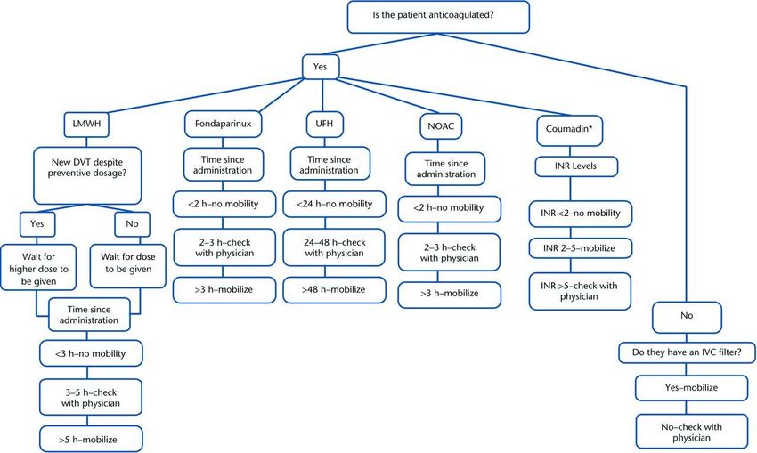
24Algorithm for Mobilizing Patients with Known Lower-Extremity Deep Vein Thrombosis
DVT = deep-vein thrombosis | LMWH = low molecular weight heparin | UFH = unfractionated heparin | NOAC = novel oral anticoagulants
INR = international normalized ratio | IVC = inferior vena cava
(Reprinted from Phys Ther. 2016;96:143-166,with permission of the American Physical Therapy Association. ©2016 American Physical Therapy Association.)
2510. Cardiovascular-Specific Labs
Cardiovascular-Specific Labs
Troponin I (cTnI) and T (cTnT)
cTnI and cTnT are two biomarkers that are sensitive, specific indicators to the myocardium of the heart. They are released when cardiac injury
occurs (6 hours after insult to 3 days), and they serve as the greatest use for diagnosing a myocardial infarction (> 0.10 ng/mL). Because hospitals
often use different assays to measure the presence of troponin, there are different diagnostic cutoff values. Newer highly-sensitive assays can
detecting circulating troponin in healthy “normal” individuals at levels as low as (N < 0.03 ng/mL) in their blood. Therefore, it is pertinent that the
clinician observes the trend of troponin levels, vigilantly monitors for cardiac symptoms, and alters the PT session accordingly.
It should be noted that troponin may also be elevated in other situations in which there is stress to the heart but not in the setting of myocardial
infarction. These instances include the following: rhabdomyolysis with cardiac damage; renal failure; inflammatory disease, such as myocarditis or
endocarditis; hypertrophic cardiomyopathy; drug toxicity; critical illness; congestive heart failure; cardiac surgery, including ablation, defibrillation,
and cardioversion; large body-surface-area burns; aortic valve disease; aortic dissection; pulmonary embolism or pulmonary hypertension; COPD;
blunt thoracic damage; or acute neurologic disease, such as stroke or subarachnoid hemorrhage.3,43-45
B-Type Natriuretic Peptide (BNP)
BNP is the strongest independent predictor of congestive heart failure (CHF), with an odds ratio of 29.60.46 Various studies have shown a
correlation that circulating BNP concentrations increases with the severity of CHF, based on the New York Heart Association (NYHA) functional
classification system. There are age-related reference values norms for males and females, but for the simplicity of this document, only
interpretative levels for a diagnosis of CHF are provided. Values tend to increase with age and are higher in women.47-49
BNP NYHA Classification
BNP 300 pg/mL46
Symptoms-based approach when determining appropriateness for activity.1,20,21
Class III – Marked limitation in activity due to symptoms, even during less-than-ordinary activity (i.e. walking
BNP > 600 pg/mL46
short distances [20–100 m]). Comfortable only at rest.
Symptoms-based approach when determining appropriateness for activity.1,20,21
BNP > 900 pg/mL46 Class IV – Severe limitations. Experiences symptoms even while at rest.
Symptoms-based approach when determining appropriateness for activity1,20,21
The following confounding factors can contribute to an elevated BNP: gender (females have higher levels); race (African-American and
Hispanic subjects have higher levels than Caucasians); anemia; atrial fibrillation. Obesity is associated with lower BNP levels.49
26Creatinine Kinase (CK)
Creatinine Kinase (CK)50 is an
Normal = 30-170 U/L
isoenzyme that is released into the
Adult Males: 52-336 U/L
blood when skeletal, brain, or cardiac
Adult Females: 38-176 U/L
muscle is injured.
Rarely present, but described as a marker for adenocarcinoma of the prostate, breast, ovary, colon,
CK1-BB brain tissue gastrointestinal tract, and for small-cell anaplastic carcinoma of lung. BB has been reported with
severe shock and/or hypothermia, infarction of bowel, brain injury, and stroke.
Commonly elevated in myocardial infarction within 3-6 hours of cardiac injury and then returns to
normal within 2-3 days (peaks 18-24 hours). Useful for diagnosing re-infarction. Might be elevated in
CK2-MB cardiac muscle
cases of carbon monoxide poisoning, pulmonary embolism, hypothyroidism, crush injuries, and
muscular dystrophy. Sensitivity and specificity are not as high as troponin levels.
Can have an increase following strenuous exercise, but not considered rhabdomyolysis.51,52
CK3-MM skeletal muscle
Intramuscular injection can increase.
2711. References
1. Goodman C, Fuller K. Pathology Implications for the Physical Therapist. 4th ed. St. Louis: Elsevier Saunders; 2015.
2. Lundberg GD. It is time to extend the laboratory critical (panic) value system to include vital values. MedGenMed. 2007;9:20.
3. Thygesen K, Alpert JS, Jaffe AS, et al. Third universal definition of myocardial infarction. Circulation. 2012;126:2020-2035.
4. McClatchey KD, Amin HM, Curry JL. Clinical laboratory medicine : self-assessment and review. 2nd ed. Philadelphia: Lippincott Williams & Wilkins; 2002.
5. Grosse SD, Odame I, Atrash HK, Amendah DD, Piel FB, Williams TN. Sickle cell disease in Africa: a neglected cause of early childhood mortality. Am J
Prev Med. 2011;41:S398-405.
6. Overfield T. Biologic variation in health and illness : race, age, and sex differences. 2nd ed. Boca Raton: CRC Press; 1995.
7. Hart CB. Race differences in long-term diabetes management in an HMO: response to Adams et al. Diabetes Care. 2006;29:1461-1462; author reply
1462.
8. Ward MM. Laboratory abnormalities at the onset of treatment of end-stage renal disease: are there racial or socioeconomic disparities in care? Arch Intern
Med. 2007;167:1083-1091.
9. Purtilo RB. Thirty-first Mary McMillan lecture. A time to harvest, a time to sow: ethics for a shifting landscape. Phys Ther. 2000;80:1112-1119.
10. Leavitt RL. Cultural competence : a lifelong journey to cultural proficiency. Thorofare, NJ: SLACK Inc.; 2010.
11. Ghazinouri R, Deshmukh S, Gorman S, et al. Lab Values Interpretation Resources. 2013;
http://c.ymcdn.com/sites/www.acutept.org/resource/resmgr/imported/labvalues.pdf. Accessed September 13, 2016.
12. Polly R, Nicole J. Understanding the transsexual patient: culturally sensitive care in emergency nursing practice. Adv Emerg Nurs J. 2011;33:55-64.
13. Fischbach FT, Dunning MB. A manual of laboratory and diagnostic tests. Ninth edition. ed. Philadelphia: Wolters Kluwer Health; 2015.
14. DeVita VT, Lawrence TS, Rosenberg SA. Devita, Hellman, and Rosenberg's cancer : principles & practice of oncology. 10th edition. ed. Philadelphia:
Wolters Kluwer; 2015.
15. Boissonnault WG. Primary care for the physical therapist : examination and triage. 2nd ed. St. Louis, Mo.: Elsevier/Saunders; 2011.
16. Capone LJ, Albert NM, Bena JF, Tang AS. Predictors of a fall event in hospitalized patients with cancer. Oncol Nurs Forum. 2012;39:E407-415.
17. Wildes TM, Dua P, Fowler SA, et al. Systematic review of falls in older adults with cancer. J Geriatr Oncol. 2015;6:70-83.
18. Carson JL, Guyatt G, Heddle NM, et al. Clinical Practice Guidelines From the AABB: Red Blood Cell Transfusion Thresholds and Storage. JAMA.
2016;316:2025-2035.
19. Peterson M. The Impact of Low Hemoglobin on the Percentage of Adverse Events During Physical Therapy in the Acute Care Setting: A Retrospective
Study. JACPT. 2015;6:29-34.
20. Paz J, West M. Acute care Handbook for Physical Therapists. 3rd ed. St. Louis: Saunders Elsevier; 2009.
21. Malone DJ, Lindsay KLB. Physical therapy in acute care : a clinician's guide. Thorofare, NJ: Slack; 2006.
22. Lindner G, Funk GC. Hypernatremia in critically ill patients. J Crit Care. 2013;28:216 e211-220.
23. Verbalis JG, Goldsmith SR, Greenberg A, et al. Diagnosis, evaluation, and treatment of hyponatremia: expert panel recommendations. Am J Med.
2013;126:S1-42.
24. Association AD. Standards in Medical Care in Diabetes-2016. Diabetes Care. 2016;39.
25. Dean E, Ross J. Discordance between cardiopulmonary physiology and physical therapy. Toward a rational basis for practice. Chest. 1992;101:1694-
1698.
26. Herridge MS, Cheung AM, Tansey CM, et al. One-year outcomes in survivors of the acute respiratory distress syndrome. N Engl J Med. 2003;348:683-
693.
27. Stiller K. Safety issues that should be considered when mobilizing critically ill patients. Crit Care Clin. 2007;23:35-53.
28. Hess CE, Nichols AB, Hunt WB, Suratt PM. Pseudohypoxemia secondary to leukemia and thrombocytosis. N Engl J Med. 1979;301:361-363.
29. Ream AK, Reitz BA, Silverberg G. Temperature correction of PCO2 and pH in estimating acid-base status: an example of the emperor's new clothes?
Anesthesiology. 1982;56:41-44.
30. Williams AJ. ABC of oxygen: assessing and interpreting arterial blood gases and acid-base balance. BMJ. 1998;317:1213-1216.
2831. DeSanti L. Involuntary weight loss and the nonhealing wound. Adv Skin Wound Care. 2000;13:11-20.
32. Luca A, Angermayr B, Bertolini G, et al. An integrated MELD model including serum sodium and age improves the prediction of early mortality in patients
with cirrhosis. Liver Transpl. 2007;13:1174-1180.
33. Kim WR, Biggins SW, Kremers WK, et al. Hyponatremia and mortality among patients on the liver-transplant waiting list. N Engl J Med. 2008;359:1018-
1026.
34. Biselli M, Gitto S, Gramenzi A, et al. Six score systems to evaluate candidates with advanced cirrhosis for orthotopic liver transplant: Which is the winner?
Liver Transpl. 2010;16:964-973.
35. Organ Procurement and Transplantation Network. In: Services UDoHaH, ed2016.
36. Stone NJ, Robinson JG, Lichtenstein AH, et al. 2013 ACC/AHA guideline on the treatment of blood cholesterol to reduce atherosclerotic cardiovascular
risk in adults: a report of the American College of Cardiology/American Heart Association Task Force on Practice Guidelines. Circulation. 2014;129:S1-45.
37. Goldman L. Disorders of lipid metabolism. 25th ed. Philadlphia, PA: Saunders Elsevier; 2016.
38. Oden A, Fahlen M, Hart RG. Optimal INR for prevention of stroke and death in atrial fibrillation: a critical appraisal. Thromb Res. 2006;117:493-499.
39. Guyatt GH, Akl EA, Crowther M, et al. Executive summary: Antithrombotic Therapy and Prevention of Thrombosis, 9th ed: American College of Chest
Physicians Evidence-Based Clinical Practice Guidelines. Chest. 2012;141:7S-47S.
40. Vandiver JW, Vondracek TG. Antifactor Xa levels versus activated partial thromboplastin time for monitoring unfractionated heparin. Pharmacotherapy.
2012;32:546-558.
41. Hillegass E, Puthoff M, Frese EM, et al. Role of Physical Therapists in the Management of Individuals at Risk for or Diagnosed With Venous
Thromboembolism: Evidence-Based Clinical Practice Guideline. Phys Ther. 2016;96:143-166.
42. Wells PS, Anderson DR, Rodger M, et al. Evaluation of D-dimer in the diagnosis of suspected deep-vein thrombosis. N Engl J Med. 2003;349:1227-1235.
43. Tanindi A, Cemri M. Troponin elevation in conditions other than acute coronary syndromes. Vasc Health Risk Manag. 2011;7:597-603.
44. Thygesen K, Alpert JS, White HD, Joint ESCAAHAWHFTFftRoMI. Universal definition of myocardial infarction. Eur Heart J. 2007;28:2525-2538.
45. Thygesen K, Mair J, Katus H, et al. Recommendations for the use of cardiac troponin measurement in acute cardiac care. Eur Heart J. 2010;31:2197-
2204.
46. Maisel AS, Krishnaswamy P, Nowak RM, et al. Rapid measurement of B-type natriuretic peptide in the emergency diagnosis of heart failure. N Engl J
Med. 2002;347:161-167.
47. Lee SC, Stevens TL, Sandberg SM, et al. The potential of brain natriuretic peptide as a biomarker for New York Heart Association class during the
outpatient treatment of heart failure. J Card Fail. 2002;8:149-154.
48. Palazzuoli A, Antonelli G, Quatrini I, Nuti R. Natriuretic peptides in heart failure: where we are, where we are going. Intern Emerg Med. 2011;6:63-68.
49. Palazzuoli A, Gallotta M, Quatrini I, Nuti R. Natriuretic peptides (BNP and NT-proBNP): measurement and relevance in heart failure. Vasc Health Risk
Manag. 2010;6:411-418.
50. Apple FS, Quist HE, Doyle PJ, Otto AP, Murakami MM. Plasma 99th percentile reference limits for cardiac troponin and creatine kinase MB mass for use
with European Society of Cardiology/American College of Cardiology consensus recommendations. Clin Chem. 2003;49:1331-1336.
51. Huerta-Alardin AL, Varon J, Marik PE. Bench-to-bedside review: Rhabdomyolysis -- an overview for clinicians. Crit Care. 2005;9:158-169.
52. Sinert R, Kohl L, Rainone T, Scalea T. Exercise-induced rhabdomyolysis. Ann Emerg Med. 1994;23:1301-1306.
29You can also read