AMERICAN THORACIC SOCIETY DOCUMENTS
←
→
Page content transcription
If your browser does not render page correctly, please read the page content below
AMERICAN THORACIC SOCIETY
DOCUMENTS

Diagnosis of Idiopathic Pulmonary Fibrosis
An Official ATS/ERS/JRS/ALAT Clinical Practice Guideline: Executive
Summary
Ganesh Raghu, Martine Remy-Jardin, Jeffrey L. Myers, Luca Richeldi, Christopher J. Ryerson, David J. Lederer,
Juergen Behr, Vincent Cottin, Sonye K. Danoff, Ferran Morell, Kevin R. Flaherty, Athol Wells, Fernando J. Martinez,
Arata Azuma, Thomas J. Bice, Demosthenes Bouros, Kevin K. Brown, Harold R. Collard, Abhijit Duggal, Liam Galvin,
Yoshikazu Inoue, R. Gisli Jenkins, Takeshi Johkoh, Ella A. Kazerooni, Masanori Kitaichi, Shandra L. Knight,
George Mansour, Andrew G. Nicholson, Sudhakar N. J. Pipavath, Ivette Buendı́a-Roldán, Moisés Selman,
William D. Travis, Simon L. F. Walsh, and Kevin C. Wilson; on behalf of the American Thoracic Society, European
Respiratory Society, Japanese Respiratory Society, and Latin American Thoracic Society
THIS OFFICIAL CLINICAL PRACTICE GUIDELINE OF THE AMERICAN THORACIC SOCIETY (ATS), EUROPEAN RESPIRATORY SOCIETY (ERS), JAPANESE RESPIRATORY
SOCIETY (JRS), AND LATIN AMERICAN THORACIC SOCIETY (ALAT) WAS APPROVED BY THE ATS, JRS, AND ALAT MAY 2018, AND THE ERS JUNE 2018

Background: This document provides clinical recommendations                     an alternative diagnosis, conditional recommendations were made
for the diagnosis of idiopathic pulmonary fibrosis (IPF). It represents          for performing BAL and surgical lung biopsy; due to lack of evidence,
a collaborative effort between the American Thoracic Society,                   no recommendation was made for or against performing
European Respiratory Society, Japanese Respiratory Society, and                 transbronchial lung biopsy or lung cryobiopsy. In contrast, for
Latin American Thoracic Society.                                                patients with newly detected ILD who have a high-resolution
                                                                                computed tomography pattern of UIP, strong recommendations
Methods: The evidence syntheses were discussed and                              were made against performing surgical lung biopsy, transbronchial
recommendations formulated by a multidisciplinary committee of                  lung biopsy, and lung cryobiopsy, and a conditional recommendation
IPF experts. The evidence was appraised and recommendations were                was made against performing BAL. Additional recommendations
formulated, written, and graded using the Grading of                            included a conditional recommendation for multidisciplinary
Recommendations, Assessment, Development, and Evaluation                        discussion and a strong recommendation against measurement of
approach.                                                                       serum biomarkers for the sole purpose of distinguishing IPF from
Results: The guideline panel updated the diagnostic criteria for IPF.           other ILDs.
Previously defined patterns of usual interstitial pneumonia (UIP)                Conclusions: The guideline panel provided recommendations
were refined to patterns of UIP, probable UIP, indeterminate for                 related to the diagnosis of IPF.
UIP, and alternate diagnosis. For patients with newly detected
interstitial lung disease (ILD) who have a high-resolution computed             Keywords: idiopathic pulmonary fibrosis; interstitial lung disease;
tomography scan pattern of probable UIP, indeterminate for UIP, or              pulmonary fibrosis

This Executive Summary is part of the full official ATS guideline, which readers may access online at http://www.atsjournals.org/doi/suppl/10.1164/
rccm.201807-1255ST. Only the Executive Summary is appearing in the print edition of the Journal. The article of record, and the one that should be cited, is:
Diagnosis of Idiopathic Pulmonary Fibrosis: An Official ATS/ERS/JRS/ALAT Clinical Practice Guideline. Am J Respir Crit Care Med 2018;198:e44–e68.
Available at http://www.atsjournals.org/doi/suppl/10.1164/rccm.201807-1255ST.

ORCID IDs: 0000-0001-7506-6643 (G.R.); 0000-0001-8247-3028 (J.L.M.); 0000-0001-8594-1448 (L.R.); 0000-0001-5258-0228 (D.J.L.);
0000-0002-9151-4829 (J.B.); 0000-0002-5591-0955 (V.C.); 0000-0001-9172-8977 (S.K.D.); 0000-0002-7206-4543 (F.M.); 0000-0003-2657-
1314 (K.R.F.); 0000-0003-2108-6248 (A.W.); 0000-0002-2412-3182 (F.J.M.); 0000-0001-7300-3219 (T.J.B.); 0000-0002-0685-0765 (D.B.);
0000-0002-8558-6711 (K.K.B.); 0000-0003-4220-2359 (A.D.); 0000-0001-5859-8744 (E.A.K.); 0000-0003-3257-102X (A.G.N.); 0000-0001-
6948-2376 (S.N.J.P.); 0000-0002-8230-0749 (I.B.-R.); 0000-0002-1022-4783 (M.S.); 0000-0003-3160-6729 (W.D.T.); 0000-0003-0497-
5297 (S.L.F.W.); 0000-0003-4429-2263 (K.C.W.).
Correspondence and requests for reprints should be addressed to Ganesh Raghu, M.D., Center for Interstitial Lung Diseases, University of Washington, 1959 NE
Pacific Street, Seattle, WA 98195. E-mail: graghu@uw.edu.
This article has an online supplement, which is accessible from this issue’s table of contents at www.atsjournals.org.
Am J Respir Crit Care Med Vol 198, Iss 5, pp 563–580, Sep 1, 2018
Copyright © 2018 by the American Thoracic Society
DOI: 10.1164/rccm.201807-1255ST
Internet address: www.atsjournals.org

American Thoracic Society Documents                                                                                                                     563
AMERICAN THORACIC SOCIETY DOCUMENTS
Contents the Patient Frequently Visits to to Ascertain the
Summary of Recommendations Exclude Potential Causes of Histopathology Diagnosis of
Introduction the ILD? UIP Pattern?
Methods Question 2: Should Patients Question 5: For Patients with
Clinical Manifestations with Newly Detected ILD of Newly Detected ILD of
Diagnosis Unknown Cause Who Are Unknown Cause Who Are
HRCT Technique Clinically Suspected of Having Clinically Suspected of Having
HRCT Patterns IPF Undergo Serological IPF, Is TBBx a Reasonable
SLB Technique Testing to Exclude CTDs as Alternative to SLB to Ascertain
Histopathology Patterns Potential Causes of the ILD? the Histopathology Diagnosis of
Diagnostic Criteria for IPF Question 3: Should Patients UIP Pattern?
Diagnostic Interventions with Newly Detected ILD of Question 6: For Patients with
Question 1: Should Patients Unknown Cause Who Are Newly Detected ILD of
with Newly Detected ILD of Clinically Suspected of Having Unknown Cause Who Are
Unknown Cause Who Are IPF Undergo Cellular Analysis Clinically Suspected of Having
Clinically Suspected of Having of Their BAL Fluid? IPF, Is Lung Cryobiopsy a
IPF Undergo a Detailed, Question 4: For Patients with Reasonable Alternative to
Prompted History of Newly Detected ILD of SLB to Ascertain the
Medication Use and Unknown Cause Who Are Histopathology Diagnosis of
Environmental Exposures at Clinically Suspected of Having UIP Pattern?
Home, Work, and Other Places IPF, Should SLB Be Performed Conclusions
Summary of For patients with newly detected ILD For patients with newly detected ILD
Recommendations of apparently unknown cause who are of apparently unknown cause who are
clinically suspected of having IPF and clinically suspected of having IPF:
Adult patients with newly detected interstitial have an HRCT pattern of probable usual d We suggest multidisciplinary discussion
lung disease (ILD) of apparently unknown interstitial pneumonia (UIP), indeterminate
(MDD) for diagnostic decision-making
cause are clinically suspected of having for UIP, or an alternative diagnosis:
(conditional recommendation, very low
idiopathic pulmonary fibrosis (IPF) if they quality of evidence).
d We suggest cellular analysis of their BAL
have unexplained symptomatic or asymptomatic
fluid (conditional recommendation, very d We recommend NOT measuring serum
patterns of bilateral fibrosis on a chest radiograph MMP (matrix metalloproteinase)-7, SPD
low quality of evidence).
or chest computed tomography (CT), bibasilar (surfactant protein D), CCL (chemokine
d We suggest surgical lung biopsy (SLB)
inspiratory crackles, and an age typically older ligand)-18, or KL (Krebs von den Lungen)-
(conditional recommendation, very low
than 60 years. Rarely, middle-aged adults 6 for the purpose of distinguishing IPF
quality of evidence).
(.40 yr and ,60 yr), especially those with from other ILDs (strong recommendation,
d The panel made no recommendation for or
risks for familial pulmonary fibrosis, may very low quality of evidence).
against transbronchial lung biopsy (TBBx).
otherwise manifest the same clinical scenario d The panel made no recommendation for
as the typical patient older than 60 years. The For comparison of the 2018 and 2011
or against lung cryobiopsy.
recommendations in this guideline are for the diagnostic recommendations, see Table 1.
patterns and distributions of images obtained For patients with newly detected ILD For an explanation of strong and
by high-resolution CT (HRCT) and, thus, of apparently unknown cause who are conditional recommendations, see Table 2.
require that patients be subjected to HRCT of clinically suspected of having IPF and have
the chest for evaluation. an HRCT pattern of UIP:
For adult patients with newly detected
Introduction
d We suggest NOT performing cellular
ILD of apparently unknown cause who are
analysis of their BAL fluid (conditional The American Thoracic Society (ATS),
clinically suspected of having IPF:
recommendation, very low quality of evidence). European Respiratory Society (ERS),
d We recommend taking a detailed d We recommend NOT performing SLB Japanese Respiratory Society (JRS), and
history of both medication use and (strong recommendation, very low quality Latin American Thoracic Society (ALAT)
environmental exposures at home, work, of evidence). collaborated to develop clinical practice
and other places the patient frequently d We recommend NOT performing TBBx guidelines for the diagnosis and
visits to exclude potential causes of ILD (strong recommendation, very low quality management of IPF in 2011 (1). New
(motherhood statement). of evidence). evidence now enables us to improve the
d We recommend serological testing to exclude d We recommend NOT performing lung diagnostic criteria. The recommendations
connective tissue disease as a potential cause cryobiopsy (strong recommendation, very in this 2018 guideline are revisions of the
of the ILD (motherhood statement). low quality of evidence). diagnostic recommendations in the 2011
564 American Journal of Respiratory and Critical Care Medicine Volume 198 Number 5 | September 1 2018Table 1. Comparison of ATS/ERS/JRS/ALAT Recommendations for the Diagnosis of IPF in the 2011 and 2018 Guidelines
2018 Guideline
HRCT Pattern of Probable UIP*, 2011 Guideline: Did Not Distinguish
Indeterminate for UIP, and among Patients with Different HRCT
Alternative Diagnosis HRCT Pattern of UIP* Patterns
BAL cellular analysis We suggest performing BAL We suggest NOT performing BAL “BAL cellular analysis should not be
cellular analysis (conditional) cellular analysis (conditional) performed in the diagnostic evaluation
of IPF in the majority of patients, but
may be appropriate in a minority of
patients.”
American Thoracic Society Documents
Surgical lung biopsy We suggest performing surgical We recommend NOT performing “Surgical lung biopsy is not required
lung biopsy (conditional) surgical lung biopsy (strong) for patients with an HRCT pattern
consistent with UIP.”
Transbronchial lung biopsy No recommendation was made We recommend NOT performing “Transbronchial biopsy should not be
either for or against transbronchial lung biopsy (strong) used in the evaluation of IPF in the
transbronchial lung biopsy majority of patients, but may be
appropriate in a minority.”
Lung cryobiopsy No recommendation was made We recommend NOT performing Not addressed
either for or against cryobiopsy cryobiopsy (strong)
AMERICAN THORACIC SOCIETY DOCUMENTS
Medical history of medication use and We recommend taking a detailed history of both medication use and “Diagnosis of IPF requires exclusion of
environmental exposures environmental exposures at home, work, and other places the patient other known causes of ILD (e.g., domestic
frequently visits to exclude potential causes of ILD (motherhood statement) and occupational environmental
exposures, connective tissue disease,
and drug toxicity).”
Serological testing to exclude We recommend serological testing to exclude connective tissue diseases as a “Diagnosis of IPF requires exclusion
connective tissue disease potential cause of the ILD (motherhood statement) of other known causes of ILD
(e.g., domestic and occupational
environmental exposures, connective
tissue disease, and drug toxicity).”
Multidisciplinary discussion We suggest multidisciplinary discussion for decision-making (conditional) “We recommend that a multidisciplinary
discussion should be used in the
evaluation of IPF.”
Serum biomarkers We recommend NOT measuring serum MMP-7, SPD, CCL-18, or KL-6 for the Not addressed
purpose of distinguishing IPF from other ILDs (strong)
Definition of abbreviations: ALAT = Latin American Thoracic Society; ATS = American Thoracic Society; CCL-18 = chemokine ligand 18; ERS = European Respiratory Society; HRCT =
high-resolution computed tomography; ILD = interstitial lung disease; IPF = idiopathic pulmonary fibrosis; JRS = Japanese Respiratory Society; KL-6 = Krebs von den Lungen-6; MMP-7 =
matrix metalloproteinase 7; SPD = surfactant protein D; UIP = usual interstitial pneumonia.
The quality of evidence for all recommendations in the 2018 guideline was very low.
*The patterns of UIP have been refined in these 2018 guidelines, compared with the 2011 guidelines.
565AMERICAN THORACIC SOCIETY DOCUMENTS
Table 2. Implications of Strong and Conditional Recommendations
Strong Recommendation (“We recommend . . .”) Conditional Recommendation (“We suggest . . .”)
For patients The overwhelming majority of individuals in this The majority of individuals in this situation would want the
situation would want the recommended course suggested course of action, but a sizeable minority
of action and only a small minority would not. would not.
For clinicians The overwhelming majority of individuals should Different choices will be appropriate for different patients,
receive the recommended course of action. and you must help each patient arrive at a management
Adherence to this recommendation according to decision consistent with her or his values and
the guideline could be used as a quality criterion preferences. Decision aids may be useful to help
or performance indicator. Formal decision aids individuals make decisions consistent with their values
are not likely to be needed to help individuals and preferences. Clinicians should expect to spend
make decisions consistent with their values and more time with patients when working toward a
preferences. decision.
For policy makers The recommendation can be adapted as policy Policy making will require substantial debates and
in most situations, including for use as involvement of many stakeholders. Policies are also
performance indicators. more likely to vary between regions. Performance
indicators would have to focus on the fact that
adequate deliberation about the management options
has taken place.
guideline (1). This guideline is intended to pulmonary fibrosis and genetic predisposition involvement is common; in some cases, the
help clinicians make an accurate diagnosis of factors for IPF, can rarely present with the craniocaudal distribution of UIP may be
IPF and to empower them to implement otherwise same clinical scenario as the typical relatively uniform (3, 4). Asymmetric
recommended courses of action in the patient older than 60 years. disease may occur in up to 25% of cases (5).
context of individual patient values and Several studies have demonstrated that the
preferences, particularly decisions regarding positive predictive value of a radiologic
which diagnostic interventions to pursue. Diagnosis diagnosis of UIP on HRCT for a pathologic
diagnosis of UIP is between 90% and 100%
HRCT Technique (6–10); however, a significant minority of
Methods The diagnostic approach to IPF is highly patients with histopathologic UIP do not
reliant on images of the lungs generated from fulfill HRCT criteria for UIP (7, 9–11).
This guideline was developed in accordance volumetric scanning of the chest. This mode Mediastinal lymphadenopathy may be
with the policies and procedures of the has essentially replaced sequential CT present in patients with UIP (12). Ground-
ATS, ERS, JRS, and ALAT (see online scanning, as it improves detection of all glass opacification may be present, but it is
supplement). abnormalities, even if subtle or focal. It also not a dominant feature and is usually
ensures precise analysis of lesion characteristics accompanied by a superimposed reticular
and distribution. Technical requirements of pattern. Rarely, small ossified nodules within
Clinical Manifestations HRCT are described in Table 3 and Table E1 areas of fibrosis may be present, and these
in the online supplement. are more common (29%) in patients with
IPF is a specific form of chronic, progressive, UIP when compared with other fibrotic lung
fibrosing interstitial pneumonia of unknown HRCT Patterns diseases (13). Patients with UIP may have
cause. The typical patient with IPF is a We advocate the use of four diagnostic features of pleuroparenchymal fibroelastosis
male, older than 60 years of age, usually with categories (Table 4) that incorporate the at the lung apices (14, 15); however, there is
a previous history of smoking tobacco, who HRCT features described above. These no clear cut-off of the proportions of each
presents with insidious onset of cough categories include “UIP pattern” (Figure 1), pattern, and these cases should be regarded
and/or exertional dyspnea, bibasilar “probable UIP pattern” (Figure 2), as UIP/IPF, if consistent with that diagnosis
inspiratory crackles, and radiologic evidence “indeterminate for UIP pattern” (Figures 3 after MDD. UIP may present as an acute
of fibrosis predominantly in the lower lobes and 4), and “alternative diagnosis” (Figure 5). exacerbation (Figure 6) or coexist in patients
without an apparent cause. Rarely, patients UIP pattern. UIP is the hallmark with emphysema (Figure E1).
with IPF may present with an acute radiologic pattern of IPF. Honeycombing is Probable UIP pattern. In the 2011
exacerbation as an initial manifestation a distinguishing feature of UIP and must be guideline, an HRCT pattern consisting of
(i.e., an unexplained worsening of dyspnea present for a definite HRCT diagnosis of subpleural, basal-predominant reticular
over a few weeks and new ground-glass UIP to be made. It can be seen with or abnormalities without honeycombing was
opacification on HRCT) with a background without peripheral traction bronchiectasis assigned the HRCT diagnosis category of
of lower lobe fibrotic lung disease (2). or bronchiolectasis. The typical distribution “possible UIP” (1). Since 2011, several
Middle-aged adults (.40 yr and ,60 yr), of UIP is subpleural with basal studies have reported that selected patients
especially patients with risks for familial predominance, although some upper lobe with a “possible UIP” pattern on HRCT
566 American Journal of Respiratory and Critical Care Medicine Volume 198 Number 5 | September 1 2018AMERICAN THORACIC SOCIETY DOCUMENTS
Table 3. High-Resolution Computed Tomography Scanning Parameters
Recommended Scanning Protocol Advantages of Updated Recommendations
1. Noncontrast examination —
2. Volumetric acquisition with selection of: A. Acquisition covering the entire lung volume (vs. analysis of
d Sub-millimetric collimation 10% of lung volume with sequential scanning)
d Shortest rotation time d No risk of missing subtle infiltrative abnormalities
d Highest pitch d Possibility of multiplanar reformations, helpful for analysis
d Tube potential and tube current appropriate to patient size: of the ILD pattern and predominant distribution of lung
∘ Typically 120 kVp andAMERICAN THORACIC SOCIETY DOCUMENTS
Table 4. High-Resolution Computed Tomography Scanning Patterns
UIP Probable UIP Indeterminate for UIP Alternative Diagnosis
Subpleural and Subpleural and Subpleural and basal predominant Findings suggestive of another
basal predominant; basal predominant; diagnosis, including:
distribution is often distribution is often Subtle reticulation; may have mild d CT features:
heterogeneous* heterogeneous GGO or distortion (“early UIP ∘ Cysts
Honeycombing with or without Reticular pattern with peripheral
pattern”)
∘ Marked mosaic
peripheral traction traction bronchiectasis or CT features and/or distribution of attenuation
bronchiectasis or bronchiolectasis lung fibrosis that do not suggest ∘ Predominant GGO
bronchiolectasis† any specific etiology (“truly ∘ Profuse micronodules
May have mild GGO indeterminate for UIP”) ∘ Centrilobular nodules
∘ Nodules
∘ Consolidation
d Predominant distribution:
∘ Peribronchovascular
∘ Perilymphatic
∘ Upper or mid-lung
d Other:
∘ Pleural plaques (consider
asbestosis)
∘ Dilated esophagus
(consider CTD)
∘ Distal clavicular erosions
(consider RA)
∘ Extensive lymph node
enlargement (consider
other etiologies)
∘ Pleural effusions, pleural
thickening (consider
CTD/drugs)
Definition of abbreviations: CT = computed tomography; CTD = connective tissue disease; GGO = ground-glass opacities; RA = rheumatoid arthritis;
UIP = usual interstitial pneumonia.
*Variants of distribution: occasionally diffuse, may be asymmetrical.
†
Superimposed CT features: mild GGO, reticular pattern, pulmonary ossification.
CT features of fibrosis for whom there is a CT findings in the presence of an decision regarding whether or not to pursue
suspicion that early UIP or probable UIP acute exacerbation. Patients with an acute a biopsy must be tailored to the clinical
is present. In such cases, it should be exacerbation of IPF have bilateral ground- situation of the individual patient. Multiple
confirmed with prone inspiratory views that glass opacification with or without biopsies should be obtained from two to
the subpleural opacities do not represent consolidation on a background of lung three lobes, because the histologic patterns
dependent atelectasis (Figure E2). fibrosis (Figure 6). In the absence of a on SLB specimens obtained from different
Alternative diagnosis. In some cases previous HRCT study, bilateral ground- segments can be discordant (e.g., coexisting
of fibrotic lung disease, there is clinical glass opacity and/or consolidation on a UIP pattern and fibrotic NSIP pattern from
suspicion of IPF, but the HRCT pattern background of a UIP pattern is highly different lobes).
suggests an alternative diagnosis. Examples suggestive of an acute exacerbation and
include bronchocentric fibrosis in the upper can be used to confirm an underlying Histopathology Patterns
lobes or profuse mosaic attenuation that IPF diagnosis in the appropriate clinical We recommend categorizing histopathologic
suggests hypersensitivity pneumonitis, context. findings of biopsies into “UIP” (Figure 7),
posterior fibrotic retraction of the hila in “probable UIP,” “indeterminate for UIP,” and
sarcoidosis, or extensive ground-glass SLB Technique “alternative diagnosis” (Table 5). Biopsies
opacification with subpleural sparing in Video-assisted thoracoscopic surgery is the designated as indeterminate for UIP
fibrotic nonspecific interstitial pneumonia preferred approach to SLB for patients demonstrate a pattern of fibrosis that does
(NSIP). Occasionally, the HRCT presentation who can tolerate single-lung ventilation, not meet criteria for UIP or any other
may be that of a UIP, probable UIP, or rather than open thoracotomy. In patients histopathologic pattern of fibrotic interstitial
indeterminate for UIP pattern, but ancillary with severe physiologic impairment or pneumonia and, in some cases, may favor an
findings suggest an alternative diagnosis. In substantial comorbidity, the risks of SLB alternative diagnosis while not categorically
such situations, an alternative diagnosis to IPF may outweigh the benefits of establishing a excluding the possibility of sampling bias in a
should be reconsidered. secure diagnosis of IPF; therefore, the final patient who ultimately proves to have UIP.
568 American Journal of Respiratory and Critical Care Medicine Volume 198 Number 5 | September 1 2018AMERICAN THORACIC SOCIETY DOCUMENTS
Figure 1. High-resolution computed tomography (CT) images demonstrating a usual interstitial pneumonia pattern. (A–C) Transverse CT section and
(D) coronal reconstruction illustrating the presence of honeycombing with subpleural and basal predominance. Note the concurrent presence of mild
ground-glass opacity. (E) Magnified view of the left lower lobe showing typical characteristics of honeycombing, consisting of clustered cystic airspaces
with well-defined walls and variable diameters, seen in single or multiple layers (arrows).
A subset of patients with previously occult guidelines and the 2011 guidelines (1) Question 1: Should Patients with
IPF may present with an acute exacerbation, and is similar to that suggested by a Newly Detected ILD of Unknown
which is commonly characterized by a task force sponsored by the Fleischner Cause Who Are Clinically Suspected
combination of a UIP pattern complicated Society (20). of Having IPF Undergo a Detailed,
by superimposed diffuse alveolar damage Prompted History of Medication Use
with or without associated hyaline and Environmental Exposures at
membranes. Diagnostic Interventions Home, Work, and Other Places the
Patient Frequently Visits to Exclude
The questions below are specifically intended Potential Causes of the ILD?
Diagnostic Criteria for IPF for patients who are “clinically suspected
of having IPF.” This classically refers to Discussion. The guideline panel recognized
Diagnosis of IPF requires the following: patients with unexplained symptomatic or there is no reasonable alternative to the
asymptomatic bilateral pulmonary fibrosis proposed course of action, so a motherhood
1. Exclusion of other known causes of ILD statement was made to take a detailed history
on a chest radiograph or chest CT scan,
(e.g., domestic and occupational of medication use and environmental
bibasilar inspiratory crackles, and an age
environmental exposures, connective exposures at home, work, and other places
typically older than 60 years. It must be
tissue disease [CTD], drug toxicity), that the patient frequently visits, to identify
recognized that the questions addressed are
and either #2 or #3 or exclude potential causes of ILD (e.g.,
not restricted to patients older than 60 years,
2. The presence of the HRCT pattern of
as middle-aged adults (.40 yr and ,60 yr), hypersensitivity pneumonitis, pneumoconiosis,
UIP (Table 4) drug toxicity). This is supported by an
especially patients with risks for familial
3. Specific combinations (Figure 8) of
pulmonary fibrosis, can rarely present with observational study that enrolled 1,084 patients
HRCT patterns (Table 4) and
the otherwise same clinical scenario as the with new-onset ILD of unknown cause
histopathology patterns (Table 5) in
typical patient older than 60 years. The reporting that 47% of the patients were
patients subjected to lung tissue
recommendations in this guideline are for identified as having hypersensitivity pneumonitis
sampling
the patterns and distributions of images on detailed assessment, suggesting that a cause
The guideline panel’s approach obtained by HRCT and, thus, require that can be found in many patients who present
to diagnosis is summarized in Figures 8 patients be subjected to HRCT of the chest with ILD (21). The panel’s clinical experience
and 9. It is based on these 2018 for evaluation. is that identification and removal of potential
American Thoracic Society Documents 569AMERICAN THORACIC SOCIETY DOCUMENTS
Figure 2. Probable usual interstitial pneumonia (UIP) pattern. (A–C) Transverse computed tomography (CT) section, (D) coronal reconstruction of both
lungs, and (E) magnified sagittal view of the right lower lobe illustrating the presence of a reticular pattern with peripheral bronchiolectasis with subpleural
and basal predominance. Depending on their orientation relative to the plane of the CT section, peripheral traction bronchiolectasis appear as tubular
(arrows) or cystic (arrowheads) structures. Note the concurrent presence of mild ground-glass opacities in the subpleural areas of both lungs and the
absence of honeycombing. UIP was proven at histology.
causative environmental factors may result in ATS/ERS/JRS/ALAT recommendations. The majority of panelists acknowledged
improved clinical outcomes. d For patients with newly detected ILD of routinely testing for CRP (C-reactive protein),
Many panelists use published apparently unknown cause who are erythrocyte sedimentation rate, antinuclear
questionnaires in their clinical practices to clinically suspected of having IPF, antibodies (by immunofluorescence),
consider environmental exposures at home, we recommend taking a detailed rheumatoid factor, myositis panel, and
work, and frequently visited places (21–23). history of both medication use and anti–cyclic citrullinated peptide. Other detailed
Such questionnaires may be tailored to cultural environmental exposures at home, work, tests are performed on a case-by-case basis
habits and geographical differences. Examples and other places the patient frequently according to associated symptoms and signs.
of pertinent exposures include mold, birds, visits to exclude potential causes of the These include muscle enzymes (creatinine
down feathers, animals, metal dusts (e.g., brass, ILD (motherhood statement). phosphokinase, myoglobin, and aldolase),
lead, steel), wood dust (e.g., pine), vegetable antisynthetase antibodies (Jo-1 and others
dust, exposure to livestock, stone polishing and if available), anti-MDA5 (melanoma
cutting, medications taken, current or recent Question 2: Should Patients with Newly differentiation-associated protein 5), anti–Mi-2,
occupations (e.g., hair dressing), and current or Detected ILD of Unknown Cause Who anti-NXP2 (nuclear matrix protein 2), anti–
recent hobbies (24–30). Although some Are Clinically Suspected of Having IPF TIF1-g (transcriptional intermediary factor
panelists use the presence of antibody in serum Undergo Serological Testing to Exclude 1-g), anti-SRP (signal recognition particle),
against specific antigen to prompt further CTDs as Potential Causes of the ILD? anti-HMGCR (3-hydroxy-3-methylglutaryl-CoA
evaluation for hypersensitivity pneumonitis, the reductase), anti-SAE (small ubiquitin-related
test is not standardized, and the specificity Discussion. Diagnosis of IPF mandates modifier–activating enzyme), anti-U1RNP
and sensitivity for the diagnosis of exclusion of other causes of ILD, including (U1 ribonucleoprotein), anti-PM/Scl75
hypersensitivity pneumonitis is unknown. The CTD-related ILD (Table E2). The guideline (polymyositis/scleroderma 75), anti-PM/Scl100,
panelists who use serum antibody testing panel concluded that foregoing serological and anti-Ku (31). If systemic sclerosis
believe that such tests may identify an antigen testing was not a reasonable alternative. (i.e., scleroderma) is suspected, additional tests
that was not suspected by clinical history and, Therefore, a motherhood statement was made include: anti–Scl-70/topoisomerase-1, anti-
therefore, may prompt further investigations for to perform routine serological testing in all centromere, anti-RNA polymerase III, anti-
the suspected etiology; also, if serum antibody patients with newly identified ILD. Although U1RNP, anti-Th/To, anti-PMScl, U3 RNP
testing is negative, the results reinforce the there was overwhelming agreement to perform (fibrillarin), and anti-Ku. If Sjögren syndrome is
conclusion that the patient does not have serological testing, there was far less agreement suspected, additional tests include: anti-SSA/Ro
hypersensitivity pneumonitis. about which serological tests to perform. (Sjögren-specific antibody A) and anti-SSB/La.
570 American Journal of Respiratory and Critical Care Medicine Volume 198 Number 5 | September 1 2018AMERICAN THORACIC SOCIETY DOCUMENTS
Figure 3. Indeterminate for usual interstitial pneumonia (UIP) pattern (early UIP pattern). (A and B) Transverse computed tomography (CT) section, (C)
coronal reconstruction of both lungs, and (D) magnified view of the right lung in supine position showing ground-glass opacity and subtle reticulation in the
subpleural areas (arrows) with a basal predominance. (E) Transverse CT section of the lower lung zones in prone position showing persistence of lung
infiltration in nondependent areas, thus excluding gravitational abnormalities. UIP was proven at histology.
If vasculitis is suspected, an additional test
includes anti-cytoplasmic antibodies. A small
minority of the panelists include all of the
detailed tests listed above as an “ILD panel” at
initial screening/baseline evaluation.
The guideline panelists do not refer all
patients with new ILD to a rheumatologist;
rather, referring only those with positive clinical
manifestations, serologies, or other characteristics
atypical for IPF (e.g., female, age ,60 yr old). In
many CTD-related ILDs, the lung disease is the
first, dominant, or only feature of the CTD
and, therefore, some patients will not fit standard
rheumatologic diagnostic criteria at presentation.
ATS/ERS/JRS/ALAT recommendations.
d For patients with newly detected ILD of
apparently unknown cause who are
clinically suspected of having IPF, we
recommend serological testing to aid in
the exclusion of CTDs as a potential
cause of the ILD (motherhood statement).
Question 3: Should Patients with
Newly Detected ILD of Unknown
Cause Who Are Clinically Suspected
of Having IPF Undergo Cellular
Figure 4. Indeterminate for usual interstitial pneumonia pattern. (A–C) Transverse computed Analysis of Their BAL Fluid?
tomography sections showing extensive lung infiltration combining honeycombing, mild to
marked ground-glass opacity, asymmetrical distribution between both lungs, and no subpleural Evidence base. Our systematic literature
predominance. search yielded 2,492 titles but did not
American Thoracic Society Documents 571AMERICAN THORACIC SOCIETY DOCUMENTS
Figure 5. Computed tomography (CT) pattern suggestive of an alternative diagnosis for lung fibrosis. (A and B) Transverse CT sections obtained at deep
inspiration showing disseminated lung infiltration, sparing some secondary pulmonary lobules in lung bases. (C) Transverse CT section obtained at
expiration confirming lobular air trapping, all findings being highly suggestive of chronic hypersensitivity pneumonitis.
identify any studies that 1) compared reviewed, and 8 were selected for analysis with other types of ILD, including
clinical outcomes among patients who (32–39) (Tables E7a–E7f). hypersensitivity pneumonitis (32, 33, 37),
underwent BAL cellular analysis to those The eight studies enrolled patients sarcoidosis (32, 36, 37), idiopathic NSIP (32,
who did not undergo BAL cellular analysis, with IPF, performed BAL, and measured 34, 37–39), cryptogenic organizing
or 2) reported the test characteristics of components of the BAL fluid, including the pneumonia (previously called bronchiolitis
BAL cellular analysis for distinguishing IPF percentage of neutrophils (32–37, 39), obliterans organizing pneumonia) (32–34,
from other ILDs. Therefore, we sought macrophages (32–36, 39), lymphocytes 37), eosinophilic pneumonia (32),
studies that compared BAL cell type (32–39), and eosinophils (32, 34–37, 39), as respiratory bronchiolitis-associated ILD
proportions among patients with IPF to well as the CD4/CD8 ratio (32, 34, 36, 37). (33), and lymphocytic interstitial pneumonia
those among patients with other types The measurements were then compared (33). Some BAL cell type proportions were
of ILD. The full text of 14 articles was with similar measurement from patients markedly different in patients with IPF
compared with patients with other ILDs
(Figure E3). Patients with IPF had a slightly
increased proportion of eosinophils
compared with healthy individuals but a
markedly lower proportion of eosinophils
than patients with eosinophilic pneumonia;
thus, patients with a markedly elevated
proportion of eosinophils are more likely to
have eosinophilic pneumonia than IPF.
Patients with IPF had a similar to slightly
higher proportion of lymphocytes and
CD4/CD8 ratio in their BAL than healthy
individuals but a markedly lower proportion
of lymphocytes and CD4/CD8 ratio in their
BAL than patients with sarcoidosis; thus,
patients with a markedly elevated proportion
of lymphocytes and CD4/CD8 ratio are
more likely to have sarcoidosis than IPF.
Conclusions. When the panel weighed
the desirable consequences of BAL cellular
analysis in patients who have an HRCT pattern
of probable UIP, indeterminate for UIP,
or an alternative diagnosis (i.e., identifying
or excluding eosinophilic pneumonia,
sarcoidosis, infection, and malignancy) versus
the undesirable consequences (i.e., risk of a
Figure 6. Acute exacerbation of idiopathic pulmonary fibrosis. (A and B) Transverse computed complication, burden, cost), the majority of the
tomography sections obtained in the upper and mid lung zones and (C and D) during acute panel concluded that the upsides of the
exacerbation showing newly developed, bilateral ground-glass opacification in both lungs on a procedure outweigh the downsides in such
background of usual interstitial pneumonia pattern. patients. There was general agreement that
572 American Journal of Respiratory and Critical Care Medicine Volume 198 Number 5 | September 1 2018AMERICAN THORACIC SOCIETY DOCUMENTS
analysis of their BAL fluid (conditional
recommendation, very low quality of
evidence).
Question 4: For Patients with Newly
Detected ILD of Unknown Cause Who
Are Clinically Suspected of Having
IPF, Should SLB Be Performed to
Ascertain the Histopathology Diagnosis
4C/FPO
of UIP Pattern?
Evidence base. Our systematic literature
search yielded 945 titles but identified no
studies that compared clinical outcomes
among patients who underwent SLB to
those who did not. Thus, we selected studies
that measured diagnostic yield of SLB using
a MDD as the diagnostic decision-maker.
The full text of 54 articles was reviewed,
and 26 were selected for analysis (40–65)
(Table E8).
Pooling studies (unweighted) indicated
that SLB obtained an adequate sample in
all patients (11 studies; 918 of 918, 100%;
95% confidence interval [CI], 99–100%),
although the panel acknowledged that this
is not always the case in clinical practice.
The proportion of SLBs that resulted in a
specific diagnosis (i.e., the diagnostic yield)
was high (26 studies; 2,338 of 2,651, 88.2%;
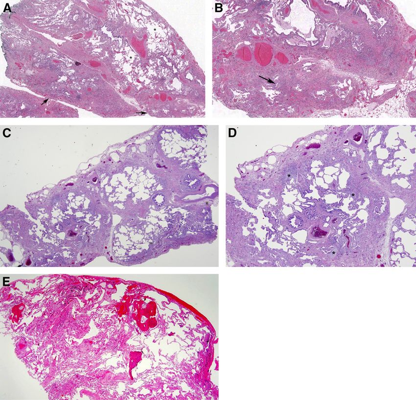
Figure 7. Histopathology demonstrating usual interstitial pneumonia (UIP). (A) Low-magnification 95% CI, 86.9–89.4%), with a minority being
photomicrograph showing classical UIP/idiopathic pulmonary fibrosis (IPF) pattern characterized deemed unclassifiable (26 studies; 313
by dense fibrosis with a predilection for subpleural and paraseptal parenchyma with associated
of 2,651, 11.8%; 95% CI, 10.6–13.1%).
architectural distortion in the form of microscopic honeycomb change (arrow) juxtaposed with
relatively unaffected lung parenchyma (*). Visceral pleura is seen in the upper portion of the figure.
Among final diagnoses, approximately one-
(B) Higher-magnification photomicrograph showing subpleural scarring and honeycomb change with third were IPF (24 studies; 752 of 2,360,
associated fibroblast foci (arrow). (C) Low-magnification photomicrograph showing probable UIP/IPF 31.9%; 95% CI, 30.0–33.8%), and many
pattern characterized by subpleural and paraseptal predominant patchwork fibrosis that is less well others were potentially treatable etiologies
developed and lacks the degree of associated architectural distortion in the form of either destructive like infection, sarcoidosis, hypersensitivity
scarring or honeycomb change illustrated in A and B. (D) Higher-magnification photomicrograph pneumonitis, eosinophilic pneumonia,
showing patchy fibrosis and fibroblast foci (*) but without the extent of scarring and honeycomb lymphangioleiomyomatosis, cryptogenic
change illustrated in A and B. (E) Indeterminate for UIP/IPF pattern in which there is mild nonspecific organizing pneumonia, and vasculitis.
fibrosis that lacks a well-developed patchy and predominantly subpleural/paraseptal distribution, Overall mortality was low (23 studies;
architectural distortion, and fibroblast foci characteristic of classical UIP/IPF. There is associated
79 of 2,268, 3.5%; 95% CI, 2.8–4.3%), but some
osseous metaplasia, a common but nonspecific finding in UIP. Although these findings are not
diagnostic, they do not preclude a diagnosis of UIP/IPF in a patient with supportive clinical and
of the deaths were probably disease related,
radiological findings. because procedure-related mortality was
lower (6 studies; 7 of 410, 1.7%; 95% CI,
0.8–3.5%). Many series reported no mortality,
BAL is appropriate when the radiologic clinically suspected of having IPF and suggesting that lower procedural mortality
differential diagnosis includes eosinophilic have an HRCT pattern of probable is possible depending on center-specific
pneumonia, sarcoidosis, or infection. In UIP, indeterminate for UIP, or an variables such as patient selection. Additional
contrast, the panel concluded that alternative alternative diagnosis, we suggest complications included exacerbations
diagnoses that can be excluded by BAL cellular cellular analysis of their BAL fluid (15 studies; 116 of 1,891, 6.1%; 95% CI,
analysis are sufficiently rare in patients who (conditional recommendation, very low 5.1–7.3%), bleeding (7 studies; 6 of 756, 0.8%;
have an HRCT pattern of UIP that the quality of evidence). 95% CI, 0.4–1.7%), severe bleeding (4 studies;
downsides of the procedure typically outweigh d For patients with newly detected ILD of 1 of 461, 0.2%; 95% CI, 0.04–1.2%),
the upsides in these patients. apparently unknown cause who are prolonged air leak (13 studies; 90 of 1,527,
ATS/ERS/JRS/ALAT recommendations. clinically suspected of having IPF and 5.9%; 95% CI, 4.8–7.2%), respiratory infection
d For patients with newly detected ILD of have an HRCT pattern of UIP, we (9 studies; 32 of 496, 6.5%; 95% CI, 4.6–9.0%),
apparently unknown cause who are suggest NOT performing cellular neuropathic pain (1 study; 3 of 66, 4.5%; 95%
American Thoracic Society Documents 573AMERICAN THORACIC SOCIETY DOCUMENTS
Table 5. Histopathology Patterns and Features
UIP Probable UIP Indeterminate for UIP Alternative Diagnosis
d Dense fibrosis with architectural d Some histologic features from d Fibrosis with or without d Features of other histologic
distortion (i.e., destructive column 1 are present but to an architectural distortion, with patterns of IIPs (e.g., absence of
scarring and/or honeycombing) extent that precludes a definite features favoring either a fibroblast foci or loose fibrosis)
d Predominant subpleural and/or diagnosis of UIP/IPF pattern other than UIP or in all biopsies
paraseptal distribution of And features favoring UIP d Histologic findings indicative of
fibrosis d Absence of features to suggest secondary to another cause* other diseases (e.g.,
d Patchy involvement of lung an alternative diagnosis d Some histologic features from hypersensitivity pneumonitis,
parenchyma by fibrosis column 1, but with other Langerhans cell histiocytosis,
d Fibroblast foci Or features suggesting an sarcoidosis, LAM)
d Absence of features to suggest d Honeycombing only alternative diagnosis†
an alternate diagnosis
Definition of abbreviations: IIP = idiopathic interstitial pneumonia; IPF = idiopathic pulmonary fibrosis; LAM = lymphangioleiomyomatosis; UIP = usual
interstitial pneumonia.
*Granulomas, hyaline membranes (other than when associated with acute exacerbation of IPF, which may be the presenting manifestation in some
patients), prominent airway-centered changes, areas of interstitial inflammation lacking associated fibrosis, marked chronic fibrous pleuritis, organizing
pneumonia. Such features may not be overt or easily seen to the untrained eye and often need to be specifically sought.
†
Features that should raise concerns about the likelihood of an alternative diagnosis include a cellular inflammatory infiltrate away from areas of
honeycombing, prominent lymphoid hyperplasia including secondary germinal centers, and a distinctly bronchiolocentric distribution that could include
extensive peribronchiolar metaplasia.
CI, 1.6–12.5%), and delayed wound healing (4 have an HRCT pattern of probable unclassifiable (seven studies; 539 of 948,
studies; 14 of 430, 3.3%; 95% CI, 2.0–5.4%). UIP, indeterminate for UIP, or an 56.9%; 95% CI, 53.7–60.0%). Among all
Conclusions. When the desirable alternative diagnosis, we suggest SLB TBBx, only one-third yielded a specific
consequences (adequate specimens in 100%, (conditional recommendation, very low diagnosis (i.e., the diagnostic yield) (seven
diagnosis made in 89%) were weighed against quality of evidence). studies; 409 of 1,133, 36.1%; 95% CI,
the undesirable consequences (surgical d For patients with newly detected ILD of 33.4–38.9%); however, it should be noted
complications including mortality, apparently unknown cause who are that there is uncertainty whether these
exacerbations, respiratory infection, bleeding, clinically suspected of having IPF and specific diagnoses were actually correct,
prolonged air leak), the guideline panel have an HRCT pattern of UIP, we because the small samples are susceptible to
concluded that the upsides of SLB outweigh recommend NOT performing SLB (strong sampling error and reduced ability to detect
the downsides for most patients with newly recommendation, very low quality of evidence). scattered histological features such as
detected ILD of uncertain etiology whose granulomas. There were no procedure-related
HRCT pattern is probable UIP, Question 5: For Patients with Newly deaths (one study; 0 of 49, 0%; 95% CI,
indeterminate for UIP, or an alternative Detected ILD of Unknown Cause Who 0–7.3%), with other complications including
diagnosis. The conclusion was strengthened Are Clinically Suspected of Having pneumothorax (one study; 5 of 49, 10.2%;
by the panel’s opinion that making a IPF, Is TBBx a Reasonable Alternative 95% CI, 4.4–21.8%) and prolonged air leak
diagnosis provides additional unquantified to SLB to Ascertain the Histopathology (one study; 3 of 49, 6.1%; 95% CI, 2.1–16.5%).
benefits, such as more accurate estimates Diagnosis of UIP Pattern? Conclusions. The panel believed that a
of prognosis, cessation of additional diagnostic major limitation of the evidence was that the
testing, and the initiation of more specific Evidence base. Our systematic literature studies did not stratify patients according to
treatment. The panel emphasized that the search yielded 945 titles but identified no HRCT pattern. It was argued that patients
decision to perform SLB should be made in studies that compared clinical outcomes whose HRCT pattern is probable UIP,
the context of a MDD by experienced among patients who underwent TBBx to indeterminate for UIP, or an alternative
clinicians. The opposite was true among those who did not. Thus, we selected studies diagnosis are significantly more likely to have
patients whose HRCT pattern is UIP, for that measured diagnostic yield of TBBx using an etiology detectable by TBBx (e.g.,
whom the panel was certain that the an MDD as the diagnostic decision-maker. sarcoidosis) than patients with an HRCT
downsides of SLB outweigh the upsides. The full text of 16 articles was reviewed, and 7 pattern of UIP. Thus, if patients had been
Because the likelihood of finding an etiology were selected for analysis (65–71) (Table E9). stratified according to their HRCT pattern,
other than UIP is small in such patients, SLB is Pooling studies (unweighted) indicated the diagnostic yield and number of SLBs
best considered confirmatory and, therefore, that TBBx obtained an adequate sample in avoided would probably have been higher
was judged by the panel to not be worth the roughly three-fourths of cases (five studies; among those with an HRCT pattern of
risk of complications. 640 of 825, 77.6%; 95% CI, 74.6–80.3%). probable UIP, indeterminate for UIP, or an
ATS/ERS/JRS/ALAT recommendations. Among the adequate samples, a specific alternative diagnosis and lower among those
d For patients with newly detected ILD of diagnosis was obtained from roughly half with an HRCT pattern of UIP.
apparently unknown cause who are (seven studies; 409 of 948, 43.1%; 95% CI, No consensus was reached on
clinically suspected of having IPF and 40.0–46.3%), with a slight majority deemed whether the desirable consequences of
574 American Journal of Respiratory and Critical Care Medicine Volume 198 Number 5 | September 1 2018AMERICAN THORACIC SOCIETY DOCUMENTS
IPF suspected* Histopathology pattern
4C/FPO
Indeterminate for Alternative
UIP Probable UIP
UIP diagnosis
UIP IPF IPF IPF Non-IPF dx
Probable UIP IPF IPF IPF (Likely)** Non-IPF dx
HRCT
pattern Indeterminate Indeterminate
IPF IPF (Likely)** Non-IPF dx
for UIP for IPF***
Alternative IPF (Likely)**
Non-IPF dx Non-IPF dx Non-IPF dx
diagnosis /non-IPF dx
Figure 8. Idiopathic pulmonary fibrosis diagnosis based upon HRCT and biopsy patterns.
*“Clinically suspected of having IPF” = unexplained symptomatic or asymptomatic patterns of bilateral pulmonary fibrosis on a chest radiograph or chest
computed tomography, bibasilar inspiratory crackles, and age greater than 60 years. (Middle-aged adults [.40 yr and ,60 yr], especially patients with
risks for familial pulmonary fibrosis, can rarely present with the otherwise same clinical scenario as the typical patient older than 60 years.)
**IPF is the likely diagnosis when any of the following features are present:
d Moderate-to-severe traction bronchiectasis/bronchiolectasis (defined as mild traction bronchiectasis/bronchiolectasis in four or more lobes including
the lingual as a lobe, or moderate to severe traction bronchiectasis in two or more lobes) in a man over age 50 years or in a woman over age 60 years
d Extensive (.30%) reticulation on HRCT and an age .70 years
d Increased neutrophils and/or absence of lymphocytosis in BAL fluid
d Multidisciplinary discussion reaches a confident diagnosis of IPF.
***Indeterminate for IPF
d Without an adequate biopsy is unlikely to be IPF
d With an adequate biopsy may be reclassified to a more specific diagnosis after multidisciplinary discussion and/or additional consultation.
dx = diagnosis; HRCT = high-resolution computed tomography; IPF = idiopathic pulmonary fibrosis; UIP = usual interstitial pneumonia.
TBBx (adequate specimens in 78%, SLB have an HRCT pattern of probable yield of lung cryobiopsy using an MDD as
avoided in 36%) outweigh the undesirable UIP, indeterminate for UIP, or an the diagnostic decision-maker. The full text
consequences (nondiagnostic in 64%, risk alternative diagnosis, the panel made of 25 articles was reviewed, and 13 were
of procedural complications) in patients no recommendation for or against selected for analysis (63, 64, 69–71, 74–81)
with an HRCT pattern of probable UIP, TBBx. (Table E10).
indeterminate for UIP, or an alternative d For patients with newly detected ILD of Pooling studies (unweighted) indicated
diagnosis. The panel made no recommend apparently unknown cause who are that lung cryobiopsy obtained an adequate
for or against TBBx as an alternative to clinically suspected of having IPF and sample in the vast majority of cases (10 studies;
SLB, meaning that until additional have an HRCT pattern of UIP, we 720 of 749, 96%; 95% CI, 94–97%). Among
evidence becomes available, TBBx should recommend NOT performing TBBx the adequate samples, a specific diagnosis was
be considered on a case-by-case basis. In (strong recommendation, very low quality obtained in more than four-fifths of cases (13
contrast, there was strong agreement that of evidence). studies; 692 of 833, 83%; 95% CI, 80–85%),
patients with an HRCT pattern of UIP with the remaining deemed unclassifiable (13
should not undergo TBBx, because the Question 6: For Patients with Newly studies; 141 of 833, 17%; 95% CI, 15–20%).
likelihood of finding an etiology other than Detected ILD of Unknown Cause Who Among lung cryobiopsy procedures, the
UIP is small and not worth the risk of Are Clinically Suspected of Having majority yielded a specific diagnosis (i.e., the
complications in such patients. IPF, Is Lung Cryobiopsy a Reasonable diagnostic yield) (13 studies; 692 of 862, 80%;
Machine learning using molecular Alternative to SLB to Ascertain the 95% CI, 77–83%).
signatures is being developed to make a Histopathology Diagnosis of UIP Overall mortality was low (seven
molecular diagnosis of UIP in TBBx Pattern? studies; 15 of 597, 2.7%; 95% CI, 1.7–4.3%),
specimens (72, 73) but is not yet available in but some deaths were likely disease
routine clinical practice, and further studies Evidence base. Our systematic literature related, because procedure-related
to validate this are pending. search yielded 945 titles but identified no mortality was even lower (three studies;
ATS/ERS/JRS/ALAT recommendations. studies that compared clinical outcomes 1 of 427, 0.2%; 95% CI, 0.04–1.3%).
d For patients with newly detected ILD of among patients who underwent lung Additional complications included
apparently unknown cause who are cryobiopsy to those who did not. Thus, we exacerbations (three studies; 1 of 82, 1.2%;
clinically suspected of having IPF and selected studies that measured diagnostic 95% CI, 0.2–6.6%), bleeding (six studies;
American Thoracic Society Documents 575AMERICAN THORACIC SOCIETY DOCUMENTS
specimens in 96%, SLB avoided in 80%), this
Patient suspected to have IPF
was offset by concern about the undesirable
consequences (nondiagnostic in 20%, risk
of procedural complications), lack of
Potential cause/associated condition
standardized procedure and approach, and
the heterogeneous rates of adverse events
No Yes noted in previous studies (82–84). The panel
identified many questions that need to be
Further evaluation answered before recommending widespread
(including HRCT) use of cryobiopsy, including: How many
specimens should be obtained to optimize
No diagnostic yield while minimizing
UIP Chest HRCT pattern Specific diagnosis complications? From which portion of the
lung should they be obtained? For how long
probable UIP, should the probe be cooled?
Yes
indeterminate, The panel concluded that it is
alternative diagnosis reasonable for experienced centers and
experts with a track record of performing
the procedure safely to continue performing
MDD lung cryobiopsy in patients whose HRCT
pattern is probable UIP, indeterminate for
Alternative UIP, or an alternative diagnosis. However,
diagnosis
the panel believed very strongly and
Surgical lung
BAL recommends that such experts work toward
biopsy*
developing a standardized procedure that
optimizes the balance between diagnostic
yield and complications. Those who have
MDD not yet begun to perform cryobiopsy should
wait until the procedure has been
standardized before implementing this into
clinical practice. In patients whose HRCT
IPF per table 6 Not IPF pattern is UIP, the panel believed that the
downsides of lung cryobiopsy outweigh the
Figure 9. Diagnostic algorithm for idiopathic pulmonary fibrosis (IPF). Patients with suspected IPF upsides. Because the likelihood of finding an
(i.e., unexplained symptomatic or asymptomatic bilateral pulmonary infiltrates on a chest radiograph etiology other than UIP is small, lung
or chest computed tomography [CT] scan, bibasilar inspiratory crackles, and age older than 60 yr), cryobiopsy is best considered a confirmatory
unexplained dyspnea on exertion, and/or cough with evidence of interstitial lung disease (ILD) should test and, therefore, was judged by the panel
be carefully evaluated for potential and/or identifiable causes of ILD, such as domestic and
to not be worth the risk of complications.
occupational environmental exposures, connective tissue disease (CTD), or drug toxicity. Middle-
aged adults (.40 yr and ,60 yr), especially patients with risks for familial pulmonary fibrosis, can
ATS/ERS/JRS/ALAT recommendations.
rarely present with the otherwise same clinical scenario as the typical patient older than 60 years. If a
d For patients with newly detected ILD of
potential cause for ILD is identified, the patient should undergo a thorough evaluation to confirm or apparently unknown cause who are
exclude other known causes, such as hypersensitivity pneumonitis, CTD, pneumoconiosis, and clinically suspected of having IPF and
iatrogenic causes (e.g., drug toxicity, irradiation). If a specific diagnosis is not made or no potential have an HRCT pattern of probable
cause for ILD is identified, further evaluation is influenced by the patterns of high-resolution CT (HRCT) UIP, indeterminate for UIP, or an
images of the chest and supportive clinical findings surfaced in the course of multidisciplinary alternative diagnosis, the panel made
discussion to ascertain or exclude the diagnosis of IPF. IPF is diagnosed if the appropriate no recommendation regarding lung
combination of HRCT patterns and histopathological patterns are present. *Surgical lung biopsy is not
cryobiopsy.
indicated in patients at high risk for intra-, peri-, or postoperative complications (e.g., severe
hypoxemia at rest and/or severe pulmonary hypertension with a diffusion capacity less than 25% after
d For patients with newly detected ILD of
correction for hematocrit; see Reference 85). Surgical lung biopsy may be unnecessary in some apparently unknown cause who are
familial cases. The panel has no recommendation for or against conventional transbronchial biopsy clinically suspected of having IPF and
and/or cryobiopsy; however, if performed, histopathology may be sufficient in selected patients (see have an HRCT pattern of UIP, we
text of Questions 5 and 6). MDD = multidisciplinary discussion; UIP = usual interstitial pneumonia. recommend NOT performing lung
cryobiopsy (strong recommendation,
very low quality of evidence).
28 of 541, 5.2%; 95% CI, 3.6–7.4%), severe (three studies; 3 of 409, 0.7%; 95% CI,
bleeding (eight studies; 5 of 674, 0.7%; 95% 0.2–2.1%). NOTE: Recommendations for questions
CI, 0.3–1.7%), prolonged air leak (two Conclusions. Although the panel related to MDD and serum biomarkers are
studies; 47 of 352, 13.4%; 95% CI, was enthusiastic about the desirable addressed in the full-text manuscript and
10.2–17.3%), and respiratory infection consequences of lung cryobiopsy (adequate online supplement (Tables E11 and E12).
576 American Journal of Respiratory and Critical Care Medicine Volume 198 Number 5 | September 1 2018You can also read