Rheumatology Connections - Cleveland Clinic
←
→
Page content transcription
If your browser does not render page correctly, please read the page content below
IN THIS ISSUE

   Understanding Behçet’s Syndrome 3 | Orbital Disease in GPA 6 | MyRheum: New Data on
 Patient-Reported Outcomes 8 | PON1, Inflammation, Disease Activity and CVD Risk in PsA 11 |
 The Intersection of Autoimmune and Infectious Disease 12 | Fever and SLE 14 | Gastric Antral
Vascular Ectasia and Antisynthetase Syndrome 16 | Practice-Changing PRECISION Subanalyses 18

                              Rheumatology
                              Connections
                               An Update for Physicians | Winter 2019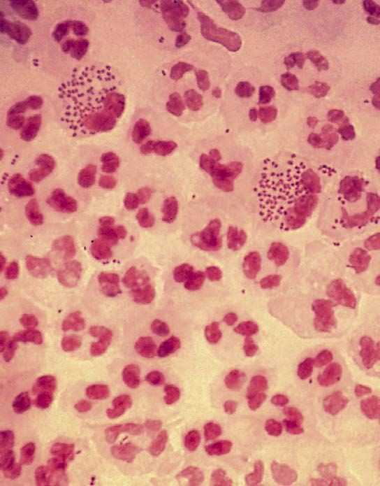
Dear Colleagues,
It is my pleasure to share with you the winter 2019
From the Chair of Rheumatic issue of Rheumatology Connections highlighting
and Immunologic Diseases the clinical, research and educational expertise in
the Department of Rheumatic and Immunologic
Diseases. This issue powerfully demonstrates the
Cleveland Clinic tripartite mission of caring for the
sick, investigating their conditions and educating
those who serve.
Two articles highlight our expertise in vasculitis. Cleveland Clinic’s Rheumatology
Dr. Rula Hajj-Ali offers an overview of Behçet’s Program is ranked among the top
syndrome, a rare vasculitis on the rise in the 2 in the nation in U.S. News &
United States (p. 3). Dr. Carol A. Langford details World Report’s “America’s Best
the optimal approach to orbital disease in Hospitals” survey.
granulomatosis with polyangiitis (p. 6).
Rheumatology Connections, published by
Another pair of articles demonstrates the research
Cleveland Clinic’s Department of Rheumatic
and clinical work generated by our unique and and Immunologic Diseases, provides information
rigorous fellowship offerings. Dr. Soumya Chatterjee on leading-edge diagnostic and management
presents a closer look at two uncommon autoimmune techniques as well as current research for
conditions — gastric antral vascular ectasia physicians.
and antisynthetase syndrome — based on two fellows’ abstracts from this year’s American College
Please direct any correspondence to:
of Rheumatology meeting (p. 16). Dr. Cassandra Calabrese, the first graduate of our combined fellowship
Abby Abelson, MD
in rheumatology and infectious disease, collaborates with a colleague in the Department of Allergy and Clinical Chair, Rheumatic and Immunologic Diseases
Immunology to demonstrate the intersection of rheumatology, immunology and infectious disease (p. 12).
Cleveland Clinic/A50
We also continue to excel in clinical and translational research. Dr. Emily Littlejohn’s research proposes a 9500 Euclid Ave.
way to distinguish flares from infection in our patients with systemic lupus erythematosus (p. 14). We also Cleveland, OH 44195
highlight the translational research of Dr. Elaine Husni on potential biomarkers for cardiovascular disease 216.444.3876
risk in patients with psoriatic arthritis (p. 11), and her clinical research on the safety of NSAIDs for patients abelsoa@ccf.org
with arthritis (p. 18). And for all our patients, we continue to drive quality and improvement through programs
Managing Editor: Deborah Booth Summers
like MyRheum, a patient-reported outcomes interface (p. 8).
Graphic Designer: Barbara Ludwig Coleman
I am honored to work with these talented rheumatologists. I hope that you find in the stories that follow an
Illustration: Brandon Stelter
opportunity to connect, collaborate or consult with our team.
Rheumatology Connections is written for
Respectfully, physicians and should be relied on for medical
education purposes only. It does not provide a
complete overview of the topics covered and
should not replace the independent judgment
of a physician about the appropriateness or risks
of a procedure for a given patient.
Abby Abelson, MD
Chair, Rheumatic and Immunologic Diseases © The Cleveland Clinic Foundation 2019
216.444.3876 | abelsoa@ccf.org
On the cover: In this image of cartilage (purple and white)
from a young mouse femur, osteoclasts (red) surround
a blood vessel filled with red blood cells (yellow). In
contrast to normal osteoclasts, the cells seen here have
only a single nucleus due to the lack of a gene involved in
osteoclast development. This research program aims to
understand how large, bone-resorbing osteoclasts form,
and whether preventing them from fusing together is a
way to control bone loss in osteoporosis, arthritis or other
conditions. Credit: Paul R. Odgren, PhD, University of
Massachusetts Medical School. Image source: NIH Image
Gallery. No changes were made.
License:
https://creativecommons.org/licenses/by-nc/2.0/legalcode
Page 2 | Rheumatology Connections | Winter 2019 For referrals, call 855.REFER.123 (855.733.3712)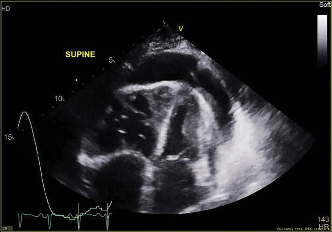
UNDERSTANDING BEHÇET’S SYNDROME
                             Multisystemic inflammatory vasculitis on the rise in the U.S.

                                                                 By Rula Hajj-Ali, MD

B
         ehçet’s disease or syndrome (BS) is a       •     Recurrent aphthous ulcerations along             In addition to changing the title to “EULAR
         multisystemic inflammatory vasculitis             with characteristic systemic manifesta-          Recommendations for the Management of
         characterized by recurrent oral and               tions, especially ocular disease (Figure         Behçet’s Syndrome,” updates include:
genital aphtha, ocular disease, skin lesions,              1C), panuveitis or retinal vasculitis.
gastrointestinal involvement, neurologic disease,                                                           •    Five new principles and one recommen-
                                                     •     Neurologic disease, including characteristic          dation for the surgical management of
vascular disease or arthritis. It is classified
                                                           central nervous system parenchymal                    arterial aneurysms.
under systemic vasculitis and is the only vascu-
                                                           findings.
litis that can affect any vessel size as well                                                                                                                    Dr. Hajj-Ali
                                                                                                            •    Adding apremilast as an option for
as the arterial and venous systems.                  •     Vascular disease, particularly pulmonary                                                              (hajjalr@ccf.org;
                                                                                                                 mucocutaneous involvement. Apremilast is
                                                                                                                                                                 216.444.9643) is
                                                           artery aneurysms, Budd-Chiari syndrome                an oral phosphodiesterase-4 inhibitor that
BS is more common in the Mediterranean and                                                                                                                       Associate Director of
                                                           and cerebral venous thrombosis.                       modulates inflammatory pathways and is          the Center for Vasculitis
Far East regions, but trend studies suggest an
                                                                                                                 approved in the U.S. for treatment of           Care and Research.
18-fold increase in prevalence in the U.S. in        The most commonly used criteria with the best
                                                                                                                 psoriasis and psoriatic arthritis — but it
recent years. The causes of this increase are        sensitivity and specificity are the International
                                                                                                                 soon may be approved for treatment of BS
unknown but could be partly attributed to popu-      Study Group (ISG) criteria for Behçet’s syndrome
                                                                                                                 as well. In a recent study presented at the
lation migration as well as increasing awareness     (Table).1 These criteria were developed to classify
                                                                                                                 2018 American College of Rheumatology
of the disorder.                                     patients for research studies. Some investiga-
                                                                                                                 Annual Meeting, apremilast demonstrated
                                                     tors propose “strong” and “weak” elements in
Some investigators regard BS as a disease.                                                                       consistent efficacy in reducing the number of
                                                     the definition of BS. A strong element, such as
Others prefer the term “syndrome” due to its                                                                     oral ulcers over placebo through week 12.3
                                                     eye disease or vascular involvement, has unique
unknown pathogenesis, lack of clinically accept-     features that distinguish BS from other patho-         •    Considering the use of anti-TNF (tumor
able laboratory screening profile or definitive      logical conditions. In contrast, weak elements,             necrosis factor) monoclonal antibodies
diagnostic test, and variability in prevalence       such as gastrointestinal involvement, point to              in patients with refractory venous throm-
and incidence. (Western patients tend to have a      more than one pathogenetic mechanism.                       bosis. Anticoagulants may be added if
milder clinical course of BS than patients from
                                                                                                                 the risk of bleeding is low in general, and
Eastern countries). Today, it is known that BS is
                                                     Treatment guidelines                                        as long as coexistent pulmonary artery
a more complicated entity affecting every tissue
                                                     BS typically runs a relapsing and remitting                 aneurysms are ruled out.
and organ in the body without exception.
                                                     course. The goal of treatment is to promptly
                                                                                                            •    Emphasizing medical treatment with
Insufficient cross-sectional studies of different    suppress inflammatory exacerbations and recur-
                                                                                                                 cyclophosphamide and corticosteroids
populations make it impossible to compare            rences to prevent irreversible organ damage.
                                                                                                                 before surgical interventions in patients
phenotypic differences around the globe. Clusters                                                                with aortic and peripheral artery aneurysms,
of various disease manifestations occur —            Treatment should be individualized according
                                                                                                                 if the situation isn’t urgent.
for example, acne with arthritis, superficial        to age, gender, type and severity of organ

thrombophlebitis with deep vein thrombosis           involvement, and patient preferences. Ocular,

(DVT), and oral ulcers (Figure 1A) with genital      vascular, neurological and gastrointestinal            Further research is needed
ulcers (Figure 1B) and erythema nodosum.             involvement may be associated with a poor              Despite ongoing efforts, recommendations

These clusters (Figure 2) could imply that the       prognosis. Disease manifestations may                  for the treatment of vascular, gastrointestinal

pathogenesis of BS is complex, with more than        ameliorate over time in many patients.                 and nervous system involvement in BS still

one mechanism. Furthermore, BS has a more                                                                   rely mostly on observational and uncontrolled
                                                     The European League Against Rheumatism                 evidence and expert opinion. While there have
aggressive and severe course in young adult
                                                     (EULAR) recently updated its 2008 evidence-            been some randomized controlled trials involving
males in the Eastern Mediterranean and Middle
                                                     based recommendations for the management of            several agents for mucocutaneous, joint and
and Far East regions as compared with Western
                                                     BS.2 An expert committee defined the problem           eye involvement, very few have been head-to-
countries. Genetic heterogeneity could explain
                                                     areas, performed a systematic literature search,       head trials. There also is a lack of research
this disparity.
                                                     and formulated a final set of recommenda-              evaluating the efficacy of different treatment
                                                     tions and research questions. The committee            strategies for BS, such as a step-up versus
How to diagnose BS                                   included experts from several countries and            step-down approach.
Diagnosing BS is based on pattern of clinical        specialists in all disciplines who care for patients
involvement, laboratory findings, tissue histology   with BS, as well as two patients with BS.
and imaging, usually in the context of:

Visit clevelandclinic.org/rheum                                                                                                   Rheumatology Connections | Winter 2019 | Page 3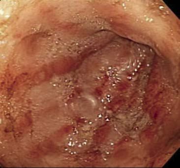
More work in BS is needed. In particular, further
                       research is warranted for controversial issues          A
                       such as the role of anticoagulation in patients
                       with thrombosis and the comparative efficacy
                       of interferon alpha and TNF in patients with eye
                       involvement.

                       References
                       1. Criteria for diagnosis of Behçet’s disease.
                         International Study Group for Behçet’s
                         Disease. Lancet. 1990;335(8697):1078-
                         1080.

                       2. Hatemi G, Christensen R, Bang D, et al.
                         2018 update of the EULAR recommendations
                         for the management of Behçet’s syndrome.
                         Ann Rheum Dis. 2018;77(6):808-818.

                       3. Hatemi G, Mahr A, Ishigatsubo Y, et al.
                         Efficacy of apremilast for oral ulcers associ-
                         ated with active Behçet’s syndrome over
                                                                               B
                         28 weeks: results from a phase III study
                         [abstract]. Arthritis Rheumatol. 2018;70
                         (suppl 10). https://acrabstracts.org/abstract/
                         efficacy-of-apremilast-for-oral-ulcers-asso-
                         ciated-with-active-behcets-syndrome-over-
                         28-weeks-results-from-a-phase-iii-study/.
                         Accessed November 6, 2018.

                                                  Figure 1. Three
                                       manifestations of Behçet’s
                                       syndrome: oral ulcerations          C
                                          (A), genital ulcerations
                                           (B) and hypopyon (C).

Page 4 | Rheumatology Connections | Winter 2019                                    For referrals, call 855.REFER.123 (855.733.3712)
TABLE. INTERNATIONAL STUDY GROUP (ISG) CRITERIA
                                       FOR BEHÇET’S SYNDROME1

              • Recurrent oral ulcers (minor aphthous, major aphthous or • •
              • herpetiform) at least three times in one 12-month period

              PLUS TWO OF THE FOLLOWING:

              • Recurrent genital ulcerations

              • Eye lesions

                  • Anterior uveitis

                  • Posterior uveitis

                  • Cells in vitreous on slit-lamp examination

                  • Retinal vasculitis observed by qualified physician

              • Skin lesions

                  • Erythema nodosum-like

                  • Pseudofolliculitis

                  • Papulopustular lesions

                  • Acneiform nodules

              • Positive pathergy test read by a physician within 48 hours of testing,
                performed with oblique insertion of a 20-22 gauge or smaller needle
                under sterile conditions.                                                                                         Figure 2. Clusters of
                                                                                                                                  disease manifestations
                                                                                                                                  in Behçet’s syndrome.

              Sensitivity: 82.4%

              Specificity: 96%

              Accuracy of the ISG: 86.7%
                                                                                                          Superficial
                                                                                                    thrombophlebitis, dural
                                                                                                    sinus thrombi and DVT

                                                                                             Orogenital
                                                                                             ulcers and                   Acne, enthesitis
                                                                                         erythema nodosum                  and arthritis

Visit clevelandclinic.org/rheum                                                                             Rheumatology Connections | Winter 2019 | Page 5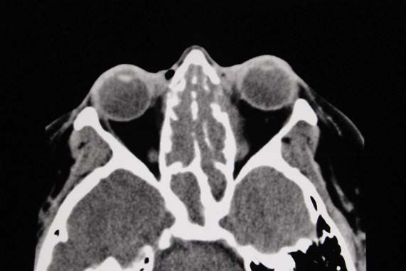
ORBITAL DISEASE IN GRANULOMATOSIS
WITH POLYANGIITIS
Active or damage and how to manage
By Carol A. Langford, MD, MHS
Two patients, similar lesions, Presenting symptoms/signs of orbital disease Surgery has limited if any role in management
different decisions? can include pressure or pain in, around or of orbital disease in GPA. Because of their
A
56-year-old female with granulomatosis behind the eye, as well as swelling of the eyelid composition of inflammatory cells and fibro-
with polyangiitis (GPA) comes to see or periorbital tissues. When the lesion abuts blasts, orbital lesions can become adherent
you for follow-up. She has had disease the ocular muscles, it can affect their function, to adjacent structures. Surgical manipulation
for nine years with past involvement of the resulting in disconjugate gaze and diplopia. of lesions abutting the optic nerve can impact
sinuses, lungs, kidney and orbit. She describes Lesions adjacent to the optic nerve can impact nerve function and result in vision loss. Because
Dr. Langford sinus congestion and discolored drainage suspi- vision. The optic nerve is particularly vulnerable of this risk, surgery is almost always avoided in
(langfoc@ccf.org; cious for infection. She has chronic periorbital at the orbital apex, where the nerve leaves the patients who have normal visual acuity.
216.445.6056)
pain and enophthalmos which are unchanged. orbit and enters the intracranial space. Lesions
is Director of the Injecting glucocorticoids directly into the orbital
Her visual acuity is normal which has not in this location can also result in other oculo-
Center for Vasculitis lesion has no proven benefit, and radiation
Care and Research as changed. In addition to starting her on antibiotics, motor cranial neuropathies with corresponding
therapy has no role in treatment.
well as Vice Chair for you perform a CT sinus/orbit that shows sinus clinical manifestations.
Research, Department mucosal thickening and a left orbital lesion that
of Rheumatic and There are two key reasons why orbital lesions
are unchanged (Figure 1). After completing the Returning to the patients
Immunologic Diseases. are challenging. The first of these is the anatomical
antibiotics, she is feeling improved and back to In the absence of other features of active GPA,
construct of the orbit in being a confined, bony
her baseline. Is there anything you should do for determining whether to treat an orbital lesion
space. Inflammatory lesions within the orbit
the orbital lesion? is based on whether this is felt to reflect active
are close to structures vital to vision, such that
inflammation or damage from scarring. This can
A 46-year-old male with a three-year history of even small lesions and any associated edema can
be difficult to determine and is largely based on
GPA comes to see you for follow-up. He has have a profound impact. The second challenge
objective evidence from a careful ophthalmologic
had past disease involving the sinus, lung, nerve is the rapid development of scarring that accom-
exam and changes in imaging. A persistent
and skin and has been in remission on metho- panies the inflammatory process and becomes
orbital lesion can occur as a result of damage
trexate. At his follow-up visit, he comments on permanent damage. This can result in chronic
from scarring and in the absence of growth is
new pressure around his left eye with doubling symptoms, signs and persistent radiologic
usually not an indication for treatment.
of his vision when looking up and to the left. changes. Over the course of time, scar tissue
On exam, his left eye appears slightly more can retract, resulting in enophthalmos which For patient one who had a known orbital lesion
proptotic with restricted left eye movement on can also impact vision in some patients. that had not changed, this was felt to reflect
left lateral gaze. CT sinus/orbit shows chronic damage, and treatment was not pursued. For
sinus mucosal thickening with This is the of
erosion first study investigating PON-1
the patient two, as there was development of a new
Treatment of orbital disease in GPA
medial orbital wall and new softactivity levels
tissue in psoriatic diseases. If the
fullness orbital lesion, this was treated as active disease.
Treatment of orbital lesions is pursued with
relationships
along the extraconal orbital space abutting the demonstrated here can be
the goal of preventing progression and with the Orbital disease in GPA can be challenging for
validated longitudinally,
medial rectus muscle (Figure 2). Is there any- this biomarker
hope of halting inflammation that has not yet both patients and physicians. Effective man-
of oxidative
thing you should do for the orbital lesion? stress may serve as an addi-
progressed to scar. The medications used for agement of these patients requires regular
tive CV risk stratification tool in systemic
active orbital disease are the same as for other assessments by an ophthalmologist and inter-
rheumatic diseases and may help iden-
Orbital disease: Why is tify it challenging? GPA manifestations and consist of glucocorticoids mittent imaging by CT or MRI, particularly in the
subsets of patients in need of more
Orbital disease is among the aggressive
most challenging combined most commonly with rituximab, setting of new symptoms. Preservation of visual
preventive cardiology strate-
disease manifestations of GPA. It can occur methotrexate or cyclophosphamide. Although acuity, minimizing chronic symptoms and avoiding
gies. The relatively young age of onset for
either de novo within the orbit psoriatic
or, most commonly, some studies raised concern about the effec- treatment-related toxicity are the cardinal objec-
diseases underscores the need
as a result of erosion of the lamina papyracea tiveness of rituximab for orbital disease, others tives, which can be achieved in many patients
for further investigation of oxidative bio-
along the medial orbital wall, allowing inflam- have supported benefit, reflecting the general through careful multidisciplinary care.
markers that may provide an incremental
matory soft tissue to extend into the orbit from difficulty in managing this manifestation. Orbital
benefit in longitudinal CV risk assessment.
the sinus. lesions can be particularly sensitive to changes
in glucocorticoid dose, and some patients
require long-term prednisone.
Page 6 | Rheumatology Connections | Winter 2019 For referrals, call 855.REFER.123 (855.733.3712)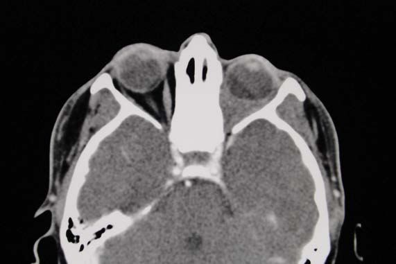
Figure 1. (Above) CT sinus/orbit shows a left orbital lesion that is filling the intraconal orbital space.

                             Figure 2. (Bottom) CT sinus/orbit shows sinus mucosal thickening with erosion of the medial orbital wall
                             and soft tissue fullness extending into the left medial extraconal orbital space.

Visit clevelandclinic.org/rheum                                                                                                Rheumatology Connections | Winter 2019 | Page 7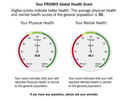
MyRheum: NEW REVELATIONS ON
                                                         PATIENT-REPORTED OUTCOMES
                                                 A step closer to understanding MCID in immune-mediated diseases

                                                                               By Abby Abelson, MD, and Chad Deal, MD

                           S
                                   tandardizing assessment of healthcare        MyRheum was deployed throughout Cleveland               and scores are standardized on a T scale with
                                   through patient-reported outcomes            Clinic’s health system, with 27 rheumatologists         a mean of 50 and standard deviation of 10.
                                   (PROs) is a national priority. Outcome       in 10 locations, in August 2016. Since then,            Results are displayed within the EHR at the time
                           measures evaluate the results of care and are        patients have used it at each rheumatology              of the visit (Figure 1). The ROS is administered
                           therefore considered the most valid metrics for      clinic visit.                                           at every visit. PHQ-9, if normal, is administered
                           measuring and comparing clinical care and driving                                                            yearly, and the remaining scales are completed
                                                                                When developing MyRheum, the team assessed
                           quality and improvement.                                                                                     at least three months apart.
            Dr. Abelson                                                         the feasibility, patient/provider compliance and
     (abelsoa@ccf.org;     There is growing recognition of the value of         utility of electronically collecting PROs using         Through June 2018, approximately 160,000
216.444.3876) is Chair     measuring PROs in patients with rheumatologic        Patient-Reported Outcomes Measurement                   MyRheum questionnaires (including nearly
     of the Department                                                          Information System (PROMIS®) Global Health              50,000 PROMIS, nearly 90,000 RAPID3, more
                           conditions. Clinical disease activity measures
      of Rheumatic and
                           are important for making treatment decisions         (GH), PHQ-9, RAPID3 or SLAQ; pain, fatigue              than 20,000 PHQ-9 and nearly 600 SLAQ) had
 Immunologic Diseases.
                           but sometimes do not measure the domains of          and physical function PROMIS domains; and               been completed by 35,700 unique patients.
                           health important to patients. PROs measured at       a review of systems (ROS). PROMIS allows                Approximately 40 percent of questionnaires
                           the point of care can enhance shared decision-       measurement of health domains important for             were completed by the patient on MyChart, at
                           making and facilitate treatment decisions,           rheumatology patients as well as comparison with        home before their visit.
                           although the ability to collect and report PROs      the general U.S. population.
                                                                                                                                        Cross-departmental comparisons of diseases
                           in real time can be challenging because of tech-
                                                                                With MyRheum, PROs are collected at the                 (PROMIS-10 T-scores) showed rheumatology
                           nology and workflow barriers.
                                                                                patient’s visit on a tablet computer or prior to        patients had the second-lowest self-reported
                           Through support from Cleveland Clinic and an         the visit through a patient portal (MyChart,            physical health score (Figure 2), demonstrating
Dr. Deal (dealc@ccf.org;
                           information technology team, the Department          Epic Systems) integrated with their electronic          the impact of rheumatic diseases and the need
 216.444.6575) is staff
      in the Department    of Rheumatic and Immunologic Diseases                health record (EHR). PROMIS domains are                 to measure PROs.
       of Rheumatic and    developed a patient-entered data (PED) system        administered using computer adaptive testing,
 Immunologic Diseases.     using validated measurements that assess
                           physical and mental function, social health
                           and well-being, and disease activity, as well
                           as a rheumatology-oriented review of systems.
                           This PED system, called MyRheum, allows the
                           clinician to evaluate patient-reported health
                           measures at the point of care, engage patients in
                           their care and make better treatment decisions
                           based on patient-centric data.

                           At the 2018 American College of Rheumatology’s
                           Annual Meeting, several staff members presented
                           compelling data on their recent experience with
                           MyRheum.

                           Developing and implementing
                           MyRheum
                           In “Development and Implementation of a
                           Patient-Reported Outcomes Measurement
                           Information System (MyRheum),” authors
                           Chad Deal, MD; Abby Abelson, MD; Leonard
                           Calabrese, DO; database designer Greg Strnad;
                           Irene Katzan, MD; and M. Elaine Husni, MD,
                           presented a behind-the-scenes look at                Figure 1. How results display to patients.
                           MyRheum.

  Page 8 | Rheumatology Connections | Winter 2019                                                                                 For referrals, call 855.REFER.123 (855.733.3712)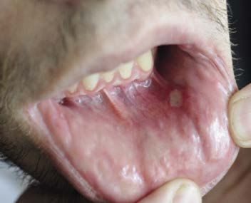
Figure 2. Comparison of
                                                                                                                                                        T-scores by department.

                                                        30                    35                      40                    45         50                  55

                                               PROMIS GH T-score Change
                               100
                                                                                                       n Worsened
                               80                                                                      n No Change
                                                                                                       n Improved
         Change in Score (%)

                               60
                                                                                                      Figure 3. Change in
                                                                                                      PROMIS GH score.
                               40

                               20

                                                 Psoriatic                               Rheumatoid
                                     Overall     Arthritis   GPA   Lupus                  Arthritis

                                                                                                          RAPID3 Weighted Score Change
                                                                                         100
                                                                                                                                                           n Worsened
                                                                                                                                                           n No Change
                                                                                         80
                                                                   Change in Score (%)

                                                                                                                                                           n Improved

                                                                                         60
                                                                                                                                                          Figure 4. Change in
                                                                                                                                                          RAPID3 score.

                                                                                         40

                                                                                         20

                                                                                         0
                                                                                                                Psoriatic                  Rheumatoid
                                                                                                      Overall   Arthritis    GPA   Lupus    Arthritis

Visit clevelandclinic.org/rheum                                                                                                      Rheumatology Connections | Winter 2019 | Page 9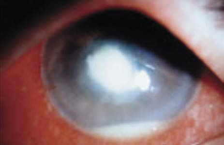
The creation of this large PRO biomarker data-       The authors found:                                      The variability in MCID implies that some
                        bank demonstrates its practicality and provides a    •     The change in PROMIS score was                    patients improve while others worsen. This
                        powerful platform for clinical care, research and          statistically significant overall (P < 0.001)     study presents an opportunity to better under-
                        value-based healthcare initiatives.                        as well as for those with either GPA (P           stand patient characteristics and therapies that
                                                                                   = 0.030) or RA (P < 0.001), but not for           may explain these changes.
                                                                                   lupus (P = 0.67) or PsA (P = 0.36).
                        Evaluating PROs in immune-mediated
                        diseases                                             •     The change in RAPID3 score was                    Capturing what matters most
                        In “Using Patient Reported Outcomes at Point               significant overall (P < 0.001), as was           The greatest advantage of measuring PROs is
                        of Care in Immune-Mediated Diseases: Minimal               the change for lupus (P = 0.048) and RA           capturing what matters most to patients and
                        Clinically Important Differences,” authors Drs.            (P < 0.001), but not for GPA (P = 0.11)           allowing clinicians to use this information at the
                        Husni, Deal, Calabrese and Abelson, Strnad                 or PsA (P = 0.17).                                point of care.
                        and biostatistician James Bena shared how
                        MyRheum helped evaluate PROs in several              •     Thresholds for clinically meaningful              Now, with MyRheum, most rheumatology

                        immune-mediated diseases.                                  change in PROMIS GH were most signifi-            providers at Cleveland Clinic start each clinical
                                                                                   cant for GPA and RA compared with PsA             visit with a review of PROs. This focuses each
                        Minimal clinically important differences (MCID)            and lupus, and in RAPID3 were most sig-           visit on what is important to the patient, which
                        are patient-derived scores that reflect changes            nificant for lupus and RA compared with           helps guide treatment decisions.
                        in clinical care that are meaningful to the                PsA and GPA.
                        patient. Little is known about MCID in many                                                                  In addition to facilitating patient engagement,

                        immune-mediated diseases, as small differ-           •     Approximately 15-20 percent of patients           MyRheum is providing rheumatology caregiv-

                        ences in PROs may be statistically significant             showed an improvement or worsening of             ers with objective, quantitative measures of

                        yet clinically unimportant for the patient.                MCID by six months; 60-70 percent had             treatment outcomes — assuring them when
                                                                                   no change between visits (Figures 3 and 4).       treatments are working.
                        Using MyRheum, data were collected from
                        patients with rheumatoid arthritis (RA), psoriatic   On average, there was improvement in PROMIS
                        arthritis (PsA), lupus and granulomatosis with       and RAPID3 scores among all immune-mediated
                        polyangiitis (GPA) who had completed PROMIS          diseases after two visits. However, this study
                        GH and RAPID3 at two separate visits, six            indicates that it is unlikely for a single MCID value
                        months apart. Paired t-tests assessed the            to be applicable across all chronic diseases.
                        changes between visits. Important differences
                        were identified by a PROMIS MCID change of
                        5 and RAPID3-weighted score of 1.2 (improve-
                        ment or worsening).

Page 10 | Rheumatology Connections | Winter 2019                                                                              For referrals, call 855.REFER.123 (855.733.3712)
PON1 ACTIVITY CORRELATES WITH
SYSTEMIC INFLAMMATION, DISEASE
ACTIVITY AND CVD RISK IN PSA
Paraoxonase-1 a potential biomarker for CVD risk
By M. Elaine Husni, MD, MPH
P
atients with psoriatic arthritis (PsA) as part of Cleveland Clinic’s Cardiometabolic PsA patients with moderate to high disease
are known to have increased cardio- Outcome Measures in Psoriatic Arthritis Study activity (defined as either a DAS28-ESR > 3.2
vascular (CV) morbidity and mortality (COMPASS). Various baseline data were or a DAS28-CRP > 2.67) showed a statistically
not completely explained by traditional CV risk assessed and recorded, including gender, significant lower arylesterase activity than those
factors. Recent research has sought to expand BMI, current disease-modifying antirheumatic with low disease activity (defined by DAS28-
the understanding of the mechanisms through pharmaceutical regimens, PsA disease activity ESR < 3.2 or DAS28-CRP < 2.67). In addition,
which PsA is linked to enhanced pathogenesis (DAS-28, CDAI, joint counts), pre-existent car- PsA patients with moderate to high disease
of atherosclerotic heart disease. Researchers diovascular disease (CVD) and CVD risk factors activity had a greater percentage of CVD risk Dr. Husni
have found evidence of increased oxidative (diabetes, dyslipidemia, hypertension, smoking), factors than those with low disease activity as (husnie@ccf.org;
stress in both psoriatic disease and athero- Framingham risk score, quality of life (QOL) measured by DAS28 scores. 216.445.1853) is
sclerosis. Paraoxonase-1 (PON1), a family of measures and labs (ESR/CRP, lipid profiles). Director of the Arthritis
and Musculoskeletal
antioxidant enzymatic proteins located on HDL
We further assessed CV disease burden by PON1 activity correlates with CV Treatment Center and
cholesterol particles, helps inhibit lipid oxida- burden and prevalent CV disease Endowed Chair of
identifying patients with metabolic syndrome,
tion. Decreased PON1 activity is considered Both PsO and PsA cohorts had significantly Translational Functional
and a subgroup of patients with PsA underwent Medicine Research at
a biomarker for increased systemic oxidative lower serum arylesterase activity when com-
carotid duplex high-resolution B-mode ultra- Cleveland Clinic.
stress and increased conversion of HDL to a pared with healthy controls (P = 0.001).
sound imaging and were screened for carotid
dysfunctional proinflammatory and proathero- Specifically, the PsA cohort demonstrated that
intima-media thickening (CIMT) and the pres-
genic state, and has been associated with the lower arylesterase activity, but not paraoxonase
ence of plaque. A subgroup underwent a second
development of cardiovascular disease. activity, of PON1 was associated with elevated
carotid duplex ultrasound two years later.
In addition, decreased PON1 enzymatic activity The serum PON1 activity level was measured disease activity measures, increasing CVD
has been demonstrated to predict the develop- by two methods: paraoxonase activity (using burden and worse QOL measures. These assoc-
ment of major adverse cardiovascular events in paraoxon as substrate) and arylesterase activity iations were not seen in the PsO cohort.
the general population. A significant reduction (using phenyl acetate as substrate). The levels While epidemiologic studies have shown that
in PON1 activity has been reported in patients of PON1 activity in the PsA and PsO cohorts patients with PsA can be seen as a high-risk
with systemic inflammatory diseases, including were compared 2-to-1 with an age- and gender- group for developing atherosclerosis, these mea-
rheumatoid arthritis (RA) and systemic lupus matched healthy human cohort. sures of HDL-associated PON1 enzymatic activity
erythematosus (SLE). may provide the mechanistic link between
My recent study with colleagues W.H. Wilson PON1 activity levels and correlation increased oxidative stress and CVD burden.
Tang, MD, and Stanley Hazen, MD, PhD, from with disease activity
Mean arylesterase activities were significantly
Cleveland Clinic’s Sydell and Arnold Miller Reference
Family Heart & Vascular Institute reported for lower in the PsO (P < 0.001) and PsA (P <
1. Husni ME, Wilson Tang WH, Lucke M, et al.
the first time on serum PON1 enzymatic activity 0.001) subjects when compared with healthy
Correlation of high-density lipoprotein-asso-
and its association with both psoriatic disease controls. In addition, the PsO cohort showed sig-
ciated paraoxonase 1 activity with systemic
activity and CV disease burden in a psoriatic nificantly lower mean arylesterase activity when
inflammation, disease activity, and cardiovas-
disease population.1 The results for psoriasis compared with the PsA cohort (P = 0.003).
cular risk factors in psoriatic disease. Arthritis
(PsO) and PsA were compared. No significant difference in median paraoxonase
Rheumatol. 2018;70(8):1240-1250.is
activity between the PsO and PsA cohorts was
detected, although median paraoxonase activity
Measuring PON1 activity level showed a trend of lower levels in the PsA and
This study, involving 343 adult patients with PsO cohorts when compared with controls.
PsO and PsA and 345 controls, was conducted
Visit clevelandclinic.org/rheum Rheumatology Connections | Winter 2019 | Page 11AT THE INTERSECTION OF
AUTOIMMUNE AND INFECTIOUS DISEASE
Case study shows value of interdisciplinary collaboration
By Cassandra Calabrese, DO, and James Fernandez, MD, PhD
C
omplex immunologic disease requires The patient’s laboratory evaluation revealed a show some patients developed meningococcal
intense collaboration across subspecial- complement deficiency assay (CH50) of 0, C7 disease even after receiving vaccinations.5
ties for optimal care. For that reason, deficiency (< 5 U/mL) and absent AH50 with-
Currently available are vaccines for the most
Cleveland Clinic’s Department of Rheumatic out evidence of humoral immune deficiency. She
common serotypes, the meningococcal con-
and Immunologic Diseases recently developed was diagnosed with primary C7 deficiency.
jugate vaccine (MenACWY) and a serogroup
the nation’s first combined fellowship in rheu-
Complement plays a key role in protection B meningococcal vaccine (MenB). While most
matology and infectious disease (ID), designed
against a variety of infections. Activation of eculizumab-associated meningococcal infections
Dr. Calabrese to train physicians to practice in both of those
terminal complement components (or mem- have been nongroupable Neisseria meningitides,
(calabrc@ccf.org; specialties.
216.978.5949) is staff brane attack complex: C5-C9) is crucial for both meningococcal vaccines should be given
in the Department of These dual-trained specialists will be well-suited controlling infections with encapsulated organ- at least two weeks before the first dose of eculi-
Rheumatic and Immuno- to engage in clinical care and research in a mul- isms, such as Neisseria spp. Patients with zumab, if possible.4,6
logic Diseases. In 2018, genetic deficiencies of terminal complement,
titude of areas, including:
she was the first gradu- Interestingly, breakthrough infection has been
such as the patient presented here, have a
ate of Cleveland Clinic’s reported,5 so antibiotic prophylaxis is also required.
• Diagnosis and management of serious significantly increased risk of recurrent invasive
combined fellowship
in rheumatology and and opportunistic infections in patients on meningococcal infection.1
infectious disease. advanced immunosuppressive regimens. Opportunities for interdisciplinary
The patient was educated on methods to reduce collaboration
• Infection prevention and vaccinology in her infection risk, including revaccination against Both the case study and the use of eculizumab
at-risk populations. all five available meningococcal serotypes every involve a significantly increased risk of systemic
three to five years. She has done well since. neisserial infection and are perfect examples of
• Diagnosis and management of patients
in primary immunodeficiency states, the opportunity for collaboration between rheu-
especially those with infectious and auto- Eculizumab therapy and its risks matology, immunology and infectious disease.
immune manifestations. Eculizumab is a monoclonal anticomplement C5
Rheumatology offers many more opportunities,
antibody that is FDA-approved for the treatment
Rheum-ID hybrids also will be well-poised to as the specialty encompasses every organ sys-
Dr. Fernandez of paroxysmal nocturnal hemoglobinuria (PNH)
(fernanj2@ccf.org; sort out the complexities of the growing list of tem and diseases with protean manifestations.
and atypical hemolytic uremic syndrome (both
216.444.6933) is staff rheumatic complications of infections, such as Patients with primary immunodeficiencies may
diseases that involve uncontrolled activation
in the Department spirochetes; blood-borne viral illnesses such as also present with autoimmune conditions, such
of Allergy and Clinical of the complement system). Not surprisingly,
HCV, HBV and HIV; and the emerging field of as connective tissue diseases, inviting even
Immunology. eculizumab is associated with a significantly
arboviruses. more interdisciplinary collaboration.
increased risk of meningococcal disease —
Below we share two situations at the intersection 1,000 to 2,000 times greater than that of a
of rheumatology/immunology and infectious healthy person.2 References
disease. 1. Audemard-Verger A, Descloux E, Ponard D,
The rheumatology community will be increasingly et al. Infections revealing complement defi-
exposed to eculizumab, as it also is used to treat ciency in adults: a French nationwide study
Case study: Patient with primary refractory lupus nephritis.3 Therefore, it is crucial enrolling 41 patients. Medicine (Baltimore).
C7 deficiency that rheumatologists become aware of the drug’s 2016;95(19):e3548.
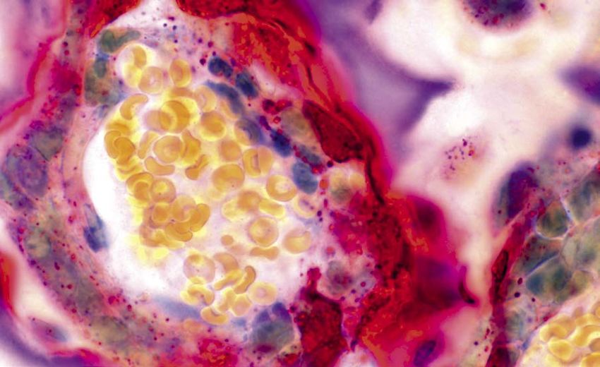
An 18-year-old female presented for evaluation of unique risk, as well as the current recommenda-
possible immunodeficiency. She was in her usual tions to offset it. 2. Benamu E, Montoya JG. Infections
state of health until two years prior, when she was associated with the use of eculizumab:
admitted for meningococcal meningitis. She was A black box warning was added to the eculi-
recommendations for prevention and
treated and recovered. One year later, she pre- zumab package insert after two of 196 PNH
prophylaxis. Curr Opin Infect Dis. 2016;
sented with flu-like symptoms and was diagnosed patients developed meningococcal infections
29(4):319-329.
again with meningitis secondary to Neisseria while using the drug during clinical trials.4 The
meningitidis (Figure). Her history is also notable insert recommends meningococcal vaccines for
for frequent upper respiratory tract infections. recipients of eculizumab, although recent data
Page 12 | Rheumatology Connections | Winter 2019 For referrals, call 855.REFER.123 (855.733.3712)3 Sciascia S, Radin M, Yazdany J, et al.
Expanding the therapeutic options for
renal involvement in lupus: eculizumab,
available evidence. Rheumatology Int.
2017;37(8):1249-1255.
4. Soliris [package insert]. Cheshire, CT: Alexion
Pharmaceuticals, Inc. 2015.
5. McNamara LA, Topaz N, Wang X, Hariri S,
Fox L, MacNeil JR. High risk for invasive
meningococcal disease among patients
receiving eculizumab (Soliris) despite receipt
of meningococcal vaccine. MMWR Morb
Mortal Wkly Rep. 2017;66(27):734-737.
6. Meningococcal ACIP Vaccine
Recommendations. Centers for Disease
Control and Prevention website. https://www.
cdc.gov/vaccines/hcp/acip-recs/vacc-specific/
mening.html. Accessed November 1, 2018.
Figure. Neisseria meningitidis
Visit clevelandclinic.org/rheum Rheumatology Connections | Winter 2019 | Page 13THE FEVER THAT CRIES WOLF
Distinguishing the causes of fevers in patients with lupus
By Emily Littlejohn, DO, MPH
A
25-year-old female with systemic Infection or flare? Using the Interestingly, where ESR elevations are strongly
lupus erythematosus (SLE) manifest- ESR-to-CRP ratio associated with disease exacerbations in SLE,1
ing with class IV lupus nephritis, Rheumatic diseases and their treatments often CRP levels do not tend to correlate with markers
lupus cerebritis, antiphospholipid syndrome, put patients at increased risk of infections. This of disease activity, such as anti-double-stranded
hemolytic anemia, oral ulcers, alopecia and leaves us keenly aware and constantly inquiring DNA antibodies and complement levels.2 The
arthritis was recently evaluated in our clinic for about infectious signs and symptoms. Herein blunted CRP response in SLE patients may be
follow-up. Current therapy included prednisone, lies one of the most common dilemmas faced due to the effects of interferon- a, a molecule
Dr. Littlejohn hydroxychloroquine and rituximab intravenous by rheumatologists: distinguishing the causes of highly expressed in lupus patients, by inhibiting
(littlee3@ccf.org; infusions, with the last dose given two months fevers in patients with rheumatic diseases. This CRP promoter activity and CRP secretion in
216.445.5559) is staff
prior to this visit. She presented after a complex is particularly true in lupus, where fevers can be hepatocytes.3
in the Department
hospitalization during which she was diagnosed a common manifestation of a lupus flare.
of Rheumatic and Since ESR rises both with lupus activity and
Immunologic Diseases. with atypical hemolytic uremic syndrome and
Research that can provide physicians with tools with infection, alone it is too nonspecific to
received eculizumab therapy.
to elucidate the cause of fevers in lupus patients distinguish between lupus flare and infection.4
At our visit the patient reported feeling chilled, is ongoing. SLE activity measures — such as CRP values of > 6.0 mg/dL in SLE patients have
with myalgia and gastrointestinal upset. Her anti-double-stranded DNA antibodies, comple- been associated with infectious processes,5-7
vital signs revealed that she was tachycardic ments and the complete blood count — can be and higher CRP levels have been observed in
to 130 beats per minute with a temperature of helpful, although these measures do not always SLE infection compared with SLE flare without
38.6°C (101.5°F). She was promptly admitted track or change with lupus activity and certainly infection.8,9 When CRP is elevated during a flare,
to the hospital where blood work revealed ele- can be abnormal in the setting of infection. CRP flares of serositis (pleuritis, pericarditis, pneu-
vated sedimentation rate (ESR) and C-reactive and ESR, both nonspecific markers of systemic monitis) and flares involving nephritis or myositis
protein (CRP), and leukopenia with stable inflammation, are potentially useful biomarkers present with a significantly higher CRP than
hemoglobin and platelets. in this frequently encountered clinical scenario. other types of SLE flares.10
In an article recently published in Lupus, the
medical records of hospitalized patients with SLE
were reviewed to assess the usefulness of the
Table. Nonspecific markers of inflammation corresponding to episodes of flare vs. infection.11 ESR-to-CRP ratio in distinguishing infection from
flare in lupus patients presenting with fever.11
Lupus flare Infection
P Value Eligible hospitalizations for this study were those
(N = 28) (N = 25)
in which patients presented with a temperature
ESR (mm/hour) 50.7 (31.3) 53.4 ± (34.5) NS of > 37.9°C (100.3°F) or with subjective fever
as a chief complaint upon admission. Collected
CRP (mg/dL) 5.4 (6.5) 11.2 (7.2) 0.0035 at admission were clinical and laboratory data,
including patient symptoms, the infectious
ESR:CRP Ratio 0.000 workup (X-rays, blood cultures, urine cultures),
basic labs (including complete blood count),
ESR and CRP.
≤2 0 (0) 3 (12.0)
We found that ESR levels were similar in
2 – 15 13 (46.4) 21 (84.0) patients with flares and infections. CRP levels
were significantly higher in infections compared
≥ 15 15 (53.6) 1 (4.0) with flares (Table). The ESR-to-CRP ratio was
positively associated with flare, where each unit
WBC > 10K/mm 3
6 (21.4) 5 (20) NS increase in the ESR-to-CRP ratio was associated
with a 13 percent increase in the odds of fever
Page 14 | Rheumatology Connections | Winter 2019 For referrals, call 855.REFER.123 (855.733.3712)etiology being attributed to SLE flare versus Figure. Relative frequency
infection. The proportion of flares versus infec-
Relative Frequency of of flares versus infections
according to the ratio of
tions varied according to the ratio of ESR to CRP
(Table and Figure), with infections predominant Flares vs. Infections erythrocyte sedimentation
rate (ESR) to C-reactive
for ratios of ≤ 2, and flares predominant for protein (CRP) (categorized
ratios of ≥ 15 (P = 0.000). 40 as ≤ 2, 2-15 or ≥ 15).
■ = Flare
Number of Hospitalizations
There was a significant
■ = Infection difference in the distribution
How the ESR-to-CRP ratio guided 30 of flares versus infections
treatment according to the ESR to
Our patient was promptly admitted to the hospital, CRP ratio (P = 0.000).11
20
where blood work revealed elevated ESR of 27
mm/hr (normal range 0-20 mm/hr) and CRP
of 2.3 mg/dL (normal < 0.9 mg/dL), with an 10
ESR-to-CRP ratio of 11.7. Complements were
normal, and anti-double-stranded DNA antibod- 0
ies also were normal. Her complete blood count ≤2 2 – 15 ≥15
showed leukopenia, with both lymphopenia and
neutropenia. Blood cultures grew methicillin-
resistant Staphylococcus epidermidis, and our 2. Bertouch JV, Roberts-Thompson PJ, et 7. Morley JJ, Kushner I. Serum C-reactive
patient received a full course of IV antibiotics, al. C-reactive protein and serological protein levels in disease. Ann N Y Acad Sci.
which resolved her fevers and normalized her indices of disease activity in systemic 1982;389:406-418.
cytopenias. lupus erythematosus. Ann Rheum Dis.
1983;42(6):655-658. 8. Honig S, Gorevic P, Weissmann G.
In summary, our patient was one with difficult- C-reactive protein in systemic lupus
to-control, multi-organ-involved lupus, receiving 3. Enocsson H, Sjöwall C, Skogh T, et al. erythematosus. Arthritis Rheum.
ongoing immunosuppression and presenting Interferon-alpha mediates suppression of 1977;20(5):1065-1070.
with a temperature of 38.6°C (101.5°F). Blood C-reactive protein: Explanation for muted
work on admission was difficult to interpret, and C-reactive protein response in lupus flares? 9. Bravo MG, Alarcón-Segovia D. C-reactive
there was a high suspicion of both infection and Arthritis Rheum. 2009;60(12):3755-3760. protein in the differential diagnosis between
SLE flare. The ESR-to-CRP ratio was a useful infection and disease reactivation in SLE.
tool in helping to guide the clinical acumen and 4. Becker GJ, Waldburger M, Hughes GR, J Rheumatol. 1981;8:291-294.
delay use of high-dose glucocorticoids or other Pepys MB. Value of serum C-reactive protein
immunosuppressive SLE medications. measurement in the investigation of fever in 10. Firooz N, Albert DA, Wallace DJ, et
systemic lupus erythematosus. Ann Rheum al. High-sensitivity C-reactive protein
This precarious situation between treating an and erythrocyte sedimentation rate in
Dis. 1980;39(1):50-52.
infection versus flare is not foreign to rheuma- systemic lupus erythematosus. Lupus.
tologists. In analyzing the usefulness of the 5. ter Borg EJ, Horst G, Limburg PC, et al. 2011;20(6):588-597.
ESR-to-CRP ratio, we hope to shed light on a C-reactive protein levels during disease
potentially useful tool that can guide manage- exacerbations and infections in systemic 11. Littlejohn E, Marder W, Lewis E, et al.
ment in this common clinical conundrum. lupus erythematosus: a prospec- The ratio of erythrocyte sedimentation
tive longitudinal study. J Rheumatol. rate to C-reactive protein is useful in
1990;17(12):1642-1648. distinguishing infection from flare
References
in systemic lupus erythematosus
1. Vilá LM, Alarcón GS, McGwin G Jr, et al.
6. Vanderschueren S, Deeren D, Knockaert patients presenting with fever. Lupus.
Systemic lupus erythematosus in a multi-
DC, et al. Extremely elevated C-reactive 2018;27(7):1123-1129.
ethnic cohort (LUMINA): XXIX. Elevation of
protein. Eur J Intern Med. 2006;17(6):430-
erythrocyte sedimentation rate is associated
433.
with disease activity and damage accrual. J
Rheumatol. 2005;32(11):2150-2155.
Visit clevelandclinic.org/rheum Rheumatology Connections | Winter 2019 | Page 15A CLOSER LOOK AT TWO RARE
AUTOIMMUNE CONDITIONS
Gastric antral vascular ectasia in systemic sclerosis
and serositis in antisynthetase syndrome
By Soumya Chatterjee, MD, MS, FRCP
A
t the 2018 American College of Methods. We conducted a retrospective chart Clinical manifestations associated with GAVE
Rheumatology Annual Meeting in review of patients with GAVE and evaluated in both SSc and non-SSc groups were tel-
Chicago, Cleveland Clinic rheumatology those diagnosed between 2012 and 2017. angiectasias, melena, hematemesis, fatigue,
fellows presented research on two rare autoim- We initially identified 145 GAVE patients and dyspnea and lightheadedness. When adjusted
mune conditions. Their findings shed new separated them into cohorts of those with SSc for pretransfusion hemoglobin, the difference
light on these little-understood diseases. and those with other diseases. We selected 37 in transfusion requirements was not statisti-
consecutive SSc and 37 consecutive non-SSc cally significant between the two groups. There
Dr. Chatterjee was no difference in use of
Gastric antral vascular
(chattes@ccf.org; NSAIDs and anticoagulants
216.444.9945) directs
ectasia in systemic
sclerosis between the two groups.
the Scleroderma Program
Second-year rheumatology There also was no difference
in the Department
of Rheumatic and fellow Rabeea Mirza, MD, in number of recurrences of
Immunologic Diseases. compared gastric antral vascu- GAVE. Two patients with
lar ectasia (GAVE) in systemic cirrhosis of the liver died.
sclerosis (SSc) with GAVE in
Further studies with larger
other diseases.
cohorts of GAVE patients
Background. GAVE is a may be helpful in under-
pathologic angioectasia with standing its natural history
a characteristic endoscopic and outcomes in specific
appearance. Rugal folds with diseases.
dilated blood vessels radiate
Dr. Mirza is a fellow
in the Department from the antrum and converge Serositis in antisynthetase
of Rheumatic and at the pylorus, resembling syndrome
Immunologic Diseases. watermelon stripes, support- First-year rheumatology fellow
ing the name “watermelon Alexis Katz, DO, studied the
stomach” (Figure 1). GAVE can prevalence of serositis in anti-
cause anemia and significant synthetase syndrome (ASS),
morbidity; hence there is need its clinical significance and its
for surveillance. Figure 1. A 55-year-old female with diffuse systemic sclerosis presenting with association with specific ASS
severe iron deficiency anemia (hemoglobin 7.5 g/dL). Upper endoscopy shows autoantibody subtypes.
GAVE has been associated
gastric antral vascular ectasias (GAVE).
with cirrhosis of the liver, Background. ASS is a relatively
autoimmune diseases (e.g., SSc, r are autoimmune disease
Dr. Katz is a fellow
rheumatoid arthritis, primary biliary cholangitis), patients from the GAVE database. Outcomes
in the Department characterized by interstitial lung disease, myositis,
of Rheumatic and end-stage renal disease, hypertension, heart were defined by number of transfusions, number
inflammatory arthritis, Raynaud phenomenon
Immunologic Diseases. failure, hypothyroidism and chronic pulmonary of recurrences of GAVE bleeding diagnosed
and mechanic’s hands. Eight autoantibodies to
disease. It also can occur after hematopoietic endoscopically, and death.
aminoacyl-transfer RNA synthetases have been
stem cell transplantation. Prevalence of SSc- Results. This study demonstrated that SSc described so far: Jo-1, PL-7, PL-12, EJ, OJ, YRS,
associated GAVE is highly variable, ranging from patients with GAVE were significantly younger KS and Zo. Morbidity and mortality are
1 to 76 percent of patients with SSc. Prevalence than those with non-SSc GAVE, and were mainly related to pulmonary complications.
of GAVE in other associated diseases and its mostly females. Patients were followed for a However, little has been reported about the
long-term outcomes are still unknown. median of five years.
Page 16 | Rheumatology Connections | Winter 2019 For referrals, call 855.REFER.123 (855.733.3712)prevalence of serositis (pleural and/or pericardial classified as trace, small, medium, large or when compared with patients with other
effusions) in ASS other than in small cohort studies tamponade, based on echocardiographic find- antibodies. Anti-PL-12 patients had a higher
(15-20 patients) and case reports. ings (Figure 2). frequency of pleural effusions relative to patients
with anti-Jo-1, anti-PL-7 and all other antibodies
Methods. Clinical data were obtained by Results. A total of 93 patients were included in
combined.
retrospective review of electronic medical this study. The mean age was 57.5 years; 63
records from 2004 to 2017. Our study included percent were females. More research is necessary to better understand,
patients diagnosed with ASS by a rheumatolo- diagnose and treat both GAVE in SSc and
Out of 90 patients with complete data available,
gist. All patients had one of the following ASS serositis in ASS. Our work is intended to raise
42.2 percent had pleural effusion(s) and 47
antibodies: Jo-1, PL-7, PL-12, EJ or OJ. Pleural awareness of these conditions, share new
percent had a pericardial effusion, of which 10
effusions were qualified as trace, small, medium insights and serve as a springboard for further
percent were moderate to large. One patient had
or large, based on chest radiographs and investigation.
tamponade physiology. Anti-Jo-1 patients were
thoracic CT scans. Pericardial effusions were
significantly less likely to have pleural effusions
Figure 2.
Echocardiogram
in a 23-year-old
female with anti-Jo-1
syndrome showing a
large circumferential
pericardial effusion.
Visit clevelandclinic.org/rheum Rheumatology Connections | Winter 2019 | Page 17PRACTICE-CHANGING
PRECISION SUBANALYSES
What rheumatologists should know
By M. Elaine Husni, MD, MPH
PRECISION trial results1,2 have challenged ibuprofen, but that celecoxib with aspirin still randomized, double-blind controlled trial
many assumptions about the use of nonselective has an equal or better safety profile (in regard published in Alimentary Pharmacology &
NSAIDs versus selective COX-2 inhibitors. to GI and renal events) relative to both agents. Therapeutics found that celecoxib had a safer
The study evaluated the trial’s on-treatment GI profile overall compared with ibuprofen or
NSAIDs are normally classified by the relative
population for both OA and RA, which consisted naproxen for patients with RA and OA.5 The
selectivity of COX-1 and COX-2 enzymes.
of 11,018 patients taking concomitant aspirin primary endpoints were clinically significant GI
Nonselective NSAIDs, such as naproxen and
and 12,935 patients not on aspirin. Propensity events (CSGIEs), including bleeding, obstruction,
Dr. Husni ibuprofen, inhibit both COX-1 and COX-2.
score weighting was used to adjust for baseline perforation events from the stomach downward or
(husnie@ccf.org; Selective NSAIDs, such as celecoxib, are COX-
characteristics, thereby increasing the validity symptomatic ulcers, and iron deficiency anemia.
216.445.1853) 2-specific and were developed to spare COX-1
of comparisons.
is Director of inhibition to allow more gastrointestinal (GI) Patients received 100 to 200 mg celecoxib
the Arthritis and
protection. Another substudy published in Rheumatology twice daily (N = 8,072), 600 to 800 mg ibu-
Musculoskeletal
Treatment Center tested the hypothesis that RA patients have a profen three times daily (N = 8,040) or 375 to
Many osteoarthritis (OA) and rheumatoid arthritis
and Endowed Chair different risk-benefit profile for the use of aspirin 500 mg naproxen twice daily (N = 7,969) as
(RA) patients rely on celecoxib, the only COX-2
of Translational in secondary CV risk prevention.4 Of 1,852 well as 20 to 40 mg esomeprazole for gastro-
Functional Medicine inhibitor still marketed in the U.S., but we
subjects with RA in PRECISION, 540 reported protection. CSGIEs occurred in 0.34, 0.74 and
Research at assumed that they faced a greater risk for car-
using low-dose aspirin for CV prevention, and 0.66 percent of patients receiving celecoxib,
Cleveland Clinic. diovascular (CV) disease.
1,312 did not. We observed major NSAID toxic- ibuprofen and naproxen, respectively. There also
PRECISION data tell us that’s not so. PRECISION ity in 79 (6.0 percent) nonaspirin users and 37 was less iron deficiency anemia in patients on
found celecoxib to be as safe as naproxen and (6.9 percent) aspirin users (P = 0.50). Thus, celecoxib than in those on naproxen or ibupro-
ibuprofen in terms of CV risk. In patients with in the RA population, low-dose aspirin users fen. Helicobacter pylori status was also studied
OA, celecoxib carries less CV risk than ibuprofen experienced the same rate of primary outcome but did not influence the outcome.
and similar risk to naproxen, and less GI risk as nonaspirin users. The risk of a major adverse
Interestingly, concomitant corticosteroid use
than ibuprofen and naproxen. Celecoxib was CV event was similar as well.
increased total GI events and CSGIEs. Our data
similar to both ibuprofen and naproxen in all-
These findings highlight the importance of show that CSGIEs are infrequent in patients
cause mortality. In patients with RA, the study
appropriately counseling arthritis patients on with OA and RA taking NSAIDs plus esomepra-
found no difference in the rates of major CV
drug safety profiles, especially when they are zole, but celecoxib has better overall GI safety
and renal adverse events among the three drugs
taking multiple medications. Remember: than ibuprofen or naproxen at these doses
but found a doubling of all-cause mortality in
regardless of concurrent low-dose aspirin or
patients who used naproxen versus celecoxib. • There were very few CV events observed
corticosteroid use.
in arthritis patients on the studied NSAIDs
Since the original results were published, many
over 18 months. As rheumatologists, we can be reassured
subanalyses have dissected the data for relevance
that patients with OA and RA who may need
to particular populations or disease states. I • Selective NSAIDs did not indicate a higher
prolonged use of NSAIDs for joint pain have
find the below studies of particular relevance for CV risk than nonselective NSAIDs.
relatively infrequent CSGIEs. These results
practicing rheumatologists who frequently help
• Using aspirin may decrease the CV safety allow us to consider celecoxib for patients at
patients balance the benefit of arthritis medica-
advantage of selective NSAIDs — although higher risk of CSGIEs.
tions with the risk of comorbidities.
there was no difference between aspirin
users and nonaspirin users in the RA Tailoring treatment for patients with
Aspirin coadministration and CV population. multisystemic disease or high risk
prevention in arthritis patients
These subgroup analyses are hypothesis gen-
who use NSAIDs
GI safety in arthritis patients erating rather than definitive, but they help
An important PRECISION substudy3 from our
Another subanalysis examined the overall GI address gaps in our knowledge related to
colleagues at Cleveland Clinic shows that adding
safety of celecoxib, ibuprofen and naproxen in chronic NSAID use in patients with OA and RA.
aspirin attenuates celecoxib’s safety advantage
arthritis patients on concomitant esomeprazole First, the overall data allow us a more tailored
over the nonselective NSAIDs naproxen and
and low-dose aspirin or corticosteroids. Our
Page 18 | Rheumatology Connections | Winter 2019 For referrals, call 855.REFER.123 (855.733.3712)You can also read