A scaffolded approach to unearth potential antibacterial components from epicarp of Malaysian Nephelium lappaceum L - Nature
←
→
Page content transcription
If your browser does not render page correctly, please read the page content below
www.nature.com/scientificreports
OPEN A scaffolded approach to unearth
potential antibacterial components
from epicarp of Malaysian
Nephelium lappaceum L.
Ali Asghar1, Yong Chiang Tan1, Mohammad Zahoor2, Syafiq Asnawi Zainal Abidin3,
Yoon‑Yen Yow1, Ezzat Khan4 & Chandrajit Lahiri1*
The emergence and spread of antimicrobial resistance have been of serious concern to human
health and the management of bacterial infectious diseases. Effective treatment of these diseases
requires the development of novel therapeutics, preferably free of side effects. In this regard,
natural products are frequently conceived to be potential alternative sources for novel antibacterial
compounds. Herein, we have evaluated the antibacterial activity of the epicarp extracts of the
Malaysian cultivar of yellow rambutan fruit (Nephelium lappaceum L.) against six pathogens namely,
Bacillus subtilis, methicillin-resistant Staphylococcus aureus (MRSA), Streptococcus pyogenes,
Pseudomonas aeruginosa, Klebsiella pneumoniae and Salmonella enterica. Among a series of solvent
extracts, fractions of ethyl acetate and acetone have revealed significant activity towards all tested
strains. Chemical profiling of these fractions, via HPLC, LC–MS and GC–MS, has generated a library
of potentially bioactive compounds. Downstream virtual screening, pharmacological prediction, and
receptor-ligand molecular dynamics simulation have eventually unveiled novel potential antibacterial
compounds, which can be extracted for medicinal use. We report compounds like catechin, eplerenone
and oritin-4-beta-ol to be computationally inhibiting the ATP-binding domain of the chaperone,
DnaK of P. aeruginosa and MRSA. Thus, our work follows the objective to propose new antimicrobials
capable of perforating the barrier of resistance posed by both the gram positives and the negatives.
Antimicrobial resistance (AMR) has been projected as one of the serious global concerns with issues in the
management of infectious diseases caused by Multidrug-Resistant (MDR) bacterial p athogens1. These pathogens
have been reported to cause 700,000 deaths each year and is estimated to cross over 10 million by 2 0502. Such
alarming rise can be attributed mostly to the prevalent misappropriation of antibiotics in human healthcare
systems3. This is delineated by the overuse and poor appropriate prescriptions coupled with the lack of new
drug development, which have reduced the efficiency of antibiotics as the resistant strains rapidly increases4.
This necessitates the urgency for unearthing alternate natural therapeutics focused on natural products to be
exploited as repertoires of natural bioactive compounds, preferably devoid of side effects and hence, the potential
for future drug development.
Natural products, frequently discovered with potent antimicrobial potential, have been used as alternatives
with the hope of eliminating the use of synthetic antibiotics, for a long time5. Besides serving a wide range of
secondary plant metabolites, for instance, alkaloids, tannins, flavonoids and phenolic compounds, with remark-
able antimicrobial e ffects6, many plant products have been reported for their antimicrobial potentials, namely,
citrus peel7, grape s eed8, cranberry p
omace9, pomegranate p eel10 and passion fruit s eed11. In this regard, the
fruit rambutan (Nephelium lappaceum L.) has gained attention due to its vast range of bioactive constituents
including, but not limited to, vitamin C, vitamin E, carotenes, xanthophylls, tannins and phenolic compounds
like geraniin, ellagic acid, quercetin, corilagin and r utin12.
While the broad range of biological activities of rambutan includes anticancer, antiviral, antidiabetic and
anti-hypercholesterolemic properties13,14, few researchers have even reported its in vitro antibacterial activities.
1
Department of Biological Sciences, Sunway University, Petaling Jaya, Malaysia. 2Department of Biochemistry,
University of Malakand, Chakdara, Pakistan. 3Jeffrey Cheah School of Medicine and Health Science, Monash
University Malaysia, Petaling Jaya, Malaysia. 4Department of Chemistry, University of Bahrain, Sakhir,
Bahrain. *email: chandrajitl@sunway.edu.my
Scientific Reports | (2021) 11:13859 | https://doi.org/10.1038/s41598-021-92622-0 1
Vol.:(0123456789)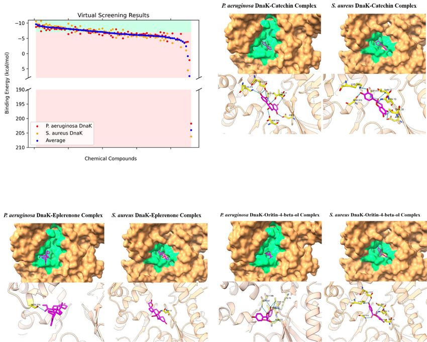
www.nature.com/scientificreports/
Zones of inhibition (mm)
Species PC SC CF EA AC ET MT WT EA(D) AC(D)
B. subtilis 30.00 ± 0.70 – – – – – – – 8.80 ± 0.65 9.39 ± 0.43
MRSA – – – – – – – 8.58 ± 0.29 –
S. pyogenes – – – – – – – – 8.35 ± 0.45
P. aeruginosa – – – – – – – – 8.00 ± 0.01
K. pneumoniae – – – – – – – – –
S. enterica – – – – – – – 6.60 ± 0.28 –
Table 1. Antibacterial activity of the N. lappaceum sequential and direct crude extracts via disc diffusion.
“–” no activity, PC: positive control (Gentamicin 10 µg), SC: solvent control (DMSO < 1%), CF: chloroform,
EA: ethyl acetate, AC: acetone, ET: ethanol, MT: methanol, WT: water, all these are sequential. EA(D): ethyl
acetate direct; AC(D): acetone direct. The data is expressed as the mean ± standard error of two independent
experiments performed in technical triplicates.
For instance, Malini & Maheshkumar15 have disclosed significant antibacterial activity of rambutan fruit sap
extracts towards Pseudomonas aeruginosa while Bhat and Al-daihan16 revealed antibacterial activities of its seeds
extracts against Staphylococcus aureus, Streptococcus pyogenes, Bacillus subtilis, Escherichia coli and P. aeruginosa.
Moreover, the antibacterial potential of rambutan peel extracts have also been reported against Vibrio cholerae,
Enterococcus faecalis, S. aureus and Staphylococcus epidermidis17. Furthermore, Sekar et al.18 comparatively evalu-
ated the antibacterial potency of red and yellow rambutan fruit peels against S. aureus and S. pyogenes, to reveal
better efficacy of the latter extracts against the tested pathogens. However, a deeper exploration of the potential
extracts to unveil new antibacterial compounds have hardly been focused.
In this study, thus, we have delineated a stepwise approach of determining the efficacy of the crude extracts
of the epicarp of yellow Malaysian rambutan against clinically important MDR bacterial pathogens e.g., B.
subtilis, methicillin-resistant S. aureus (MRSA), S. pyogenes, P. aeruginosa, K. pneumoniae and S. enterica. To
this end, through HPLC, LC–MS and GC–MS analyses, we have prepared chemical profiling of the ethyl acetate
and acetone fractions of the crude extracts with promising antibacterial activities. This revealed their potential
chemical determinants which were screened virtually to pharmacologically unveil novel bioactive compounds
in parallel to molecular dynamics simulation. This enabled us to elect compounds like catechin, eplerenone and
oritin-4-beta-ol, which computationally inhibit the important chaperone protein, DnaK of P. aeruginosa and
MRSA. Essentially, this report serves to unveil these compounds as novel alternatives to cope with the multidrug-
resistant gram-positive and -negative pathogens.
Results
Variable antibacterial activity of crude extracts by disc diffusion. We have preliminarily screened
the antibacterial activities of the yellow-variety Malaysian Rambutan epicarp crude extracts through disc dif-
fusion assay. We have only used freshly prepared solutions of crude extracts for all the tested pathogens (TP).
During such trials, the solvent control (SC), DMSO, did not exercise antibacterial activity against the TP, as
manifested by a no inhibition zone. Moreover, none of the sequential extracts exhibited activity against the TP
(Table 1), while in the case of direct extracts, the picture was different. Extracts of ethyl acetate (EA) displayed
visible activity against MRSA, B. subtilis and S. enterica but did not show activity against the rest of the TP (RTP).
Again, acetone (AC) extracts exhibited markedly prominent activity against B. subtilis along with visible activity
against S. pyogenes and P. aeruginosa, which was not observed among the RTP (Table S1).
Antibacterial screening of crude extracts via Broth dilution. Next, we have used the broth dilu-
tion method for evaluating the antibacterial potential of yellow fruit epicarp crude extracts and calculating the
viability percentage of every TP (Table 2). This, in turn, helped us to illustrate the percentages of antibacterial
potential of EA and AC fractions from the sequential & direct extracts, against the TP, at a concentration of
250 µg/ml. For the sequential extracts, the fraction from AC exhibited the highest percentage (90) of antibacte-
rial activity against P. aeruginosa followed by 71% against MRSA while that from EA showed a 59% effect against
S. pyogenes. No significant results were observed either against the RTP (Fig. S3A–F) or for the remaining solvent
extracts namely, chloroform (CF), ethanol (ET), methanol (MT) and aqueous (water, WT) extracts against all
TP (Table S2). In the case of direct extracts with the six solvents used, fractions of EA exhibited inhibition of
80% against B. subtilis along with 60, 62, 73 and 72% for MRSA, P. aeruginosa, S. enterica and K. pneumonia,
respectively, without any positive results against S. pyogenes. Notably, all tested pathogens were inhibited by AC
fractions and the percentage of antibacterial effects were 75, 90, 70, 70, 60 and 75, respectively for MRSA, B.
subtilis, S. pyogenes, P. aeruginosa, S. enterica and K. pneumonia (Fig. S1A–D).
The results of EA and AC fractions portrayed notable antibacterial efficiency. We have, thus, utilized them
for the identification of bioactive compounds via HPLC, LC–MS and GC–MS analyses.
Revelation of antioxidants from crude extracts using HPLC–UV. At first, we have conducted
HPLC–UV, for preliminary identification of the basic antioxidants present in the Malaysian yellow-rambutan
epicarp extracts. All compounds with known antioxidant capacities were identified in comparison with standard
Scientific Reports | (2021) 11:13859 | https://doi.org/10.1038/s41598-021-92622-0 2
Vol:.(1234567890)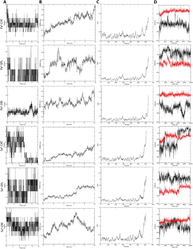
www.nature.com/scientificreports/

                                   % Inhibition of all tested pathogen
                                   Microorganisms              S.C                 P.C                 EA(S)       EA(D)       AC(S)       AC(D)
                                   B. subtilis                 02 ± 0.00           100 ± 0.00               –      80 ± 1.98       –       90 ± 1.42
                                   MRSA                        02                  100                      –      60 ± 1.49   71 ± 1.91   75 ± 2.04
                                   S. pyogenes                 02                  100                 59 ± 1.33       –           –       70 ± 1.40
                                   P. aeruginosa               02                  100                      –      62 ± 1.83   90 ± 1.58   70 ± 2.02
                                   K. pneumonia                02                  100                      –      72 ± 1.79       –       75 ± 2.61
                                   S. enterica                 02                  100                      –      73 ± 1.84       –       60 ± 2.74

                                  Table 2.  Screening of antibacterial effect of sequential and direct extracts by broth dilution method. EA(S):
                                  ethyl acetate sequential; AC(S): acetone sequential; EA(D): ethyl acetate direct; AC(D): acetone direct; S.C:
                                  solvent control: DMSO (< 1%), P.C: positive control (Gentamicin 10 µg); – : Low activity (less than 50%).
                                  The data is expressed as the mean ± standard error of two independent experiments performed in technical
                                  triplicates.

                                   Sample extract     RT (min)       Identified compounds       Peak area
                                                      12.9           Malic acid                   40.441
                                   EA (S)              4.8           Vitamin C                  1717.201
                                                       6.2           Chlorogenic acid            190.156
                                                       2.9           Malic acid                    4.938
                                                       4.8           Vitamin C                  1226.316
                                                       6.2           Chlorogenic acid            134.587
                                   AC (S)
                                                       8.8           Epigallocatechin gallate    205.717
                                                      10.7           Quercetin                    85.112
                                                      20.7           Catechin hydrate             44.169

                                  Table 3.  Identified compounds in N. lappaceum ethyl acetate and acetone sequential fractions using HPLC–
                                  UV.

                                  phenolic compounds. The identified compounds and their quantification, along with their specific peak posi-
                                  tion and retention time (Rt) in the chromatogram, are shown in Table 3 and Fig. S2. We identified only three
                                  compounds in the EA extract namely, malic acid, vitamin C and chlorogenic acid along with three more in the
                                  AC extract. These are epigallocatechin gallate, catechin hydrate and quercetin.
                                     EA (S): Ethyl acetate sequential fraction; AC (S): Acetone sequential fraction.

                                  Exploration of other chemical determinants through Liquid Chromatography–Mass Spec‑
                                  trometry (LC–MS) Analysis. For a fast, mass-directed exploration of the compounds possibly present in
                                  the rambutan epicarp, we have subjected the EA and AC sequential crude extracts to LC–MS analysis (Figs. S3
                                  & S4). Our analysis revealed the presence of 54 and 44 compounds, respectively, in the above-mentioned EA
                                  and AC extracts (Tables S3 & S4). We have matched them with the identity of known molecules on the Metlin
                                  database, keeping a threshold of Molecular Formula Generator (MFG) scores above 86% along with a ± 2% dif-
                                  ference. We further explored the compounds above the mentioned cut-off for their biological activities and car-
                                  ried forward for virtual screening. Notably, 31 compounds from both sets of EA and AC extracts have not been
                                  reported to date with any antibacterial activities (Tables S3 & S4).

                                  Identification of volatile constituents by Gas chromatography–mass spectrometry (GC–
                                  MS). Hereafter, to identify any volatile organic compounds, present in the EA and AC extracts of Malaysian
                                  yellow-variety N. lappaceum epicarp, we have exposed them for GC–MS analysis (Fig. S5a–d). Most of the com-
                                  pounds from EA and AC fractions, extracted directly, have been reported with antibacterial activities (Table 4).
                                  On the contrary, the compounds of these fractions from sequential extraction have not been reported for any such
                                  activity. The chromatogram of these compounds showed mentionable area % scores (above 0.5%) for 3-Methyl-
                                  1,2-diazirine (compound 1) and Card-20(22)-enolide, 3-[(6-deoxy-3,4-O-methylenehexopyranos-2-ulos-l-yl)
                                  oxy]-5,11/14-trihydroxy-12 -oxo-, (3-beta, 5-alpha, 11-alpha) (compound 2) in the EA extract while, the AC
                                  extract showed the presence of Silane, [[(3alpha,5beta,20S)-pregn-11-ene-3,11,17,20-tetrayl] tetrakis(oxy)] tet-
                                  rakis [trimethyl] and 2,2-Bis[4-[(4,6-dichloro-1,3,5-triazin-2-yl) oxy] phenyl]-1,1,1,3,3,3-hexafluoropropane
                                  (Table 4). Of these, compound 2 is known as Eplerenone (Fig. 1) and was found to be an important one in the
                                  upcoming analyses.

                                  Short listing of antibacterial compounds via virtual screening and pharmacokinetics. We
                                  have inspected the quality of DnaK protein homology models, all of which had good steric properties (Fig. S6).
                                  Moreover, we have validated the method for virtual screening via the redocking approach, with minimal RMSD

Scientific Reports |   (2021) 11:13859 |                     https://doi.org/10.1038/s41598-021-92622-0                                                     3

                                                                                                                                                       Vol.:(0123456789)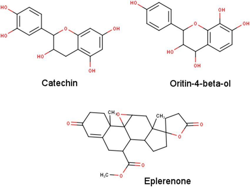
www.nature.com/scientificreports/
No Extracts Identified compounds Molecular Formula R.T. (Min) Area % Antibacterial activity report
1 3-Methyl-1,2-diazirine C2H4N2 3.02 0.776 Not reported
EA (S) Card-20(22)-enolide, 3-[(6-deoxy-3,4-O-methylenehexopyranos-2-ulos-l-yl)
2 C30H40O11 3.00 3.073 Not reported
oxy]-5,11/14-trihydroxy-12 -oxo-, (3- beta.,5-alpha., 11-alpha.)
Silane, [[(3alpha,5beta,20S)-pregn-11-ene-3,11,17,20-tetrayl] tetrakis(oxy)]
3 C33H66O4Si4 41.85 5.257 Not reported
tetrakis [trimethyl]
AC (S)
2,2-Bis[4-[(4,6-dichloro-1,3,5-triazin-2-yl) oxy] phenyl]-1,1,1,3,3,3-hexafluor-
4 C21H8Cl4F6N6O2 3.00 4.241 Not reported
opropane
1 Phenol, 2,4-bis(1,1-dimethylethyl) C14H22O 10.74 38.698 19
2 Curlone C15H22O 14.44 4.233 20
3 Ar-tumerone C15H20O 18.65 0.857 21
4 Stigmasterol C29H48O 17.87 1.66 22
5 EA (D) n-Hexadecanoic acid C16H32O2 48.78 3.2 23
6 3,7,11-Tridecatrienenitrile, 4,8,12-trimethyl C16H25N 23.94 2.567 Not reported
7 2-Methoxy-1,3-dioxolane C4H8O3 39.4 0.563 Not reported
5H-Cyclopropa (3,4) benz(1,2-e) azulen-5-one, 1,1a-à,1b-á,4,4a,7a-à,7b,8,9,9a-
8 decahydro-7b-à,9-á,9a-à-trihydroxy-3-hydroxymethyl-1,1,6,8-à-tetramethyl- C41H66O8 58.69 2.34 Not reported
4a-methoxy-, 9,9a-didecanoate
1 Phenol, 2-methoxy-3-(2-propenyl) C10H12O2 10.73 0.678 Not reported
2 Phenol, 2,4-bis(1,1-dimethylethyl) C14H22O 14.44 0.912 19
3 Alpha-Tocopherol C31H52O3 44.65 1.612 24
AC(D)
4 Curlone C15H22O 18.65 1.156 20
5 1,3-Dioxolane, 2-pentadecyl C21H40O4 57.36 3.06 Not reported
6 Ar-tumerone C15H20O 17.87 4.081 21
Table 4. Compounds existing in N. lappaceum ethyl acetate and acetone (sequential & direct) extract
identified by GC–MS analysis. EA(S): Ethyl acetate sequential; AC(S): Acetone Sequential; EA(D): Ethyl acetate
direct; AC(D): Acetone direct.
Figure 1. 2D Chemical Structures of Catechin, Oritin-4-beta-ol, and Eplerenone generated using MarvinSketch
version 20.16 (http://www.chemaxon.com/)31.
values (1.715 Å for 4B9Q and 1.427 Å for 4JNE) between reference and docked poses (Fig. S8). Upon virtual
screening of the 91 chemical compounds obtained via chromatographic analyses, we considered 41 of them,
with a binding energy of less than − 7 kcal/mol, as good binders (Table S5; Figs. 1, 2A). Among these, we have
chosen potential drug candidates based on their predicted pharmacokinetic properties. For example, we prior-
itized good gastrointestinal (GI) absorption, bad BBB permeability, and non-P-glycoprotein (PGP) substrates
for absorption properties. For metabolism, we prioritized non-cytochrome P450 inhibitors. Besides, we avoided
violations of drug-likeness rules. To cater to the need, we considered five druggability rules, namely, the Lipinski,
Ghose, Veber, Egan, and Muegge r ules25–29. Lastly, we also prioritized higher bioavailability scores. The Abbot
Bioavailability Score utilized herein was to predict chances of drug bioavailability to be more than 10% upon
oral intake30.
Among the virtually screened compounds, we found that catechin (C), eplerenone (E) and oritin-4-beta-ol
(O) stood out to be good binders with their average binding energies being − 8.205, − 7.980 and − 7.190 kcal/
Scientific Reports | (2021) 11:13859 | https://doi.org/10.1038/s41598-021-92622-0 4
Vol:.(1234567890)www.nature.com/scientificreports/
A. B.
C. D.
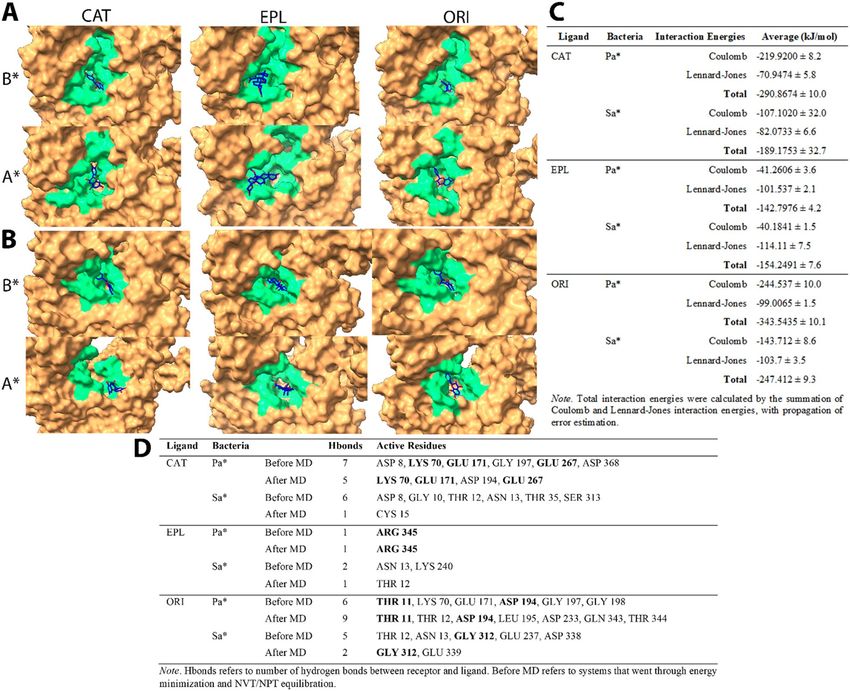
E. ACTIVE RESIDUES
Chemicals DnaK Hbonds Active Residues
Catechin P. aeruginosa 8 ASP 8, THR 11, THR 12, GLY 197, GLU 267, LYS 270, ASP 368
S. aureus 7 ASP 8, THR 12, ASN 13, THR 35, GLY 171, LYS 241, SER 313
Eplerenone P. aeruginosa 1 SER 274
S. aureus 1 ASN 13
Oritin-4-beta-ol P. aeruginosa 9 THR 11, THR 12, ASN 13, LYS 70, GLY 197, GLY 198
S. aureus 9 THR 12, ASN 13, THR 35, GLU 237, LYS 241, GLY 312
Note. Hbonds refers to number of hydrogen bonds between receptor and ligand.
Figure 2. Results of virtual screening targeting P. aeruginosa and S. aureus DnaK proteins. (A) Distribution of
binding energies plotted via M atplotlib32 against screened compounds. Top binders with good pharmacological
properties, namely (B) Catechin, (C) Eplerenone, and (D) Oritin-4-beta-ol, generated using UCSF ChimeraX33.
P2Rank predicted binding pockets were colored in green for visualization of ligands (magenta) binding to
the receptor DnaK proteins (brown), with active sites (yellow) labelled. (E) Intermolecular hydrogen bonds
tabulated with active residues listed.
Scientific Reports | (2021) 11:13859 | https://doi.org/10.1038/s41598-021-92622-0 5
Vol.:(0123456789)www.nature.com/scientificreports/
Figure 3. MD simulation of (A) P. aeruginosa and (B) S. aureus DnaK (brown)-Catechin(blue) complexes, of
which P2Rank predicted druggable pocket residues were colored in green for better visualization. (C) Tabulated
interaction energy values of DnaK-Catechin complexes. (D) Tabulated changes in number of hydrogen bonds
and active residues before and after MD. Note B*—Before MD, A*—After MD, Pa*—P. aeruginosa, Sa*—S.
aureus, CAT—Catechin, EPL—Eplerenone, ORI—Oritin-4-beta-ol. All data were generated using G ROMACS34
in-built functions.
mol, respectively for S. aureus (Sa) and P. aeruginosa (Pa) DnaK proteins. C, E, O also exhibited good predicted
pharmacological properties except that C is a PGP substrate (Fig. 1, Table S5). The binding conformations of
C (Fig. 2B), E (Fig. 2C), and O (Fig. 2D) to both DnaK proteins of Sa (SaD) and Pa (PaD) showed potential
structural competitive inhibition of ATP binding at the docking pocket. Moreover, we observed rich electrostatic
interactions (Fig. 2E) in C and O, but not in E, having only one intermolecular hydrogen bond.
Validation of inhibitory effects of selected compounds by Molecular Dynamics Simulation. To
this end, we carried out Molecular Dynamics (MD) simulations for 10 ns for C, E, and O Ligand-SaD/PaD com-
plexes to observe ligand-receptor interactions. Throughout MD simulations, the ligands were retained in the
docking pocket of respective DnaK receptors, except for C in the SaD system (CSaD) of which the ligand seemed
to be escaping from the initial binding pocket (Fig. 3A, B). Moreover, the upper part of the DnaK NBD domain
was completely disintegrated in CSaD. Besides, the total number of receptor-ligand intermolecular hydrogen
bonds were maintained stably at around 4 and 5 in P. aeruginosa DnaK complexed with C (CPaD) and O (OPaD)
respectively, and 4 in S. aureus DnaK complexed with O (OSaD) (Fig. 4A). Moreover, both the E complexes of
SaD (ESaD) and PaD (EPaD) have maintained the total number of hydrogen bonds at around 1. However, in
CSaD, we observed a sharp decline in the number of intermolecular hydrogen bonds at the 5 ns time point from
around 4 to between 0 and 1, which can explain the escape of ligand from its initial docking pocket. We also
observed stable active residues in CPaD (LYS 70, GLU 171, GLU 267), EPaD (ARG 345), and OPaD (THR 11,
ASP 194) complexes, as well as in OSaD (GLY 312) complex (Fig. 3D).
In all MD simulation systems, we found that the root-mean-square fluctuations of the DnaK receptor main-
tained at around 0.5 nm (5 Å) except for the C-terminal end where the disordered regions were localized
Scientific Reports | (2021) 11:13859 | https://doi.org/10.1038/s41598-021-92622-0 6
Vol:.(1234567890)www.nature.com/scientificreports/
Figure 4. Receptor-ligand interactions over the course of MD simulation, of which (A) number of hydrogen
bonds between receptor and ligand, (B) RMSD values of Catechin, (C) RMS fluctuation-per-residue of receptor
macromolecule, and (D) receptor-ligand interaction energies (Black: Coulombic interaction energies, Red:
Lennard–Jones energies) were computed over the course of MD. Note Pa*—P. aeruginosa, Sa*—S. aureus,
CAT—Catechin, EPL—Eplerenone, ORI—Oritin-4-beta-ol. All data were generated using G ROMACS34 in-built
functions.
Scientific Reports | (2021) 11:13859 | https://doi.org/10.1038/s41598-021-92622-0 7
Vol.:(0123456789)www.nature.com/scientificreports/
(Fig. 4C). Besides, we observed that the RMSD of C and E were maintained at 0.5 nm in the PaD receptor, as
well as higher RMSD values of around 0.8 nm in CSaD and ESaD (Fig. 4B). The ligand RMSD were relatively
lower in O, compared to others, which was 0.4 nm in both SaD and PaD cases. The interaction energies of all
systems maintained stably throughout the simulation, except for the CSaD complex of which a sharp decrease
of Coulomb potential can be observed at around 6 ns time point (Fig. 4D). In general, E maintained the lowest
total interaction energies, followed by C and O (Fig. 3C).
Discussion
Over the years, the commendable development in the field of virtual screening has enabled time- and cost-
efficient drug discovery along with r epurposing35. Herein, we have carried out a scaffolded approach to antimi-
crobial drug discovery from a yellow variety of Malaysian N. lappaceum L. fruit epicarp crude extracts. The first
upstream set of experimental work comprised the extraction of the plant product, followed by characterization
of their antimicrobial property and chromatographic identification of chemical compounds from therein. This
was coupled with a downstream set of computational analyses comprising virtual screening and pharmacological
predictions of extracted chemical compounds against potential drug targets. To this end, molecular dynamic
simulation has taken a step forward to uncover new potent bioactive compounds which can target both gram-
negative and -positive bacteria at the same time. Our study delineates a method to uncover potent chemicals
which might have contributed to the antibacterial activities of plant products like Nephelium lappaceum epicarp,
to be further utilized for drug discovery, repurposing, or other ab initio synthetic enhancements.
Extraction is the key stage to obtain the diverse bioactive chemical compounds from plant products. These
chemical determinants display different solubility with different organic solvents such that screening with dif-
ferent solvents helps to bring forth the best one for further exploration36. Thus, we have utilized several organic
solvents to explore the extraction of biologically active constituents. Herein, we initiated a sequential extraction
process of utilizing solvents like chloroform (CF), ethyl acetate (EA), acetone (AC), ethanol (ET), methanol (MT)
and water (WT), in order of their increasing polarity. Our study revealed that the yellow variety of Malaysian N.
lappaceum epicarp crude extracts exhibited varied inhibitory activities against the six tested MDR pathogens,
namely, B. subtilis, methicillin-resistant S. aureus (MRSA), S. pyogenes, P. aeruginosa, K. pneumoniae and S.
enterica. Essentially, the EA(S) and AC(S) fractions notably inhibited the Gram-positive S. pyogenes and MRSA
and the Gram-negative P. aeruginosa while the remaining solvent fractions responded moderately or poorly.
This provided a strong clue for us to proceed for further direct extraction from EA(S) and AC(S) crude extract
fractions (CEF). Thereafter, following the chromatographic analyses of HPLC, LC–MS and GC–MS of these
EA(S) and AC(S) CEF, we obtained different results from the direct extract fractions of EA and AC. Interestingly,
we found the CEF of EA(D) and AC(D) to be more efficient via broth dilution than the disc diffusion methods
(Tables 1 and 2). This could be attributed to the following fact. The different constituents of the CEF need to
diffuse slowly in agar from a liquid to solid interphase in the agar diffusion method compared to the complete
liquid interphase for broth microdilution.
It is important to note that despite similar reports to our findings by Mohamed et al.37; Thitilertdecha
et al.38and Tadtong et al.39, a comprehensive chemical profiling to unearth plausible determinants, potential
enough against the MDR pathogens, is lacking to date. Thus, based on the prominent antibacterial effects of the
EA(S) and AC(S) extracts of N. lappaceum fruit epicarp, we perceived these two fractions to harbor important
bioactive molecules. Therefore, we subjected the sequential extracts of EA(S) and AC(S) fractions to HPLC
analysis. The results confirmed the presence of some standard phenolic compounds with antioxidant proper-
ties namely, malic acid, vitamin C, chlorogenic acid, epigallocatechin gallate, quercetin and catechin hydrate
(Table 3). Notably, we found ethyl acetate and acetone as the competent solvents to extract total flavonoid and
phenolic compounds36. Nazir et al.40,41, however, reported the afore-mentioned compounds in various other
organic solvent extracts of Silybum marianum and Elaeagnus umbellate.
To this end, an extensive spectrum of chemical classes was revealed after LC–MS analysis and included,
terpenes, alkaloids, polyunsaturated and monounsaturated fatty acids among others, that were present in both
the extracts. Among these, only 21 of the 54 compounds (with above 86% MFG scores) of the EA(S) fractions
have been reported to possess antibacterial activities (Table S3). Interestingly, most of them have been newly
reported (within the last five years) including L242, L343, L644, L1046, L1247, L1648, L2049, L2251, L2652, L2853,
L3154, L4658, L4859, L5160, L5361 and L5459. Others are known for some time, namely, L745, L2150, L3555, L3756 and
L4557. Similarly, the AC(S) extract fractions contained only 10 from 44 compounds with reported antibacterial
activities (Table S4). Of these, except for L 2455 and L
3558, all were reported recently. These included L 244, L562,
L1048, L1352, L1954, L3658, L3859 and L 4261. Thus, possibilities exist for those 35 and 26 compounds from EA(S)
and AC(S) fractions, respectively, with no matched identity with the library (Table S6 & S7) to be medically
important, though, further characterization is required to evaluate their usages.
We have authenticated a further revelation of important biomolecules through GC–MS (Table 4) besides
the HPLC and LC–MS analyses mentioned above in Tables 3 & S3–S4 respectively. Notably, we analyzed both
the sequential and direct extracts of EA and AC fractions through GC–MS. Unlike the LC–MS reported com-
pounds, however, about 50% of the chemical components, unearthed through GC–MS, are unknown for their
antibacterial activity. For instance, in the case of sequential extracts, for both the EA(S) and AC(S) fractions, only
4 molecules were detected (with area % scores above 0.5%) without any a priori antibacterial activity (Table 4).
Likewise, for the direct extract fractions of EA(D), only 5 out of the 8 compounds detected (with area % scores
above 0.5%) were known to possess such activity. These are D GEA119, D GEA220, D GEA321, D GEA422 and
DGEA523. However, for the AC(D) extract fraction, 4 out of the total 6 compounds detected, were reported to
possess the antibacterial effect. These are D GAC219, DGAC324, DGAC420 and DGAC621.
Scientific Reports | (2021) 11:13859 | https://doi.org/10.1038/s41598-021-92622-0 8
Vol:.(1234567890)www.nature.com/scientificreports/
With a set of 91 compounds obtained through chromatographic analyses, we have conducted a compu-
tational analysis for virtual screening through molecular docking to shortlist a selective set of chemicals via
pharmacokinetics consideration (Table S5). Pharmacokinetics is an important criterion when it comes to drug
discovery and drug design, especially about bioavailability and toxicity. Herein, we have considered several
parameters for absorption, metabolism, drug-likeness, and bioavailability for selecting the ideal drug for poten-
tial pharmacological application in the future. For instance, good GI absorption can allow absorption into the
bloodstream during oral consumption, while bad blood–brain barrier (BBB) permeability can avoid interrup-
tion to the central nervous system63. P-glycoprotein (PGP) substrates are being actively effluxed from the cells
thereby resulting in low absorption into the blood circulation64,65. Besides these, cytochrome P450 enzymes are
crucial in the metabolism of most clinical drugs. Hence, the inhibition of cytochrome P450 enzymes can lead
to decreased drug metabolism and possibly, adverse health complications, due to drug-drug interaction upon
co-prescription with other drugs66,67. Moreover, the drug-likeness rules, such as the Lipinski rule of five, work
by predicting pharmacological behavior upon oral administration based on the chemical properties of potential
drugs25. Lastly, bioavailability takes consideration of both absorption and distribution of the drugs, of which the
eventual presence in blood circulation upon oral consumption is evaluated.
DnaK protein belongs to the 70 kDa heat shock protein (HSP70) family, which functions as a molecular
chaperone, mediated by its ATPase a ctivities68. DnaK protein has been reported to be central in mediating
bacterial stress responses. Among these, DnaK mutants have manifested an increase in antimicrobial suscepti-
bilities and a decrease in survivability in the h ost69–71. Moreover, our previous work on whole-genome analysis
(WGA) of protein interaction network (PIN) reported that DnaK protein was crucial in mediating quorum
sensing in multidrug-resistant Proteus mirabilis72. Furthermore, WGA analyses of PIN from MDR pathogens
like P. aeruginosa, S. aureus, S. enterica, S. pneumoniae, P. mirabilis, Acinetobacter baumannii, Escherichia coli
and Mycobacterium tuberculosis revealed DnaK to be among the top 10 crucial proteins indispensable for the
acteria73. Also, the ATP-binding pocket of the DnaK chaperone has been indicated to
cellular integrity of the b
be druggable and shown promise to cope with MDR in both gram negatives and positives as observed from an
unpublished work of the same group of researchers. Hence, DnaK protein has been selected for the in-silico study,
herein, as a promising drug target for MDR bacteria by inhibiting its ATP binding pocket, which can result in
its impairment of chaperone function.
Through our computational screening of the chemical libraries of the N. lappaceum L. fruit epicarp extrac-
tions, we have shortlisted Catechin (C), Eplerenone (E), and Oritin-4-beta-ol (O) as the promising antimicrobi-
als in combating the MDR pathogens by dint of their capacity in targeting the DnaK protein and having good
pharmacological profiles. Despite being a PGP substrate, C has manifested a strong binding affinity to DnaK and
therefore, can result in effective DnaK functional inhibition with a small amount. Otherwise, PGP inhibitors
like C can be co-prescribed easily as it has a good metabolic profile. Moreover, C has been well-characterized
for its antibacterial activities and known for its ability to cause leakage of bacterial cellular contents along with
increased intracellular reactive oxygen species production in both gram negatives and p ositives74,75. However,
the biological targets of C have not been described. As DnaK protein is crucial in bacterial stress response, by
inhibiting the DnaK chaperone function, the bacterial cellular and biomolecular integrity can be effected upon
receiving environmental oxidative stress. Herein, we showed that in P. aeruginosa, C could bind stably to the
ATP-binding pocket of DnaK throughout the MD simulation with 3 stable active residues (LYS 70, GLU 171,
and GLU 267), while maintaining the ATP-bound conformation of the DnaK protein without the necessity for
ATP binding (Figs. 2B, 3A). This reflected the inability of the ATP molecules to bind the CPaD (Catechin-bound
DnaK protein of P. aeruginosa) as also a complete halting of the normal DnaK chaperone function via confor-
mational changes ensuing ATP hydrolysis. However, C could not inhibit SaD (DnaK of S. aureus) the same way,
due to its inability to maintain the integrity of the NBD domain and thereby escaping from the binding pocket.
It is this binding pocket that allows subsequent binding of ATP molecules on DnaK to continue the chaperone
function. On the contrary, herein, we present the discovery of two novel potential compounds, E and O, whose
antibacterial activities have not been reported and/or described earlier. Notably, E has been widely utilized in
cardiovascular implications and as diuretics52,76. O, however, has not been explored to confer any biological
significance. Despite that, it is notable that the chemical structure of O is analogous to C (Fig. 1), with the sites
of hydroxylation being slightly different.
Throughout the molecular dynamics simulation (MDS) processes, we can only observe 1 or 2 hydrogen bonds
in EPaD and ESaD, which suggested weak protein–ligand electrostatic interactions. This can be explained by the
chemical structure of E, being crowded with carbonyls and ethers which are weak bases, and hydroxyl groups are
lacking. The ligand, however, has been retained in the docking pocket throughout MDS. This probably suggests
that hydrophobic (van der Waals) interactions were dominant in this case. This was reflected through the inter-
molecular interaction energies (Fig. 4D), of which the Lennard–Jones potentials were much higher than Coulomb
potentials in Eplerenone-DnaK (ED) complexes, while the reverse was observed in for C and O. Moreover, the
binding conformation of E in PaD did not “cover-up” completely at the binding site of phosphate groups of the
ATP for which further wet-lab confirmation is required. Furthermore, among the three ligands simulated, O
manifested the best binding capabilities to both PaD and SaD with rich intermolecular electrostatic interactions
and the highest total interaction energies. After MDS, the active residues THR 11 and ASP 194 were retained
in OPaD, while GLY 312 was retained in OSaD. Again, despite being structurally analogous to C, O manifested
good predicted pharmacological properties in all the aspects considered. Therefore, with better binding capa-
bilities to DnaK receptor and pharmacological properties, herein we report O to be a more potent antibacterial
compound compared to the well-known C, which is active against both the gram-positive and -negative bacteria.
In the end, E, the compound not reported earlier to exhibit antibacterial properties against the tested promis-
ing pathogens MRSA and P. aeruginosa, demands a separate focus. Importantly, E has been found in both the
EA(S) and AC(S) CEF. However, the EA(S) showed no activity compared to the AC(S) CEF. This might probably
Scientific Reports | (2021) 11:13859 | https://doi.org/10.1038/s41598-021-92622-0 9
Vol.:(0123456789)www.nature.com/scientificreports/
be attributed to the interference of other chemicals in that EA(S) which might not have been the case for AC(S),
probably, facing no interference and thus, showing activities. Thus, E can be a probable candidate as projected
through our in-silico studies comprising screening of pharmacological properties followed by molecular dynam-
ics simulation.
Conclusion
Our findings reinstate the promising antibacterial effects, of the yellow variety of Malaysian Rambutan (N. lap-
paceum L.) fruit epicarp crude extracts, against selected Gram-positive and Gram-negative MDR pathogens.
In this context, particularly ethyl acetate and acetone (sequential and direct) extracts demonstrated remarkable
antibacterial effects toward at least MRSA and P. aeruginosa among the six tested MDR pathogens, while remain-
ing fractions including, chloroform, ethanol, methanol and water did not exhibit such potential. Nevertheless,
we present the epicarp of N. lappaceum as a novel source for antibacterial compounds projecting catechin,
eplerenone and oritin-4-beta-ol with high potential for the development of pharmaceutically valuable future
drugs. Further studies are mandatory to separate the specifically mentioned three compound(s) responsible for
the desired effects and to develop our knowledge on the other unseen potentials in N. lappaceum.
Materials and methods
Solvents. For the preparation of crude extracts, we have used all solvents, of HPLC grades. In the order of
increasing polarities, these were Chloroform (99.9%, Sigma-Aldrich, LiChrosolv, Malaysia), Ethyl acetate, Ace-
tone (99.5% Chemiz, Malaysia), Ethanol, Methanol (99.8%, ChemAR, Systerm, Malaysia) and double distilled
Milli-Q Type 1 water (MilliporeMerck, Germany). For LC–MS and GC–MS studies, we have used the solvents
of MS grades.
Plant product. We purchased the yellow variety fruits of N. lappaceum L. from the local marketplace, Bandar
Sunway, Selangor, Malaysia. We prepared the Herbarium voucher and deposited at Sunway University, Selangor
Darul Ehsan, Malaysia. Thereafter, for our research, we carefully observed the fruit characters of rambutan and
selected the epicarp according to the Descriptor for Rambutan77.
Tested microorganisms.For our study, we obtained six clinical isolates from the Department of Biologi-
cal Sciences, Sunway University, Malaysia. These were Streptococcus pyogenes (ATCC-49399), Bacillus subtilis
(ATCC-11774), methicillin-resistant Staphylococcus aureus (MRSA) (MTCC-381123), Pseudomonas aeruginosa
(ATCC-10145), Klebsiella pneumoniae (ATCC-700603) and Salmonella enterica (ATCC-14028). We tested all
these six bacterial strains to be resistant to at least five of the ten antibiotics tested for their resistivity/sensitivity
profile and thus, considered them to be multidrug-resistant (Table S8).
Preparation of crude extracts. We prepared the epicarp crude extracts following the method of Do
et al.78 using the solvents mentioned earlier for the direct extracts. For the sequential method of extraction,
we used the mentioned solvents in the order of their increasing polarity viz. chloroform < ethyl acetate < ace-
tone < ethanol < methanol < water. In both cases, essentially, we removed the peels of N. lappaceum from the fruit
and washed thoroughly with running, followed by, distilled water to remove contaminants and thereafter dried
using a freeze-dryer. We ground the dried peels into a fine powder using an electric grinder. To produce differ-
ent fractions of crude extracts, we extracted 10 g of powder in 100 ml of selected solvents. Thereafter, we mixed
the solution thoroughly by using an incubator shaker (Yihder LM-530D Incubator Shaker, Taiwan) for 24 h.
To separate supernatant, we centrifuged the solution (Eppendorf 5810 R Centrifuge, Germany) at 4000 rpm
for 10 min at 4 °C to eliminate the leftover fine sediments. Finally, we concentrated the solvent extracts using a
Rotary evaporator, and further with a vacuum concentrator until a viscous extract was obtained. We stored all
the extracts at 4 °C for future experiments.
Potential in‑vitro antibacterial activities of yellow rambutan fruit epicarp extracts. Disc diffu-
sion assay. We consistently swabbed the seed culture of the tested pathogen on an agar plate. Then, we sepa-
rately impregnated sterilized blank paper discs with different crude extract fractions and placed them on the agar
plate. We incubated the plates at 37ºC for 16 h. We noted the antibacterial activity by measuring the diameter of
the inhibition zone. We used gentamicin (10 µg/disc) as positive control and kept DMSO (< 1%) as a negative
control. We ensured that all the experiments had technical triplicates and we performed them twice to render
two biological replicates.
Broth dilution assay. We used a broth micro-dilution method to evaluate the minimum inhibitory concentra-
tion (MIC) values of crude extracts using Clinical & Laboratory Standards Institute (CLSI) procedures. Essen-
tially, we added each extract (5 μl) into the wells of a 96 well plate comprising 1 05 CFU/ml bacterial cells. We
incubated the 96 well plates at 37 °C for 16 h. We kept the final concentrations ranging from 250 to 2000 µg/
ml. In each test, we included three controls comprising, gentamicin 10 µg/ml (as positive), DMSO < 1% final
concentration (as solvent) and bacterial inoculum (as negative). We have taken the MIC value as the lowest
concentration of the tested extract showing inhibitory effect against the pathogens, recorded via the Microplate
reader (TECAN, Infinite-M200-PRO). We confirmed all tests, having technical triplicates, twice. We observed
promising results for both the fractions of ethyl acetate and acetone extracts with which we carried out all chro-
matographic analyses.
Scientific Reports | (2021) 11:13859 | https://doi.org/10.1038/s41598-021-92622-0 10
Vol:.(1234567890)www.nature.com/scientificreports/
Statistical tests. In the present study, we performed all the tests in triplicates and expressed the data obtained as
the mean ± standard deviation (S.D). We determined the P values using the student’s T-test, two-tailed distribu-
tion, (*) is P ≤ 0.05. These have been reflected in Tables 1 and 2 and Figure S2.
Exploration of chemical constituents through chromatographic analyses. High‑performance
Liquid Chromatography (HPLC). We used the ethyl acetate and acetone extracts as samples for qualitative
phytochemical screening via HPLC via the Agilent-1260 infinity system, according to the reported method of
Zeb79. Briefly, we mixed one-gram sample extract in methanol and water (1:1; 20 mL; v/v) and heated at 70˚C
for 1 h in a water bath. We centrifuged this mixture at 4000 rpm for 10 min and filtered 2 ml of the supernatant
into HPLC vials through Whatman filter paper. We performed the separation via the Agilent-Zorbax-Eclipse
column (XDB-C18). Column gradients system comprised solvent B and C. Solvent B consisted of deionized
water: methanol: acetic acid having a ratio of 180: 100: 20; v/v while solvent C had deionized water: methanol:
acetic acid in the ratio of 80: 900: 20; v/v. We started the gradient system by solvent B for 100%, 85%, 50% and
30% at 0, 5, 20 and 25 min followed by solvent C (100%) from 30 to 40 min. Elution occurred after 25 min. We
set the ultraviolet array detector (UVAD) at 280 nm for the antioxidants analysis and documented the chroma-
togram using retention times. We carried out the UV spectra of compounds and accessible standards along with
quantification by taking the per cent peak area. We measured the quantity of the antioxidants by the formula:
Ax × Cs µg ml × V (ml)
Cx = (1)
As × Sample wt. in g
Cx = Sample concentration; As = Standard peak area; Ax = Sample peak area; Cs = Standard concentration (0.09 µg/
ml).
Liquid chromatography and mass spectrometry (LC–MS). We analyzed a mixture of standards and new metabo-
lites found in the ethyl acetate and acetone fractions via LC–MS, exactly as per the method reported by Yap
et al.80. To eradicate systematic errors, we used a reference solution with the two ions, with m/z of 121.0508 and
92,266.0097, being selected for mass calibration. Finally, we ran the mass spectra for the compounds present in
ethyl acetate (EA) and acetone (AC) fractions against the database of NIST (National Institutes of Standard and
Technology, Gaithersburg, MD, USA) Mass Spectral Search Program-2009 version 2 for the documentation of
homologous compounds over Agilent Mass-Hunter Qualitative Analysis B.05.00 software.
Gas chromatography–mass spectrometry (GC–MS). We subjected the ethyl acetate and acetone fractions to gas
chromatography-mass spectrometry (GC–MS) analysis, using Agilent technologies model 7890B GC System
coupled with Pegasus HT High Throughput TOFMS (Leco Corp., MI, USA). We injected an aliquot of an extract
of 1 ml into the GC–MS apparatus. Next, we used Agilent J&W HP-5MS (phenylmethyl siloxane, length 30 m,
Dia. 0.32 mm, Film, 0.25 µm) analytic column to separate components under an inert atmosphere of helium
(1.5 mL/min). Other standardized parameters utilized during the process: oven temperature of 80 °C (2 min)
was increased to a temperature of 300 °C at the rate of 3 °C/min, solvent delay time was 5 min, inlet line tempera-
ture was 225 °C, and ion source temperature was 250 °C. Mass spectra were taken at 70 eV and the acquisition
mode-scan was 20–1000 amu while sixty-four (64) minutes was the GC run time. We achieved the interpretation
of mass spectrum and the documentation of phytochemicals present in the fractions via the database of NIST
libraries.
Virtual screening of chemical determinants from chromatographic analyses. In silico protein
model generation. We have chosen S. aureus (Sa) and P. aeruginosa (Pa) as gram-positive and gram-negative
bacterial representatives for computational analyses of DnaK protein binding. 3D structures of DnaK proteins,
from the aforesaid species, were generated via homology modelling using MODELLER version 9.2481. DnaK has
two conformations, namely, the open or ATP-bound and the closed or ADP-bound conformation65. Herein, we
focused on the open conformation of DnaK, to identify potential competitive inhibitors of ATP to prevent the
proper functioning of DnaK protein.
We have obtained the protein sequences of Sa and Pa DnaK from UniProtKB with accession IDs of Q2FXZ2
and A6VCL882, respectively. To search for suitable homology modelling templates, we utilized both NCBI
BLASTp and the MODELLER in-built build_profile.py81,83. For Pa DnaK (PaD), the templates were full-length
ATP-bound E. coli (Ec) DnaK protein structures (PDB ID: 5NRO, Chain: A, Query Coverage (QC): 94%, Percent
Identity (PI): 79.50%, Resolution (R): 3.25 Å; PDB ID: 4JNE, Chain: A, QC: 94%, PI: 78.80%, R: 1.96 Å; and
PDB ID: 4B9Q, Chain: A, QC: 94%, PI: 77.96%, R: 2.40 Å). For Sa DnaK (SaD), besides the afore-mentioned Ec
DnaK (EcD) models, we selected one additional template, from Geobacillus kaustophilus DnaK protein (PDB ID:
2V7Y, Chain: A), due to the high percentage of sequence identity expected as per the gram-positive character
of S. aureus and G. kaustophilus. As this template structure was in the closed conformation and we were only
interested in the open conformation, only the Nucleotide Binding Domain (NBD, residues 1 to 350 in tem-
plate model) which does not differ much in both conformations, were taken into consideration for homology
modelling, and the remaining C-terminal residues modelling were guided by the Ec models to shape an open
conformation. Therefore, the templates for SaD were (PDB ID: 2V7Y, Chain: A, Template Residues: 1–350, QC:
57%, PI: 83.19%, R: 2.37 Å; PDB ID: 5NRO, Chain: A, QC: 93%, PI: 56.19%, R: 3.25 Å; PDB ID: 4JNE, Chain:
A, QC: 92%, PI: 55.54%, R: 1.96 Å; and PDB ID: 4B9Q, Chain: A, QC: 94%, PI: 55.43%, R: 2.40 Å). The template
sequences were aligned with the target sequence for homology modelling via the built-in function of MODEL-
LER (Fig. S6A). 5 homology models were generated for each protein of SaD and PaD, and the models with the
Scientific Reports | (2021) 11:13859 | https://doi.org/10.1038/s41598-021-92622-0 11
Vol.:(0123456789)www.nature.com/scientificreports/
lowest DOPE (discrete optimized protein energy) scores were selected for downstream virtual screening for
both. We then validated the SaD and PaD homology models via Swiss-Model Structure Assessment and SAVES
v5.0 servers84 (Fig. S6).
Druggable pocket validation. To validate the druggability of the ATP docking pocket, we have conducted ligand
binding site prediction using P2Rank from PrankWeb s erver85. P2Rank predicts the chemical druggability on
protein solvent-accessible surface via a non-templated machine learning approach. The ATP binding pocket was
predicted to be druggable and ranked first in both cases of SaD and PaD (Table S9; Fig. S7). Thus, we further
considered these pockets from the SaD and PaD complexes to be targeted for virtual screening.
Molecular docking with chemical determinants. We utilized the POAP p ipeline86 for an in silico virtual screen-
ing of the chemical compounds obtained through different chromatographic separation. We have obtained the
SMILES notations of these compounds, and generated their 3D models (in mol2 format) through the POAP
Ligand Preparation pipeline. To this end, we utilized Chimera to generate physiological protonation states of the
ligands, and PDBQT files were prepared87. We also carried out ligand optimizations via the POAP Ligand Prepa-
ration pipeline utilizing the MMFF94 force field which is being optimized for drug-like organic molecules and
molecular docking88. Out of the 50 conformers, generated for each ligand through the Weighted Rotor Search
approach, only the best conformers were retained. Finally, we have subjected the ligands to energy minimization
for 5000 steps by the conjugate algorithm.
We have prepared the macromolecule receptors, of the SaD and PaD proteins, using AutoDockTools. We
utilized AutoDock 4.2, aided by the POAP pipeline, for the virtual screening p rocess89. For AutoDock param-
eters, we have set 100 generations of Lamarckian Genetic Algorithm for each protein–ligand complex. To fit
in the previously predicted pocket, we adjusted the docking grids into squares of 24 Å with x, y, z coordinates
of 17.647, 75.43, 27.766, and 18.069, 74.299, 28.532, for SaD and PaD, respectively. For the silicon-containing
compound among the set of ligands, we separately carried out molecular docking with AD4.1_bound parameter
file, obtained from AutoDock, wherein we added the parameters for silicon atoms (Rii = 4.3; eii = 0.402)90.
We have validated the docking methodology via redocking of experimentally confirmed and deposited struc-
tures with the reference ligand (Fig. S8). Therein, we have retrieved the ATP-bound E. coli DnaK crystallized
structures from PDB (PDB ID: 4B9Q, CHAIN ID: A; PDB ID: 4JNE, CHAIN ID: A). The molecular docking
search grids were squares of 24 Å, in x, y, z coordinates of 108.958, 73.922, 100.622, and 19.081, 76.887, 31.16,
for 4B9Q and 4JNE respectively.
Pharmacological properties screening. Using SwissADME91, we have carried out predictions on the pharmaco-
logical properties, encompassing pharmacokinetics, drug-likeness, and molecular information, for each chemi-
cal compound.
Molecular dynamics simulation. Ensuing virtual and pharmacological screenings, we rationally selected poten-
tial drug candidates to undergo molecular dynamics (MD) simulation via GROMACS version 2019.334. We have
utilized the CHARMM36 force field of version July 2020, along with the TIP3P water model, for macromol-
ecule processing92. We used Avogadro software for mol2 format conversion and complete protonation (protona-
tion of non-polar atoms)93. We also used a Perl script, sort_mol2_bonds.pl, written by Justin Lemkul for bond
order arrangements in ligand mol2 files. Then, we generated the topologies of the ligand models through the
CGenFF server, and utilized a python script (cgenff_charmm2gmx.py) to convert topologies for CHARMM to
GROMACS94. We carried out solvation in a dodecahedron box ranged 1.0 Å from the protein–ligand complex.
The system was then ionized to achieve electrostatic neutralization. Subsequently, we subjected the system to
energy minimization via the steepest descent algorithm until convergence at a maximum force of less than
1000 kJ mol−1 nm−1 (Fig. S9). Herein, we have monitored the potential energy shifts of the systems.
We carried out equilibration of the systems via NVT and NPT ensembles for 50,000 steps (100 ps), with tem-
perature, pressure, and density shifts being monitored therein. Subsequently, we have carried out the production
MD simulations for 5,000,000 steps (10 ns) to observe protein–ligand interactions. We computed the RMSD
(Root Mean Square Deviation) values of ligands and receptors, number of hydrogen bonds between ligands and
receptors, and ligand-receptor interaction energies (Coulombic interaction energies and Lennard–Jones ener-
gies) throughout the MD simulations. We have also computed thetotal interaction energies, and estimated the
errorsvia error propagation by addition.
Generation of graphical illustrations. We utilized Matplotlib, a python library, to tabulate binding energies of all
screened compounds31. We generated all 3D structural images using UCSF ChimeraX32, and 2D chemical struc-
tures using MarvinSketch33. Finally, we retrieved the figures for MD simulation analyses from the GROMACS
in-built functions34.
Received: 9 December 2020; Accepted: 10 June 2021
Scientific Reports | (2021) 11:13859 | https://doi.org/10.1038/s41598-021-92622-0 12
Vol:.(1234567890)www.nature.com/scientificreports/
References
1. Maheshwari, M., Ahmad, I. & Althubiani, A. S. Multidrug resistance and transferability of blaCTX-M among extended-spectrum
β-lactamase-producing enteric bacteria in biofilm. J. Glob. Antimicrob. Resist. 6, 142–149 (2016).
2. Dockrill, P. The 12 deadliest drug-resistant bacteria have officially been ranked. Science Alert. https://www.sciencealert.com/the-
12-deadliest-drug-resistant-bacteria-have-officially-been-ranked (2017).
3. Obeng-Nkrumah, N. et al. High levels of extended-spectrum beta-lactamases in a major teaching hospital in Ghana: the need for
regular monitoring and evaluation of antibiotic resistance. Am. J. Trop. Med. Hygiene 89(5), 960–964 (2013).
4. Barbieri, R. et al. Phytochemicals for human disease: An update on plant-derived compounds antibacterial activity. Microbiol. Res.
196, 44–68 (2017).
5. Cheesman, M. J. et al. Developing new antimicrobial therapies: are synergistic combinations of plant extracts/compounds with
conventional antibiotics the solution?. Pharmacogn. Rev. 11(22), 57 (2017).
6. Djeussi, D. E. et al. Antibacterial activities of selected edible plants extracts against multidrug-resistant Gram-negative bacteria.
BMC Complement. Altern. Med. 13(1), 164 (2013).
7. Mandalari, G. et al. Antimicrobial activity of flavonoids extracted from bergamot (Citrus bergamia Risso) peel, a byproduct of the
essential oil industry. J. Appl. Microbiol. 103(6), 2056–2064 (2007).
8. Katalinić, V. et al. Polyphenolic profile, antioxidant properties and antimicrobial activity of grape skin extracts of 14 Vitis vinifera
varieties grown in Dalmatia (Croatia). Food Chem. 119(2), 715–723 (2010).
9. Vattem, D. A. et al. Antimicrobial activity against select food-borne pathogens by phenolic antioxidants enriched in cranberry
pomace by solid-state bioprocessing using the food grade fungus Rhizopus oligosporus. Process Biochem. 39(12), 1939–1946 (2004).
10. Kanatt, S. R., Chander, R. & Sharma, A. Antioxidant and antimicrobial activity of pomegranate peel extract improves the shelf life
of chicken products. Int. J. Food Sci. Technol. 45(2), 216–222 (2010).
11. Oliveira, D. A. et al. Nanoencapsulation of passion fruit by-products extracts for enhanced antimicrobial activity. Food Bioprod.
Process. 104, 137–146 (2017).
12. Nguyen, N. M. P. et al. In vitro antioxidant activity and phenolic profiles of tropical fruit by-products. Int. J. Food Sci. Technol.
54(4), 1169–1178 (2019).
13. Zhuang, Y. et al. Protective effects of rambutan (Nephelium lappaceum) peel phenolics on H 2O2-induced oxidative damages in
HepG2 cells and d-galactose-induced aging mice. Food Chem. Toxicol. 108, 554–562 (2017).
14. Suhendi, A. Potential activity of Rambutan (Nepheliumlappaceum L.) fruit peel extract as antidiabetic and antihypercholesterolemia
(2015).
15. Malini, C. & Maheshkumar, R. Evaluation of bioactive potential in rambutan fruit (Nephelium lappaceum) samples using pathogens.
Glob. J. Eng. Appl. Sci. 3(3), 138–142 (2013).
16. Bhat, R. S. & Al-daihan, S. Antimicrobial activity of Litchi chinensis and Nephelium lappaceum aqueous seed extracts against some
pathogenic bacterial strains. J. King Saud Univ. Sci. 26(1), 79–82 (2014).
17. Thitilertdecha, N., Teerawutgulrag, A. & Rakariyatham, N. Antioxidant and antibacterial activities of Nephelium lappaceum L.
extracts. LWT-Food Sci. Technol. 41(10), 2029–2035 (2008).
18. Sekar, M. et al. Comparative evaluation of antimicrobial properties of red and yellow rambutan fruit peel extracts. Annu. Res. Rev.
Biol. 15, 3869–3874 (2014).
19. Padmavathi, A. R., Abinaya, B. & Pandian, S. K. Phenol, 2, 4-bis (1, 1-dimethylethyl) of marine bacterial origin inhibits quorum
sensing mediated biofilm formation in the uropathogen Serratia marcescens. Biofouling 30(9), 1111–1122 (2014).
20. Gul, P. & Bakht, J. Antimicrobial activity of turmeric extract and its potential use in food industry. J. Food Sci. Technol. 52(4),
2272–2279 (2015).
21. Lee, H.-S. Antimicrobial properties of turmeric (Curcuma longa L.) rhizome-derived ar-turmerone and curcumin. Food Sci.
Biotechnol. 15(4), 559–563 (2006).
22. Yusuf, A. J. et al. Antimicrobial activity of stigmasterol from the stem bark of Neocarya macrophylla. J. Med. Plants Econ. Dev. 2(1),
1–5 (2018).
23. de Dieu Tamokou, J. et al. Antioxidant and antimicrobial activities of ethyl acetate extract, fractions and compounds from stem
bark of Albizia adianthifolia (Mimosoideae). BMC Complement. Altern. Med. 12(1), 99 (2012).
24. Tintino, S. R. et al. Action of cholecalciferol and alpha-tocopherol on Staphylococcus aureus efflux pumps. EXCLI J. 15, 315 (2016).
25. Lipinski, C. A. Lead-and drug-like compounds: the rule-of-five revolution. Drug Discov. Today Technol. 1(4), 337–341 (2004).
26. Ghose, A. K., Viswanadhan, V. N. & Wendoloski, J. J. A knowledge-based approach in designing combinatorial or medicinal
chemistry libraries for drug discovery. 1. A qualitative and quantitative characterization of known drug databases. J. Comb. Chem.
1(1), 55–68 (1999).
27. Veber, D. F. et al. Molecular properties that influence the oral bioavailability of drug candidates. J. Med. Chem. 45(12), 2615–2623
(2002).
28. Egan, W. J., Merz, K. M. & Baldwin, J. J. Prediction of drug absorption using multivariate statistics. J. Med. Chem. 43(21), 3867–3877
(2000).
29. Muegge, I., Heald, S. L. & Brittelli, D. Simple selection criteria for drug-like chemical matter. J. Med. Chem. 44(12), 1841–1846
(2001).
30. Hann, M. M. & Keserü, G. M. Finding the sweet spot: the role of nature and nurture in medicinal chemistry. Nat. Rev. Drug Discov.
11(5), 355–365 (2012).
31. Hunter, J. D. Matplotlib: a 2D graphics environment. IEEE Ann. Hist. Comput. 9(03), 90–95 (2007).
32. Goddard, T. D. et al. UCSF ChimeraX: Meeting modern challenges in visualization and analysis. Protein Sci. 27(1), 14–25 (2018).
33. MarvinSketch (version 20.16), 2020, ChemAxon. http://www.chemaxon.com/.
34. Abraham, M. J. et al. GROMACS: high performance molecular simulations through multi-level parallelism from laptops to super-
computers. SoftwareX 1, 19–25 (2015).
35. Abdella, M., Abdella, B. & Lahiri, C. Rediscovering and repurposing natural microbial macromolecules through computational
approaches. Microb. Nat. Macromol. 65, 373–400 (2020).
36. Bakari, S. et al. Proximate analysis, mineral composition, phytochemical contents, antioxidant and antimicrobial activities and
GC-MS investigation of various solvent extracts of cactus cladode. Food Sci. Technol. 37(2), 286–293 (2017).
37. Mohamed, S., Hassan, Z. & Hamid, N. A. Antimicrobial activity of some tropical fruit wastes (guava, starfruit, banana, papaya,
passionfruit, langsat, duku, rambutan and rambai). Pertanika 17, 219–219 (1994).
38. Thitilertdecha, N. et al. Identification of major phenolic compounds from Nephelium lappaceum L. and their antioxidant activities.
Molecules 15(3), 1453–1465 (2010).
39. Tadtong, S. et al. Antibacterial activities of rambutan peel extract. J. Health Res. 25(1), 35–37 (2011).
40. Nazir, N. et al. Phytochemical analysis, molecular docking and antiamnesic effects of methanolic extract of Silybum marianum
(L.) Gaertn seeds in scopolamine induced memory impairment in mice. J. Ethnopharmacol. 210, 198–208 (2018).
41. Nazir, N. et al. Phytochemical analysis and antidiabetic potential of Elaeagnus umbellata (Thunb.) in streptozotocin-induced
diabetic rats: pharmacological and computational approach. BMC Complement. Altern. Med. 18(1), 332 (2018).
42. Song, F. et al. Antibacterial effect of fosfomycin tromethamine on the bacteria inside urinary infection stones. Int. Urol. Nephrol.
65, 1–10 (2019).
Scientific Reports | (2021) 11:13859 | https://doi.org/10.1038/s41598-021-92622-0 13
Vol.:(0123456789)You can also read