Brown spider venom toxins: what are the functions of astacins, serine proteases, hyaluronidases, allergens, TCTP, serpins and knottins?
←
→
Page content transcription
If your browser does not render page correctly, please read the page content below
REVIEW OPEN ACCESS
ISSN 1678-9199
www.jvat.org
Brown spider venom toxins:
what are the functions of astacins,
serine proteases, hyaluronidases,
allergens, TCTP, serpins and knottins?
Luiza Helena Gremski1 , Fernando Hitomi Matsubara1 , Hanna Câmara da Justa1 , Zelinda Schemczssen-Graeff1,
Antonielle Beatriz Baldissera1, Pedro Henrique de Caires Schluga1 , Isabel de Oliveira Leite1,
Marianna Boia-Ferreira1, Ana Carolina Martins Wille2 , Andrea Senff-Ribeiro1 , Silvio Sanches Veiga1*
1
Department of Cell Biology, Federal University of Paraná (UFPR), Curitiba, PR, Brazil.
2
Department of Molecular Structural Biology and Genetics, State University of Ponta Grossa (UEPG), Ponta Grossa, PR, Brazil.
Abstract
Accidents caused by the bites of brown spiders (Loxosceles) generate a clinical condition that
often includes a threatening necrotic skin lesion near the bite site along with a remarkable
Keywords: inflammatory response. Systemic disorders such as hemolysis, thrombocytopenia, and
Astacins acute renal failure may occur, but are much less frequent than the local damage. It is
Serine proteases already known that phospholipases D, highly expressed toxins in Loxosceles venom,
Serpins can induce most of these injuries. However, this spider venom has a great range of
Knottins toxins that probably act synergistically to enhance toxicity. The other protein classes
TCTP remain poorly explored due to the difficulty in obtaining sufficient amounts of them
Hyaluronidases for a thorough investigation. They include astacins (metalloproteases), serine proteases,
knottins, translationally controlled tumor proteins (TCTP), hyaluronidases, allergens and
Allergens
serpins. It has already been shown that some of them, according to their characteristics,
Spider venom may participate to some extent in the development of loxoscelism. In addition, all of
these toxins present potential application in several areas. The present review article
summarizes information regarding some functional aspects of the protein classes listed
above, discusses the directions that could be taken to materialize a comprehensive
investigation on each of these toxins as well as highlights the importance of exploring
the full venom repertoire.
* Correspondence: veigass@ufpr.br
https://doi.org/10.1590/1678-9199-JVATITD-2020-0188
Received: 17 January 2021; Accepted: 9 March 2021; Published online: 12 July 2021
On-line ISSN 1678-9199 © The Author(s). 2021 Open Access This article is distributed under the terms of the Creative Commons Attribution 4.0 International License (http://
creativecommons.org/licenses/by/4.0/), which permits unrestricted use, distribution, and reproduction in any medium, provided you give appropriate credit to the original author(s) and
the source, provide a link to the Creative Commons license, and indicate if changes were made. The Creative Commons Public Domain Dedication waiver (http://creativecommons.org/
publicdomain/zero/1.0/) applies to the data made available in this article, unless otherwise stated.
Gremski et al. J Venom Anim Toxins incl Trop Dis, 2021, 27:e20201088                                                            Page 2 of 15

    Background                                                                 Through omics analyzes (especially proteomics and
    Accidents involving brown spider bites are endemic in South and         transcriptomics), it was shown that Loxosceles venoms have
    Southeast regions of Brazil, where they have caused more than           two groups of toxins: the highly expressed ones, and those
    80,000 notifications over the past ten years [1]. Brown spiders         expressed in lower amounts (Figure 1). A study regarding
    have this name due to the characteristic brown color displayed          the transcripts encoded in the Loxosceles intermedia venom-
    by their bodies. They are cosmopolitan spiders that are found in        producing glands showed that among the toxins produced
    all continents, but are more adapted to hot or temperate regions        in large quantities, knottins (inhibitor cystine knot peptides
    with temperatures ranging from 8 to 43°C [2,3]. These spiders           or ICKs), comprise about 56% of the transcripts that encode
    belong to the Sicariidae family and the Loxosceles genus, whose         toxins, whereas astacins (metalloproteases) represent about
    name alludes to the fact that these animals have their legs curled      23% of these transcripts, and phospholipases D (dermonecrotic
    or bent during the rest period (Loxosceles means folded/slanted         toxins) account for about 20%. Together, the other families of
    legs) [4–6]. There are more than 150 species of Loxosceles spiders      toxins identified to date – hyaluronidases, serine proteases,
    described in the literature [7].                                        serpins (serine protease inhibitors), allergens and translationally
       They are sedentary animals with nocturnal habits, not                controlled tumor proteins (TCTP) – account for about 1.3% of
    aggressive, that organize irregular webs, and prefer to inhabit         the toxin encoding transcripts [25].
    dark places [2,3,8]. However, few species have clinical significance.      Among the toxins with high expression, phospholipases
    In the United States of America, mainly in the South region, and        D are undoubtedly the most studied and well characterized
    in Central America the spiders Loxosceles reclusa, L. arizonica         molecules from the biochemical and functional standpoints.
    and L. deserta prevail. In South America Loxosceles intermedia, L.      They are highly conserved toxins among the various species of
    laeta and L. gaucho are predominant, which are of great medical         Loxosceles spiders described in the literature, comprising a family
    importance, especially as they present endemic dissemination            of toxins with intra- and inter-species occurrence coverage.
    in some regions of Brazil, Chile and Peru [3,6,8–10].                   These toxins represent approximately 16% of the transcripts
       Accidents caused by the bites of brown spiders (Loxosceles) are      from the venom-producing glands of L. laeta [26], about 15% of
    clinically designated as loxoscelism and are characterized by the       the transcripts produced by glands of L. similis [27], in addition
    appearance, in most victims, of skin lesions at the bite site such      to the 9% of total transcripts in the glands of L. intermedia [25].


    as necrosis, edema, ecchymosis and erythema, which spread out           They are related to the uncontrolled activation of inflammatory
    to neighboring regions (gravitational spread – the hallmark of          response that appears after envenoming, which results in the
    loxoscelism). Additionally, a massive inflammatory response             skin injury, in addition to triggering the systemic deleterious
    at the site of the injury and/or its vicinity is reported, with the     processes previously mentioned. Herein, phospholipases D will
    participation of neutrophils that seem to be responsible for the        not be explored, since updated data on them were extensively
    damage of tissues seen during envenomation [2,3,11,12]. This            reviewed in recent publications [6,17,28,29].
    clinical condition is histologically characterized as an aseptic           In the present study, we will discuss the literature related to
    coagulative necrosis, which consists of a massive destruction of        the least studied toxins found in the Loxosceles venoms, which
    skin structures without direct involvement of infectious agents         have little data available on their participation in the biology of
    [13–15]. This picture has a very intriguing mechanism, since the        venoms. Two families of toxins belonging to the group of highly
    venom does not directly activate the leukocytes involved in the         expressed toxins (knottins and astacins) will be addressed as
    tissue destruction, but instead causes an initial activation of the     well as the other families of toxins belonging to the group of
    endothelium, which in turn indirectly activates the leukocytes          low-expressed toxins (hyaluronidases, allergens, TCTP, serine
    [16,17]. Signs like itching and skin rash in the vicinity of the bite   proteases, and serpins) (Figure 1). Although these toxins are
    site are also reported, which suggest an allergenic component           not individually involved in all venom deleterious activities like
    in the venom [3,6,8,10,18,19].                                          phospholipases D, recent studies point out that some of these
       At the systemic level, less frequent but more severe                 molecules have a relevant participation in the framework of
    alterations are reported, which include intravascular hemolysis,        loxoscelism pathophysiology, in addition to being a potential
    thrombocytopenia, and acute renal failure. These signs can              target for the development of biotechnological resources or for
    evolve and even lead patients to death [6,8,10,20,21]. Even in          the understanding of molecular and cellular processes.
    small amounts, this venom causes severe effects on patients. It is
    speculated that a few microliters of venom are injected during the
    bite, which contains between 20 and 200 micrograms of proteins          Methodology
    [3,6,22,23]. The venom of Loxosceles is produced by two venom-          This systematic review was elaborated based on articles retrieved
    producing glands located in the cephalothorax region of the             from the electronic databases PubMed and Google Scholar.
    spiders. These glands display a holocrine secretion mechanism,          Original articles, review studies and case reports published until
    being the venom produced by epithelial cells organized in a             January 15, 2021 were considered after being identified using
    secretory epithelial monolayer, which release a large number of         as keywords “Loxosceles” and the class of a specific toxin (i.e.,
    secretory vesicles containing the synthesized toxins towards the        metalloprotease, serine protease, hyaluronidase, allergen, TCTP,
    apical domain of the cells and then to the gland lumen [3,24].          serpin or ICK peptide) combined. After the identification of the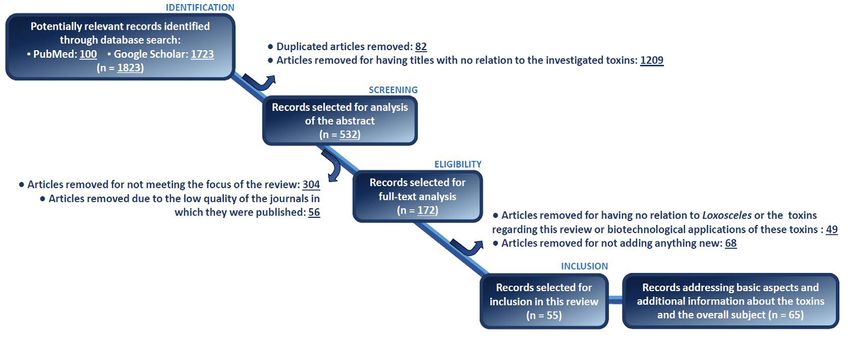
Gremski et al. J Venom Anim Toxins incl Trop Dis, 2021, 27:e20201088                                                              Page 3 of 15

    articles, exclusion criteria were sequentially applied in order to     core synthesized by endothelial cells [33,34], in addition to
    select appropriate articles regarding the aim of this review. In the   proteolytic activity on fibrinogen [35]. In addition, it was found
    first step, articles repeated between the databases accessed were      that a metalloprotease of L. gaucho venom acts upon red cell band
    removed as well as articles with titles showing no relation to the     3 transmembrane protein [36]. Then, two important descriptions
    toxins investigated. Only articles in English and Spanish were         were made: the first showing the biological conservation of these
    considered for further analyses whereas theses and dissertations       metalloproteases in venoms of different species of Loxosceles
    shown by the search engine Google Scholar were also disregarded.       spiders, such as L. rufescens [37], L. gaucho, L. deserta and L. laeta
       Subsequently, the articles selected in the previous step were       [38], indicating the importance of metalloproteases in the biology
    screened based on their abstract, resulting in the removal of          of these animals. Another important description used extract of
    articles that do not contain information supporting the aim of         L. intermedia venom-producing glands, discarding criticisms of
    this review or were published in low-quality journals (with no         a possible contamination of venoms by the digestive secretions
    impact factor assigned). The remaining articles were submitted         of spiders used in the previous articles, which were obtained by
    to full-text appreciation followed by discarding of articles that      electrostimulation, and then proving that metalloproteases are
    do not involve the context intended (i.e., characterization of         components of venoms and not digestive contaminants [39].
    specific toxins present in the venom of Loxosceles spiders) or that       The classification of these metalloproteases as astacins was
    do not add any new information to the literature of Loxosceles         first described by the cloning of an isoform of these enzymes
    toxins herein described. Finally, the remaining 55 articles were       from a cDNA library obtained from the venom-producing
    included in this review. Other 65 articles were biased, selected       gland of L. intermedia, which identified a metalloprotease
    and included since they were fundamental to set the background         with 30 kDa, containing a sequence signature of astacins. This
    and historically, structurally, biochemically or biologically          metalloprotease — which was named LALP (from Loxosceles
    characterize each class of toxin. Figure 2 summarizes the steps        astacin-like protease) — was obtained in its recombinant form by
    adopted in order to produce this review.                               heterologous expression using bacterial model and proved to have
                                                                           gelatinolytic, fibronectinolytic and fibrinogenolytic properties
    Astacins and Serine Proteases                                          [40]. Transcriptomic studies of the venom-producing glands of
    The existence of proteases in brown spider venoms was originally       L. laeta, which showed the presence of 8.2% of transcripts for


    reported over 40 years ago. The first descriptions showing the         metalloproteases [26], and L. intermedia, which showed about
    presence of proteases in the brown spider venoms were made             9.8% of the transcripts for the same family of toxins, bring
    using the crude venom of Loxosceles reclusa, which showed              into view further evidence that strongly suggests the presence
    proteolytic activity on larvae of Heliothis viresceus and Musca        of metalloproteases in different brown spider venoms [25].
    domestica [30]. These data on proteolytic activities in brown          Metalloproteases are also expressed in high amounts in the
    spider venoms were confirmed by the use of synthetic substrates        venom gland of L. gaucho, as found by a transcriptome analysis
    derived from the L-aminoacyl-beta-naphthylamide, which                 [41]. A recent study analyzed the Peruvian L. laeta transcripts
    were hydrolyzed by L. reclusa venom [31]. The discovery of             focusing on LALPs, and found 9 putative sequences coding for
    metalloproteases (astacins) in the venoms of brown spiders             astacine-like metalloproteases highly similar to LALP1 from L.
    has the participation of several Brazilian researchers, and was        intermedia [42]. In addition, the authors compared the activities
    described almost twenty years later, in studies that initially used    of Brazilian and Peruvian L. laeta venom upon fibrinogen and
    the crude venom from Loxosceles intermedia. Through kinetics           gelatin/collagen, and concluded that the Peruvian venom have a
    experiments of protein substrate degradation using fibronectin         higher activity upon these molecules than the Brazilian one [42].
    or fibrinogen incubated with crude venom, results showed the           A proteomic analysis of the L. intermedia crude venom using
    presence of proteolytic activity on these proteins (Figure 3).         mass spectrometry also described these enzymes as components
       Zymograms with copolymerized gelatin identified proteases           of the venom [43].
    with molecular masses between 30 and 35 kDa, and zymograms                Finally, astacins were described as forming an intra and
    with copolymerized fibronectin or fibrinogen showed                    inter-species family of toxins in the brown spider venoms,
    proteases with 22-28 kDa. In both cases, only divalent metal           which contain at least six different isoforms as showed through
    chelators such as ethylenediaminetetraacetic acid (EDTA)               an elegant experiment using two-dimensional zymogram
    and 1,10-phenanthroline — but not inhibitors of other classes          copolymerized with gelatin [44,45]. These data were later
    of proteases such as phenylmethylsulfonyl fluoride (PMSF),             confirmed by molecular cloning of members of metalloprotease
    aprotinin, leupeptin, and pepstatin A — blocked these                  family, strengthening the existence of various isoforms of these
    proteolytic activities, confirming that the proteases involved         proteases in the brown spider venoms [46]. The existence of
    on such as activities are metalloproteases [32]. The presence          families of astacins in the venoms of brown spiders was also
    of metalloproteases in the venoms of brown spiders were also           demonstrated by the use of a monoclonal antibody produced
    demonstrated by other authors, who showed metalloprotease-             against a L. intermedia LALP isoform, which cross-reacted
    dependent proteolytic activities in L. intermedia venom on the         with crude venoms of L. laeta and L. gaucho and neutralized
    substrates entactin and on heparan-sulfate proteoglycan protein        the proteolytic effects of these enzymes in the venoms [47].
Gremski et al. J Venom Anim Toxins incl Trop Dis, 2021, 27:e20201088 Page 4 of 15
Figure 1. Overview of the toxins present in the venom of Loxosceles spiders. Venom components can be divided into two major groups: (i) highly expressed toxins
(phospholipases D, knottins or ICK peptides and metalloproteases); and (ii) low-expressed toxins (translationally controlled tumor proteins – TCTP, allergens,
hyaluronidases, serine proteases and serine protease inhibitors). Phospholipases D can induce all the main effects associated with the whole venom, in addition
to displaying insecticidal activity. Recombinant phospholipase D can trigger a dermonecrotic lesion, the hallmark of Loxoscelism (light blue panel – source: Vuitika et
al. [123]). In comparison to phospholipases D, all the other venom components have been less studied. Knottins, also known as ICK peptides, are associated
with insecticidal activity. Metalloproteases have been linked up to the hydrolysis of extracellular matrix elements facilitating the spread of other toxins, as well as
to the induction of deleterious effects on endothelial cells worsening the tissue damage caused by the venom itself (source: da Silveira et al. [40]). TCTP proteins
act as a histamine-releasing factor, degranulating mast cells and triggering inflammatory events after the envenoming (source: Justa et al. [19]). Allergens also
participate in the inflammatory process, stimulating the degranulation of mast cells and increasing vascular permeability (source: Justa et al. [19]). Hyaluronidases
hydrolyze hyaluronic acid and then elicit the gravitational spread of the dermonecrotic lesion (source: Ferrer et al. [68]). Serine proteases hydrolyze gelatin and
may be involved with the extracorporeal digestion of prey (source: Veiga et al. [51]). Serpins (serine protease inhibitors) have been poorly characterized to
date, but have already shown to be able to inhibit components (serine proteases) of the complement system. Center image from Chaim et al. [114].
Figure 2. Flow chart highlighting the methodology used in order to produce this systematic review.Gremski et al. J Venom Anim Toxins incl Trop Dis, 2021, 27:e20201088 Page 5 of 15
Figure 3. Experimental strategies used to study some toxins present in the venom of Loxosceles spiders. (A) De Castro et al. [110] reported the obtainment of
native toxins (knottins or ICK peptides) purified from L. intermedia crude venom by means of sequential chromatographic approaches. The native toxins were then
biologically characterized as insecticidal molecules and their amino acid sequences were identified by sequencing. (B) Hyaluronidases from L. intermedia were first
studied by Da Silveira et al. [66] using biochemical methods (zymography). These analyses showed molecules displaying hydrolytic activity in the venom upon hyaluronic
acid and chondroitin sulfate. Toxin identification using biochemical assays was also described for metalloproteases. Feitosa et al. [32] showed that the crude venom
of L. intermedia degraded human fibronectin in proteolytic digestion kinetic experiments. Further analyses showed that metalloprotease inhibitors blocked crude
venom ability to hydrolyze fibronectin, which together with zymography experiments using gelatin as a substrate, pointed out the presence of metalloproteases in
the studied venom. (C) Identification and characterization of toxins can also be performed using transcriptome analysis, followed by recombinant protein-production
techniques and studies for the characterization. Sade et al. [87] and Boia-Ferreira et al. [88] reported biological and biochemical characterization of a recombinant
L. intermedia TCTP expressed in bacterial model identified in the venom-producing gland transcriptome. Ferrer et al. [68] produced a recombinant hyaluronidase
in bacterial model followed by in vitro and in vivo analysis; the sequence coding this hyaluronidase was also identified in the transcriptome of L. intermedia venomous
gland. In addition, transcriptomic findings revealed a sequence of an allergen encoded in L. intermedia venom-producing gland. Based on this information, Justa et al.
[19] recombinantly expressed this toxin using baculovirus-infected insect cells and characterized its functionality. Venom producing glands (C) from Chaim et al. [114].
The biological conservation and the presence of astacin isoforms heparan-sulfate proteoglycan, involved in endothelial cell biology
in different brown spider venoms have already pointed out the and blood vessel stability. This mechanism was previously
participation of these proteolytic enzymes in the biological investigated [34,35] and could induce signs related to blood
events related to brown spider venom. However, what are the vessel disorders after envenoming, such as increased capillary
true biological functions of these molecules? permeability, edema, and ecchymosis. Moreover, the action of
The first aspect that can be discussed is undoubtedly the astacins on extracellular matrix components involved in blood
participation of astacins as regulating molecules of extracorporeal vessel stability, blood clotting, platelet adhesion and aggregation
digestion performed on prey captured by these spiders, which are — such as fibronectin, fibrinogen and entactin — could explain
carnivorous and considered very efficient predators that ingest other events that occur after envenoming, such as hemorrhage
food in liquid form [48]. The presence of these endo-proteases in and even the reported difficulty in healing the skin lesions
the venom helps to degrade protein components of the obtained triggered by accidents [32–35].
preys, and thus, facilitate the feeding of these spiders. Another The proteolytic action of brown spider venoms has also
possibility is the proteolytic action of these metalloproteases been demonstrated on basement membranes, structures with
on the components of blood vessel endothelial cells, such as enormous importance in the organization of various tissues [49].Gremski et al. J Venom Anim Toxins incl Trop Dis, 2021, 27:e20201088 Page 6 of 15
Together with the proteolytic action on soluble components of treatment with thrombin, they acquire proteolytic activity [51].
the extracellular matrix, these activities could explain some toxic They are enzymes with an optimal pH between 7 and 8 and,
effects caused by the venoms as previously discussed. By making interestingly, they do not have a wide spectrum of proteolysis.
the extracellular matrix loosened and disorganized, astacins These enzymes showed proteolytic activity restricted to gelatin,
could also promote the spread of other toxins to the nearby with no catalytic activity on proteins such as BSA, hemoglobin,
blood vessels and other parts of the bodies of injured victims, IgG, casein or laminin [51]. The presence of serine proteases in
enhancing deleterious systemic effects during envenoming. brown spider venoms was reinforced by transcriptomic analysis
Finally, metalloproteases present in the venoms could act as of L. laeta venom-producing gland, which indicated the presence
molecular scissors that cleave precursor molecules of the venom of serine proteases transcripts comprising about 0.5% [26], and
and/ or confined to the hosts, thus activating the toxic activities also of L. intermedia venomous gland, which revealed about
seen after accidents. An example that illustrate this mechanism 0.3% of transcripts encoding serine proteases [25]. These data
is that of zymogens, produced by leukocytes, which could be indicate inter-species conservation, strengthening the biological
activated during the unregulated inflammation that occurs after importance of these proteases, although in both cases they
accidents, as previously hypothesized [40]. are low expressed in comparison to other toxins such as the
Another interesting work that deserves to be cited phospholipases D and ICKs.
and is based on the participation of astacins in the The results based on transcriptomic analysis were confirmed
toxicity of brown spider venoms used linear sequences by proteomic studies, which showed the presence of serine
of an astacin from the venom of L. intermedia LALP-1 proteases in the crude venom of L. intermedia, indicating that
(SLGRGCTDFGTILHE, ENNTRTIGPFDYDSIMLYGAY, and these molecules are indeed components of brown spider venoms
KLYKCPPVNPYPGGIRPYVNV) in the construction of a chimeric [43]. As reported for astacins, the purification of native serine
recombinant antigen containing, in addition to LALP sequences, proteases from Loxosceles spiders’ crude venoms is virtually
the sequences of a hyaluronidase and a phospholipase D present unviable and the production of these enzymes in heterologous
in this venom. This chimera was antigenic and triggered the models has proved to be quite difficult. For these reasons,
production of antibodies that neutralized the dermonecrotic the biological functions of serine proteases in the venoms of
activity in rabbits’ skin and inhibited mice lethality induced by brown spiders have not been determined yet, leading to only
the crude venom [50]. Another hybrid immunogen consisting speculative hypothesis regarding their functionality. Based on
of hydrophilic regions from the metalloprotease LgALP1 from these theoretical assumptions, as described for the astacins,
L. gaucho and from a phospholipase D of the same species the serine proteases could act as molecular scissors that work
was constructed, expressed and used to produce antibodies by activating precursors molecules in the crude venoms or in
in mice. This antiserum neutralized dermonecrotic (in vivo) victim’s body, and/or digestive enzymes that participate in the
as well as fibrinogenolytic and platelet aggregation (in vitro) extracorporeal digestion occurring shortly after the envenoming
activities elicited by Loxosceles venoms [41]. Astacins, as well of the prey. However, the precise functions of serine proteases
as other toxins present in the venom of Loxosceles spiders, in the envenoming are still to be determined.
are not feasible to be obtained in their native form through
purification from crude venom because of the low yield of the
Hyaluronidases
venom extractions. Moreover, these enzymes are difficult to
be recombinantly obtained in their soluble and active form Hyaluronidases are found in several animal venoms including
[40,46]. Due to this difficulty in isolating these molecules, the those of spiders [52–56] snakes [57], caterpillars [58], and bees
knowledge regarding their participation in the envenoming [59,60]. Hyaluronidase activity in brown spider venom was first
process is based on theoretical possibilities and/or hypotheses reported in the venom of L. reclusa [61]. Later, other description
related to their biochemical properties, pointing out the lack regarding hyaluronidase in a brown spider venom was made by
of robust evidence for final conclusions. Wright et al. [62]. In this study, a hyaluronidase was purified
Other proteases described in brown spider venoms belong to from the venom glands of L. reclusa and exhibited activity on
the family of the serine proteases. In this case, literature data hyaluronic acid (optimum pH 5.0 – 6.6) and chondroitin sulfate,
are even scarcer and are restricted to a few published articles. being the former the preferred substrate. The authors also tested
The first description of serine proteases as part of brown spider the in vivo activity of this enzyme and observed the development
venoms was made using crude venom of L. intermedia, which of a mild erythema in guinea pigs after 6 hours that subsided
after treatment with thrombin, activated two molecules with over the next 24 hours. Although a complete neutralization
gelatinolytic activity at regions of 85 kDa and 95 kDa in a of purified L. reclusa hyaluronidase by the gamma globulin
zymogram analysis. These proteases were inhibited by serine fraction of a specific antivenom raised against L. reclusa crude
proteases inhibitors as PMSF, aprotinin, benzamidine, soybean- venom was observed in vitro, the whole antiserum exhibited
trypsin inhibitor and leupeptin, but not by other protease only a slight inhibitory effect on the spreading action of the
inhibitors as EDTA, 1-10’-phenanthroline, iodoacetamide and venom [62]. The presence of antibodies in the antivenom that
pepstatine-A. In addition, such data indicated that these toxins recognize L. reclusa purified hyaluronidase was confirmed later
are present in the forms of inactive zymogens, and that after by immunodiffusion studies [63].Gremski et al. J Venom Anim Toxins incl Trop Dis, 2021, 27:e20201088 Page 7 of 15
At that time, toxinologists already understood that hyaluronidases are of interest also because they belong to the same
hyaluronidases in spider venoms were not a toxic element class of mammal hyaluronidases, as mentioned earlier. The use of
per se, but probably act as a spreading factor [64,65]. Later, a recombinant human PH20 hyaluronidase (rHuPH20; Halozyme
hyaluronidase activity was described in the venom of various Therapeutics, Inc.) to overcome the resistance to bulk fluid flow
Loxosceles species, e.g. L. deserta, L. gaucho, L. intermedia and L. in the subcutaneous space and favor drug delivery, dispersion,
laeta, and this activity appeared in a hyaluronic acid zymogram and absorption, is currently FDA-approved. It acts by degrading
as a 44 kDa enzyme [38]. L. intermedia hyaluronidase was further hyaluronic acid, facilitating the route of administration and
characterized by da Silveira et al. (Figure 3B) [66]. These authors optimizing the dosage of subcutaneous therapies [72].
showed that this hyaluronidase is a hydrolase characterized as an A recombinant human hyaluronidase was also proved to
endo-b-N-acetyl-D-hexosaminidase, has an optimal activity at be effective and secure as a facilitating agent for subcutaneous
6.0-8.0 pH and hydrolyze both hyaluronic acid and chondroitin immunoglobulin in a retrospective, multicenter study (fSCIG;
sulfate in vitro and hyaluronic acid in vivo [66]. Proteomic and HyQviaR ®) in elderly patients with primary or secondary
transcriptomic analyses identified few sequences that correspond immunodeficiencies [73]. They are also widely applied in the
to hyaluronidase, evidencing that they are low-abundance toxins field of dermatology, to degrade hyaluronic acid filler to reverse
in Loxosceles venom, comprising 0.13% of L. laeta and 0.05% of cutaneous augmentation with this glycan [74]. In short, the
L. intermedia venom gland transcripts [25,26,67]. production of recombinant spider venom hyaluronidases is a
The role of Loxosceles venom hyaluronidases as spreading promising alternative since they can contribute to understand the
factors was first demonstrated when a recombinant L. intermedia role of these glycosidases in the venom and to the development
hyaluronidase was produced [68]. Dietrich’s hyaluronidase, as of specific therapies to treat loxoscelism, besides having various
it was named, was expressed in E. coli cells and subjected to potential applications for the pharmaceutical industry [75,76].
in vitro refolding in order to obtain a soluble and active 6xHis
tagged enzyme with ~45 kDa (Figure 3C). This recombinant Allergens
enzyme retained linear antigenic determinants from native Accidents involving bites caused by bees and ants, in addition
hyaluronidases of Loxosceles crude venom as demonstrated by to exposures to animals such as cockroaches and mites can
immunoassays. Finally, Dietrich’s hyaluronidase increased the area generate serious allergic reactions such as fever, edema, vertigo
of dermonecrosis and enhanced edema induced by a recombinant and anaphylactic shock [77–81]. In spiders, little is known about
phospholipase D, as well as triggered the gravitational spreading of allergenic molecules and their biological activities. However, in
the lesion. These data proved the role of Loxosceles hyaluronidases accidents involving spiders from the Loxosceles genus, symptoms
as a spreading factor of other toxins near the bite site [68]. Two at the cutaneous tissues as itch, erythema, edema, cutaneous rash,
other spider hyaluronidases were produced as recombinant and in some cases acute generalized exanthematous pustulosis
toxins: CsHyal (from Cuppienius salei), which was produced are common, suggesting allergic reactions in some instance
in E. coli and further refolded, and BvHyal (from Brachypelma [6,28,82,83]. The presence of toxins with hyaluronidase activity
vagans), which was expressed using baculovirus system in insect in the venoms of Loxosceles spiders, which are strong allergenic
cells [69,70]. CsHyal potentiated the insecticidal activity of factors in bee venoms, was initially described by studying the
neurotoxins in invertebrate preys, and authors speculated that crude venom of L. reclusa [62]. Later, toxins characterized as
this glycosidase may act as a spreading factor that enhance the hyaluronidases were identified in venoms of other Loxosceles
activity of neurotoxic venom compounds [70]. species, as mentioned earlier [37,38,66], however there was never
Recently, a novel isoform of Loxosceles intermedia venom a direct correlation of these toxins with allergenic activities in
hyaluronidase was produced in a baculovirus-infected insect cells these venoms.
system and named LiHyal2 [71]. This recombinant glycosidase The presence of allergenic factors in the venoms of Loxosceles
was produced as an active glycosylated enzyme and the biological spiders was also demonstrated in the study regarding the
characterization of LiHyal2 confirmed its ability in acting as transcriptome of L. intermedia venom glands, in which 0.2 %
a spreading factor. of transcripts encoding toxins were described as being allergens
By using two linear epitopes of Dietrich’s hyaluronidase [25]. The similarity of these allergenic toxins from L. intermedia
(NGGIPQLGDLKAHLEKSAVDI and ILDKSATGLRIIDWEAWR) with other allergens present in the venoms of the spider Lycosa
combined with epitopes of other Loxosceles toxins (e.g. astacin- sigoriensis and the scorpoion Opisthacanthus cayaporum as well
like protease and phospholipase D), Lima et al. [50] produced a as mite allergens of Ixodes scapularis and Argas monolakensis
recombinant multiepitopic protein named rMEPLox (recombinant suggests the involvement of this toxin in the possible allergic
MultiEpitopic Protein derived from Loxoscelic toxins). Antibodies responses seen after accidents. Finally, the existence of an allergen
against this protein efficiently neutralized hyaluronidase activity in the venoms of Loxosceles spiders was greatly suggested by
of L. intermedia venom [50]. These results represent one of the a study using molecular biology techniques, which reported
various possibilities of using Loxosceles hyaluronidases as biotools cloning of an allergen named as LALLT (Loxosceles allergen-like
for therapeutical applications. These enzymes are related to toxin) [19]. This toxin was cloned, expressed in the baculovirus/
several physiological and pathological processes. In fact, venom insect cells system, purified and some of its biological activitiesGremski et al. J Venom Anim Toxins incl Trop Dis, 2021, 27:e20201088 Page 8 of 15
were reported (Figure 3C). The allergen from L. intermedia performed by using circular dichroism spectroscopy and showed
venom has a molecular mass of 42 kDa and presents epitopes the proper folding features of the recombinant protein [88].
that cross reacted with anti-venom sera developed using crude Envenomation by Loxosceles spiders can cause hypersensitivity
venoms from L. laeta and L. gaucho, suggesting that these and allergic reactions. The cutaneous symptoms generated by the
molecules are conserved in different species of the Loxosceles venom include erythema, edema, itching and pain. Rattmann
genus. This biological conservation was also revealed by analysis et al. [89] demonstrated that the L. intermedia venom triggers
involving multiple sequences alignment of cDNA-deduced mast cell activation and histamine-dependent effects. Initial
amino acid sequences for LALT orthologues from L. laeta and inflammation events, such as increased vascular permeability,
from L. gaucho venom, reinforcing the existence of a family of were related to the participation of histaminergic and serotonergic
allergens in the venoms of brown spiders [19]. receptors. As mentioned earlier, many symptoms observed
LALLT has 18 cysteine residues and belong to the CAP during loxoscelism can be mimicked by phospholipase D toxins
superfamily, showing significant identity to other allergens from (PLDs), the most characterized and studied family of toxins in
spiders, scorpions, mites and ticks. Experiments of biological Loxosceles venoms. However, recombinant PLDs are not able to
characterization of LALLT showed that this recombinant induce paw edema with the same intensity as the crude venom,
molecule caused edema in the skin of rabbits, increased vascular pointing to histaminergic events in the increased formation of
permeability and triggered paw edema in mice, besides increasing edema during envenomation [6,87].
calcium influx and inducing release of beta-hexosaminidase Brown spider venom was shown to be capable of causing
from mast cells (RBL-2H3) in vitro. Finally, LALLT caused regulated release of mast cell mediators, mainly histamine,
degranulation of rat mesentery mast cells [19]. In this same study, responsible for inducing vasodilation in experiments with
histological analysis of the skin of rabbits exposed to recombinant rat aorta using a chamber for an isolated organ [89]. Paludo
LALLT revealed edema and an infiltrate of inflammatory cells on et al. [90] identified the presence of histamine in the venom
the dermis, findings that are common in the histopathological in sufficient quantities to exert inflammatory effects. Despite
analysis of samples from patients with allergies caused by this, the dialyzed venom, without the presence of histamine,
arthropods bites [84]. was still capable of exerting a certain histamine-dependent
All the data exposed here support the existence of allergens in inflammatory effect, due to some other component present in the
the venom of Loxosceles spiders, providing valuable information venom, acting directly on mast cells [89,90]. First Sade et al. [87]
that can assist in the treatment of accidents in the hospital or and then Boia-Ferreira et al. [88] demonstrated that the TCTP
outpatient settings. The fact that these toxins belong to the group of L. intermedia participates in the exacerbated inflammatory
of low expressed toxins in Loxosceles venoms has to be taken into process resulting from accidents: LiRecTCTP causes in vivo
account, since their low concentrations can make these allergenic increased vascular permeability and edema in mice, in a time
responses uncommon, restricted to more susceptible patients. and concentration dependent manner [87,88]. Therefore, these
results suggest the LiTCTP may be the first and fastest component
Translationally Controlled Tumor Protein (TCTP) to induce edema formation in loxoscelism pathophysiology [87].
Loxosceles intermedia TCTP protein, LiRecTCTP, was identified Boia-Ferreira et al. [88] also demonstrated that LiRecTCTP is
in the cDNA library of the brown spider venom gland of L. capable of activation mast cells (de-sensitized RBL-2H3 cells)
intermedia [25]. This protein exhibits a high degree of similarity leading to degranulation in vitro.
with tick TCTPs (~70%), which are described as histamine Expression profile analysis via quantitative real-time PCR
release factors [85–87]. The complete sequence identified in showed that LiRecTCTP induced the cellular expression of
the cDNA library contains 536 bp, which encodes a 172-amino cytokines involved in allergic and parasitic processes such as
acid protein with a predicted molecular mass of 22.3 kDa and a IL-3, IL-4, and IL-13 in cultured RBL-2H3 cells. In vivo assays
pI of 4.7 (mature TCTP) [87]. Additionally, the transcriptome showed that when LiRecTCTP was injected in mice together
study of the L. intermedia venom gland identified 0.2% of the with inhibitors of histamine receptors (H1, H2, H3 and H4) a
total transcripts corresponding to TCTP protein transcripts reduction in vascular permeability and edema was observed
[25]. In 2012, Sade et al. [87] performed cloning, heterologous when compared to isolated toxin, confirming that this toxin
expression, purification and functional characterization of the is responsible for inducing these deleterious histaminergic
L. intermedia TCTP (Figure 3C). The recombinant protein, effects [88]. H1 and H2 receptors inhibitors (prometazine and
expressed in E. coli with a 6 His-tag at the N-terminus, was called thioperamide, respectively) have been shown to significantly
LiRecTCTP. Purification involved two chromatography steps - reduce the effects of LiRecTCTP on increasing vascular
an affinity chromatography using Ni-NTA agarose resin with permeability. The degranulation inhibitor cromolyn, in turn,
subsequent ion exchange chromatography (DEAE-sepharose) was able to abrogate the edematogenic effect prompted by
[87]. Recently Boia-Ferreira et al. [88] have standardized a new LiRecTCTP. Furthermore, experiments of dermonecrosis using
purification protocol with higher yield and purity using the Akta rabbits demonstrated a synergism between LiRecTCTP and a
purified system and affinity chromatography (Ni-NTA agarose). recombinant phospholipase D toxin. These data emphasizes
Secondary structures and solubility analyses of LiRecTCTP were LiRecTCTP relevant participation in the inflammatory andGremski et al. J Venom Anim Toxins incl Trop Dis, 2021, 27:e20201088 Page 9 of 15
histaminergic cutaneous effects of loxoscelism for acting as a protease inhibitor from L. intermedia [100]. The reasons for the
histamine release factor and thus contributing to the systemic presence of serine protease inhibitors in the venom of Loxosceles
dispersion of other venom components [88]. spiders, as well as the physiological targets of these molecules are
TCTP-related proteins were also identified in the venom of still unknown. It was proposed that these toxins, through their
other Loxosceles spiders (L. laeta and L. gaucho) by immunoblot inhibitory activities on proteases, could protect the integrity of
cross-reactivity assays [91]. Literature on TCTP from spiders is other venom components, and thus increase the useful life of
scarce but some sequences were identified in the venom of spiders venom toxins exposed to an external proteolytic environment,
from different species [92–95], and in gland secretions of ixodid for instance when the venom is released to protect the spider
tick parasites, which are also arachnids [96]. Concerning the against predators or to kill their prey [43].
biological and evolutionary purpose of this toxin to be present An interesting fact that suggests the biological importance
in Loxosceles venom we must highlight that L. intermedia TCTP of serine protease inhibitors for Loxosceles spider venom is
does not present a signal peptide for endoplasmic reticulum that toxins found in the venom of L. laeta are similar to serine
translocation, this toxin can be secreted by exosomes and also protease inhibitors from different animals as Mus musculus,
by the holocrine secretion pathway [24], as other constituents Aedes aegypti, Branchiostoma lanceolatum, Gallus gallus and
of whole venom [87]. Boophilus microplus [26]. This also is valid for serine protease
Studies on TCTP as a venom toxin are very few and its inhibitors described in the L. intermedia venom, which are quite
biological and evolutionary role as a venom component in similar to inhibitors found in mammals such as Mus musculus
prey capture remains still unknown [97]. In contrast, as a and Pan troglodytes, or in the tick Ambliomma americanum
multifunctional protein involved in several biological processes, [25]. These toxins fit into the families of molecules with low
its biotechnological potential is enormous and yet to be further expression in the venoms of Loxosceles spiders [25] and perhaps
explored. that is why they have been little studied so far. However, the
identification of transcripts coding for serpins present in the
Serine Protease Inhibitors (Serpins) venom-producing glands, and the production of recombinant
Another group of toxins found in the venoms of Loxosceles molecules as tools, will contribute and help elucidate the functions
spiders includes protease inhibitors of the serine protease for these inhibitors, as well as contribute to structural analysis
family characterized as serpins [28,98]. The first evidence in to understand the relation between structure and function
the literature pointing out the existence of protease inhibitors in of these toxins. Finally, but not less important is the possible
Loxosceles spiders was described using crude venom of L. reclusa, uses of these molecules, since there are numerous examples
which showed the presence of a potent inhibitory activity on the of biotechnological applications of analogs of serpins in the
complement-dependent hemolysis (an event highly dependent control of blood clotting, anti-tumor activity and viral infection
of serine proteases). This component of the venom showed treatments [98,101–105].
properties such as not being dialyzable, but can be excluded from
the venom by means of a gel filtration chromatography using ICK Peptides or Knottins
Sephadex G-75, in addition to being stable under a broad range Inhibitory Cystine Knot (ICK) peptides are single-chain molecules
of pH [99]. Serine protease inhibitors were initially described enriched in cysteine residues, which establish intramolecular
in the venoms of brown spiders by means of transcriptome disulfide bonds. The disulfide bonds are organized in a specific
analyzes of L. laeta venom-producing glands. In this study, pattern in which two of them together with the peptide backbone
transcripts encoding serine protease inhibitors corresponded to form a ring that is crossed by the third disulfide bond. This
0.6% of the total transcriptome [26]. Serine proteases inhibitors disulfide bonds’ arrangement creates a pseudo-knot framework,
were additionally identified through proteomic analyzes using which is why these peptides are also known as knottins [106–
crude venom of L. intermedia that was submitted to sequential 108]. A large number of studies have already shown that ICK
chromatography steps by using cation exchange and reverse peptides display insecticidal activity [109–111]. For this reason,
phase to purify proteins and peptides that were identified by the foremost function regarding ICK peptides in spider venoms
mass spectrometry MS/MS. These studies reported the presence concern the predation for feeding purposes, especially insects
of molecules in the venom characterized as trypsin inhibitor-like [112–114]. It is also due to this insecticidal activity that ICK
protein and serine protease inhibitor protein [43]. peptides have been biotechnologically explored in order to
The presence of serine proteases inhibitor transcripts in the develop alternative bioinsecticides to the harmful chemical
venom glands of Loxosceles spiders was later identified in the compounds still used [113,115].
transcriptome analysis of the venom-producing gland of L. The ICK peptides from Loxosceles spiders were first studied
intermedia, which showed the presence of 0.1% of toxin encoding by De Castro et al. [110] who fractioned the crude venom
transcripts identified as serine protease inhibitors [25]. Finally, of L. intermedia and identified a fraction with insecticidal
a further biochemical characterization supporting the presence activity (Figure 3A). Further chromatographic steps sequentially
of serine protease inhibitors in the venom of Loxosceles spiders performed allowed the purification of three peptides named
is currently being carried out by using a recombinant serine LiTx1, LiTx2 and LiTx3 with insecticidal activity (inducedGremski et al. J Venom Anim Toxins incl Trop Dis, 2021, 27:e20201088 Page 10 of 15
flaccid paralysis) on the larvae of economic interest Spodoptera the target of U2-SCRTX-Lit2, Meissner et al. [119] performed
frugiperda. By using amino acid sequencing and molecular molecular docking and dynamics analyses using a voltage-gated
biology methodologies, De Castro et al. [110] obtained the sodium channel from Spodoptera litura (tobacco cutworm),
coding sequence for these peptides, which revealed that these whose structure has already been determined. The choice of this
peptides are produced as prepropeptide precursors (signal target was also due to the fact that µ-HXTX-Mg2a was able to
peptide, propeptide and mature peptide). Later, the authors cause paralysis in S. litura and was associated with the inhibition
included a sequence related to a fourth isoform (LiTx4) on the of voltage-gated sodium channels (SlNaVSC) in synaptosome
GenBank, which has not had its insecticidal activity tested yet. preparations obtained from cockroaches by binding to the site
The transcriptome of the venom-producing glands of L. 3 of these channels [116]. Bioinformatics data showed that U2-
intermedia published by Gremski et al. [25] revealed that the SCRTX-Lit2 presents amino acid residues arranged in a pattern
majority of the sequences expressed regarding toxins was that suggests affinity to the site 3 of the SlNaVSC and revealed
related to ICK peptides (55.9%). Sequences with high identity that the peptide may act as a steric blocker, hiding the gate access
with the LiTx1-4 peptides represented 53.5%, and 2.4% of that of these channels [119]. Sequence analyses comparisons have
transcripts showed significant similarity with a neurotoxic pointed out that the peptides LiTx3 and U2-SCRTX-Li1b may
ICK peptide from the spider Macrothele gigas [116]. In addition act on voltage-gated sodium channels as well [110,117].
to the annotation of the sequences mentioned, Gremski et al. Another study regarding ICK peptides from L. intermedia
[25] analyzed the venom protein content by SDS-PAGE, which was carried out by Matsubara et al. [111]. In this study, the
suggested that ICK peptides are massively predominant in the peptide U2-SCRTX-Li1b was recombinantly expressed in the
venom of L. intermedia. periplasm of bacterial cells. This strategy was selected because
Matsubara et al. [117] investigated an ICK peptide sharing the periplasm of E. coli provides the molecular machinery
86% sequence identity with LiTx3, which was named U2- that assists in the correct formation of disulfide bridges, in
sicaritoxin-Li1b (U2-SCRTX-Li1b) in agreement with the rational contrast to the reducing environment of the cytoplasm that
nomenclature developed by King et al. [118]. The authors cloned disadvantages the establishment of these structures [120]. After
the sequence and expressed the peptide in bacterial cells, resulting purification, recombinant U2-SCRTX-Li1b was able to cause
in the production and purification of the first recombinant ICK long-lasting paralysis in sheep blowflies (Lucilia cuprina), which
peptide from Loxosceles’ venoms. Using the recombinant U2- was irreversible even after 72 hours. Therefore, U2-SCRTX-Li1b
SCRTX-Li1b and hyperimmune sera raised against different constitutes the first recombinant ICK peptide from Loxosceles
Loxosceles spider venoms, the authors performed immunoassays spiders to have its activity determined [111]. Furthermore, the
that showed antigenic cross-reactivity, pointing out that ICK authors carried out a screening of sequences encoding ICK
peptides constitute a family of toxins widespread throughout peptides in other two Loxosceles species (L. gaucho and L.
the genus [117]. An additional ELISA cross-reactivity analysis laeta) from the total RNA produced in the venom glands of the
using polyclonal antibodies and the recombinant peptide U2- spiders. This screening of venom-gland transcripts resulted in
SCRTX-Li1b or whole venom performed by Buch et al. [91] the obtainment of sequences encoding orthologues of LiTx1-4
suggested that ICK peptides are present in the venoms of L. peptides, with identities ranging from 83% to 100% compared
intermedia, L. laeta and L. gaucho, reinforcing once more that to the sequences encoding ICK peptides of L. intermedia. All
these peptides belong to a conserved family of toxins in Loxosceles sequences encoding ICK peptides found contain 10 cysteine
spiders. Interestingly, Buch et al. [91] also carried out western residues in their mature sequence and exhibit the same predicted
blotting analysis that did not show cross-reactivity between the disulfide bond connectivity pattern [111].
recombinant peptide and polyclonal antibodies that recognize L. In 2017, Trevisan-Silva et al [67] published a revealing
laeta and L. gaucho venoms or between the polyclonal antibodies proteomic analysis of the whole venom of L. intermedia by using
that recognize the peptide U2-SCRTX-Li1b and L. laeta and L. a multi-protease, multi-dissociation, bottom-up-to-top-down
gaucho whole venoms. approach. This study identified ICK peptides from Loxosceles
Meissner et al. [119] studied another ICK peptide from L. venoms at a proteomic level for the first time, resulting in the
intermedia, whose sequence was identified in the venom gland identification of peptides with correspondence to LiTx in high
transcriptome [25]. This peptide – U2-SCRTX-Lit2 – shares abundance as depicted by the L. intermedia transcriptome [25].
52% identity with the toxin μ-hexatoxin-Mg2a (µ-HXTX- ICK peptides have also been described as toxins that help
Mg2a) from M. gigas, in addition to the fact that both peptides spiders in defending against their predators. In addition,
contain 10 cysteine residues that establish the same disulfide due to the anthropic action, many species of spiders have
bond connectivity pattern. A great deal of ICK peptides have had their natural habitats destroyed and, consequently, they
been described as highly specific to insects, interacting with ion have been recurrently found in peridomiciliar environments,
channels or membrane receptors in their nervous system and which facilitates accidents with humans [3,6]. Due to natural
then resulting in paralysis and death. In order to investigate interactions with predators and episodic interactions withGremski et al. J Venom Anim Toxins incl Trop Dis, 2021, 27:e20201088 Page 11 of 15
humans, many species of spiders have evolved ICK peptides Conclusion
with harmful properties to these organisms. Loxosceles bites Much learning has been gathered about astacins, serine proteases,
result in a mild stinging that usually is painless in humans [3,6], knottins, TCTP, hyaluronidases, allergens and serpins (Table 1).
which can suggest the existence of molecules with anesthetic However, we are still at an initial phase in understanding the full
or analgesic effects in the venom. Some ICK peptides in other role of these proteins in the brown spider venom and how they
spiders have already proved to display analgesic effects on can work together to affect the tissue of victims. Novel strategies
animal models such as the peptide PcTx1 (µ-TRTX-Pc1a) from must be undertaken to overcome the barrier of obtaining enough
the tarantula Psalmopoeus cambridgei, which interacts with amount of these toxins to enable further investigation and
acid sensing ion channels and results in analgesic properties comprehension of the pathophysiology of loxoscelism. Then, it
in rat models for acute pain when administered intrathecally will be possible to use this information to improve therapeutic
or intracerebroventricularly [107,121]. Hence, given the great strategies for treating affected patients. In addition, a deeper
diversity of ICK peptide-coding sequences and the painless knowledge on functional and structural aspects of these poorly
aspect of Loxosceles spider bites, the search for peptides with explored toxins will certainly reveal new possible applications
possible analgesic activities remains a promising idea. in diverse areas.
Table 1. A summary of the studies involving each family of toxins present in the venom of Loxosceles spiders approached in this review.
Toxin References Major findings
L. intermedia venom was able to degrade fibronectin and fibrinogen, but not
laminin or types I and IV collagens. This activity was blocked by EDTA and
Feitosa et al. [32], 1998
1,10-phenantroline. Zymogram analyzes of venom detected a 35 kDa enzyme with
gelatinolytic activity and a fibronectinolytic and fibrinogenolytic band at 28 kDa.
A 30 kDa metalloprotease was cloned and produced a recombinant protein in a
prokaryotic expression system. It was named LALP1, from Loxosceles astacin-like
Da Silveira et al. [40], 2007 protease, because it was structurally and functionally related to the astacin family
of metalloproteases. LALP1 induced de-adhesion of endothelial cell cultures and
degraded fibronectin and fibrinogen.
Two novel cDNAs encoding astacins were cloned from L. intermedia venom glands
(LALP2 and LALP3). The venoms of L. intermedia, L. laeta and L. gaucho showed
Metalloproteases Trevisan-Silva et al. [122], 2010 immunologically-related toxins with LALP1 and toxins with gelatinolytic activity with the
(Astacins) same electrophoretic mobilities. The screening of mRNAs from L. laeta and L. gaucho
venom glands revealed members of the astacin family (LALP4 and LALP5, respectively).
Based on the analysis of subproteomes of LALPs from L. intermedia, L. laeta and L.
Trevisan-Silva et al. [44], 2013 gaucho, authors showed that LALPs comprise a large family of toxins in Loxosceles
venom, and that each venom has distinct proteolytic activities.
LALP3 was expressed using a SUMO tag in Escherichia coli Shuffle T7 Express LysY
cells. Immunoassays showed that LALP1 and LALP3 share linear epitopes and LALP3
Morgon et al. [46], 2017
shares conformational epitopes with native venom astacins. Molecular modeling of
LALP3 revealed the zinc binding and Met-turn motifs forming the active site.
L. laeta venom gland transcripts were analyzed with a focus on LALPs and nine
Medina-Santos et al. [41], 2019 possible LALPs isoforms from Peruvian L. laeta venom were identified and validated
by in silico and in vitro experiments.
Serine protease activity was detected in the venom of L. intermedia after treatment
Serine proteases Veiga et al. [51], 2000 with trypsin. These gelatinolytic molecules presented electrophoretic mobility of 85
and 95 kDa in a zymogram analysis.
L. intermedia venom hyaluronidases were characterized as endo-β-N-acetyl-D-
Da Silveira et al. [66], 2006 hexosaminidases that hydrolyze hyaluronic acid (HA) and chondroitin sulfate (CS). Lytic
activities upon these GAGs were observed by zymogram analyzes at 41 and 43 kDa.
A recombinant hyaluronidase (Dietrich’s hyaluronidase) from L. intermedia venom
was expressed and refolded. It was able to degrade HA and CS, cross-reacted with
Ferrer et al. [68], 2013
Hyaluronidases native venom toxins and increased the dermonecrotic effect of a Loxosceles PLD,
confirming its activity as a spreading factor.
A novel hyaluronidase of L. intermedia venom was produced in a baculovirus-
insect cell expression system as a fully active enzyme with post-translationally
De-Bona et al. [71], 2021
modifications (i.e., N-linked carbohydrates). LiHyal2, as it was named, potentialized
dermonecrosis, edema and vascular permeability induced by a Loxosceles PLD.You can also read