Multi omic analyses in Abyssinian cats with primary renal amyloid deposits
←
→
Page content transcription
If your browser does not render page correctly, please read the page content below
www.nature.com/scientificreports
OPEN Multi‑omic analyses in Abyssinian
cats with primary renal amyloid
deposits
Francesca Genova1,50,51, Simona Nonnis1,50, Elisa Maffioli1, Gabriella Tedeschi1,
Maria Giuseppina Strillacci1, Michela Carisetti1, Giuseppe Sironi1, Francesca Anna Cupaioli2,
Noemi Di Nanni2, Alessandra Mezzelani2, Ettore Mosca2, Christopher R. Helps3,
Peter A. J. Leegwater4, Laetitia Dorso5, 99 Lives Consortium* & Maria Longeri1*
The amyloidoses constitute a group of diseases occurring in humans and animals that are
characterized by abnormal deposits of aggregated proteins in organs, affecting their structure and
function. In the Abyssinian cat breed, a familial form of renal amyloidosis has been described. In this
study, multi-omics analyses were applied and integrated to explore some aspects of the unknown
pathogenetic processes in cats. Whole-genome sequences of two affected Abyssinians and 195
controls of other breeds (part of the 99 Lives initiative) were screened to prioritize potential disease-
associated variants. Proteome and miRNAome from formalin-fixed paraffin-embedded kidney
specimens of fully necropsied Abyssinian cats, three affected and three non-amyloidosis-affected
were characterized. While the trigger of the disorder remains unclear, overall, (i) 35,960 genomic
variants were detected; (ii) 215 and 56 proteins were identified as exclusive or overexpressed in the
affected and control kidneys, respectively; (iii) 60 miRNAs were differentially expressed, 20 of which
are newly described. With omics data integration, the general conclusions are: (i) the familial amyloid
renal form in Abyssinians is not a simple monogenic trait; (ii) amyloid deposition is not triggered by
mutated amyloidogenic proteins but is a mix of proteins codified by wild-type genes; (iii) the form is
biochemically classifiable as AA amyloidosis.
The amyloidoses are a heterogeneous group of diseases widespread in humans and animals and are character-
ized by the abnormal deposition of aggregated and misfolded proteins, named amyloid, mainly in the extra-
cellular space of various organs and tissues. Amyloid proteins are characterized by a high degree of β-pleated
sheet secondary structure, forming insoluble fibrillary masses that are resistant to p
roteolysis1. The diagnosis of
amyloidosis is based on amyloid detection by histology (via biopsy or necropsy). Amyloid deposits are staining
positive with Congo red and show typical birefringence on polarization microscopy. The chemical identity of
the prevalent protein in the deposits gives the name to a myloidosis2.
In humans, 36 different extracellular fibril protein types have been identified and related to different disease
types. The well-known types are: AL amyloidosis, characterized by the deposition of the immunoglobulin light
chains; ATTR amyloidosis, characterized by both wild type and mutated misfolded transthyretin proteins (TTR)3;
and AA amyloidosis, characterized by the deposition of circulating serum amyloid A (SAA) as fibrillar protein
(AA) in tissues2. Moreover, variations in genes leading to mutated proteins such as fibrinogen α, apolipoprotein
A-I (ApoA-I), apolipoprotein A-II, apolipoprotein A-IV (ApoA-IV), and lysozyme can lead to hereditary sys-
temic forms2. Also, the formation of the β-amyloid peptide in Alzheimer’s Disease (AD) is widely recognized
and investigated4.
In animals, systemic amyloidosis is rare and mainly characterized by a preponderance of protein AA in the
deposits. Cases are reported as secondary to chronic inflammatory, infectious or neoplastic diseases in domes-
ovine5 and ovine6, wild mammals (Gazella dorcas7, Panthera tigris altaica8, Panthera leo9,
tic animals, such as b
Mustela nigripes10), and birds11,12. Cases of probable AL have been recorded in horses [13; 14], cats, and dogs,
1
Department of Veterinary Medicine, University of Milan, 26900 Lodi, Italy. 2Institute of Biomedical Technologies,
National Research Council of Italy (CNR-ITB), 20090 Segrate, Italy. 3Langford Vets, University of Bristol, Langford BS40
5DU, UK. 4Department of Clinical Sciences of Companion Animals, Utrecht University, 3508 TD Utrecht, The
Netherlands. 5Pathology Service for Large Animals, Ecole Nationale Vétérinaire Oniris, 44300 Nantes, France.
50
These authors contributed equally: Francesca Genova and Simona Nonnis. 51A list of authors and their affiliations
appears at the end of the paper. 52Teri L. Lear is deceased. *email: maria.longeri@unimi.it
Scientific Reports | (2021) 11:8339 | https://doi.org/10.1038/s41598-021-87168-0 1
Vol.:(0123456789)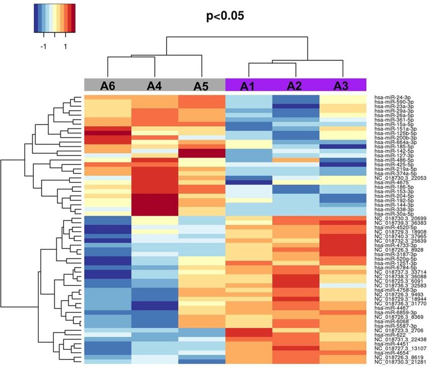
www.nature.com/scientificreports/
Cat ID Sample ID Status Age at death Gender Type Analysis Amyloid affected tissues
Kidneys, spleen, lymph-nodes, gut associated lymphoid tissue,
A1 PN# 1/08 A Affected 2 M FFPE Proteomics and miRNAs
parathyroids
A2 PN# 160/09 Affected 5 F FFPE Proteomics and miRNAs Kidneys
A3 PN# 233/02 Affected 4 NF FFPE Proteomics and miRNAs Kidneys
A4 PN# 119/11 Healthy 5,8 NF FFPE Proteomics and miRNAs = = =
A5 PN# 131/12 Healthy 6 F FFPE Proteomics and miRNAs = = =
A6 PN# 44/14 Healthy 12 NF FFPE Proteomics and miRNAs = = =
Kidneys, Spleen, lymph-nodes, intestinal lamina propria, liver,
S1 Fact#18,778 Affected 4 F Skeletal muscle Genomics
thyroid and parathyroids
S2 Fact#16,515 Affected 6 F Skeletal muscle Genomics Kidneys
Table 1. Abyssinian samples used in the study. “Sample_ID”: Fcat# is the accession number at the whole
genome sequence feline 99 Lives initiative, PN# is the protocol number at the Department of Veterinary
Medicine, University of Milan. “Cat_ID” is the identification number used in the main text, Fig. 4 and Fig. 5.
“Status” is the health status related to amyloidosis. “Age at death” is reported as years, months. “Gender”:
M = Male, F = Female, NF = Neutered Female. “Type” is the specimen DNA has been extracted from. “Analysis”
are the omic analyses the sample has been subjected to. “Amyloid affected tissues” are the tissues with amyloid
deposits at necropsy.
which are associated with p lasmacytomas15. In non-humans, evidence of genetic predisposition to develop AA
amyloidosis has been reported in the Shar-Pei dog breed16,17, in captive cheetahs (Acinoynx jubatus18), black-
footed cats (Felis nigripes19), and domestic cat (Felis catus).
The domestic cat is reported to spontaneously develop amyloidosis at a young age and often without evidence
of a pre-existent inflammatory condition. The disease has been described as primary and familial in two breeds:
the Abyssinian, with a mainly renal form20,21, and the Siamese with a mainly hepatic form22,23. The three consist-
ently reported features in Abyssinian renal amyloidosis are (i) the occurrence in related cats, (ii) the systemic
deposition of AA protein with a significantly higher presence in kidneys, and (iii) the early death from renal
failure. The trait is described as autosomal, and the influence of sex on the onset or development of the disease
has not been reported in cats. The prevalence of the disease in any cat breed is unknown. Despite the “familial”
transmission23, the mode of inheritance remained u ndetermined23,24. In a pioneering s tudy25, the pre-eminent
peptide in the amyloid renal deposits of an affected Abyssinian cat with a diagnosis of AA amyloidosis was
sequenced. The complete primary structure of the SAA protein showed the amino-terminal residues were het-
erogeneous among different species of mammals, indicating a high tolerance for mutations in this r egion26. The
cDNA sequencing of the N-terminal region in affected Abyssinians, Siameses, and domestic shorthair c ats24–27
revealed some non-synonymous substitutions, however association of any genomic variation with the disease
was not established.
In the last decade, mass spectrometry (MS)-based methods for human amyloid typing have been developed.
Unlike the immunohistochemistry approach, which requires a panel of precursor specific antibodies and sub-
stantial observer expertise, MS determines objective identification of the proteins, allowing characterization of
the deposits and the predominant fibril t ype28. Several proteomic approaches are now considered new clinical
standards for amyloid typing, either on formalin-fixed paraffin-embedded (FFPE) or unfixed frozen specimens.
Macrodissection or laser microdissection techniques are used to identify amyloid fibril composition of the
major amyloid types, such as ATTR, AL, A A29, as well as rarer amyloid types: hereditary g elsolin30, ApoA-I31
and ApoA-IV32.
Studies in humans also highlighted the potential role of miRNAs in the accumulation of amyloid fi brils33,34. In
cats, miRNA profiling is a rather recent achievement. The kidney has been profiled per s e35 and some healthy and
affected tissues/organs have been profiled to underlie their miRNA differential e xpression36,37. A more detailed
characterization of the kidney miRNAome in cats was achieved by sequencing a feline kidney cell line before
and after infection with mink enteritis virus38 and, more recently, using normal feline tissues39.
On tissues from complete necropsied cats, the following analyses were performed: (i) the whole genome vari-
ation of two affected Abyssinians versus 195 not affected cats of different breeds, (ii) the proteome from FFPE
kidney specimens of three affected and three non-affected Abyssinians, and (iii) the miRNAome from the same
six FFPE specimens. Data of this multi-omics investigation were integrated to hypothesize potential molecular
mechanisms underlying the renal familial amyloidosis of the Abyssinian cat.
Methods
Eight pedigreed Abyssinian cats subjected to complete necropsy at University Services during the past decade
were included in the study; their tissues were donated by consenting owners for research (Table 1). All subjects
were enrolled and classified according to their pathology reports, including gross necropsy and histology. The
status was categorized as “affected” when the cat died at five years of age or younger, with a pathology report of
amyloidosis confirmed by renal tissue positivity to Congo red staining with and without 0.25% acidified potas-
sium permanganate pre-treatment and excluding other previous or intercurrent conditions, including acute and
chronic inflammatory processes. In veterinary pathology, treatment with potassium permanganate solution of
Congo Red allows identifying amyloid AA because amyloid AA becomes discolored, loses its affinity with Congo
Scientific Reports | (2021) 11:8339 | https://doi.org/10.1038/s41598-021-87168-0 2
Vol:.(1234567890)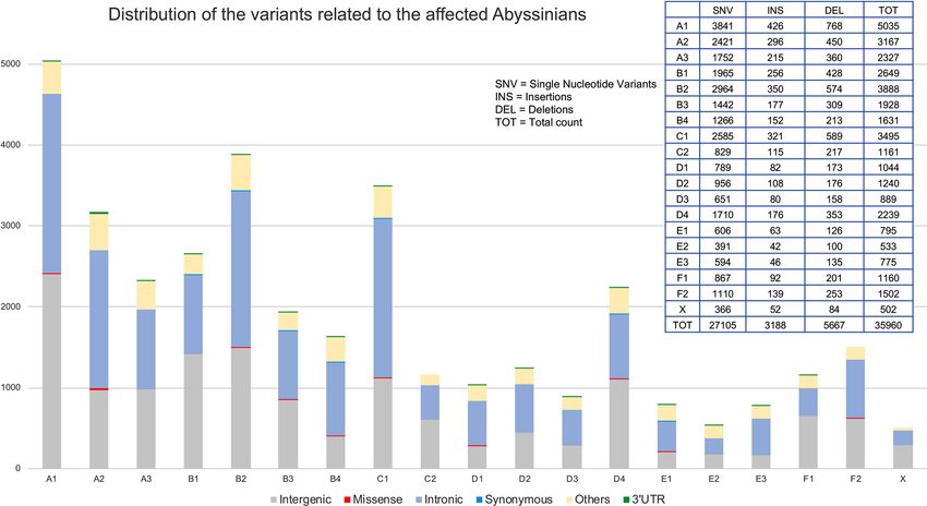
www.nature.com/scientificreports/
Red, and loses birefringence properties40. The subjects classified as “healthy” were three cats older than five years
at death and with a report excluding amyloidosis and other disorders affecting kidney or both acute and chronic
inflammatory systemic processes (controls). The cats were not first-degree relatives, but all belonged to related
genetic lines where amyloidosis frequently occurred. Age, gender, specimens, analyses, and amyloidosis affected
organs for each cat are also reported in Table 1.
Genomic DNAs from frozen skeletal muscles of two affected females (S1, S2) were whole-genome sequenced
(WGS) at the University of Missouri in the context of the 99 Lives initiative and archived following the Institu-
tional Animal Care and Use Committee protocol study protocols 9056, 9178, and 9642.
Formalin-Fixed Paraffin-Embedded (FFPE) kidney specimens of six cats, three with amyloidosis (one male,
one female, one neutered female; A1, A2, A3 respectively) and three healthy (non-amyloidosis affected: one
female, two neutered females; A5, A4, A6 respectively), were available at the Department of Veterinary Medicine,
University of Milan and were analyzed individually for both proteomics and miRNAomics.
WGS data were produced as previously d escribed41 and are accessible as part of the feline 99 Lives initiative
(www.f eline genet ics.m
issou
ri.e du/9 9Live s). Details of the whole genome sequencing, sequence processing, vari-
ant calling, and NCBI accession numbers have been previously r eported42. The WGS data were aligned to the
V9.0 Felis catus reference assembly43. WGS of 195 additional cats of different breeds and random-bred cats from
the 99 Lives project (Control Population—Supplementary Table 1) were used for genomic variants detection. The
Control Population included two other Abyssinians, one was the reference genome, and the other was enrolled in
the 99 Lives Project for a different trait. All the WGS data were available via the 99 Lives project as Variant Call
Format (VCF) file, retrieved from the Felis catus reference genome vs 9.0. The file included 84,005,772 variants.
The function “Activate Variants by Genotype Count Threshold” available through the SNP & Variation Suite
v8.4 of the Golden Helix SVS software (Golden Helix, Inc., Bozeman, MT, www.goldenhelix.com) was used to
detect the disease-associated variants. The VCF file was used as input, all the samples were set as “individual
unrelated samples”, the two affected Abyssinians were set on “TRUE” and all the other cats were set on “FALSE”
to be considered as controls. As the Control Population could theoretically include disease carriers, thresholds
were set to consider only the variants present in both the affected cats, either with a heterozygous or homozygous
genotype, and present in the remaining 195 cats with genotype frequency < 2% (counting both homozygous
and heterozygous). The resulting variants were annotated and investigated for their potential function with the
Ensembl Variant Effect Predictor (VEP), using the vs 9 of the cat genome a ssembly44. Missense, frameshift, and
3′UTR variants affecting the miRNA binding site were integrated with proteome and miRNAome data.
Proteome analysis was performed on kidney FFPE blocks. Two slices of 10 μm were cut from each block of
the three affected and three healthy cats, Table 1. Specimens were mounted on glass slides and deparaffinized
by incubating through two changes of xylene for 10 min each. The slices were rehydrated through a series of
graded ethanol for 5 min each (100%, 95%, 70%), distilled water for 5 min, and then incubated in 50 mM Tris
HCl buffer with mini protease inhibitors cocktail, for at least 30 min. Hydrated tissue slices were harvested with
a scalpel blade and homogenized using a potter homogenizer in extraction buffer containing 8 M urea, 20 mM
Hepes pH 8, with mini protease inhibitors cocktail. The homogenate was centrifuged (15 min, + 4 °C) to pellet
non-homogenized tissue and cellular debris. The supernatant containing proteins were quantified with Brad-
ford’s protein assay. Extracted proteins were subjected to reduction, alkylation, and protein digestion by using
sequence-grade trypsin (Roche) overnight at 37 °C. The digestion was quenched by the addition of 2 μL of 98%
formic acid, into the sample solution45. The proteolytic digest was desalted using a reversed-phase Zip-Tip C18
pipette tip (Millipore)46 and evaporated at 65 °C in a SpeedVac. Before Liquid Chromatograph Mass Spectrom-
eter (LC–MS), samples were reconstituted in 0.1% formic acid and the Liquid Chromatography-electrospray
Ionization-tandem Mass Spectrometry (LC–ESI–MS/MS) analysis was performed as p ublished47.
MS spectra were searched against the Felis catus reference NCBI sequence database (release March 2019) by
MaxQuant (version 1.6.0.1) using the following parameters: initial maximum allowed mass deviation of 15 ppm
for monoisotopic precursor ions and 0.5 Da for MS/MS peaks, trypsin enzyme specificity, a maximum of two
missed cleavages, carbamidomethyl cysteine as a fixed modification, N-terminal acetylation, and methionine
oxidation, as variable modifications. False protein identification rate (1%) was estimated by searching MS/MS
spectra against the corresponding reversed-sequence (Decoy) database. The minimum required peptide length
was set to 6 amino acids and the minimum number of unique peptides supporting protein identification was
set to 1. Quantification in MaxQuant was performed using label-free quantification algorithms (LFQ) based on
the extracted ion intensity of precursor ions47. This extraction procedure does not distinguish between fibrillar
and fibril associated soluble molecules, and this type of MS analysis cannot ascertain the locations of the specific
proteins with confidence, because the aim was to characterize the proteome of the affected kidney.
Only proteins detectable and quantifiable in at least 2 out of 3 biological replicates were considered as posi-
tively identified in a sample and used for statistical analyses. Statistical analyses of Max Quant results were
performed using the Perseus software module (version 1.4.0.8, www.biochem.mpg.de/mann/tools/) to iden-
tify proteins differentially expressed among the different c onditions48. Proteins were considered differentially
expressed if they were present only in one condition or showed a significant t-test difference (p-value ≤ 0.05).
Bioinformatic analyses were carried out by Panther software (Version 12.1)49 and DAVID software (release
6.8)50 to cluster enriched annotation groups of Cellular Component (GOCC), Biological Process (GOBP),
Molecular Function (GOMF) and Pathways within the set of proteins up-regulated or present only in affected
cats. The functional grouping was based on p-value ≤ 0.05 and at least three counts51. The mass spectrometry
proteomics data have been deposited to the ProteomeXchange Consortium via the P RIDE52 partner repository
with the dataset identifier PXD024140.
Kidney miRNAome analysis was performed on five sections of 10 μm sliced from the same six blocks used for
the proteome analysis (Table 1). Total RNA was isolated using the miRNeasy FFPE Kit, with minor modification
to the manufacturer instructions (Qiagen Handbook 06/2015). Briefly, after deparaffinization, the incubation
Scientific Reports | (2021) 11:8339 | https://doi.org/10.1038/s41598-021-87168-0 3
Vol.:(0123456789)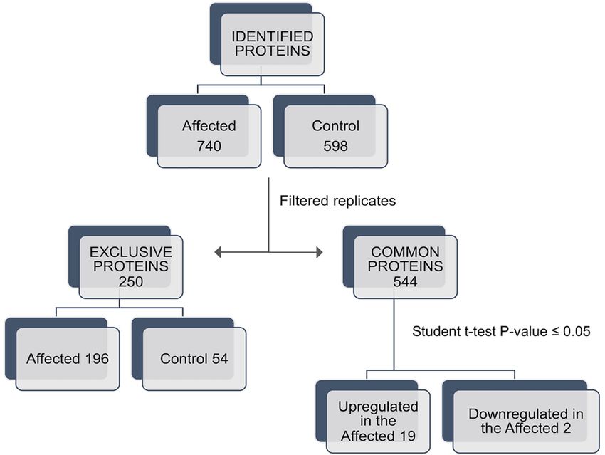
www.nature.com/scientificreports/
Figure 1. Overview of the different types of variants identified by Whole Genome Sequencing in the two
affected Abyssinian cats compared to the Control Population with the Golden helix SVS Software. Variants
distribution is reported for each cat chromosome. Variants referred to the class of “Other” included 5′UTR
variants, downstream and upstream gene variants, non-coding transcript exon variants, and splice region
variants.
to dewax tissue slices was increased up to 8 min, and gently shaking was applied. Samples were then eluted in
25 µl of RNase-free water. Isolated RNAs were quantified with the Qubit RNA HS (High Sensitivity) Assay Kits
(#Q32855, Life Technologies) and their Quality Control (QC) was performed by Bioanalyzer Agilent RNA 6000
Nano Assay (#5067–1511, Agilent Technologies) in Agilent 2100 Bioanalyzer.
Approximately 200 ng of isolated RNA from individual kidneys was sequenced using the small RNA-seq
kit by Illumina and the Illumina NextSeq500 platform, according to the Company’s protocol. The read length
obtained was 1 × 75 bp for a total of 30 million reads per sample. The QC of the reads was assessed with FastQC
(https://www.bioinformatics.babraham.ac.uk/projects/fastqc/) and the adapter sequences were removed using
Cutadapt53. MiRNA mapping and profiling were obtained with m iRDeep254 and miRNAs were matched to the
human homologs present in the miRbase database vs22.155, as the cat miRNAome is still not available in the
online databases.
MiRNAs with less than 10 reads per sample were removed from the dataset. Raw counts were normalized
with the trimmed mean of the M-value method (TMM) to reduce errors and expressed as log2 counts per mil-
lion. All the filtered miRNAs were used to evaluate the sample distributions through a multidimensional scaling
(MDS) plot performed with R Studio routine (https://rstudio.com/products/rpackages/). Finally, the differential
expression between miRNAs in affected versus control cats was assessed with a moderated t-test by limma, and
nominal P-values were adjusted by the Benjamini–Hochberg method. For this analysis, only miRNAs having
the genomic region supported by the highest number of reads were considered, and the significant miRNAs
(P-value < 0.05) were represented using a heatmap. The heatmap was created with the R package "gplots", version
3.03 (https://rstudio.com/products/rpackages/).
MiRNA precursor sequences (pre-sequences) obtained with miRDeep2 that did not match any human miR-
NAs were aligned on miRbase database vs22.1 against C. familiaris, B. taurus, S. scrofa, M. musculus, and again
H. sapiens. (BLASTN function).
For the identification of miRNA genes, the pre-sequences were aligned to the cat reference genome vs9.0
(BLAST tool of Ensembl genome browser 97). MiRNet d atabase56 was used to predict if the identified Abyssin-
ian miRNAs could target and regulate the proteins detected by proteomics. The prediction was made in human
miRNAome since the cat was not available online, and the kidney was specified as the tissue in which the inter-
actions take place. The identification of miRNAs binding sites on the mutated 3′UTR regions was performed
through a customized algorithm retrieved from TargetScan vs7.257.
Results
Genome variant detection. Comparing the variants of the two affected Abyssinians to those of the 195
cats of the Control Population, 35,960 potential disease-associated variants were detected, the majority (31,045)
mapped to intergenic or intronic regions (Fig. 1). The second most common variants (4623) were represented by
Scientific Reports | (2021) 11:8339 | https://doi.org/10.1038/s41598-021-87168-0 4
Vol:.(1234567890)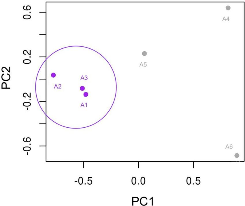
www.nature.com/scientificreports/
Figure 2. Venn diagram of the proteins identified in FFPE kidney tissues from cats affected by renal
amyloidosis (AMY) and healthy (CTR). Only proteins present and quantified in at least 2 out of 3 repeats in
each group were considered as positively identified.
Figure 3. Workflow of the proteomic approach. A shotgun proteomic analysis was performed on FFPE
kidney tissue from cats affected by renal amyloidosis (Affected) and healthy (Control). Statistical analyses were
performed using the Perseus software (version 1.4.0.8, www.biochem.mpg.de/mann/tools/).
the category of “Other” variants (Fig. 1) that included 5′UTR variants, downstream and upstream gene variants,
non-coding transcript exon variants, and splice region variants. The 35,960 variants comprised 27,105 single
nucleotide variants (SNV), 5667 deletions and 3188 insertions (Fig. 1). Considering the coding gene variants,
101 missense variants and one frameshift deletion were identified. Also, 84 variants mapping in 3′UTRs were
detected and selected for additional analyses, given that SNVs in 3′UTR regions can modify miRNA binding
sites, which can compromise gene expression58 (Supplementary Table 2). Overall, 186 variants were considered
for additional analyses, in which 23 were homozygous and 113 heterozygous in both the affected cats and 50
were homozygous in one affected cat and heterozygous in the other (Supplementary Table 2).
Kidney Proteome Analysis. Specimens, used for both proteome and miRNAome analyses (Table 1), came
from the same six cats and in which both acute and chronic inflammatory processes were excluded with total
necropsy and multi-organ histology. In affected cats, the kidneys were characterized by massive amyloid depos-
its. Focal to diffuse amyloid deposits had a typical salmon-pink color on Congo red staining in bright field and
showed the characteristic apple-green birefringence under polarized light (Supplementary Fig. 1A, B). In the
kidneys of the affected cats, amyloid deposition was found in interstitium, glomeruli, and blood vessels wall.
Other organs of the affected cats with amyloid deposition are listed in Table 1.
Birefringence was not detected in potassium permanganate treated sections. Amyloid deposits were not
detected in Congo red-stained sections of healthy cats observed both in bright field and under polarized light.
To characterize the protein component of the deposits, a quantitative label-free shotgun proteomic approach
was adopted. The MS/MS data identify 740 proteins in kidneys with amyloid deposits and 598 proteins in the
control kidneys. Proteins (n = 544) were expressed in both pathological and healthy tissues, while 196 proteins
were exclusively present in the deposits and 54 proteins were expressed only in the controls (Fig. 2). Out of
the 544 proteins common to the healthy and disease samples, 21 were differentially expressed (p-value ≤ 0.05,
Student’s t-test): 19 proteins were up-regulated, and two proteins were down-regulated in cats suffering from
amyloidosis (Fig. 3).
Scientific Reports | (2021) 11:8339 | https://doi.org/10.1038/s41598-021-87168-0 5
Vol.:(0123456789)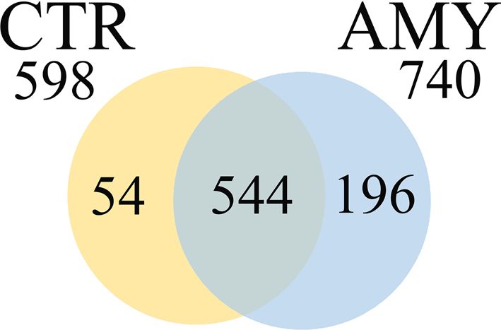
www.nature.com/scientificreports/
Figure 4. MDS plot realized with R Studio (https://www.rstudio.com/products/rpackages/), showing the
cluster of the affected cats, based on the 258 miRNAs expression (log2 counts per million). Affected individuals
(purple) are grouped on the left, while the healthy individuals are represented by grey dots on the right.
The exclusive and the differentially expressed proteins are listed in Supplementary Tables 3 and 4. Among
proteins exclusively detected in pathological kidneys, SAA, ApoA-IV, and TTR were identified, with SAA being
prevalent (Supplementary Table 3). By DAVID and Panther analyses, up-regulated and exclusively expressed
protein in amyloidosis affected cats were related to main cellular components such as ribosome and extracellular
matrix, as well as to cellular pathways involving oxidoreductase activity, the TCA cycle, and blood coagulation
(Supplementary Table 5 and 6). These proteins were clustered in the following functional networks according
to GO analysis (Supplementary Fig. 2: extracellular space region and matrix, metabolism, inhibition of serine
protease activity and coagulation, antioxidant activity, impact on the structural constituent of ribosome, and
translation.
Kidney miRNAome Analysis. The quality of the total miRNA isolation was overall good and is shown
in the length and quality plots of Supplementary Fig. 3. Weak peaks, corresponding to a small amount of other
longer types of RNAs, were noticed for samples A4 and A5 that underwent a less efficient deparaffinising treat-
ment (Supplementary Fig. 3).
For smallRNA-seq, the number of reads per sample ranged from 27,607,431 to 52,126,090 (mean value
40,267,032), with the highest number of reads per samples in the range of the 18–23 bp in length, which is in
accordance with the standard 18-22 bp length of a mature miRNA sequence. Samples A1 and A4 showed more
reads with 10–17 bp length, which could be addressed to the presence of non-miRNA short RNAs. Overall, a
good sequencing quality was obtained (Supplementary Fig. 4). Sequencing data in fastq format were deposited in
the ArrayExpress database at EMBL-EBI (www.e bi.a c.u k/a rraye xpres s/) under accession number E-MTAB- 8556.
The library size, the miRNA density, and the read count were evaluated before and after the normalization
with the TMM method and are reported in Supplementary Fig. 5. A total of 854 different miRNA pre-sequences
were identified with miRDeep2, corresponding to 618 unique mature sequences, reported with the same miRNA
name (Supplementary Table 7). After filtering and normalization with miRDeep2, only 258 of 618 miRNA mature
sequences were considered. The total read count for these miRNAs ranged from 7 reads (hsa-miR-4529-3p) to
29,052,681 reads (hsa-miR-10a-5p), with 58 miRNAs having less than 1,000 reads, 146 miRNAs ranging between
1,000 and 100,000 reads, and 45 miRNAs with more than 100,000 reads (Supplementary Table 7). Out of the
258 miRNAs, 71 had no match with human miRNAs according to miRDeep2, therefore the name assigned by
the software (prefix “NC”) was retained. The remaining 187 were named according to the human nomenclature
(prefix “hsa-mir”) (Supplementary Table 7). The manual BLAST of these 71 NC_miRNA pre-sequences showed
eight new matches with humans that were not identified by miRDeep2 and were therefore added to the 187 “hsa-
mir” for a total of 195 human miRNAs (Supplementary Table 8). Two miRNAs matched only to a bovine and a
swine miRNA, while 61 miRNAs remained “NC” and therefore provisionally considered feline-specific miRNAs.
All the filtered miRNAs (n = 258) were used to evaluate the sample distribution with an MDS plot, and the
affected cats clustered in a separate group (Fig. 4). The differential expression analysis showed 60 significant miR-
NAs (P-value < 0.05—Supplementary Table 9) with seven miRNAs significant also after the Benjamin-Hochberg
correction (Adj. P-value < 0.05; Table 2).
The results shown by the MDS (Fig. 4) were confirmed with the heatmap based on the P-values of the 60
significant miRNAs. The plot showed four perfectly distinct blocks, with 27 miRNAs down-regulated in the
affected samples and the remaining 33 miRNAs with the opposite trend (Fig. 5).
Out of the 60 miRNAs, 37 were already known in feline literature38,39 and three had human homologous.
The remaining 20 miRNAs were considered new and exclusive to the cat (Supplementary Table 10). Twenty-one
Scientific Reports | (2021) 11:8339 | https://doi.org/10.1038/s41598-021-87168-0 6
Vol:.(1234567890)www.nature.com/scientificreports/
miRNA_ID (miRDeep2) P-Value Adj. P-Val Read count
NC_018730.3_21281 0.0001 0.0189 69,021
NC_018727.3_13107 0.0002 0.0189 144
hsa-miR-4451 0.0002 0.0189 10,373
NC_018731.3_22438 0.0005 0.0318 192,694
NC_018726.3_8619 0.0007 0.0318 183
hsa-miR-4654 0.0007 0.0318 29
hsa-miR-186-5p 0.0011 0.0426 575,679
Table 2. List of the seven miRNAs with significant differential expression in affected and control cats also after
the Benjamini–Hochberg correction (Adjusted P-value < 0.05).
Figure 5. Heatmap created using the R package "gplots", version 3.03 (https://rstudio.com/products/rpack
ages/) with the 60 significant miRNAs (nominal P-val < 0.05). The grey bar and the purple bar represent the
healthy (A4, A5, and A6) and the affected (A1, A2, A3) Abyssinians, respectively. The shades of blue refer to
down-regulated miRNAs while the shades of red refer to up-regulated miRNAs.
miRNA genes were already annotated, 37 aligned in non-annotated regions and one did not align to the cat
reference genome V9.043 (Supplementary Table 10).
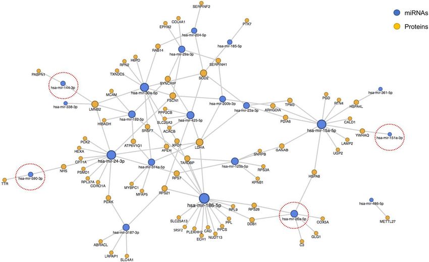
Omics integration. Considering their interactions, 89 proteins are suggested to have regulation by 22 miR-
NAs (Supplementary Table 11). Hsa-miR-186-5p, hsa-miR-15a-5p and hsa-miR-24-3p had the largest number
of targets (n = 18). Almost all the proteins in the network were expressed exclusively in the affected tissues, with
only 18 out of 89 proteins exclusively present in the healthy controls (Supplementary Table 11). A clearer over-
view of the interactions that exclusively characterized the affected kidneys excluded these 18 proteins (Fig. 6).
While most of the miRNAs regulated both the proteins related to the affected and the control cats, four down-
regulated miRNAs are suggested to interact only with proteins exclusive or up-regulated in the affected tissues
(sa-miR-26a-5p, hsa-miR-144-3p, hsa-miR-151a-3p and hsa-miR-590-3p, red circles in Fig. 6).
Searching for miRNA targets within the 84 3′UTR regions harboring the variants found by WGS, 14 miRNAs
had their binding site in the mutated 3′UTR of 15 different genes (Supplementary Table 12).
Discussion
Amyloidosis samples from cats are rare specimens, as not many breeders will take the expense to confirm their
cat’s diagnosis by biopsy or necropsy, thus the cats with an accurate diagnosis are limited. As in most species, AA
amyloid is related to long-term inflammation, the criterion adopted for the enrollment was the absence of acute/
chronic inflammatory or tumor lesions in fully necropsied cats. The diagnosis of amyloidosis was determined
by the histological specimens applying the Congo red staining routine protocols. In veterinary medicine, this
method is still the gold standard for the detection of amyloid, and its sensitivity to potassium permanganate
pre-treatment was used to preliminary identify amyloid deposits based on the fact that amyloid AA affinity for
Congo red is lost in tissue sections incubated in potassium permanganate solution.
Scientific Reports | (2021) 11:8339 | https://doi.org/10.1038/s41598-021-87168-0 7
Vol.:(0123456789)www.nature.com/scientificreports/
Figure 6. Network plot showing the interactions in pathological kidney tissue among the proteins exclusively
present or up-regulated in the affected specimens and the miRNAs identified in the study, according to miRNet
database. MiRNAs circled in red represent the only four miRNAs regulating exclusively the proteins related to
the affected cats and none of those found in the controls.
The study was almost exclusively on females (five of six). An effect of sex on amyloid deposition has never
been described in cats, even a total uniformity in sex may have helped to further reduce the background vari-
ability. Some observed differences could also have been related to age. The study design needed a difference of
age between the affected (younger) and the controls (older). Age-matched non-amyloidosis controls might have
added to the significance of the observations. But young cats carrying unknown amyloid predisposition genes
without lesions detectable with the classical histology at a very early stage could have been wrongly enrolled in
the healthy group. Age differences in organ transcriptomes in both healthy and affected (as described in TTR
amyloidosis in m ice59) might still have accounted for some of the findings. Proteins and miRNAs differentially
detected in affected compared to healthy were related to the pathology, but they could also have been modulated
by differences in age and other factors (diet, stress, etc.) which was impossible to control and manage because of
the different provenances of the cats. All the cats enrolled belonged to closely related lines, segregating for the
disease. Abyssinians are quite homogeneous g enetically60. Therefore, it can be assumed that these cats shared a
large part of their gene pool and this helped to reduce much of the variability not related to the disease.
Genome. In the present analysis genes that are known to be mutated in human inherited amyloidosis forms,
such as APP, and TTRhad variants matching the reference sequence. A great part of the variants detected in the
affected cats mapped to intronic or intergenic regions. Since the function of these variants is still quite unknown
in cats, future studies will improve the understanding of these polymorphisms. Several missense variants in cod-
ing genes, almost unique to the cases compared to the controls, were detected. These results must be confirmed
on a larger number of samples, but they can be helpful to define a mutation profile of the amyloidosis affected
Abyssinians. Two genes within those characterizing the affected cats (Supplementary Table 2) were reported in
the literature as related to AA amyloidosis: TREM1 and TNFRSF1A.
TREM1 encodes an immune receptor, belongs to the Immunoglobulin superfamily, and is expressed on mono-
nuclear phagocytes. Soluble TREM1 levels were recently demonstrated to be significantly higher in patients with
AA amyloidosis61. The mutations in the TREM1 gene in the two affected Abyssinians could bring to the protein
loss of function. Macrophages are instrumental in both accumulation and clearance of A A61, but TREM1 muta-
tion effect on amyloid deposition results unclear. Mutation in TNFRSF1A was recorded in one case of human AA
secondary amyloidosis in kidney. The patient was a heterozygous mutant for two genes, including TNFRSF1A,
developing an autoinflammatory syndrome characterized solely by amyloid d eposits62.
The genomic analysis also showed some variants in the affected cats which mapped in genes directly involved
in the development of diverse kidney syndromes. The Maltase—Glucoamylase gene (MGAM) had both one
insertion and one deletion in the 3′UTR region, with both the variants being homozygous in the two affected
cats (Supplementary Table 2). Despite no evidence supporting a correlation between MGAM and amyloidosis is
reported in the literature, the protein codified by this gene has been identified as an important biomarker in
acute kidney injury, due to its notably higher concentration in patients affected by this c ondition63. FANCD2
and FANCI Associated Nuclease 1 gene (FAN1) also had a missense variant homozygous in both the affected
Abyssinians. Mutations in this gene resulted in chronic kidney disease64. According to the GeneCards database
(https://www.genecards.org), other suggestive genes were DAZ Interacting Zinc Finger Protein 1 Like (DZIP1L),
involved in the development of polycystic kidney disease, and GREB1 Like Retinoic Acid Receptor Coactivator
(GREB1L), related to renal hypoplasia and/or renal agenesis.
Scientific Reports | (2021) 11:8339 | https://doi.org/10.1038/s41598-021-87168-0 8
Vol:.(1234567890)www.nature.com/scientificreports/
These data could lead to considering that the onset of amyloidosis or the kidney major target in the Abyssin-
ian cat can be facilitated by the presence of a compromised renal background.
Proteome. The analysis performed by LC–MS/MS mass spectrometry showed that affected kidney tissues
were characterized by well-known potentially amyloidogenic proteins as immunoglobulins, ApoA-IV, TTR, and
SAA, the latter was prevalent. These findings are consistent with the data reported in the literature on kidney
amyloid deposits, both in humans and cats. All the genes coding for these proteins had the reference alleles.
As shown in Supplementary Table 3, one of the most abundant proteins, exclusively identified in the affected
tissues, was the constant region of the immunoglobulin G2 heavy chain. Although the presence of immunoglobu-
lins heavy chains is not common in amyloidosis, rare instances of amyloid fibrils derived from the immunoglobu-
lin heavy chain, designated heavy chain amyloidosis (AH), have been r eported65,66. In this study, the abundant
presence of this immunoglobulin cannot be easily attributed to an ongoing inflammation process, because of
the criteria applied to the cat enrollment. Furthermore, the GO analysis conducted on the up-regulated and
exclusively expressed proteins in affected cats did not show an enrichment of the pathway of the inflammatory
immune response, excluding this scenario.
Interestingly, SAA was exclusively detected in the kidney of the affected cats. The Felis catus protein data-
base contains several isoforms of whole or partial SAA protein, as also reported in Supplementary Table 3.
The proteomic analysis identified three different isoforms of the SAA protein, two of which corresponding to
partial sequences, according to the databank. The presence of abundant wild type AA in the deposits but not
constant high SAA concentration, as experienced in the practice and reported in a longitudinal story on affected
Abyssinians67, has suggestive similarities both with the suggested mechanism of rapid incorporation of wild
type AA into the deposits drastically reducing the circulating SAA concentration, recorded in a mouse model68
and with the unresolved issue of a short seeding and nucleation that could make unnecessary chronic high SAA
concentrations for docking and misfolding in the t issues69.
Besides, Supplementary Table 3 reports the presence of TTR protein, which, in humans can cause familial
forms of amyloidosis, although, until now, there is no experimental evidence of TTR amyloidogenicity in cats.
The GO analysis clustered the up-regulated and exclusively expressed proteins in the affected Abyssinians
into five distinct classes: extracellular space region and matrix (ECM), metabolism, inhibition of serine protease
activity and coagulation, antioxidant activity and impact on the structural constituent of ribosome, and transla-
tion, (Supplementary Fig. 2, Supplementary Table 5, Supplementary Table 6), suggesting a mixed panorama of
proteins representative of different phases and aspects of the progressive tissue damage occurring from the onset
to the late disease. These results are also consistent with previous studies reporting that the protein composition
of insoluble deposits in the extracellular spaces can include ECM c omponents70 and suggesting that the ECM
modulates the level of amyloidogenic p eptides71. These effects could be related to direct regulation of the amyloi-
dogenic protein expression by ECM signaling molecules and/or to modulation of protein processing by the ECM
related cytoskeletal network in the cytoplasmic compartment72. Furthermore, the results of the present study
showed that renal amyloidosis affects catabolic processes. This is consistent with previous findings suggesting
that ATP, at concentrations of physiological significance to both intra and extracellular environments, actively
participates in fibrillogenesis and is involved in a different form of amyloidosis73.
Those that can be considered as potential secondary effects were represented in the remaining GO categories.
The present study reported an increase in categories related to serine-type endopeptidase inhibitor activity with
significant enrichment in proteins involved in this process and the blood coagulation pathway. Bleeding mani-
festations is a multifactorial event, which can occur in human patients with an advanced stage of amyloidosis.
In general, the vascular infiltration of amyloid fibrils can disrupt vascular integrity, and bleeding manifestations
in patients with renal amyloidosis may be augmented by acquired hemostatic abnormalities associated with
renal dysfunction74. Moreover, acquired deficiency of serine endopeptidase factor X (Stuart factor) is the most
common coagulation factor deficiency that has been identified in patients with a myloidosis75, probably due to
the apparent ability of amyloid fibrils to bind factors X 74.
The increased oxidoreductase and antioxidant activity in affected kidneys (Supplementary Table 5, Supple-
mentary Table 6) could be related to the progressive deposition of amyloid fibrils in organs. Indeed, increased
levels of oxidative stress around the amyloid deposits have been detected in a variety of amyloid diseases76.
However, it is still poorly understood whether oxidative stress is involved in the progression of amyloidosis.
Then again, Panther data analysis showed that renal amyloidosis in cats influenced both ribosomes and
translation processes. These data agreed with previous studies demonstrating that in vivo protein aggregate
accumulation was mainly composed of ribosomal c omponents77.
miRNAome. MiRNAs found in affected kidneys were used in gene target prediction study and the corre-
spondence of miRNA target messengers and proteins founded were evaluated, giving a deeper insight into the
cellular mechanisms of the disorder. The broad range of expression of miRNA analysed was already reported
in the literature57 and it was shown to be related to the functional roles of miRNAs in a specific tissue or in a
particular functional frame, which could influence their higher or lower expression.
Four significantly down-regulated miRNAs mainly characterized the interactions that took place exclu-
sively in the pathological tissues (Fig. 6). They target two known “amyloidogenic” proteins (TTR and PABPN1)
and proteins whose solubility was changed by the deposit’s presence78. Hsa-miR-144-3p and hsa-miR-590-3p
regulated the expression of TTR and Poly(A) Binding Protein Nuclear 1 (PABPN1) respectively, with the first
one reported as involved in the TTR amyloidosis d eposits58 and the second one in the amyloid-like fibrils of
79
the Oculopharyngeal Muscular Dystrophy . Hsa-miR-26a-5p regulated six genes including the one coding for
Complement C3 (C3), whose upregulation has been reported facilitating the deposit c learance80, and that coding
Scientific Reports | (2021) 11:8339 | https://doi.org/10.1038/s41598-021-87168-0 9
Vol.:(0123456789)www.nature.com/scientificreports/
for the Heat shock 70 kDa protein (HSPA8), which was one of the proteins demonstrated to lose their solubility
following an extracellular accumulation of amyloid in a mouse model of Alzheimer-type amyloidosis81. YWHAQ
was also among the proteins reported by Xu et al.78 and it was regulated by hsa-miR-151a-3p, the fourth down-
regulated miRNA.
The proteins—miRNAs network showed, once again, different phases of the disease. The two nodes of the
network belonging to hsa-miR-26a-5p and hsa-miR-151a-3p were representative of the cell response to the
mechanisms activated by the deposition of amyloid plaques, while the nodes represented by hsa-miR-144-3p
and hsa-miR-590-3p were suggestive of more direct potential triggers in the disease process.
The TargetScan database algorithm was used to predict which binding site of the differentially expressed
miRNAs occurred in the mutated 3′UTR regions of the genes identified through the genomic analysis. Fifteen
genes were mutated in a 3′UTR binding site.
Among these genes, two were the most interesting based on their functions. The variant in the 3′UTR region
of the Matrix Metalloproteinase 1 (MMP1) gene occurs in the binding site of miR-374a-5p, which was among
the down-regulated miRNAs. MMP1 is known for its role in the proteolysis of AA fibril proteins82. However,
like the majority of the 3′UTR variants identified, the variant characterizing the 3′UTR of MMP1 was a SNV and
one single base mismatch could not always completely compromise the binding of the miRNA. On the other
hand, an interesting result was noticed for the already mentioned MGAM gene, which was also characterized by
missense variants. In this case, the 3′UTR variant had a consistent insertion of 22 bp, occurring in the binding
site of miR-6784-5p.
The latter result was engaging considering the integration of all three omics. MGAM was the only gene
common to the three different analyses. It had both one insertion and one deletion in the 3′UTR region, both
homozygous in the two affected cats (Supplementary Table 2). In particular, the insertion in the 3′UTR of MGAM
occurred in the binding site of miR-6784-5p, one of the up-regulated miRNAs in the affected. Moreover, the
MGAM protein was among the proteins identified exclusively in the affected cats. Despite no evidence support-
ing a correlation between MGAM protein and amyloidosis was reported in the literature, it was identified as an
important biomarker in acute kidney injury, due to its notably higher concentration in patients affected by this
condition63. Therefore, the 3′UTR variant may cause the missed binding with miR-6784-5p and the consequent
loss of the protein expression silencing, explaining the exclusive presence of MGAM protein in the affected
tissues.
Conclusions. The multi-omics data integration emphasized the presence of different genes involved in
weakening and predisposing the kidney to the development of different syndromes, also creating a favorable
background for the insurgence of amyloidosis and its side effects. Major limits of the present study were the low
number of samples (due to their rarity), the difference in age (due to the features of the disease), the absence of
a transcriptome analysis (impracticable on FFPE), and the tools used for some data analysis (essentially based
on human references). Beyond that, the metadata available through this study can be an important hint for the
reconstruction of the molecular basis of feline and other amyloidoses.
Overall, the present data demonstrated Abyssinian amyloidosis as polygenic with several low-penetrance
genetic variants. This finding is consistent with a recent study where evidence for a complex genetic basis for the
disease in Oriental shorthair cats was found83. The genomic results suggested a complex molecular background
characterizing feline renal amyloidosis, with different mutated genes directly interacting with AA fibrils pathway,
and other mutated genes more involved in the kidney impairment.
The biochemical characterization of the kidney deposits by mainly AA and some other well-known amyloi-
dogenic proteins, such as TTR and ApoA-IV, confirmed what was reported in the classical feline literature and
classifies the Abyssinian form within the “AA amyloidoses”25. This is consistent with the loss of affinity for Congo
Red stain in potassium permanganate treated tissue sections of affected cats. All these classic amyloidogenic
proteins resulted to be wild type in the genomic analysis. This might suggest their massive accumulation in the
deposits as a secondary event.
Despite in a recent study on FFPEs of two unknown or mixed breed cats with systemic amyloidosis, proteome
analysis showed positivity to SAA but also APOE and not ApoA-IV84, differences in breed and absence of healthy
controls, information on age and sex make the comparison between the data of the two works incongruous.
The present work shows how a multi-omics approach can be useful for complex trait studies also in animals
and be a source for further annotations and meta-analyses. A list of 102 missense variants and 84 3′UTR variants
potentially related to Abyssinian feline amyloidosis is now available and can be used for a population screen-
ing in future studies to confirm possible associations. Proteomics gave further support in characterizing the
depositions in the affected tissues, confirming already known amyloidogenic proteins and outlining a protein
differential expression profile.
Finally, miRNAs differential expression profile was defined, and 20 novel feline miRNAs were identified.
Data availability
All the six FFPE samples (PN#) were out of the archives at the Department of Veterinary Medicine—University
of Milan—Organismo Preposto al Benessere degli Animali OPBA-56–2016 approval; all methods were carried
out following relevant guidelines and regulations. All the genomics data that support the findings of this study
are available through the 99Lives Initiative, are published in the NCBI SRA and the accession numbers are also
available in the Supplementary material of Buckley, R.M. et al.42; at https://w
ww.m
dpi.c om/2 073-4 425/1 1/6/6 82/
s1. The samples S1 and S2 are NCBI biosample# SAMN05980344 and SAMN08924116. All the mass spectrometry
proteomics data have been deposited to the ProteomeXchange Consortium via the PRIDE partner repository.
Scientific Reports | (2021) 11:8339 | https://doi.org/10.1038/s41598-021-87168-0 10
Vol:.(1234567890)www.nature.com/scientificreports/
The dataset identifier is PXD024140. All the miRNAs sequencing data in fastq format are available on the Array-
Express database at EMBL-EBI (www.ebi.ac.uk/arrayexpress/), under the accession number E-MTAB- 8556.
Received: 28 April 2020; Accepted: 19 March 2021
References
1. Holub, D. et al. Mass spectrometry amyloid typing is reproducible across multiple organ sites. BioMed Res. Int. 2019, 1–9 (2019).
2. Benson, M. D. et al. Amyloid nomenclature 2018: recommendations by the International Society of Amyloidosis (ISA) nomen-
clature committee. Amyloid 25, 215–219 (2018).
3. Merlini, G. et al. Effects of Tafamidis on Transthyretin stabilization and clinical outcomes in patients with non-Val30Met Tran-
sthyretin amyloidosis. J. Cardiovasc. Trans. Res. 6, 1011–1020 (2013).
4. Allsop, D. & Mayes, J. Amyloid β-peptide and Alzheimer’s disease. Essays Biochem. 56, 99–110 (2014).
5. Elitok, O. M., Elitok, B. & Unver, O. Renal amyloidosis in cattle with inflammatory diseases. J. Vet. Intern. 22, 450–455 (2008).
6. Ménsua, C. et al. Pathology of AA amyloidosis in domestic sheep and goats. Vet. Pathol. 40, 71–80 (2003).
7. Rideout, B. A. et al. Renal medullary amyloidosis in Dorcas gazelles. Vet. Pathol. 26, 129–135 (1989).
8. Schulze, C. et al. Generalized AA-amyloidosis in Siberian tigers (Panthera tigris altaica) with predominant renal medullary amyloid
deposition. Vet. Pathol. 35, 70–74 (1998).
9. Williams, J. H., Van Wilpe, E. & Momberg, M. Renal medullary AA amyloidosis, hepatocyte dissociation and multinucleated
hepatocytes in a 14-year-old free-ranging lioness (Panthera leo). J. S. Afr. Vet. Assoc. 76, 90–98 (2005).
10. Garner, M. M., Raymond, J. T., O’Brien, T. D., Nordhausen, R. W. & Russell, W. C. Amyloidosis in the black-footed ferret (Mustela
nigripes). J. Zoo Wildlife Med. 38, 32–41 (2007).
11. Guo, J.-T., Aldrich, C. E., Mason, W. S. & Pugh, J. C. Characterization of serum amyloid A protein mRNA expression and second-
ary amyloidosis in the domestic duck. Proc. Natl. Acad. Sci. 93, 14548–14553 (1996).
12. Ovelgönne, J. H., Landman, W. J., Gruys, E., Gielkens, A. L. & Peeters, B. P. Identical amyloid precursor proteins in two breeds of
chickens which differ in susceptibility to develop amyloid arthropathy. Amyloid 8, 41–51 (2001).
13. Andel, A. C. J., Gruys, E., Kroneman, J. & Veerkamp, J. Amyloid in the horse: a report of nine cases. Equine Vet. J. 20, 277–285
(1988).
14. Østevik, L., Gunnes, G., de Souza, G. A., Wien, T. N. & Sørby, R. Nasal and ocular amyloidosis in a 15-year-old horse. Acta Vet.
Scand. 56, 50 (2014).
15. Platz, S. J., Breuer, W., Geisel, O., Linke, R. P. & Hermanns, W. Identification of λ light chain amyloid in eight canine and two feline
extramedullary plasmacytomas. J. Comp. Pathol. 116, 45–54 (1997).
16. Segev, G. et al. Renal amyloidosis in dogs: a retrospective study of 91 cases with comparison of the disease between Shar-Pei and
non-Shar-Pei dogs. J. Vet. Intern. Med. 26, 259–268 (2012).
17. Olsson, M. et al. Thorough investigation of a canine Autoinflammatory Disease (AID) confirms one main risk locus and suggests
a modifier locus for amyloidosis. PLoS ONE 8, e75242 (2013).
18. Zhang, B. et al. Characterization of the cheetah serum amyloid A1 Gene: critical role and functional polymorphism of a cis-acting
element. J. Hered. 99, 355–363 (2008).
19. Terio, K. A., O’Brien, T., Lamberski, N., Famula, T. R. & Munson, L. Amyloidosis in Black-footed Cats (Felis nigripes). Vet. Pathol.
45, 393–400 (2008).
20. Chew, D. J., DiBartola, S. P., Boyce, J. T. & Gasper, P. W. Renal amyloidosis in related Abyssinian cats. J. Am. Vet. Med. Assoc. 181,
139–142 (1982).
21. Boyce, J. T., DiBartola, S. P., Chew, D. J. & Gasper, P. W. Familial renal amyloidosis in Abyssinian cats. Vet. Pathol. 21, 33–38 (1984).
22. van der Linde-Sipman, J. S., Niewold, T. A., Tooten, P. C. J., de Neijs-Backer, M. & Gruys, E. Generalized AA-amyloidosis in Siamese
and Oriental cats. Vet. Immunol. Immunop. 56, 1–10 (1997).
23. Niewold, T. A., van der Linde-Sipman, J. S., Murphy, C., Tooten, P. C. & Gruys, E. Familial amyloidosis in cats: Siamese and Abys-
sinian AA proteins differ in primary sequence and pattern of deposition. Amyloid 6, 205–209 (1999).
24. van Rossum, M. et al. Analysis of cDNA sequences of feline SAAs. Amyloid 11, 38–43 (2004).
25. DiBartola, S. P., Benson, M. D., Dwulet, F. E. & Cornacoff, J. B. Isolation and characterization of amyloid protein AA in the Abys-
sinian cat. Lab. Invest. 52, 485–489 (1985).
26. Kluve-Beckerman, B., Dwulet, F. E., DiBartonla, S. P. & Benson, M. D. Primary structures of dog and cat amyloid A proteins:
comparison to human AA. Comp. Biochem. Phy. B 94, 175–183 (1989).
27. Johnson, K. H. et al. Amino acid sequence variations in protein AA of cats with high and low incidences of AA amyloidosis. Comp.
Biochem. Physiol. B 94, 765–768 (1989).
28. Lavatelli, F. & Vrana, J. A. Proteomic typing of amyloid deposits in systemic amyloidoses. Amyloid 18, 177–182 (2011).
29. Vrana, J. A. et al. Classification of amyloidosis by laser microdissection and mass spectrometry-based proteomic analysis in clinical
biopsy specimens. Blood 114, 4957–4959 (2009).
30. Klein, C. J. et al. Mass spectrometric-based proteomic analysis of amyloid neuropathy type in nerve tissue. Arch. Neurol. 68, 1
(2011).
31. Rowczenio, D. et al. Amyloidogenicity and clinical phenotype associated with five novel mutations in apolipoprotein A-I. Am. J.
Pathol. 179, 1978–1987 (2011).
32. Sethi, S. et al. Medullary amyloidosis associated with apolipoprotein A-IV deposition. Kidney Int. 81, 201–206 (2012).
33. Weng, L. et al. Dysregulation of miRNAs in AL amyloidosis. Amyloid 18, 128–135 (2011).
34. Patel, N. et al. MicroRNAs can regulate human APP levels. Mol. Neurodegener. 3, 10 (2008).
35. Ichii, O. et al. MicroRNA expression profiling of cat and dog kidneys. Res. Vet. Sci. 96, 299–303 (2014).
36. Weber, K., Rostert, N., Bauersachs, S. & Wess, G. Serum microRNA profiles in cats with hypertrophic cardiomyopathy. Mol. Cell.
Biochem. 402, 171–180 (2015).
37. Cong, W. et al. Global miRNA expression profiling of domestic cat livers following acute Toxoplasma gondii infection. Oncotarget
8, 1 (2017).
38. Sun, J. et al. MicroRNA profile analysis of a feline kidney cell line before and after infection with mink enteritis virus. Gene 539,
224–229 (2014).
39. Laganà, A. et al. Discovery and characterization of the feline miRNAome. Sci. Rep. 7, 9263 (2017).
40. Wright, J. R., Calkins, E. & Humphrey, R. L. Potassium permanganate reaction in amyloidosis. Lab. Invest. 36, 274–281 (1977).
41. Gandolfi, B. et al. COLQ variant associated with Devon Rex and Sphynx feline hereditary myopathy. Anim. Genet. 46, 711–715
(2015).
42. Buckley, R. M. et al. Werewolf, there wolf: variants in Hairless associated with hypotrichia and roaning in the Lykoi cat breed.
Genes 11, 682 (2020).
Scientific Reports | (2021) 11:8339 | https://doi.org/10.1038/s41598-021-87168-0 11
Vol.:(0123456789)You can also read