RQCH AND RQCP CATALYZE PROCESSIVE POLY-ALANINE SYNTHESIS IN A RECONSTITUTED RIBOSOME-ASSOCIATED QUALITY CONTROL SYSTEM
←
→
Page content transcription
If your browser does not render page correctly, please read the page content below
Published online 13 July 2021                                                    Nucleic Acids Research, 2021, Vol. 49, No. 14 8355–8369
                                                                                                       https://doi.org/10.1093/nar/gkab589

RqcH and RqcP catalyze processive poly-alanine
synthesis in a reconstituted ribosome-associated
quality control system
Hiraku Takada1,2,3,* , Caillan Crowe-McAuliffe4 , Christine Polte4 , Zhanna Yu. Sidorova5,6 ,
Victoriia Murina2,3 , Gemma C. Atkinson8 , Andrey L. Konevega5,7,8 , Zoya Ignatova 4 ,
Daniel N. Wilson 4,* and Vasili Hauryliuk 2,3,9,10,*
1
 Faculty of Life Sciences, Kyoto Sangyo University, Kamigamo, Motoyama, Kita-ku, Kyoto 603-8555, Japan,

                                                                                                                                                              Downloaded from https://academic.oup.com/nar/article/49/14/8355/6320412 by guest on 20 October 2021
2
 Department of Molecular Biology, Umeå University, 90187 Umeå, Sweden, 3 Laboratory for Molecular Infection
Medicine Sweden (MIMS), Umeå University, 90187 Umeå, Sweden, 4 Institute for Biochemistry and Molecular
Biology, University of Hamburg, 20146 Hamburg, Germany, 5 Petersburg Nuclear Physics Institute named by B.P.
Konstantinov of National Research Centre “Kurchatov Institute”, 188300 Gatchina, Russia, 6 Russian Research
Institute of Hematology and Transfusiology of FMBA, 191024 Saint Petersburg, Russia, 7 Peter the Great St.
Petersburg Polytechnic University, 195251 Saint Petersburg, Russia, 8 National Research Centre “Kurchatov
Institute”, 123182 Moscow, Russia, 9 Department of Experimental Medical Science, Lund University, 221 00 Lund,
Sweden and 10 University of Tartu, Institute of Technology, 50411 Tartu, Estonia

Received May 18, 2021; Revised June 21, 2021; Editorial Decision June 22, 2021; Accepted June 24, 2021

ABSTRACT                                                                        tively, our findings provide mechanistic insight into
                                                                                the role of RqcH and RqcP in the bacterial RQC path-
In the cell, stalled ribosomes are rescued through
                                                                                way.
ribosome-associated protein quality-control (RQC)
pathways. After splitting of the stalled ribosome, a
C-terminal polyalanine ‘tail’ is added to the unfin-                            INTRODUCTION
ished polypeptide attached to the tRNA on the 50S                               In all cells, the process of synthesizing proteins by ‘reading’
ribosomal subunit. In Bacillus subtilis, polyalanine                            the coded instructions in mRNA is called translation, and it
tailing is catalyzed by the NEMF family protein RqcH,                           is carried out by the molecular machine called the ribosome.
in cooperation with RqcP. However, the mechanis-                                Damaged or truncated mRNAs are harmful to cells because
tic details of this process remain unclear. Here we                             they sequester ribosomes from active protein production
demonstrate that RqcH is responsible for tRNAAla se-                            and can result in the synthesis of cytotoxic truncated pro-
lection during RQC elongation, whereas RqcP lacks                               teins (1). Therefore, diverse ribosome rescue pathways have
                                                                                evolved in all domains of life to disassemble stalled ribo-
any tRNA specificity. The ribosomal protein uL11 is
                                                                                somal complexes (2–4). The first ribosome rescue pathway
crucial for RqcH, but not RqcP, recruitment to the                              to be identified in bacteria was the trans-translation sys-
50S subunit, and B. subtilis lacking uL11 are RQC-                              tem (5). The system consists of transfer-messenger RNA
deficient. Through mutational mapping, we identify                              (tmRNA; also referred to as small stable RNA A, ssrA)
critical residues within RqcH and RqcP that are im-                             that serves both as an mRNA template (6) and a tRNA
portant for interaction with the P-site tRNA and/or                             mimic (7). This RNA component of tmRNA is assisted
the 50S subunit. Additionally, we have reconstituted                            by small protein B (SmpB) (8). Acting as a tRNA, alanyl-
polyalanine-tailing in vitro and can demonstrate that                           tmRNA mediates the addition of alanine to ribosome-
RqcH and RqcP are necessary and sufficient for pro-                             stalled nascent polypeptide chains (7). Next, decoding of
cessivity in a minimal system. Moreover, the in vitro                           the ORF encoded within the tmRNA results in the addi-
reconstituted system recapitulates our in vivo find-                            tion of the short C-terminal ssrA peptide tag (6). This, in
                                                                                turn, marks the aberrant proteins for degradation by ClpA,
ings by reproducing the importance of conserved
                                                                                ClpXP and FtsH proteases (9). In addition to the trans-
residues of RqcH and RqcP for functionality. Collec-                            translation system, Escherichia coli possess two alternative

* To
   whom correspondence should be addressed. Tel: +46 70 60 90 493; Email: vasili.hauryliuk@med.lu.se
Correspondence may also be addressed to Hiraku Takada. Tel: +81 80 7800 6466; Email: hiraku.takada@cc.kyoto-su.ac.jp
Correspondence may also be addressed to Daniel N. Wilson. Tel: +49 40 42838 2841; Email: daniel.wilson@chemie.uni-hamburg.de


C The Author(s) 2021. Published by Oxford University Press on behalf of Nucleic Acids Research.
This is an Open Access article distributed under the terms of the Creative Commons Attribution License (http://creativecommons.org/licenses/by/4.0/), which
permits unrestricted reuse, distribution, and reproduction in any medium, provided the original work is properly cited.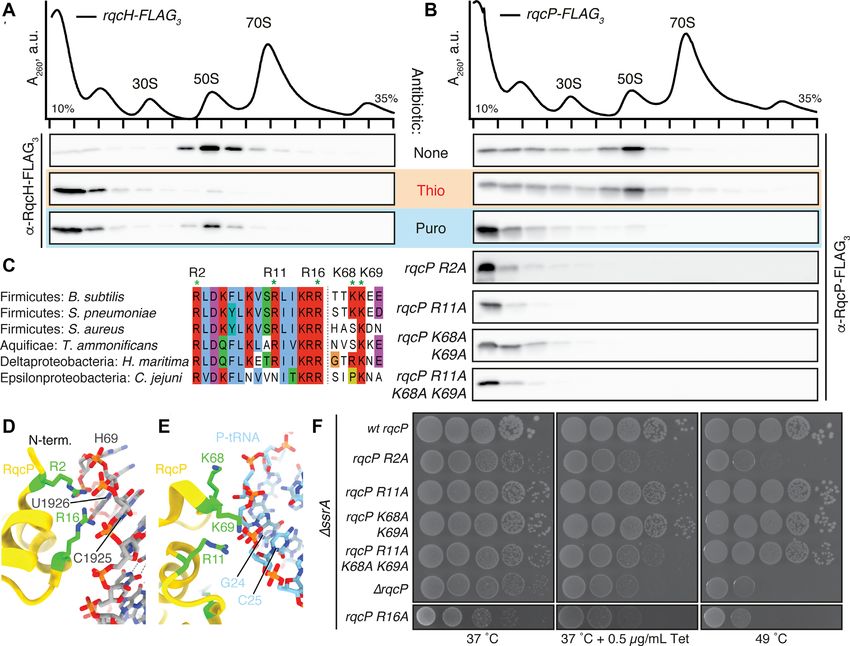
8356 Nucleic Acids Research, 2021, Vol. 49, No. 14
rescue systems that are related to or rely on the canonical that, in turn, act as degron tags recognized by the ClpP pro-
stop codon-dependent translation termination machinery tease (41). The rqcH gene genetically interacts with ssrA, as
involving class-1 release factors RF1 and RF2 (2,3). While the double deletion mutant ssrA and rqcH displays an in-
alternative rescue factor A (ArfA) recruits RF2 to release creased sensitivity to antibiotics that inhibit translation and
the stalled polypeptide in the absence of the A-site stop displays a synthetic growth defect at 45◦ C (41). This genetic
codon (10), ArfB acts as a peptidyl hydrolase itself (11). Un- interaction suggests that the two ribosome rescue systems
like the trans-translation system, which is universally con- have complementary functions in B. subtilis.
served in bacteria (12,13), ArfA and ArfB have patchy evo- Cryo-EM structures of RqcH-50S complexes isolated us-
lutionary distributions, and are missing, for example, in the ing in vivo pull-down approaches have established that the
model firmicute bacterium Bacillus subtilis or the human NFACT-N domain of RqcH is the key structural element
pathogen Francisella tularensis (14). Instead, these species mediating tRNAAla recognition (42,44), thus rationalizing
rely on release factor-dependent alternative rescue systems the earlier report of D97A/R98A substitution (RqcHDR ) in
ArfT (F. tularensis (15)) and BrfA (B. subtilis (16)). the domain ablating RQC functionality (41). Indeed, RqcH
Downloaded from https://academic.oup.com/nar/article/49/14/8355/6320412 by guest on 20 October 2021
The translation apparatus of both archaea and the eu- appears to interact with both tRNAAla isoacceptors, namely
karyotic cytoplasm lack the trans-translation and Arf/Brf tRNAAla(UGC) and tRNAAla(GGC) (42) (note that the latter
ribosome rescue systems. Instead, they rely on ribosome- was annotated as tRNAAla(IGC) in the original report due
associated protein quality control, RQC (17,18). RQC to the hybridization with the microarray probe designated
is executed by the concerted action of Ribosome qual- as Ala-IGC, Supplementary Table S1)––and the conserved
ity control complex subunits 1 (Rqc1) and 2 (Rqc2, aka 97 DR98 residues have been suggested to contribute to this
NEMF/SDCCAG1 in humans, Caliban in Drosophila and specificity by interacting with the anticodon–stem–loop of
Tae2 in yeast), E3 ubiquitin ligase Ltn1/Listerin and the these tRNAs, in particular the G35 at the central position
AAA ATPase Cdc48/p97 (19,20). Unlike the bacterial sys- of the anticodon (42,44) (Figure 1B,C). A similar variant
tems described above that operate on the 70S ribosomes, in yeast Rqc2 could bind to 50S-P-tRNA complexes, but
eukaryotic RQC operates on the large 60S subunit (19,20) not support CAT tailing (28). The structures also revealed
which can be generated by splitting the stalled ribosome that the M domain of RqcH is tethered to ribosomal pro-
by Dom34/Pelota-Hbs1 and ABCE1/Rli1 (21–25) or, al- tein uL11 located at the L7/L12 stalk base, suggesting that
ternatively, the RQC-trigger/ASC-1 complex (26,27). In this interaction is critical for RqcH function. Furthermore,
yeast, the resulting 60S-P-tRNA complex is a substrate analysis of the RqcH-50S RQC pull-down complexes led
for the mRNA-independent addition of C-terminal exten- to the discovery of RqcP, an Hsp15-family protein (42,44).
sions of poly-alanine co-polymerised with threonine (CAT By analysing the ensemble of generated cryo-EM struc-
tails) (28). CAT tailing progresses the stalled nascent pep- tures, a putative mechanism of RqcH/RqcP-driven poly-
tide through the exit tunnel, presenting lysine residues in alanine synthesis on the large 50S subunit was proposed
the nascent peptide that are substrates for ubiquitinylation (Figure 1D) (42). By associating with 50S-peptidyl-tRNA
by Ltn1 (29). Off the ribosome, CAT tails promote protein complexes, RqcP stabilizes the peptidyl-tRNA in the P-site,
aggregation (30,31) and serve as a degron tag promoting thus allowing efficient recruitment of RqcH:tRNAAla to
degradation by the proteasome (32). Recently, it has been the A-site. Consequent spontaneous transpeptidation and
shown that C-terminal tails mediated by the RQC machin- RqcP dissociation allow the 50S-bound tRNAs to sample
ery in mammalian cells and Drosophila melanogaster are different ribosomal binding sites. This results in departure
composed predominantly of alanine, with minor contribu- of the deacylated E-site tRNA and allows the peptidyl-
tion from other amino acids (33–35). tRNA extended with C-terminal alanine residue to adopt
Diverse bacterial species of phyla across the bacterial tree a hybrid A/P-like state. Rebinding of RqcP stabilizes the
of life encode NEMF-family fibronectin-binding protein A peptidyl-tRNA in the P-site, resulting in a complex with P-
(FpbA) proteins homologous to eukaryotic Rqc2 proteins site tRNA, RqcH and RqcP. This latter complex was the
(36). For decades, the exact molecular function of these pro- best-resolved state observed in cryo-EM reconstructions,
teins was unclear. Enterococcus faecalis EfbA (37), Liste- which we termed state B. An additional state, which was
ria monocytogenes FbpA (38,39) and Streptococcus pneumo- similar to state B, but with partially dissociated RqcH, was
niae PavA (40) were originally recognized as virulence fac- termed state B* and assumed to represent a transition to-
tors. The situation changed in 2019 when B. subtilis FpbA wards RqcH dissociation.
homologue YloA was demonstrated to be a functional ana- Here, we employ biochemical and structural techniques
logue as well as a homologue of eukaryotic Rqc2, which to dissect the role of RqcH and RqcP during bacterial RQC.
led to renaming of the protein to bacterial Rqc2 homolog, We show that RqcH can specifically select tRNAAla in the
RqcH (41). RqcH, like other NEMF family proteins, con- absence of the 50S subunit and that RqcP does not con-
tains a conserved core domain architecture of NFACT- tribute to this selection specificity. Indeed, we show that
N (NFACT means domain found in NEMF, FbpA, Cal- mutation of the conserved DR motif to AA leads to loss
iban, and Tae2), helix-hairpin-helix (HhH), coiled coils of this tRNAAla specificity, supporting the importance of
(CC), a small -hairpin-containing middle (M)-domain be- the motif for distinguishing tRNAAla from other tRNA
tween the two helices of the CC, and an NFACT-R do- species. A cryo-EM structure of a RqcHDR mutant in com-
main (28,36,41–43) (Figure 1A). RqcH recruits tRNAAla to plex with the peptidyl–tRNA–50S complex reveals that the
50S-peptidyl-tRNA complexes generated by an as yet un- NFACT-N domain of the RqcHDR mutant has disengaged
known molecular players that split stalled 70S ribosomes, from the anticodon stem loop of the P-site tRNA, thus pro-
and drives the addition of C-terminal poly-alanine tails viding a structural basis for the loss in specificity. In ad-
Nucleic Acids Research, 2021, Vol. 49, No. 14 8357
Downloaded from https://academic.oup.com/nar/article/49/14/8355/6320412 by guest on 20 October 2021
Figure 1. Bacterial ribosome-associated RQC is mediated by RqcH and RqcP. (A) RqcH domain composition. Amino acid positions are numbered as
per B. subtilis RqcH. (B) Sequence alignment of RqcH homologs from diverse bacteria. (C) Interaction between RqcH (HhH domain in lilac, NFACT-N
domain in tan, 97 DR98 motif in green) and P-tRNA (transparent cyan) in state B (PDB 7ASA (42)). (D) Overview of proposed RQC elongation cycle
mediated by RqcH and RqcP (42). Beginning with a 50S with trapped P-tRNA, binding and dissociation cycles of RqcH and RqcP, accompanied by
movement of tRNAs through the A-, P- and E-sites, results in mRNA- and 30S-independent elongation.
dition, we demonstrate that uL11 is critical for RqcH re- on a 10–40% sucrose gradient in overlay buffer (60 mM
cruitment to the 50S subunit and B. subtilis lacking uL11 NH4 Cl, 1 mM Mg(OAc)2 , 0.25 mM EDTA, 3 mM -
is RQC-deficient. We also identify critical residues within mercaptoethanol, 20 mM Tris–HCl pH 7.5) in a zonal rotor
RqcH and RqcP that are important for recruitment to the (Ti 15, Beckman, 18 h at 21 000 rpm). The peak containing
peptidyl-tRNA 50S complexes. Furthermore, we establish pure 50S subunits was pelleted by centrifugation (48 h at 35
an in vitro-reconstituted polyalanine-tailing assay, which we 000 rpm), and the final ribosomal preparation was dissolved
use to demonstrate that RqcH and RqcP are necessary and in HEPES:Polymix buffer with 5 mM MgOAc.
sufficient for processivity of polyalanine tailing. The in vitro-
reconstituted polyalanine-tailing assay opens the way for Growth assays
further studies to dissect the role of other factors involved
in bacterial RQC. B. subtilis 168 wild-type and deletion strains were pre-grown
on LB plates overnight at 30◦ C. Fresh individual colonies
were used to inoculate liquid LB medium cultures (OD600
MATERIALS AND METHODS adjusted to 0.01) at 37◦ C. Log phase cultures (OD600 of
Strains, plasmids (and details regarding the construction ≈0.4) diluted to OD600 of 0.1 were used to prepare 10- to
thereof) as well as oligonucleotides and synthetic DNA se- 105 -fold serial dilutions which were then spotted onto LB
quences used in this study are provided in Supplementary agar plates with or without 1 mM IPTG. The plates were
Table S2. Detailed description of protein and tRNA purifi- scored after 18 hours incubation at either 37◦ C or 49◦ C.
cation procedures is provided in the Supplementary meth-
ods. Immunoprecipitation of FLAG3 -tagged proteins
The experiments were performed as described earlier (42).
Multiple sequence alignment Strains expressing FLAG3 -tagged proteins were pre-grown
RqcH and RqcP sequences were extracted from the previ- on LB plates overnight at 30◦ C. Fresh individual colonies
ously reported dataset (42), aligned with MAFFT v7.164b were used for inoculation and grown in LB medium. 3×
with the L-ins-i strategy (45) and alignments were visualized 1 liter cultures were grown at 37◦ C to OD600 = 0.8. Cells
with Jalview (46). were collected by centrifugation (8000 rpm for 10 min at
4◦ C, JLA-16.25 Beckman Coulter rotor), pellets frozen in
liquid nitrogen and stored at –80◦ C. Cell pellets were re-
Preparation of B. subtilis 50S ribosomal subunits
suspended in 8 ml of cell opening buffer (95 mM KCl, 5
B. subtilis 70S was purified as described earlier (47), di- mM NH4 Cl, 20 mM HEPES (pH 7.5), 1 mM DTT, 15 mM
luted with low-magnesium HEPES:Polymix buffer (1 mM Mg(OAc)2 , 0.5 mM CaCl2 , 8 mM putrescine, 1 mM spermi-
MgOAc) and incubated on ice for 30 minutes to promote dine, 1 tablet of cOmplete™ EDTA-free Protease Inhibitor
the dissociation of subunits. The sample was then resolved Cocktail (Roche) per 50 ml of buffer) and disrupted using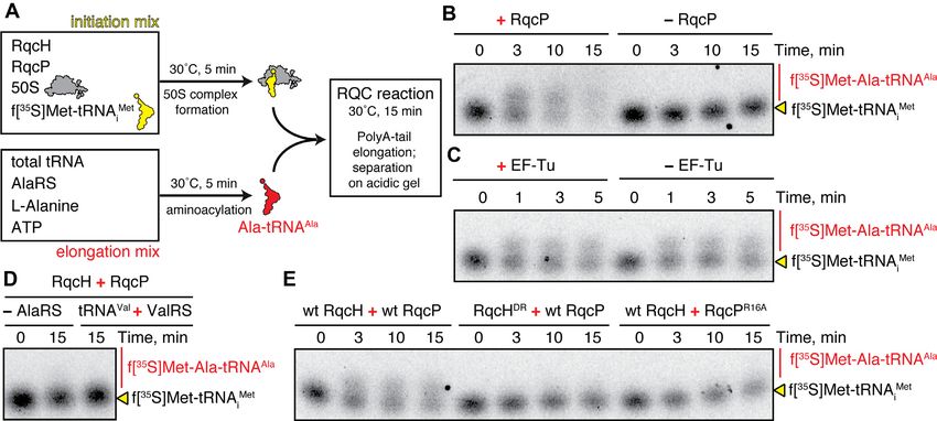
8358 Nucleic Acids Research, 2021, Vol. 49, No. 14
FastPrep homogeniser (MP Biomedicals) with 0.1 mm Zir- classification without angular sampling (53). The majority
conium beads (Techtum) in 6 cycles by 20 s with 3 min chill of particles were grouped into a single class containing a
on ice. Cell debris was removed by centrifugation at 14 800 50S ribosomal large subunit, P-tRNA, RqcP and weak sur-
rpm for 20 min 4◦ C in F241.5P rotor using a 149 Microfuge rounding density. This class was selected for another 3D re-
22R centrifuge (Beckman Coulter). The supernatant was finement, followed by partial signal subtraction with a mask
combined with 100 l of ANTI-FLAG M2 Affinity Gel around the A, P and E sites (54). Another 3D classification
(Sigma) pre-equilibrated in cell opening buffer, and incu- was then performed without angular sampling, the regular-
bated for 1.5 h at 4◦ C on a turning wheel (Fisherbrand™ ization parameter T set to 50, and the resolution used in
Multi-Purpose Tube Rotators). The samples were loaded on the expectation step limited to 7 Å. Classes of interest were
Micro Bio-Spin columns (Bio-Rad) pre-equilibrated in cell re-extracted with the original pixel size, refined, and post-
opening buffer, and washed 10 times with 1 ml of cell open- processed. FSCs were assessed using the ‘gold-standard’ ap-
ing buffer by gravity flow. RqcH-FLAG3 was eluted by ad- proach (55). Local resolution was estimated with ResMap
dition of 200 l opening buffer containing 0.1 mg/ml poly- (56). For analysis, parts of an existing model of a bacterial
Downloaded from https://academic.oup.com/nar/article/49/14/8355/6320412 by guest on 20 October 2021
FLAG peptide (Biotool, Bimake) for 45 min on a turning RQC complex (PDB: 7AS8) were fitted, by domain, into
wheel. All incubations, washes and elutions were performed density using ChimeraX (57).
at 4◦ C. The eluted sample was collected by centrifugation at
2000 rpm for 1 min 4◦ C in a F241.5P rotor using a 149 Mi-
Electrophoretic mobility shift assay (EMSA)
crofuge 22R centrifuge (Beckman Coulter). One aliquot of
the eluted sample was resolved on SDS-PAGE, the other Experiments were performed as described earlier (58).
was blotted on cryo-EM grids, and the remaining sample Before performing the experiment, stock mRNA(MVF) (5 -
was used for tRNA-array analyses. For SDS-PAGE anal- GGCAAGGAGGAGAUAAGAAUGGUUUUCUAAUA-
yses, 20 l aliquots of samples were mixed with 5 L of 3 ) was incubated for 2 min at 60◦ C to denature possible
5× SDS loading buffer and heated at 95◦ C for 15 min, and secondary structures. Reaction mixtures (10 L) in
denatured samples were loaded on 12% SDS-PAGE. SDS- HEPES:Polymix buffer (47) with 5 mM Mg2+ were assem-
gels were stained by ‘Blue-Silver’ Coomassie Staining (48) bled by adding either E. coli tRNAVal or B. subtilis tRNAAla
and washed with water for 6 h or overnight before imaging or tRNALys (0.1 M final concentration) as well as mRNA
with LAS4000 (GE Healthcare). mRNA(MVF) competitor (1 M final concentration),
followed by the addition of RqcH-HTF (either wild-type
or D97A R98A substituted). After incubation for 5 min
Preparation of cryo-EM grids
at 37◦ C, 4 L of 50% sucrose was added per sample, and
Eluted pull-down samples were kept on ice and loaded on the samples were electrophoretically resolved on a 12%
grids within 2 h after preparation without freezing. The con- Tris:borate:EDTA gel at 4◦ C (160–180 V) for 1–1.5 h.
centration of ribosomes in the samples was estimated from Gels were stained with SYBR Gold nucleic acid stain
SDS-PAGE gels by comparison of ribosomal band intensi- (Life Technologies) for 30 min, followed by visualization
ties in eluted samples with the bands from loaded ribosomes using a Typhoon Trio Variable Mode Imager (Amersham
with known concentration (Supplementary Figure S1). The Biosciences). Bands were quantified using ImageJ (59). The
concentration of ribosomes in elution of RqcHDR -FLAG3 efficiency of complex formation (effective concentration,
was approximately 50 nM. Samples were loaded on Quan- EC50 ± standard deviation) was calculated using the 4PL
tifoil 2/1 Cu300 grids three times with manual blotting fol- model (Hill equation) as per Sebaugh (60) using eight data
lowed by the final blotting using the Vitrobot (FEI). Vit- points; each experiment was performed at least two times.
robot blotting was performed at 100% humidity, 4◦ C, 5 s
blot time, 1 s wait time and 0 s drain time; the resultant sam-
Immunoprecipitation of purified His6 -TEV-FLAG3 -tagged
ple was vitrified by plunge-freezing in liquid ethane. Grids
RqcH variants with B. subtilis total tRNA
were imaged on a Titan Krios (FEI) operated at 300 kV at a
nominal magnification of 165 000× and a pixel size of 0.82 1 M purified His6 -TEV-FLAG3 -tagged RqcH variants
Å with a Gatan K2 Summit camera with a 4 s exposure and (wild-type and D97A/R98A) and 10 M B. subtilis total
20 frames using the EPU software. tRNA were mixed on ice in IP buffer (HEPES:Polymix
buffer pH 7.5, 1 mM DTT, 5 mM MgOAc; 200 l total vol-
ume). The sample was mixed with 50 l of ANTI-FLAG
Cryo-EM data analysis
M2 Affinity Gel (Sigma) pre-equilibrated in IP buffer, and
Unless otherwise specified, all processing was performed incubated for 0.5 h at 4◦ C on a turning wheel (Fisherbrand™
in RELION 3.1 (49). 2510 raw micrograph stacks were Multi-Purpose Tube Rotators). The samples were loaded on
motion-corrected with MotionCor2 (50) and the CTF was Micro Bio-Spin columns (Bio-Rad) pre-equilibrated in IP
estimated with CTFFIND4 (51) (Supplementary Figure S2, buffer, and washed 4 times with 0.5 ml of IP buffer by grav-
Table S3). Micrographs with an estimated CTF fit that was ity flow. RqcH-His6 -TEV-FLAG3 was eluted by addition of
significant beyond 4 Å were selected for further processing. 200 l IP buffer containing 0.1 mg/ml poly-FLAG peptide
crYOLO was used to pick particles (52), resulting in 145 (Biotool, Bimake) for 30 min on a turning wheel. All incu-
631 initial particles which were extracted with 3× down- bations, washes and elutions were performed at 4◦ C. The
sampling. After 2D classification, the remaining 125 573 eluted sample was collected by centrifugation at 2000 rpm
particles were used to create an ab initio model, which was for 1 min 4◦ C in a F241.5P rotor using a 149 Microfuge
subsequently used as a reference for 3D refinement and 3D 22R centrifuge (Beckman Coulter). RNA was extracted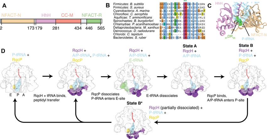
Nucleic Acids Research, 2021, Vol. 49, No. 14 8359
twice with 5:1 acidic phenol:chloroform, precipitated with on each array, the IP samples originated from larger cell
ethanol, and resuspended in ddH2 O. Samples containing amounts and this was also considered by calculating the
150 ng of total RNA were separated on 8 M urea-containing final ratios. Enrichment of tRNA species in in vivo and
8% polyacrylamide gels. The gel was stained with SYBR in vitro IP samples was calculated over corresponding
Gold (Life technologies) for 30 min, followed by visualiza- total tRNA pool, i.e. either tRNA from cellular lysates or
tion using a Typhoon Trio Variable Mode Imager (Amer- purified total B. subtilis tRNA, respectively. To assess the
sham Biosciences). spread of the data, covariance of the signal is calculated
for each single isoacceptor (represented with grey bars
on the corresponding figures) which is further used to
tRNA Northern blotting
compute the confidence intervals, i.e. the spread of the
For deacylation immunoprecipitated samples were incu- data for all isoacceptors. In the case of experiments with
bated with 125 mM Tris–HCl, pH 9.0, 0.1 M EDTA, 0.5% three replicates, the confidence intervals between replicates
(w/v) SDS at room temperature for 45 min, before neutral- 1 and 2, 2 and 3, 1 and 3 were 98%, 99% and 99% (for
Downloaded from https://academic.oup.com/nar/article/49/14/8355/6320412 by guest on 20 October 2021
ization with an equal volume of 1 M NaOAc, pH 5.5. RNA Figure 2E), 95%, 97% and 92% (Figure 2F), 96%, 96%
was extracted twice with 5:1 acidic phenol:chloroform, pre- and 97% (Figure 3B), 95%, 97% and 92% (Figure 3C)
cipitated with ethanol, and resuspended in ddH2 O. Sam- and, finally, 94%, 98% and 97% (Figure 5F). In the case of
ples containing 150 ng of total RNA were separated on 8M experiments with two replicates, the confidence intervals
urea-containing 8% polyacrylamide gels followed by elec- between replicates 1 and 2 were 95% (Figures 3D and 3E).
troblotting to Zeta-probe membranes (Bio-Rad). The blots
were sequentially probed for B. subtilis tRNAAla , B. subtilis Sucrose gradient fractionation and Western blotting
tRNALys and E. coli tRNAVal using 32 P-labeled oligonu-
cleotides (Supplementary Table S2). The probes for B. sub- Experiments were performed as described earlier (42), with
tilis tRNAAla were designed to hybridize with the conserved minor modifications. 50S ribosomal protein L3 was de-
TC-loop of both tRNAAla isoacceptors. Signals were de- tected using anti-L3 primary antibodies provided by Fu-
tected by phosphorimaging using a Typhoon FLA 9500 jio Kawamura (1:20,000 dilution) combined with goat anti-
biomolecular imager. In parallel, a second gel was subjected rabbit IgG-HRP secondary antibodies (Sigma-Aldrich,
to SYBR Gold (Life Technologies) nucleic acid staining A0545; 1:10 000 dilution). ECL detection was performed
for 30 min, followed by visualization using a Typhoon Trio using WesternBright™ Quantum (K-12042-D10, Advansta)
Variable Mode Imager (Amersham Biosciences). western blotting substrate and ImageQuant LAS 4000 (GE
Healthcare) imaging system.
tRNA microarrays
Poly-Ala synthesis in a reconstituted B. subtilis RQC system
tRNA microarrays were performed similarly to
as previously described (42). A detailed protocol Initiation (2 M B. subtilis 50S, 3 M RqcH-HTF, 3 M
for the microarrays is published on protocols.io RqcP, 2 M f[35 S]Met-tRNAi Met in HEPES:Polymix buffer
(dx.doi.org/10.17504/protocols.io.hfcb3iw). In vivo and in pH 7.0 1 mM DTT and 7.5 mM MgCl2 ) and elongation
vitro co-immunoprecipitated tRNA samples as well as the (10 M B. subtilis total tRNA (tBulk), 2 M AlaRS, 1
corresponding total tRNA controls (tRNA from cell lysate mM ATP, 200 M Alanine, 1 mM DTT and 7.5 mM
for in vivo pulldown experiments or purified total B. subtilis MgCl2 in HEPES:Polymix buffer pH 7.0) mixtures were
tRNA for in vitro pulldown experiments, respectively) separately prepared on ice. After 5 min incubation at 30◦ C,
were incubated with 100 mM Tris-HCl pH = 9.0 at room the two mixtures were combined and incubated at 30◦ C.
temperature for 45 min, thereafter precipitated with one 10 l aliquots were taken (either after 15-min long incu-
volume of 100 mM NaCl 100 mM NaOAc pH 4.8 as well bation or throughout the time course), quenched with 10
as 2.7-volumes of absolute ethanol, resuspended in ddH2 O l of loading dye (7 M urea, 0.05% bromophenol blue and
and tRNAs were subjected to labeling of the invariant 100 mM NaOAc, pH 5) and resolved on acidic urea-PAGE
3 -NCCA-ends. The IP samples were labeled with Cy3- in 1× TBE (8 M urea, 6.5% PAGE). The gel was exposed
labeled RNA/DNA while the total tRNA control samples overnight, and the imaging plate was scanned on Typhoon
were labeled with Atto647-labeled RNA/DNA hybrid FLA 9500 (GE Healthcare).
oligonucleotide. Labeled tRNA samples were loaded as
pairs (i.e. Cy3-labeled IP and Atto647-labeled total tRNA) RESULTS AND DISCUSSION
on the same microarray containing 24 replicates of full-
RqcH specifically selects tRNAAla from the tRNA pool in the
length tDNA probes recognizing the 36 B. subtilis tRNA
absence of the 50S subunit
isoacceptors (see Supplementary Table S1) and hybridized
for 16 h at 60◦ C. The fluorescence signals of microarrays Previous studies indicated that B. subtilis RqcH appears
were recorded with a GenePix 4200A scanner (Molecular to specifically interact with tRNAAla and that this speci-
Devices) and statistically analyzed with in-house scripts ficity may be mediated by the highly conserved 97 DR98
with Python version 3.7.0. Each fluorescent signal was residues (41,42,44). To investigate this further, we used Elec-
normalised to intrinsic standards as described (61) and trophoretic Mobility Shift Assays (EMSAs) to study com-
presented as ratios of the IP complexes against total plex formation between B. subtilis RqcH – wild-type and
tRNA (i.e. Cy3 vs Atto647 signals). Thereby, while equal D97A/R98A (DR) variant – and individual native deacy-
amounts of IP samples and total tRNAs were analyzed lated tRNAs purified from B. subtilis bulk tRNA (Figure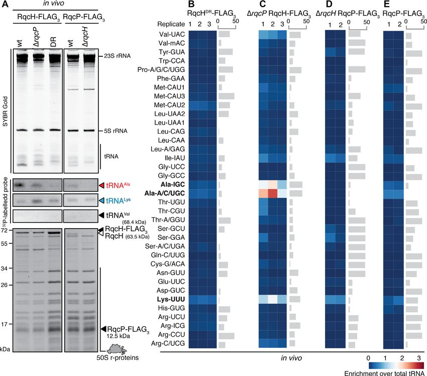
8360 Nucleic Acids Research, 2021, Vol. 49, No. 14
Downloaded from https://academic.oup.com/nar/article/49/14/8355/6320412 by guest on 20 October 2021
Figure 2. RqcH can specifically select tRNAAla from the tRNA pool off the 50S ribosome. (A, B) Complex formation between increasing concentrations
of either wild-type (A) or DR variant (D97A R98A double substitution) (B) B. subtilis RqcH-HTF and 0.1 M of either B. subtilis tRNAVal , B. subtilis
tRNAAla or E. coli tRNAVal was monitored by EMSA. 1 M of synthetic mRNA(MVF) RNA oligonucleotide was used as a nonspecific competitor.
Representative full-size EMSA gels are provided as Supplementary Figure S3. (C, D) SYBR Gold staining (C) and Northern blotting (D) analyses of
RqcH-HTF:tRNA complexes isolated through co-IP of either wild-type or DR variant of RqcH-HTF preincubated with total B. subtilis tRNA, tBulk.
For validation of northern blot probe specificity see Supplementary Figure S4. (E, F) tRNA microarray analyses of tRNAs isolated through co-IP of either
wild-type (E) or DR variant (F) of RqcH-HTF preincubated with B. subtilis total tRNA. Grey bars represent an example of covariance analysis between
replicates 1 and 3 (E) and replicates 2 and 3 (F). The Ala-IGC tRNA array probe hybridizes with tRNAAla(GGC) , Ala-A/C/UGC probe hybridizes with
tRNAAla(UGC) and Lys-UUU hybridizes with tRNALys(UUU) ; the full reference table is provided as Supplementary Table S1. The colour key indicates the
fold-enrichment of tRNAs in pulldown samples over total tRNA.
2A, B and Supplementary Figure S3). We tested B. subtilis affinities (KD /EC50 ), the method is not suitable for measur-
tRNAAla as the native RQC tRNA substrate, and, as con- ing the on- and off-rates of RqcH:tRNA complex formation
trols, B. subtilis tRNALys and E. coli tRNAVal ; tRNAAla and (k+1 and k–1 ). Alternatively, it is also possible that the high
tRNAVal used for EMSA were not further purified into in- selectivity is only evident when RqcH is recruited to the 50S
dividual isoacceptors. While the wild-type RqcH displays a subunit, and the preference for tRNAAla actually reflects the
preference for tRNAAla , it also binds the control tRNAs, specificity of RqcH:50S RQC complex, rather than that of
though with lower affinity: EC50 tRNAAla of 190 nM ver- the RqcH itself.
sus EC50 tRNALys of 310 nM and EC50 tRNAVal of 390 To address the first of these possibilities, we performed
nM (Figure 2A). The preference for tRNAAla is lost in the a set of in vitro pulldown experiments using total B. sub-
RqcHDR variant, which binds all the three tRNA species tilis tRNA preparation (total tRNA bulk, tBulk) and puri-
with the same (low) affinity, analogous to that observed for fied RqcH (C-terminally extended by the His6 -TEV-FLAG3
the wild-type protein binding to non-cognate tRNA (i.e. (HTF) tag). The purified HTF-tagged wild-type RqcH or
tRNAVal and tRNALys ; EC50 240–310 nM). The modest RqcHDR were incubated with total tRNA, and the asso-
specificity of RqcH for tRNAAla observed in EMSAs is sur- ciated tRNA species were probed by (i) Northern blotting
prising given the pronounced selectivity for tRNAAla in B. (Figure 2C, D; for validation of Northern blot probe speci-
subtilis RQC (41,42). The higher specificity for tRNAAla in ficity see Supplementary Figure S4), and (ii) tRNA mi-
vivo is only evident in kinetic competition with the other croarray (Figure 2E, F). Both experiments were in agree-
tRNA species. To assess this, time-resolved studies will be ment with each other, demonstrating strong preference of
necessary; while our EMSA can quantify the equilibrium the wild-type RqcH for tRNAAla . In the microarrays, we de-
Nucleic Acids Research, 2021, Vol. 49, No. 14 8361

                                                                                                                                                       Downloaded from https://academic.oup.com/nar/article/49/14/8355/6320412 by guest on 20 October 2021
Figure 3. While RqcP itself does not have a specific preference for tRNAAla , loss of RqcP moderately compromises tRNAAla selection by RqcH in vivo.
(A) SYBR Gold staining (top), Northern blotting (middle) and SDS PAGE (bottom) analyses of 50S RQC complexes isolated through co-IP of either
RqcH-FLAG3 or RqcP-FLAG3 from lysed B. subtilis. (B–E) tRNA microarray analyses of 50S RQC complexes isolated through co-IP of either RqcH-
FLAG3 (B and C, three independent biological replicates) or RqcP-FLAG3 (D and E, two independent biological replicates) from lysed B. subtilis. Grey
bars represent an example of covariance analysis between either replicates 1 and 3 (B), 2 and 3 (C), or 1 and 2 (D,E). The Ala-IGC tRNA array probe
hybridizes with tRNAAla(GGC) , Ala-A/C/UGC probe hybridizes with tRNAAla(UGC) and Lys-UUU hybridizes with tRNALys(UUU) ; the full reference table
is provided as Supplementary Table S1. The colour key indicates the fold-enrichment of tRNAs in pulldown samples over total tRNA.

tected both tRNAAla(GGC) and tRNAAla(UGC) isoacceptors                      in the RqcHDR sample (Figure 3A and Supplementary Fig-
(hybridizing to the Ala-IGC and Ala-A/C/UGC probes,                         ure S5). Collectively, our results demonstrate that RqcH re-
respectively, see Supplementary Table S1). The signal for                   cruitment to the 50S subunit is not necessary for the tRNA
tRNAAla is abrogated in the RqcHDR variant. Notably, in                     selection by RqcH.
the case of RqcHDR , the tRNALys signal on the Northern                        We also probed the role of the second RQC elonga-
blot (and to a lesser extent that of tRNAVal ) is stronger                  tion factor, RqcP, in tRNA selection using northern blot-
than that for the wild-type protein (Figure 2D). A possi-                   ting, SYBR Gold staining (Figure 3A and Supplementary
ble explanation is that the RQC-cognate tRNAAla efficiently                 Figure S5) and tRNA microarrays (Figure 3C–E). Com-
out-competes tRNALys for binding to wild-type RqcH, but                     pared to the rqcP+ sample, in the RqcH-FLAG3 :50S pull-
not to RqcHDR . Finally, we validated the in vitro tRNA                     down from rqcP B. subtilis, there is a moderate, but de-
specificity results in vivo. Both northern blotting (Figure                 tectable decrease in abundance of both tRNAAla isoaccep-
3A) and tRNA microarray (Figure 3B) analyses of in vivo                     tors, as well as an increase of the tRNALys signal (Figure
FLAG3 pulldown RqcH:50S complexes demonstrate that,                         3C). This could be either a direct effect of intrinsic speci-
indeed, tRNAAla specificity is lost in RqcHDR . Note that                   ficity of RqcP towards tRNALys , or an indirect effect of
tRNAAla signal is clearly resolved from other RNA species                   the compromised RQC elongation through RqcP depletion
on SYBR Gold gels, and the corresponding band is lacking                    leading to accumulation of ‘initiation’ RQC complexes con-
8362 Nucleic Acids Research, 2021, Vol. 49, No. 14
taining tRNALys . Therefore, we next probed the intrinsic still somewhat distorted, perhaps as a result of other re-
tRNA selectivity of RqcP by analysing RqcP-FLAG3 pull- gions of RqcH, such as a N-terminus of the coiled-coil do-
downs from both the RQC-incompetent rqcH strain (Fig- main, that contact the tRNA (Figure 4F, G) (42,44). Col-
ure 3D) and RQC-competent rqcH+ strain (Figure 3E). lectively, the structure suggests that the mutation of 97 DR98
While we see no tRNA specificity in the rqcH pulldown motif to 97 AA98 destabilizes the interaction of the NFACT-
samples, there is a weak signal for selection of tRNALys N domain of RqcHDR with the anticodon stem loop of the
of a similar magnitude and preference tRNAAla in rqcH+ P-tRNA, thereby supporting the hypothesis that this inter-
pulldowns. This strictly-RqcH-dependent specificity signal action is critical for the specificity of RqcH for tRNAAla . In
in the in vivo RqcP-FLAG3 pulldowns can be explained by the available structures so far, RqcH 97 DR98 has been ob-
the tRNA specificity of RqcH present in RqcP-FLAG3 - served either to interact with G35 at the central position of
RqcH-50S (but not RqcP-FLAG3 -RqcH-50S) RQC com- the tRNA anticodon, which is common to several B. subtilis
plexes. Our RqcP pulldown results also suggest that the tRNAs, or not interact with the tRNA at all. It is therefore
decrease in tRNAAla abundance observed in the RqcH- unlikely that these conformations alone can account for the
Downloaded from https://academic.oup.com/nar/article/49/14/8355/6320412 by guest on 20 October 2021
FLAG3 :50S pulldown from the rqcP strain is an indirect tRNA specificity of RqcH. RqcH and tRNAAla may form a
effect of compromised RQC elongation, with the increase complex with an alternative conformation when not bound
of the tRNALys signal possibly reflecting the nature of the in complex with the 50S subunit.
tRNA in the initial stalled 70S complexes.
Ribosomal protein uL11 is important for RQC functionality
Cryo-EM structures of an in vivo formed RqcHDR -50S
In the cryo-EM structure of the RqcH-50S RQC complex,
complex
we observed strong interaction between the M domain of
To gain the structural insight into the specific role of RqcH and the uL11 stalk base (Figure 5A), as observed
D97/R98 residues in tRNAAla recognition by RqcH, we previously for the wild-type RqcH 50S complexes (42,44).
determined the structure of a RqcHDR -50S RQC complex This interaction appeared to be critical for RqcH function
by cryo-EM. Following an established affinity purification since treatment with the antibiotic thiostrepton that inter-
procedure (42), we generated 50S RQC complexes from acts with uL11 (63), and has an overlapping binding site
B. subtilis expressing C-terminally FLAG3 -tagged RqcHDR with RqcH M domain (inset to Figure 5A), destabilised
(D97A/R98A double substitution in NFACT-N domain) interaction of RqcH with the 50S RQC complex (42). To
(Supplementary Figure S2). As observed previously, RqcH directly investigate the importance of uL11 for RQC func-
apparently dissociated from many particles during sample tion, we constructed a B. subtilis strain in which uL11 is ex-
preparation, but with focussed classification we nonethe- pressed under the control of an IPTG-inducible Phy−spank
less obtained a volume with RqcH stably bound (Supple- promoter (64), while the genomic copy of rplK encoding
mentary Figure S2, Table S3). The main state observed is uL11 is disrupted (rplK). This allowed conditional deple-
broadly similar to the previously defined state B, consist- tion of uL11 in bacteria that are grown in the absence of
ing of a 50S subunit with peptidyl-tRNA in approximately an inducer. uL11 depletion results in a pronounced growth
the P-site, nascent chain, RqcP, and RqcH (Figure 4A). Al- defect as seen in Figure 5B. When we combined the deple-
though the overall resolution of this complex was 3.2 Å, tion of uL11 with rqcH disruption, we did not observe a
this mostly reflected the core of the 50S subunit, as RqcH further synthetic growth defect. This lack of genetic inter-
and other bound factors were less well resolved (Supple- action is consistent with RQC functionality being already
mentary Figure S6). Compared to the previously observed lost in the uL11-depleted strain. By contrast, simultaneous
state B (42), the RqcHDR NFACT-N and HhH domains loss in functionality of both RQC and trans-translation ri-
are swung away from the tRNA anticodon in the RqcHDR - bosome rescue systems renders B. subtilis sensitive to ele-
bound structure (Figure 4B). These domains are also espe- vated temperature and antibiotics targeting protein synthe-
cially poorly resolved, indicating flexibility (Figure 4A, Sup- sis, such as tetracycline (41,42,44). This synergetic growth
plementary Figures S6 and S7). Such flexibility is likely due defect is commonly used for probing RQC functionality in
to the 97 DR98 motif interacting with the tRNA anticodon an ssrA background (41,42,44). Indeed, we observed a
stem in the RqcH wild-type state B structure, possibly stabi- strong genetic interaction of rplK with ssrA: uL11 deple-
lizing the major conformation (Figure 4C). Nonetheless, we tion in ssrA rplK genetic background results in the syn-
were able to fit the individual domains of RqcHDR into the thetic lethality of the strain (Figure 5B). This is likely due
map to create a model with sufficient confidence to exam- to the proteotoxic stress due to a defunct RQC and trans-
ine domain-level movements. The RqcHDR complex is very translation, combined with the general translation defect
similar to state B* that was also observed in the wild-type due to loss of uL11.
RqcH reconstructions (42) (Supplementary Figure S7). The Next, we assessed the effect of uL11 on the recruitment of
P-tRNA in the RqcHDR complex had a poorly resolved an- RQC factors to the 50S RQC complex using sucrose gradi-
ticodon (Figure 4D). Previously, wild-type RqcH has been ent centrifugation and Western blotting against the FLAG-
shown to make close contact with the P-tRNA, unwind- tagged RqcH or RqcP factors (Figure 5C). While RqcP re-
ing the anticodon stem and forming interactions with the mains associated with uL11 50S, RqcH is lost from the
splayed anticodon (42,44). In comparison, the RqcHDR - 50S fractions in the rplK strain (Figure 5C), strongly sug-
bound tRNA anticodon stem was not as distorted (Figure gesting that uL11 plays a key role in RqcH recruitment to
4E). However, compared to a canonical P-tRNA or free 50S, and, therefore, RQC function. In good agreement with
crystallised tRNA (62), the RqcHDR -bound P-tRNA was decreased affinity of RqcH to 50S lacking uL11, character-Nucleic Acids Research, 2021, Vol. 49, No. 14 8363
Downloaded from https://academic.oup.com/nar/article/49/14/8355/6320412 by guest on 20 October 2021
Figure 4. Cryo-EM structures of an in vivo-formed RqcHDR -50S complex. (A) View of RqcHDR -FLAG3 bound to 50S with P-tRNA and RqcP. The state
broadly resembles state B described in (42). (B) Comparison of RqcHDR (purple) with wild-type RqcH (pink) state B. (C) Close views, rotated compared
to C, showing the position of the mutated 97 DR98 residues. Transparent density (top panel) or model (bottom panel) is shown for the P-tRNA anticodon
stem. (D) View of the P-tRNA on the RqcHDR -bound 50S. The anticodon stem is poorly resolved, likely due to flexibility. Part of the RqcH CC N-terminus
is shown. (E) Comparison of the P-tRNA in the RqcHDR volume (light blue) with the P-tRNA in the RqcH WT-bound volume (state B, grey) (42). (F)
As in E, except the RqcHDR -FLAG3 P-tRNA is compared to a canonical P-tRNA bound in the P-site of the ribosome (tan, PDB 6CFJ) (74). (G) As in
E and F, except the RqcHDR -FLAG3 P-tRNA is compared to crystallised yeast tRNAPhe (reddish, PDB 1EHZ) (62).
ization of RqcH-FLAG3 pulldown samples revealed rela- with the 50S subunit (Figure 6A), whereas here we show that
tively low abundance of both rRNA and r-proteins in com- RqcP remains unaffected (Figure 6B). By contrast, the ad-
parison to pulldowns from wild-type strain (Figure 5D, E, a dition of puromycin, which mediates release of the polypep-
side-by-side comparison is shown on Supplementary Figure tide chain from the P-tRNA (65), led to a modest reduction
S5). By contrast, the strong band consistent with tRNA re- in RqcH binding (Figure 6A), as observed previously (42),
mained (Figure 5D), in agreement with RqcH being able to but more strikingly, resulted in a dramatic reduction of the
specifically recruit tRNAAla in the absence of the 50S sub- RqcP band within the 50S fraction (Figure 6B). Since re-
unit. tRNA microarray analysis of the RqcH-FLAG3 pull- lease of the nascent polypeptide chain by puromycin is likely
down samples from B. subtilis rplK revealed that indeed to have a destabilizing effect on the binding of P-tRNA to
RqcH specifically interacts with both tRNAAla isoacceptors the 50S subunit, these findings suggest that binding of RqcP,
(Figure 5F), as seen previously for tRNA microarray anal- and to a lesser extent RqcH, is stabilized by the presence of
ysis of RqcH 50S complexes (42). the peptidyl-tRNA in the P-site.
Polypeptide release dissociates RqcP from RqcP-RqcH-50S Stable interaction of RqcP with 23S rRNA H69, but not with
RQC complexes P-site tRNAAla is essential for RQC functionality
Our finding that uL11 is critical for RqcH, but not RqcP, In the 50S RQC complex, RqcP forms a network of contacts
interaction with the 50S subunit (Figure 5C) suggested that with P-site tRNA, but also the 23S rRNA helix 69 (H69)
binding of RqcP to the 50S subunit is independent of RqcH. (42,44). Interactions with H69 are mediated by conserved
This is consistent with cryo-EM analysis where P-tRNA- Arg2 and Arg16 of RqcP, whereas contacts with P-tRNA
50S complexes containing RqcP but lacking RqcH are ob- are formed by Arg11, Lys68 and Lys69 (Figure 6C–E). To
served (Supplementary Figure S2) (42). To pursue this fur- date, only Arg16 of RqcP was assessed experimentally for
ther, we employed sucrose density gradient centrifugation its functional role, and the residue was shown to be crucial
and Western blotting to monitor the association of both because Arg16Ala substitution abolished RqcP association
RqcH and RqcP in the presence of the antibiotics thiostrep- with the 50S subunit and, when introduced in the ssrA
ton and puromycin (Figure 6A,B). As before (42), we ob- background, rqcP R16A phenocopies ssrA rqcP double
served that thiostrepton abolished the interaction of RqcH deletion (42).8364 Nucleic Acids Research, 2021, Vol. 49, No. 14
Downloaded from https://academic.oup.com/nar/article/49/14/8355/6320412 by guest on 20 October 2021
Figure 5. Ribosomal protein uL11 is important for RqcH recruitment to 50S and for RQC functionality. (A) Overview of wild-type RqcH (state B, PDB
7AS8, EMD-11889) bound to the 50S-P-tRNA complex with focus on the L7/L12 stalk base. Right, close view of the RqcH M hairpin interacting with
uL11. Antibiotic thiostrepton (Thio, in red) is modeled based on PDB 3CF5 (63). (B) B. subtilis strains expressing ribosomal uL11 (rplK) under the control
of IPTG-inducible Phy−spank promoter in wild-type, rplK, rplK ssrA and rplK rqcH backgrounds were grown in either uL11-inducing (LB sup-
plemented with 1 mM IPTG, left) or non-inducing (LB, right) conditions. Plates were scored after 18 hours at 37◦ C. (C) Sucrose gradient sedimentation
and anti-FLAG3 immunoblotting of either RqcH-FLAG3 or RqcP-FLAG3 expressed in rplK background. (D–F) SYBR Gold staining (D), SDS PAGE
(E) and tRNA microarray analyses (F) of 50S RQC complexes isolated through co-IP of RqcH-FLAG3 from lysed rplK B. subtilis. Three independent bi-
ological replicates of tRNA microarray analyses are shown. Grey bars represent an example of covariance analysis between replicates 1 and 3. The Ala-IGC
tRNA array probe hybridizes with tRNAAla(GGC) , Ala-A/C/UGC probe hybridizes with tRNAAla(UGC) and Lys-UUU hybridizes with tRNALys(UUU) ;
the full reference table is provided as Supplementary Table S1. The colour key indicates the fold-enrichment of tRNAs in pulldown samples over the total
tRNA.
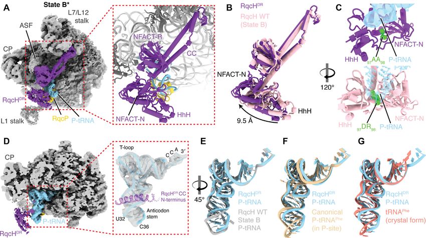
We first probed the importance of Arg2, Arg11, Lys68 the loss of RqcP (rqcP), thereby re-emphasizing the im-
and Lys69 in RqcP association with the 50S through su- portance of the interaction of RqcP with H69 of the 23S
crose gradient sedimentation experiments. As seen in Fig- rRNA for RQC functionality. By contrast, there appears
ure 6B, the association with 50S was compromised for all to be redundancy in the interactions between RqcP and
of the tested RqcP variants, suggesting that the contacts the P-site tRNA: ssrA B. subtilis strains expressing single
of RqcP with both H69 and the P-site tRNA are both im- R11A-substituted and the double K68A/K69A-substituted
portant for establishing a stable interaction with the large RqcP variants display wild-type-like phenotypes, only with
subunit. Next, we assessed the functional importance of the triple R11A/K68A/K69A substitution resulting in a
Arg2, Arg11, Lys68 and Lys69 in vivo by following the growth defect (Figure 6F). The apparent discrepancy be-
growth of ssrA B. subtilis strains expressing substituted tween the loss of interaction of the R11A and K68A/K69A
RqcP variants. The cells were grown either in normal con- variants with the 50S subunit (Figure 6B) and the lack of
ditions (37◦ C), at elevated temperature (49◦ C), or in the effect in the growth assays (Figure 6F) is likely due to the
presence of low concentrations of tetracycline (Figure 6F). non-equilibrium nature of sucrose gradient sedimentation
Similar to R16A (42), the R2A substitution phenocopies experiments, i.e. the lack of association does not indicate anNucleic Acids Research, 2021, Vol. 49, No. 14 8365
Downloaded from https://academic.oup.com/nar/article/49/14/8355/6320412 by guest on 20 October 2021
Figure 6. Interaction of RqcP with 23S rRNA H69, but not with P-site tRNAAla , is essential for RQC functionality. (A, B) Sucrose gradient sedimentation
and anti-FLAG3 immunoblotting analysis of B. subtilis strains expressing either wild-type RqcH-FLAG3 (A) or RqcP-FLAG3 , wild-type and substituted
variants (B). As indicated on the figure, the antibiotics were added after cell lysis: thiostrepton (Thio, 50 M) and puromycin (Puro, 1 mM). (C) Sequence
alignment of RqcP/Hsp15 homologs from diverse bacteria. (D, E) View of RqcP from state B interacting with (D) 23S rRNA H69 or (E) the P-tRNA
(PDB 7AS8). Residues selected for mutation are coloured green. 23S rRNA nucleotides are numbered according to E. coli numbering. (F) Synthetic growth
defects caused by amino acid substitutions in rqcP, or rqcP deletion in a ssrA background. 10-fold serial dilutions were spotted onto LB agar plates and
incubated for 18 hours at 37◦ C (left), 49◦ C (right) or 37◦ C in the presence 0.5 g/ml tetracycline (Tet, middle).
absence of the interaction in the cell, but rather means that sized that the fMet–tRNA–50S complex would mimic the
the complex is not retained during the 3-hour-long centrifu- peptidyl-tRNA 50S complexes that arise during RQC and
gation. thereby act as a substrate for RqcH and RqcP (Figure 7A).
In parallel, we assembled a separate reaction containing
Ala-tRNAAla from B. subtilis total tRNA that had been in-
RqcP is a processivity factor essential for poly-alanine syn- cubated with B. subtilis alanyl-tRNA synthetase (AlaRS),
thesis during RQC elongation ATP and L-alanine at 30◦ C. Since the Mg2+ concentra-
Previous studies have shown using in vivo approaches that tion is a major determinant of tRNA affinity to the ribo-
RqcP phenocopies RqcH in the ssrA background (42,44) some (69,70), we increased the concentration to 7.5 mM in
and that deletion of rqcP leads to a loss of poly-alanine tail- our HEPES:Polymix-based buffer system (47) to find the
ing of a non-stop reporter construct (44), collectively indi- optimal balance between tRNA affinity (for efficient Ala-
cating that RqcP has a critical role in bacterial RQC. How- tRNAAla recruitment to 50S RQC complex) and dynamics
ever, it still remains unclear whether RqcH and RqcP are (for efficient RQC elongation), and adjusted the pH to 7.0 in
necessary and sufficient to mediate polyalanine tailing of order to stabilise aminoacyl-tRNA. After preincubation the
50S–peptidyl-tRNA complexes. To investigate this, we un- two mixtures were combined, the resultant reaction was fur-
dertook to establish an in vitro polyalanine tailing system ther incubated for 30 minutes at 30◦ C, tRNA species were
using only purified RQC factors, as has been previously per- resolved in a bis-tris gel and then visualised by phospho-
formed for canonical translation (66,67). To initiate RQC, imaging.
we loaded 50S subunits with radioactively labeled formy- In the reaction lacking RqcP, we only detect 35 S-labeled
lated initiator tRNA, 35 S-fMet-tRNAi Met , which has pref- fMet-tRNAi Met , whereas in the presence of RqcP there is
erential affinity to the ribosomal P-site (68). We hypothe- an up-shift in the 35 S-labeled fMet signal, which is consis-8366 Nucleic Acids Research, 2021, Vol. 49, No. 14
Downloaded from https://academic.oup.com/nar/article/49/14/8355/6320412 by guest on 20 October 2021
Figure 7. RqcP is a processivity factor essential for poly-Ala synthesis in a biochemically reconstituted RQC system. (A) Schematics of the experimental
setup. Separate initiation and elongation mixtures were prepared in HEPES:Polymix buffer (7.5 Mg2+ pH 7.0) and incubated at 30◦ C for 5 min before
combining. After an additional 15 minute-long incubation, tRNA species were resolved in a bis-tris gel and visualised by phosphoimaging. (B) Formation
of Ala-tRNAAla in the reconstituted RQC system is abrogated upon omission of RqcP. (C) Addition of canonical translation elongation factor EF-Tu and
1 mM GTP does not affect the efficiency of Ala-tRNAAla synthesis. (D) No poly-Ala synthesis is observed either upon omission of AlaRS or in the presence
of tRNAVal , Valyl-tRNA synthetase (ValRS) and L-valine. (E) Neither RqcHDR nor RqcPR16A support Ala-tRNAAla formation in the reconstituted RQC
system.
tent with the polyalanine tailing of the fMet (Figure 7B), gation due to the DR substitution in the NFACT-N do-
thus directly demonstrating the role of RqcP as an essen- main of RqcH or loss of RqcP? It is tempting to speculate
tial processivity factor for RQC elongation. No effect of the that tRNALys is enriched in these complexes due to ribo-
efficiency of poly-Ala synthesis was observed in the RQC somal stalling on lysine residues triggering RQC. Indeed,
reaction that was additionally supplemented with canon- a recent analysis of ribosome profiling data suggests that
ical translation elongation factor EF-Tu and 1 mM GTP the presence of P-site Lys residues encoded by AAA codons
(Figure 7C), suggesting that EF-Tu does not play a role are associated with a moderate slowing down of translation
in delivery of Ala-tRNA to the 50S subunit and that tail- elongation in E. coli (71). However, no Lys-associated ri-
ing proceeds independently of GTP. Moreover, no poly- bosomal stalling signal was detected in a B. subtilis study
alanine tailing was observed when the aminoacylation mix using 5Pseq (72). Therefore, while tempting, this hypoth-
was replaced with the presence of tRNAVal , valyl-tRNA esis requires further investigation. Second is the question
synthetase (ValRS), ATP and L-valine (Figure 7D), which is of a potential C-terminal-tailing processivity factor in eu-
consistent with the strong preference of RqcH for tRNAAla . karyotic RQC. Here we directly demonstrate in vitro that
Similarly, no activity was detected when AlaRS was omitted S4 homologue Hsp15-family protein RqcP is, indeed, es-
(Figure 7D). Finally, our reconstituted RQC system faith- sential to drive the poly-alanine tailing by 50S-associated
fully reproduces the effects of loss-of-function substitutions RqcH/Rqc2/NEMF in a minimal bacterial system. This in-
in RqcH (RqcHDR variant compromised in tRNAAla selec- dicates that an analogous yet-to-be-discovered factor could
tion) and RqcP (RqcPR16A variant compromised in inter- be also cooperating with Rqc2/NEMF in archaea and eu-
action with the H69 rRNA element), since no poly-alanine karyotes. Additionally, the establishment of a reconstituted
tailing was observed when replacing the wild-type RqcH bacterial RQC system provides researchers with a power-
and RqcP with these variants (Figure 7E). Collectively, our ful tool for activity-driven discovery of additional bacte-
biochemical results establish that the combination of RQC rial RQC factors, either by directly testing candidate fac-
factors RqcP and RqcH is both necessary and sufficient to tors identified through structural and genetic studies or
drive polyalanine synthesis on isolated 50S subunits in the through fractionation of cellular lysates. Currently, it is un-
absence of canonical translation elongation factors. clear which cellular factors split the stalled 70S ribosomes
to generate the 50S RQC substrate and which factors termi-
nate the RQC-mediated poly-alanine addition to release the
CONCLUSIONS AND PERSPECTIVE
tagged polypeptide. Our genetic experiments suggest that
This study supports the proposed molecular mechanism of ribosome-splitting factor HflX (73) is not essential for RQC
C-terminal alanine tailing mediated by the concerted ac- in live cells since we detect no synthetic growth defect upon
tion of RqcH and RqcP (Figure 1D) (42). Importantly, it the simultaneous deletion of hflX and ssrA (Supplementary
also opens up several research directions for follow-up in- Figure S8). A fully reconstituted biochemical RQC system
vestigations. What is the significance of tRNALys enrich- could be instrumental for deconvoluting the redundancies
ment in 50S RQC complexes compromised in RQC elon- and overlapping functions of the factors involved. Finally,Nucleic Acids Research, 2021, Vol. 49, No. 14 8367
an analogous eukaryotic RQC system could be established 4. Yip,M.C.J. and Shao,S. (2021) Detecting and rescuing stalled
using 60S ribosomal subunits, Rqc2/NEMF and aminoa- ribosomes. Trends Biochem. Sci.,
https://doi.org/10.1016/j.tibs.2021.03.008.
cylated tRNAs to enable activity-driven discovery of addi- 5. Keiler,K.C., Waller,P.R. and Sauer,R.T. (1996) Role of a peptide
tional RQC factors. tagging system in degradation of proteins synthesized from damaged
messenger RNA. Science, 271, 990–993.
6. Tu,G.F., Reid,G.E., Zhang,J.G., Moritz,R.L. and Simpson,R.J.
DATA AVAILABILITY (1995) C-terminal extension of truncated recombinant proteins in
Escherichia coli with a 10Sa RNA decapeptide. J. Biol. Chem., 270,
The cryo-EM map of the RqcHDR -50S complex and the 9322–9326.
associated molecular model have been deposited in the 7. Komine,Y., Kitabatake,M., Yokogawa,T., Nishikawa,K. and
Protein Data Bank and Electron Microscopy Data Bank Inokuchi,H. (1994) A tRNA-like structure is present in 10Sa RNA, a
with the accession codes EMPIAR-10726, EMD-13017 small stable RNA from Escherichia coli. Proc. Natl. Acad. Sci. U.S.A.,
91, 9223–9227.
and PDB-7OPE, respectively. The tRNA microarray data 8. Karzai,A.W., Susskind,M.M. and Sauer,R.T. (1999) SmpB, a unique
have been deposited in Gene Expression Omnibus (GEO) RNA-binding protein essential for the peptide-tagging activity of
Downloaded from https://academic.oup.com/nar/article/49/14/8355/6320412 by guest on 20 October 2021
database under the accession number GSE174254. SsrA (tmRNA). EMBO J., 18, 3793–3799.
9. Gottesman,S., Roche,E., Zhou,Y. and Sauer,R.T. (1998) The ClpXP
and ClpAP proteases degrade proteins with carboxy-terminal peptide
SUPPLEMENTARY DATA tails added by the SsrA-tagging system. Genes Dev., 12, 1338–1347.
10. Chadani,Y., Ito,K., Kutsukake,K. and Abo,T. (2012) ArfA recruits
Supplementary Data are available at NAR Online. release factor 2 to rescue stalled ribosomes by peptidyl-tRNA
hydrolysis in Escherichia coli. Mol. Microbiol., 86, 37–50.
11. Chadani,Y., Ono,K., Kutsukake,K. and Abo,T. (2011) Escherichia
ACKNOWLEDGEMENTS coli YaeJ protein mediates a novel ribosome-rescue pathway distinct
from SsrA- and ArfA-mediated pathways. Mol. Microbiol., 80,
We thank Michael Hall for help with cryo-EM data collec- 772–785.
tion, Marcus J.O. Johansson for help with tRNA northern 12. Gueneau de Novoa,P. and Williams,K.P. (2004) The tmRNA website:
blotting assays, and Fujio Kawamura for providing anti-L3 reductive evolution of tmRNA in plastids and other endosymbionts.
Nucleic Acids Res., 32, D104–D108.
primary antibodies. The electron microscopy data was col- 13. Keiler,K.C., Shapiro,L. and Williams,K.P. (2000) tmRNAs that
lected at the Umeå Core Facility for Electron Microscopy, encode proteolysis-inducing tags are found in all known bacterial
a node of the Cryo-EM Swedish National Facility, funded genomes: A two-piece tmRNA functions in Caulobacter. Proc. Natl.
by the Knut and Alice Wallenberg, Family Erling Persson Acad. Sci. U.S.A., 97, 7778–7783.
14. Burroughs,A.M. and Aravind,L. (2019) The origin and evolution of
and Kempe Foundations, SciLifeLab, Stockholm Univer- release factors: Implications for translation termination, ribosome
sity and Umeå University. rescue, and quality control pathways. Int. J. Mol. Sci., 20, 1981.
15. Goralski,T.D.P., Kirimanjeswara,G.S. and Keiler,K.C. (2018) A new
mechanism for ribosome rescue can recruit RF1 or RF2 to nonstop
FUNDING ribosomes. mBio, 9, e02436-18.
16. Shimokawa-Chiba,N., Müller,C., Fujiwara,K., Beckert,B., Ito,K.,
Swedish Research Council (Vetenskapsrådet) grants [2017- Wilson,D.N. and Chiba,S. (2019) Release factor-dependent ribosome
03783 to V.H and 2019-01085 to G.C.A.]; Deutsche rescue by BrfA in the Gram-positive bacterium Bacillus subtilis. Nat.
Forschungsgemeinschaft [WI3285/8-1 to D.N.W.]; Ragnar Commun., 10, 5397.
Söderbergs Stiftelse (to V.H.); Umeå Centre for Micro- 17. Joazeiro,C.A.P. (2019) Mechanisms and functions of
ribosome-associated protein quality control. Nat. Rev. Mol. Cell
bial Research, UCMR (to H.T.); European Union from Biol., 20, 368–383.
the European Regional Development Fund through the 18. Inada,T. (2020) Quality controls induced by aberrant translation.
Centre of Excellence in Molecular Cell Engineering [2014- Nucleic Acids Res., 48, 1084–1096.
2020.4.01.15-0013 to V.H.]; EU Horizon 2020 program 19. Brandman,O., Stewart-Ornstein,J., Wong,D., Larson,A.,
(Marie Skłodowska-Curie) [764591 to Z.I.]; Estonian Re- Williams,C.C., Li,G.W., Zhou,S., King,D., Shen,P.S., Weibezahn,J.
et al. (2012) A ribosome-bound quality control complex triggers
search Council [PRG335 to V.H.]; G.C.A. and V.H. groups degradation of nascent peptides and signals translation stress. Cell,
were supported by a project grant from the Knut and Alice 151, 1042–1054.
Wallenberg Foundation [2020-0037 to G.C.A.]; D.N.W. and 20. Defenouillere,Q., Yao,Y., Mouaikel,J., Namane,A., Galopier,A.,
V.H. groups were supported by the Deutsche Zentrum für Decourty,L., Doyen,A., Malabat,C., Saveanu,C., Jacquier,A. et al.
(2013) Cdc48-associated complex bound to 60S particles is required
Luft- und Raumfahrt [DLR01Kl1820 to D.N.W.]; Swedish for the clearance of aberrant translation products. Proc. Natl. Acad.
Research Council [2018-00956 to V.H.] within the RIBO- Sci. U.S.A., 110, 5046–5051.
TARGET consortium under the framework of JPIAMR. 21. Kuroha,K., Zinoviev,A., Hellen,C.U.T. and Pestova,T.V. (2018)
Funding for open access charge: Swedish Research Council Release of ubiquitinated and Non-ubiquitinated nascent chains from
(Vetenskapsrådet) [2017-03783 to V.H.]. stalled mammalian ribosomal complexes by ANKZF1 and Ptrh1.
Mol. Cell, 72, 286–302.
Conflict of interest statement. None declared. 22. Pisareva,V.P., Skabkin,M.A., Hellen,C.U., Pestova,T.V. and
Pisarev,A.V. (2011) Dissociation by Pelota, Hbs1 and ABCE1 of
mammalian vacant 80S ribosomes and stalled elongation complexes.
REFERENCES EMBO J., 30, 1804–1817.
1. Keiler,K.C. and Feaga,H.A. (2014) Resolving nonstop translation 23. Shao,S., von der Malsburg,K. and Hegde,R.S. (2013)
complexes is a matter of life or death. J. Bacteriol., 196, 2123–2130. Listerin-dependent nascent protein ubiquitination relies on ribosome
2. Müller,C., Crowe-McAuliffe,C. and Wilson,D.N. (2021) Ribosome subunit dissociation. Mol. Cell, 50, 637–648.
rescue pathways in bacteria. Front. Microbiol., 12, 652980. 24. Shoemaker,C.J. and Green,R. (2011) Kinetic analysis reveals the
3. Sitron,C.S. and Brandman,O. (2020) Detection and degradation of ordered coupling of translation termination and ribosome recycling
stalled nascent chains via ribosome-associated quality control. Annu. in yeast. Proc. Natl. Acad. Sci. U.S.A., 108, E1392–E1398.
Rev. Biochem., 89, 417–442.You can also read