DDX3 depletion represses translation of mRNAs with complex 5 UTRs
←
→
Page content transcription
If your browser does not render page correctly, please read the page content below
5336–5350 Nucleic Acids Research, 2021, Vol. 49, No. 9                                                                         Published online 27 April 2021
doi: 10.1093/nar/gkab287

DDX3 depletion represses translation of mRNAs with
complex 5 UTRs
Lorenzo Calviello 1,† , Srivats Venkataramanan 1,† , Karol J. Rogowski2 , Emanuel Wyler2 ,
Kevin Wilkins1 , Malvika Tejura1 , Bao Thai1 , Jacek Krol3 , Witold Filipowicz4 ,
Markus Landthaler2,5,* and Stephen N. Floor 1,6,*
1
  Department of Cell and Tissue Biology, University of California, San Francisco, San Francisco, CA 94143, USA,
2
  Berlin Institute for Medical Systems Biology, Max-Delbrück-Center for Molecular Medicine in the Helmholtz
Association, 13125 Berlin, Germany, 3 Institute of Molecular and Clinical Ophthalmology Basel, Basel, Switzerland,

                                                                                                                                                                Downloaded from https://academic.oup.com/nar/article/49/9/5336/6255701 by guest on 30 July 2021
4
  Friedrich Miescher Institute for Biomedical Research, Basel, Switzerland, 5 IRI Life Sciences, Institut für Biologie,
Humboldt Universität zu Berlin, Philippstraße 13, 10115 Berlin, Germany and 6 Helen Diller Family Comprehensive
Cancer Center, University of California, San Francisco, San Francisco, CA 94143, USA

Received October 12, 2020; Revised April 02, 2021; Editorial Decision April 05, 2021; Accepted April 08, 2021

ABSTRACT                                                                                 4), a function that is essential in all eukaryotes (5). Dys-
                                                                                         function in DDX3 is linked to numerous diseases, includ-
DDX3 is an RNA chaperone of the DEAD-box family                                          ing medulloblastoma (3,6–12), many other cancer types
that regulates translation. Ded1, the yeast ortholog                                     (5) and de novo developmental delay (13–16). Previous
of DDX3, is a global regulator of translation, whereas                                   work studied how translation is altered by DDX3 variants
DDX3 is thought to preferentially affect a subset of                                     found in medulloblastoma (3,12), which are exclusively mis-
mRNAs. However, the set of mRNAs that are regu-                                          sense variants that preferentially target conserved residues.
lated by DDX3 are unknown, along with the relation-                                      In contrast, hematological cancers like natural killer/T-
ship between DDX3 binding and activity. Here, we                                         cell lymphoma (17,18) and others (19,20) also have fre-
use ribosome profiling, RNA-seq, and PAR-CLIP to                                         quent variants in DDX3X, but they are mostly truncat-
define the set of mRNAs that are regulated by DDX3                                       ing or frameshift variants resulting in decreased expression.
in human cells. We find that while DDX3 binds highly                                     Changes in gene expression occurring as a result of de-
                                                                                         creased DDX3 levels remain incompletely understood.
expressed mRNAs, depletion of DDX3 particularly af-
                                                                                            Inactivation of Ded1 in yeast leads to polysome collapse
fects the translation of a small subset of the tran-                                     and global downregulation of translation (21,22). More re-
scriptome. We further find that DDX3 binds a site                                        cent work showed that Ded1 is required for translation of
on helix 16 of the human ribosomal rRNA, placing                                         most transcripts in yeast using genome-wide approaches
it immediately adjacent to the mRNA entry channel.                                       (1,23). In contrast, DDX3 depletion seems to only affect
Translation changes caused by depleting DDX3 lev-                                        translation of a subset of expressed transcripts (2,4,24–26).
els or expressing an inactive point mutation are dif-                                    Despite the importance of DDX3 to normal function and
ferent, consistent with different association of these                                   its alteration in diverse disease states, the set of genes that
genetic variant types with disease. Taken together,                                      depend on DDX3 for translation is not clearly defined.
this work defines the subset of the transcriptome that                                   Moreover, it has been challenging to relate DDX3 binding
is responsive to DDX3 inhibition, with relevance for                                     to functional effects on bound mRNAs, and it was unclear
                                                                                         if DDX3 functions outside of translation initiation given
basic biology and disease states where DDX3 is al-
                                                                                         that binding was detected in coding sequences and 3 UTRs
tered.                                                                                   (3,12).
                                                                                            Here, we depleted DDX3 protein levels and measured al-
INTRODUCTION                                                                             terations to translation and RNA abundance using ribo-
Translation initiation is affected by mRNA regulatory el-                                some profiling and RNA-seq. We also characterized DDX3
ements. The DEAD-box RNA chaperone DDX3 and its                                          binding by PAR-CLIP, using the presence of T>C muta-
yeast ortholog Ded1 facilitate translation initiation on mR-                             tions as a diagnostic hallmark of protein–RNA interac-
NAs with structured 5 untranslated regions (UTRs) (1–                                   tions. We observed robust interactions between DDX3 and

* To
   whom correspondence should be addressed. Tel: +1 415 476 3275; Fax: +1 415 502 7338; Email: stephen@floorlab.org
Correspondence may also be addressed to Markus Landthaler. Email: markus.landthaler@mdc-berlin.de
†
    The authors wish it to be known that, in their opinion, the first two authors should be regarded as Joint First Authors.


C The Author(s) 2021. Published by Oxford University Press on behalf of Nucleic Acids Research.
This is an Open Access article distributed under the terms of the Creative Commons Attribution License (http://creativecommons.org/licenses/by/4.0/), which
permits unrestricted reuse, distribution, and reproduction in any medium, provided the original work is properly cited.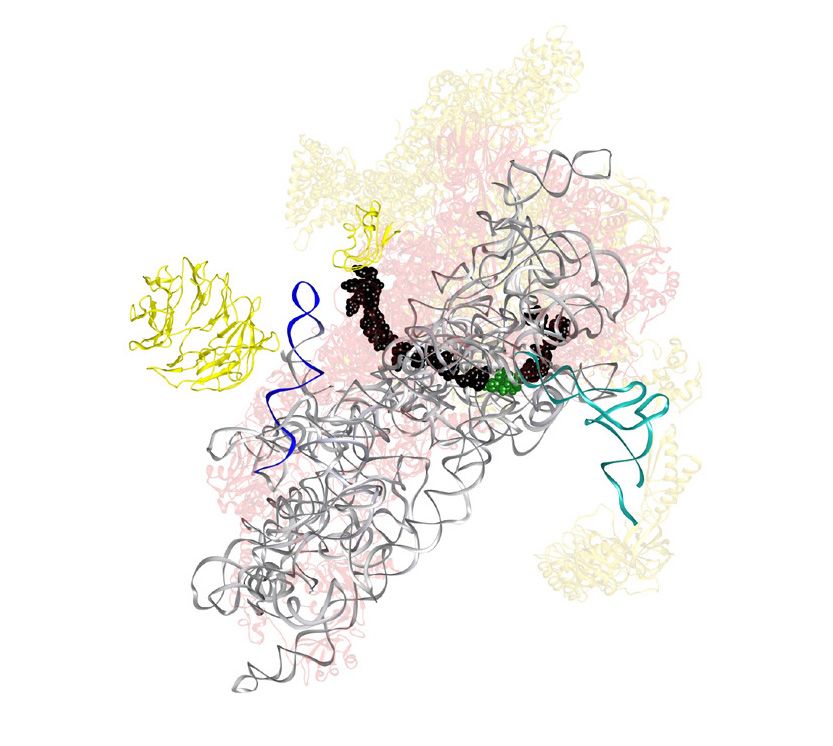
Nucleic Acids Research, 2021, Vol. 49, No. 9 5337

transcript 5 UTRs, as well as a specific and conserved site   genes, using the Bioconductor packages GenomicFeatures,
on the 18S ribosomal rRNA. We found that transcripts with      GenomicFiles and GenomicAlignments (31). Genomic and
structured 5 UTRs are preferentially affected by DDX3.        transcript regions where extracted using Ribo-seQC (32).
We used in vitro and cellular reporter systems to conclude     Only reads mapping for more than 25nt were used.
that decreases in ribosome occupancy upon DDX3 deple-             Differential analysis was using DESeq2 (33). Concordant
tion are driven by 5 UTRs. Taken together, our results sup-   changes were defined using an FDR cutoff of 0.01 for RNA-
port a model for DDX3 function where interactions with         seq and Ribo-seq individually and ensuring the same direc-
the small ribosomal subunit facilitate translation on mes-     tionality in the estimated fold changes.
sages with structured 5 UTRs, which, when inactivated,           Changes in translation efficiency were calculated using
pathologically deregulates protein synthesis.                  DESeq2 by using assay type (RNA-seq or Ribo-seq) as an
                                                               additional covariate. Translationally regulated genes were
MATERIALS AND METHODS                                          defined using an FDR cutoff of 0.05 from a likelihood ra-
                                                               tio test, using a reduced model without the assay type co-
NGS data pre-processing                                        variate, e.g. assuming no difference between RNA-seq and

                                                                                                                              Downloaded from https://academic.oup.com/nar/article/49/9/5336/6255701 by guest on 30 July 2021
Ribo-seq fastq files were stripped of the adapter sequences    Ribo-seq counts (34).
using cutadapt. UMI sequences were removed and reads              For both RNA-seq and Ribo-seq, only genes with Base-
were collapsed to fasta format. Reads were first aligned       Mean >8 or more than the bottom 10% of the library
against rRNA (accession number U13369.1), and to a col-        were used. GO enrichment analysis was performed with the
lection of snoRNAs, tRNAs and miRNA (retrieved using           topGO package (version 2.38.1; available from BioConduc-
the UCSC table browser) using bowtie2 (27) in the ‘local’      tor), using the Fisher test with default parameters.
alignment mode.                                                   The Random Forest regression was run using the ran-
   Remaining reads were mapped to the hg38 version of the      domForest package (version 4.6-14; available from CRAN)
genome (without scaffolds) using STAR 2.6.0a (28) sup-         with default parameters. Lasso regression was performed
plied with the GENCODE 32 .gtf file. A maximum of three        on scaled variables using the glmnet package (35). The fol-
mismatches and mapping to a maximum of 50 positions was        lowing features for each gene were used:
allowed. De-novo splice junction discovery was disabled for
                                                               • TPM values using RNA-seq (in log scale);
all datasets. Only the best alignment per each read was re-
                                                               • Baseline TE levels, defined as ratio of Ribo to RNA reads;
tained. Read counts for all libraries are in Supplementary
                                                               • Baseline RNA mature levels, defined as length-
Table S3.
                                                                 normalized ratio of RNA-seq reads in introns versus
                                                                 exons;
PAR-CLIP peak calling                                          • GC content, length (in log scale) and ribosome density in:
Peak calling for PAR-CLIP reads was performed with PAR-          5 UTRs, a window of 25nt around start and stop codons,
alyzer v1.5 (29) in the ‘EXTEND BY READ’ mode using              CDS regions, non-coding internal exons, introns, and 3
the following parameters:                                        UTRs;
                                                               • Additional sequence features, including density of motif
BANDWIDTH = 3                                                    scores (calculated using the Biostrings package) for the
CONVERSION = T>C                                                 following motifs, partially taken from (36):
MINIMUM READ COUNT PER GROUP = 5
MINIMUM READ COUNT PER CLUSTER = 5                             - TOP: a binary variable indicating if gene belongs to core
MINIMUM READ COUNT FOR KDE = 5                                   TOP mRNA, as defined in (37)
MINIMUM CLUSTER SIZE = 8                                       -     PRTE(pyrimidine-rich       translational   element):
MINIMUM CONVERSION LOCATIONS FOR CL                              ‘[CU][CU][CU][CU][U][CU][CU][CU]’
 USTER = 1                                                     - TISU(Translator Initiator of Short 5 -UTR):
MINIMUM CONVERSION COUNT FOR CLUSTER                             ‘[CG][A][A][CG][A][U][G][G][C][G][G][C]’
 =3                                                            - CERT (cytosine-enriched regulator of trans-
MINIMUM READ COUNT FOR CLUSTER INCL                              lation):           ‘[CG][CGU][CGU][CG][CGU][C][CG
 USION = 5                                                       U][C][CA][GU][C][CGU][CGUA][CG][C]’
MINIMUM READ LENGTH = 18                                       - PQS: propensity to create G quadruplexes, calculated us-
MAXIMUM NUMBER OF NON CONVERSION MI                              ing the pqsfinder R package (38)
 SMATCHES = 0
                                                                  Feature importance (measured by mean decrease in accu-
Peaks with >10 reads were retained for subsequent analysis.
                                                               racy for the random forest model) and correlation between
   Coverage-normalized T>C conversions on rRNA for po-         predicted and measured test data were calculated on a 10-
sitions with 2000 reads or more (Supplementary Figure          fold cross-validation scheme.
S4A) were mapped onto the 18S rRNA sequence from PDB
entry 6FEC and visualized using UCSF Chimera (30).             Meta-transcript profiles
                                                               PARalyzer peaks (and peaks from the POSTAR2 repository
Differential expression analysis
                                                               (39)) were mapped on transcript coordinates using one cod-
Count matrices for Ribo-seq and RNA-seq were built using       ing transcript per gene: such transcript was chosen to have
reads mapping uniquely to CDS regions of protein-coding        the longest 5 UTR and the most common annotated start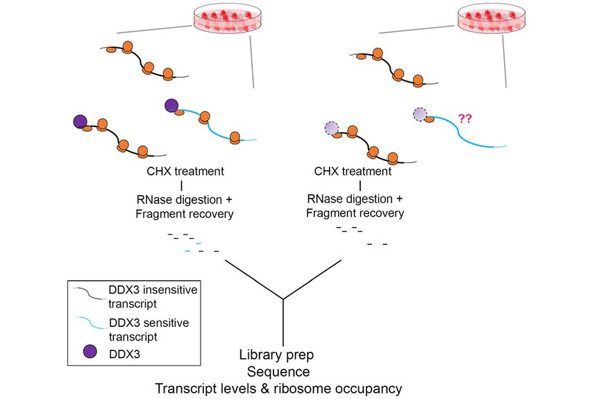
5338 Nucleic Acids Research, 2021, Vol. 49, No. 9
codon for that gene. Transcript positions were converted replicates each, siTOOLs Biotech; sequences in Supplemen-
into bins using 15 bins for each 5 UTR, 30 bins for each tary Table S5) were washed with PBS containing 100 g/ml
CDS and 20 bins for each 3 UTR. Peak scores were nor- cycloheximide to trap ribosomes, flash frozen on liquid ni-
malized for each transcript (to sum up to 1), and values were trogen, lysed in lysis buffer (20 mM Tris–HCl pH 7.4, 150
summed for each bin to build aggregate profiles, as in Fig- mM NaCl, 5 mM MgCl2 1% (v/v) Triton X-100, 25 U/ml
ure 4B. When plotting profiles for different RBPs. the aggre- TurboDNase (Ambion), harvested, centrifuged at 20 000 g
gate profiles were further normalized to a sum of 1. To build for 4 min at 4◦ C and supernatants were stored at –80◦ C.
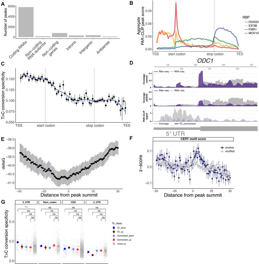
the average meta-transcript profile in Figure 4C, conversion Thawed lysates were treated with RNase I (Ambion) at 2.5
specificity values were averaged per transcript bin. To cre- U/l for 45 min at room temperature with slow agitation.
ate shuffled profiles in Figure 4F and Supplementary Figure Further RNase activity was stopped by addition of SU-
S4, 5 random positions for each peak were taken from the PERase:In (Ambion). Next Illustra MicroSpin Columns S-
same bound UTR. 400 HR (GE Healthcare) were used to enrich for ribosome
complexes. RNA was extracted from column flow throughs
with TRIzol (Ambion) reagent. Precipitated nucleic acids
Downloaded from https://academic.oup.com/nar/article/49/9/5336/6255701 by guest on 30 July 2021
De novo motif finding were further purified and concentrated with Zymo-Spin
The STREME algorithm (40) was used to perform de novo IIC column (Zymo Research). Obtained RNA was depleted
motif finding, using PAR-CLIP peaks from the POSTAR2 of rRNAs with Ribo-Zero Gold Kit (Human/Mouse/Rat)
repository, selecting peaks from HEK 293 cells called with kit (Illumina), separated in 17% urea gel and stained with
PARalyzer as above, and selecting for peaks in UTRs and SYBR Gold (Invitrogen). Gel slices containing nucleic acids
intronic regions in protein-coding genes. The following pa- 27–30 nucleotides long were excised and incubated in a ther-
rameters were used: momixer with 0.3 M NaCl at 4◦ C overnight with constant
–rna –minw 5 –maxw 15 –pvt 0.05 –totallength 1e7 –time agitation to elute RNA. After precipitation nucleic acids
18000 –patience 6 were treated with T4 polynucleotide kinase (Thermo Scien-
tific). Purified RNA was ligated to 3 and 5 adapters, reverse
transcribed and PCR amplified. The amplified cDNA was
Additional transcript features analysis sequenced on a HiSeq2000 (Illumina).
To compare read mapping locations within transcripts, a Degron RP: DDX3X-mAID tagged HCT 116 (one 15 cm
window of 25nt around the start codon was subtracted from dish at 80–90% confluency per replicate, two replicates) cells
annotated 5 UTRs and CDS. 5 UTRs and CDS regions expressing OsTIR1 were transfected with either wild-type
in genomic and transcriptomic space were retrieved using DDX3X or DDX3X R326H. 24 hours post-transfection,
Ribo-seQC. Counts on 5 UTR and CDS were first aver- media was changed and fresh media with 500 M indole
aged between replicates. The ratio 5 UTR to CDS of these 3-acetic acid (IAA) was added to cells. Un-transfected cells
counts were calculated for each gene, in the siRNA and con- were treated with either DMSO or IAA. Forty-eight hours
trols condition. The log2 of the ratio siDDX3/control for after auxin addition, cells were treated with 100 g/ml cy-
those values represents the skew of counts towards 5 UTR cloheximide (CHX) for two minutes to trap ribosomes and
in the siDDX3 condition: harvested and lysed as described in (43). Briefly, cells were
washed with PBS containing 100 g/ml CHX and lysed
5 UT R ri bosome skew in ice-cold lysis buffer (20 mM Tris–HCl pH 7.4, 150 mM
NaCl, 5 mM MgCl2 , 1 mM DTT, 100 g/ml CHX, 1%
RP5 UT R RP5 UT R (v/v) Triton X-100, 25 U/ml TurboDNase (Ambion). 240
= log2 /
RPC DS
si DDX3
RPC DS
contr ol
l lysate was treated with 6 l RNase I (Ambion, 100 U/l)
for 45 min at RT with gentle agitation and further diges-
RNA in silico folding was performed on 5 UTRs se- tion halted by addition of SUPERase:In (Ambion). Illustra
quences using RNAlfold (41) with default parameters. Av- Microspin Columns S-400 HR (GE healthcare) were used
erage G values per nucleotide were calculated averaging to enrich for monosomes, and RNA was extracted from
the G values of each structure overlapping that nucleotide. the flow-through using Direct-zol kit (Zymo Research). Gel
%GC content and T>C transition specificity (defined as slices of nucleic acids between 24 and 32 nts long were ex-
ConversionEventCount / (ConversionEventCount + Non- cised from a 15% urea–PAGE gel. Eluted RNA was treated
ConversionEventCount)) for each PAR-CLIP peak were de- with T4 PNK and preadenylated linker was ligated to the 3
rived using the clusters.csv output file from PARalyzer. Gviz end using T4 RNA Ligase 2 truncated KQ (NEB, M0373L).
was used to plot tracks for RNA-seq, Ribo-seq and PAR- Linker-ligated footprints were reverse transcribed using Su-
CLIP over different transcripts. The Wilcoxon rank sum perscript III (Invitrogen) and gel-purified RT products cir-
was used for statistical testing and Cliff’s delta was used to cularized using CircLigase II (Lucigen, CL4115K). rRNA
calculate effect sizes as described (42). depletion was performed using biotinylated oligos as de-
Source code to reproduce figures can be found at: scribed in (44) and libraries constructed using a different
https://github.com/lcalviell/DDX3X RPCLIP reverse indexing primer for each sample.
Ribosome profiling PAR-CLIP experiments
Knockdown RP: Flp-In T-REx HEK 293 cells transfected Flp-In T-REx HEK 293 cells expressing FLAG/HA-tagged
with control siPool and with DDX3X-targeting siPools (two DDX3X (45) were labeled with 100 M 4-thiouridine for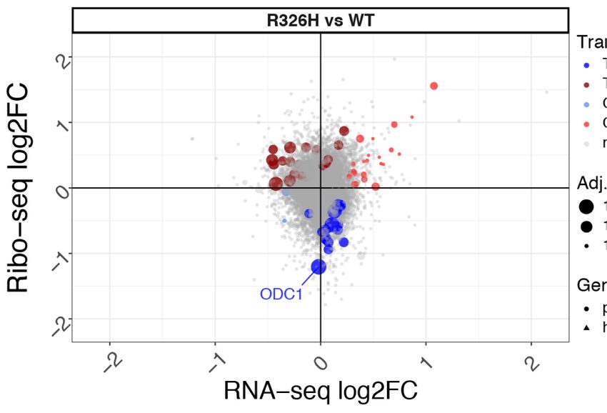
Nucleic Acids Research, 2021, Vol. 49, No. 9 5339
16h. PAR-CLIP was performed generally as described further incubation for 30 minutes. RNA was precipitated
(46,47). Briefly, cells were UV-crosslinked with 0.15 J/cm2 by the addition of 200 l 0.3 M NaOAc pH 5.3, 15 g Gly-
at 365 nm, and stored at –80◦ C. Obtained cell pellets coBlue co-precipitant (Thermo-Fisher Scientific) and 750
were lysed in three times the cell pellet volume of NP- l 100% EtOH. Precipitated RNA was further purified over
40 lysis buffer (50 mM HEPES-KOH at pH 7.4, 150 mM the RNA Clean & Concentrator-25 columns (Zymo Re-
KCl, 2 mM EDTA, 1 mM NaF, 0.5% (v/v) NP-40, 0.5 search). Glyoxal gel was run to assess the integrity of the
mM DTT, complete EDTA-free protease inhibitor cock- RNA before subsequent capping and 2 O-methylation.
tail (Roche)), incubated 10 min on ice and centrifuged 20 g of total RNA was used in a 40 l capping reaction
at 13000 rpm for 15 min at 4◦ C. Supernatants were fil- with 0.5mM GTP, 0.2 mM S-adenosylmethionine (SAM),
tered through 5 m syringe filter. Next, lysates were treated 20 units of Vaccinia capping enzyme (New England Bio-
with RNase I (Thermo-Fisher Scientific) at final con- labs), 100 units of 2 -O-Me-transferase (New England Bio-
centration of 0.25 U/l for 10 min at room tempera- labs) and 25 units RNasin Plus RNase inhibitor (Promega).
ture. Immunoprecipitation of the DDX3/RNA complexes The reactions were incubated at 37◦ C for 1 h, followed
was performed with FLAG magnetic beads (Sigma). Af- by purification over the RNA Clean & Concentrator-25
Downloaded from https://academic.oup.com/nar/article/49/9/5336/6255701 by guest on 30 July 2021
ter IP and washing, the protein-bound RNAs were 3 de- columns (Zymo Research) and elution in DEPC H2 O. Gly-
phosphorylated and 5 -end phosphorylated using T4 PNK oxal gel was run to assess the integrity of the RNA before
with 0.01% Triton X-100, and the NIR fluorescent adap- proceeding to in vitro translation reactions.
tor (5 -OH-AGATCGGAAGAGCGGTTCAGAAAAAA
AAAAAA/iAzideN/AAAAAAAAAAAA/3Bio/-3 ) was Transfection of siRNA for in vitro translation
ligated to the RNA using truncated RNA ligase 2 K227Q
HEK 293T cells in 150mM plates were transfected with 20
(NEB) overnight at 16◦ C, shaking at 1600 rpm. Crosslinked
l of siRNA (against DDX3 or a non-targeting control; se-
protein–RNA complexes were resolved on a 4–12% Nu-
quences in Supplementary Table S5) using Lipofectamine
PAGE gel (Thermo-Fisher Scientific) and transferred to a
2000 (Thermo Fisher Scientific), following manufacturer’s
nitrocellulose membrane. Protein–RNA complex migrat-
instructions. Cells were harvested for preparation of cellu-
ing at an expected molecular weight were excised, and
lar extracts after 48 h.
RNA by proteinase K (Roche) treatment and phenol–
chloroform extraction. Purified RNA was further ligated
to 5 adapters, reverse transcribed and PCR amplified. The Generation of DDX3 mutant translation extracts
amplified cDNA was sequenced on a NextSeq 500 device DDX3 WT and R326H mutant constructs were synthesized
(Illumina). and cloned downstream of a CMV promoter (Twist Bio-
sciences). 40 g of plasmids were transfected into HCT
116 cells using Lipofectamine 2000 (Thermo Fisher Sci-
In vitro transcription, capping, and 2 -O methylation of re- entific), following manufacturer’s instructions. Cells were
porter RNAs treated with 500 M indole-3-acetic acid (IAA) 24 h post-
Annotated 5 UTRs for selected transcripts were cloned transfection and harvested for preparation of cellular ex-
upstream of Renilla Luciferase (RLuc) under the control tracts after a further 48 h.
of a T7 promoter, with 60 adenosine nucleotides down-
stream of the stop codon to mimic polyadenylation. 5 Preparation of cellular extracts for in vitro translation
UTR sequences are in Supplementary Table S4. Untrans-
Three to five 150 mm plates of HEK 293T or HCT 116 cells
lated regions were cloned using synthetic DNA (Integrated
were trypsinized and pelleted at 1000g, 4◦ C. One cell-pellet
DNA Technologies) or by isolation using 5 RACE (RLM-
volume of lysis buffer (10 mM HEPES, pH 7.5, 10 mM
RACE kit, Invitrogen). Template was PCR amplified using
KOAc, 0.5 mM MgOAc2 , 5 mM DTT, and 1 tablet Com-
Phusion polymerase from the plasmids using the following
plete mini EDTA free protease inhibitor (Sigma) per 10 ml)
primers, and gel purified, as described (42).
was added to the cell pellet and was incubated on ice for
pA60 txn rev: TTT TTT TTT TTT TTT TTT TTT TTT
45 min. The pellet was homogenized by trituration through
TTT TTT TTT TTT TTT TTT TTT TTT TTT TTT TTT
a 26G needle attached to a 1 ml syringe 13–15 times. Ef-
TTT CTG CAG
ficiency of disruption was checked by trypan blue staining
pA60 txn fwd: CGG CCA GTG AAT TCG AGC TCT
(>95% disruption target). The lysate was cleared by cen-
AAT ACG ACT CAC TAT AGG
trifugation at 14000g for 1 min at 4◦ C, 2–5 l was reserved
100 l in vitro transcription reactions were set up at
for western blot analysis, and the remainder was aliquoted
room temperature with 1–5 g of purified template, 7.5 mM
and flash frozen in liquid nitrogen.
ACGU ribonucleotides, 30 mM Tris–Cl pH 8.1, 125 mM
MgCl2 , 0.01% Triton X-100, 2 mM spermidine, 110 mM
Antibodies
DTT, T7 polymerase and 0.2 U/l units of Superase-In
RNase inhibitor (Thermo-Fisher Scientific). Transcription Primary antibodies used in this study include anti-DDX3
reactions were incubated in a PCR block at 37◦ C for 1 h. 1 (Bethyl A300-474A; Figure 1), rabbit polyclonal anti-
l of 1 mg/ml pyrophosphatase (Roche) was added to each DDX3 (custom made by Genemed Synthesis using pep-
reaction, and the reactions were subsequently incubated in tide ENALGLDQQFAGLDLNSSDNQS; Figure 5; (26)),
a PCR block at 37◦ C for 3 h. 1 unit of RQ1 RNase-free anti-actin HRP (Santa Cruz Biotechnology, sc-47778), anti-
DNase (Promega) was added to each reaction followed by FLAG HRP (Sigma, A8592).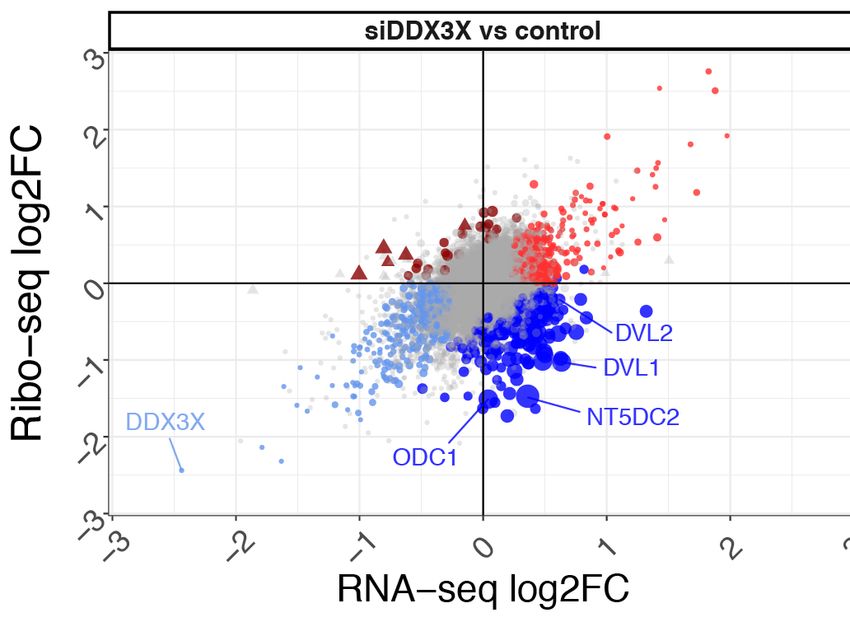
5340 Nucleic Acids Research, 2021, Vol. 49, No. 9
In vitro translation neuronal branching belonged to the translationally down-
regulated set (Supplementary Figure S1B). To confirm the
5 l in vitro translation reactions were set up with 2.5
effects of DDX3 depletion on the translated transcriptome,
l of lysate and 20 ng total RNA (0.84 mM ATP, 0.21
we also established a cell line to rapidly and efficiently de-
mM GTP, 21 mM creatine phosphate, 0.009 units/ml crea-
grade endogenous DDX3 in human male-derived colorec-
tine phosphokinase, 10 mM HEPES pH 7.5, 2 mM DTT,
tal cancer HCT 116 cells, a near-diploid cell line amenable
2 mM MgOAc, 100 mM KOAc, 0.008 mM amino acids,
to genome engineering, upon addition of auxin (Supple-
0.25 mM spermidine, 5 units RNasin Plus RNase in-
mentary Figure S2A; (51). As with the siRNA knockdown,
hibitor (Promega) as described (48). Reaction tubes were
induced degradation of DDX3 predominantly resulted in
incubated at 30◦ C for 45 min, and expression of the re-
a marked decrease in the translation of a subset of cellu-
porter was measured using the Renilla Luciferase Assay
lar messages (Supplementary Figure S2B). Translation ef-
System (Promega) on a GloMax Explorer plate reader
ficiency (TE) changes were more similar than steady state
(Promega).
RNA levels upon siRNA knockdown or chemical degrada-
tion (Supplementary Figure S2C, Supplementary Table S1),
Downloaded from https://academic.oup.com/nar/article/49/9/5336/6255701 by guest on 30 July 2021
In cell translation even though these experiments were performed in different
cell lines (HEK 293T versus HCT 116) and with different
DDX3X-mAID tagged HCT-116 cells expressing OsTIR1
depletion approaches, affirming DDX3 function in transla-
were plated into 96-well plates at ∼80% confluency. Cells
tion regulation.
were pre-treated with DMSO or 500 M indole 3-acetic
Our data suggest that DDX3 directly affects translation
acid (IAA) for 48 h. Cells were transfected with a mixture
of a subset of mRNAs. However, ribosome profiling mea-
of 10 ng in vitro transcribed firefly luciferase RNA with a
sures ribosome density, which can be affected by changes to
minimal 5 UTR (FLuc) and between 50–200 ng renilla lu-
translation initiation, translation elongation, or ribosome
ciferase RNA (RLuc) downstream of selected 5 UTRs us-
stalling. DDX3 is thought to regulate translation through
ing the TransIT mRNA Transfection Kit (Mirus Bio) per
transcript 5 UTRs, and we found genes that are regu-
manufacturer’s instructions. Twenty-four hours post trans-
lated by their 5 UTRs such as ODC1 in the translation-
fection, cells were lysed and expression of the reporters
ally downregulated set (Figure 1C; (50,52). To test whether
were measured using the Dual-Luciferase Reporter As-
altered translation initiation contributes to the impact of
say System (Promega) on a GloMax Explorer plate reader
DDX3 knockdown on ribosome density, we cloned DDX3-
(Promega). The Renilla Luciferase signal in each well was
sensitive 5 UTRs from this and previous work (3,25,53) up-
normalized to the Firefly Luciferase signal to control for
stream of a Renilla luciferase reporter and compared them
cell number, transfection efficiency, and basal translation
to a control reporter that is not sensitive to DDX3 deple-
levels.
tion. Since DVL2 has many annotated 5 UTRs that over-
lap, we cloned the prevalent isoforms in HEK 293T cells
RESULTS using 5 RACE, which yielded a short and long isoform
(Materials and Methods). We then made translation ex-
Identifying mRNAs that depend on DDX3 for efficient trans- tracts from HEK 293T cells transfected with a nontarget-
lation ing control siRNA or a DDX3 siRNA (Figure 1E). Next,
We performed ribosome profiling and RNA-seq to deter- reporter RNAs were in vitro transcribed, capped, and 2 -
mine the set of transcripts that are affected by depletion O methylated and used for in vitro translation in wild-type
of DDX3. DDX3X is an essential gene (26,49), so we tran- or DDX3-depleted lysate. We found that the 5 UTRs from
siently knocked down its expression using siRNA and col- the DDX3-sensitive mRNAs ODC1, PRKRA, RAC1 and
lected ribosome protected footprints in duplicate experi- DVL2 also conferred DDX3 dependence to the luciferase
ments (Figure 1A, B, Supplementary Figure S1A). Knock- reporter (Figure 1F). However, other reporter RNAs, such
down efficiencies were ∼90% and ∼70% in replicates (Fig- as ATF5 or and RPLP1, did not change in the in vitro
ure 1B). Measuring changes in both RNA abundance and translation upon DDX3 knockdown. RPLP1 was identi-
ribosome occupancy enabled us to distinguish between dif- fied as an mRNA with uORF occupancy changes upon
ferent modes of DDX3-mediated regulation. We found that mutant DDX3 expression in prior work (3), while ATF5
depletion of DDX3 affects ribosome occupancy of a mi- was previously implicated in DDX3-dependent translation
nority of messages (Figure 1C). Most changes in ribosome (25,53). To compare in vitro translation with translation
occupancy upon DDX3 depletion were decreases, broadly in cellular contexts, we transfected the same reporter mR-
suggesting that the function of DDX3 is to increase ri- NAs into HCT 116 cells with DDX3 depleted using our
bosome occupancy. Genes such as DVL2, NT5DC2 and degron system (Figure 1G, Supplementary Figure S2D).
ODC1, which is described to be translationally-controlled The results of the in vitro and cellular translation assays
(50), decreased in ribosome occupancy upon DDX3 deple- show a high degree of concordance, with the exception of
tion (Figure 1C, D, Supplementary Table S1). Diverse bio- the CCNE1 5 UTR, which was previously shown to re-
logical pathways were affected by DDX3 depletion, reveal- quire DDX3 for efficient translation (25). Therefore, based
ing the enrichment of histone mRNAs among the few exam- on these reporter experiments, we interpret ribosome occu-
ples of translationally upregulated transcripts, which might pancy changes upon DDX3 depletion as a result of mis-
reflect a resistance to a widespread translation suppression regulated translation initiation dynamics and refer to them
rather than an increase in protein synthesis. Genes related to as translation efficiency (TE).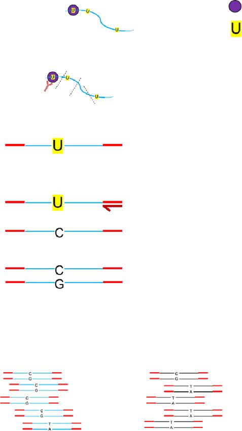
Nucleic Acids Research, 2021, Vol. 49, No. 9 5341

          A                                                                                           B

                                                                                                      C

                                                                                                                                                                       Downloaded from https://academic.oup.com/nar/article/49/9/5336/6255701 by guest on 30 July 2021
          D                                                NT5DC2
                                   100               300         500           700              900
                                            200            400          600               800
                      0.4       Ribo−seq   RNA−seq
           Coverage
            control

                      0.2

                       0
                                                                                                      E                siNTC            siDDX3X
                      0.4       Ribo−seq   RNA−seq
           Coverage
            siDDX3

                      0.2

                       0

          F                                                                                           G
                                                            **   ***   ***    **     **                                   *       ***    *   *    *   **

                            0                                                                                  0

Figure 1. Translational changes upon DDX3 depletion. (A) A workflow of the ribosome profiling experiments. (B) siRNA knockdown efficiency of DDX3
analyzed by western blot. Mock: untreated. NTC: nontargeting control siRNA. (C) The log2 fold change (log2 FC) in ribosome profiling or RNA levels
are plotted for all genes. Number genes in each category are indicated with selected genes labeled. Point size indicates the P-value of a significant change in
translational efficiency. (D) Tracks showing RNA-seq (RNA) or ribosome profiling (RP) reads that map to the NT5DC2 mRNA. NT5DC2 is an mRNA
with differential ribosome occupancy in (C). (E) Western blot analysis of nontargeting (siNTC) or siDDX3 translation lysate samples with antibodies
indicated. (F) Renilla luciferase luminescence from in vitro transcribed reporter RNAs translated in vitro in siNTC or siDDX3 HEK 293T lysates. (G)
Reporter RNAs as in (F) transfected into HCT 116 cells with or without IAA treatment to induce the DDX3 degron.

Defining the features that mediate DDX3-dependent transla-                                                served changes = 0.54, Supplementary Figure S3A), with
tion                                                                                                      few features driving the model performance (Figure 2A,
                                                                                                          Materials and Methods), such as baseline translation lev-
The in vitro and cellular translation experiments implicated
                                                                                                          els, GC content in coding sequences and 5 UTRs, and den-
translation initiation and transcript 5 UTRs in mRNAs
                                                                                                          sity of the CERT motif. The CERT motif is a cytosine-
that are sensitive to DDX3 depletion. To determine which
                                                                                                          rich element that has been implicated in eIF4E- and eIF4A-
features contribute to quantitative changes in translation
                                                                                                          dependent translation through incompletely understood
upon knockdown of DDX3, we used known translational-
                                                                                                          mechanisms (54,55). Interestingly, mRNAs that are sensi-
control elements to generate a random forest regression
                                                                                                          tive to DDX3 depletion appear to be poorly translated in
model. A model with 28 features (Materials and Methods)
                                                                                                          HEK 293T cells (Figure 2B). A reduced random forest re-
was able to moderately predict the translation changes upon
                                                                                                          gression model only using the four most relevant features
DDX3 knockdown (correlation between predicted and ob-
5342 Nucleic Acids Research, 2021, Vol. 49, No. 9
A C
Downloaded from https://academic.oup.com/nar/article/49/9/5336/6255701 by guest on 30 July 2021
D
B
Figure 2. DDX3-sensitive transcripts have complex 5 leaders. (A) Strength of individual random forest model features that predict TE with R = 0.54
(Supplementary Figure S2A, Materials and Methods). Features in blue are plotted in (B). (B) The translation efficiency (TE) in wild-type cells, CERT
motif score, GC-content of 5 UTRs, and GC content of coding sequences in the indicated gene sets from Figure 1C were computed and are plotted as
a density plot based on their importance in the random forest model. Vertical lines are medians. P-values for Wilcoxon rank sum and effect sizes from
Cliff’s delta (d) versus mixed ns indicated. (C) The fold-change of the ratio in ribosome occupancy in the 5 UTR versus the coding sequence upon DDX3
depletion as a density plot for transcripts in the indicated sets. A larger 5 UTR skew value means that there are more ribosomes in the 5 UTR compared
to the coding sequence. P-values from Wilcoxon rank sum and effect sizes from Cliff’s delta (d) versus mixed ns indicated. (D) An example gene (HMBS)
with increases in 5 UTR ribosomes versus coding sequence ribosomes with tracks as in Figure 1D.Nucleic Acids Research, 2021, Vol. 49, No. 9 5343
performed similarly; conversely, a model built without us- Helix 16 (h16) is on the small ribosomal subunit facing
ing sequence predictors or baseline translation levels could incoming mRNA, which might provide DDX3 access to
not recapitulate translation downregulation effects (Supple- resolve mRNA secondary structures to facilitate inspection
mentary Figure S3B, S3C). 5 UTR and coding sequence by the scanning 43S complex (Figure 3C). The crosslink
GC content may be indications of increased RNA structure site on h16 is just opposite an RRM domain that has
in these regions, possibly relevant for other aspects of cyto- been assigned as eIF4B, another factor crucial in ribosome
plasmic RNA processing (Discussion). recruitment and scanning (56,58,59). This is consistent with
To test the ability of the random forest to select relevant observations that eIF4B and Ded1 cooperate in translation
features predictive of DDX3-mediated translation changes, initiation on mRNAs (58). Recently, it has been proposed
we compared it to lasso regression, another method used that this RRM domain may belong to eIF3g, another
to perform feature selection among a set of correlated pre- translation initiation factor (60), which is consistent with a
dictors (Materials and Methods). The two methods largely reported interaction between eIF3 and DDX3 (26).
agreed in pinpointing relevant features, with the random In addition to ribosomal RNA binding, we found that
forest slightly outperforming the lasso (Supplementary Fig- DDX3 interacts primarily with coding transcripts (Figure
Downloaded from https://academic.oup.com/nar/article/49/9/5336/6255701 by guest on 30 July 2021
ure S3D, E). The features predicted to drive DDX3 sensitiv- 4A, Supplementary Table S2). To identify where DDX3
ity also showed different distributions among the regulated binds mRNAs, we aggregated peaks across all expressed
mRNAs, especially in the translationally downregulated set, transcripts in a metagene analysis. We found that DDX3
indicating that the features identified by the random forest primarily contacts transcript 5 UTRs, with a small num-
are indeed different between sets of transcripts (Figure 2B). ber of reads mapping in the coding sequence and 3 UTR
We measured the enrichment of ribosomes in transcript 5 (Figure 4B). A large peak was also observed at the start
UTRs, under the hypothesis that depletion of DDX3 might codon, which could reflect kinetic pausing during subunit
lead to defective scanning and ribosome accumulation (1), joining while DDX3 is still bound to the initiating ribosome
or selective ribosome depletion on coding sequences. In- (61). We used available CLIP data to compare the binding
deed, we found more ribosomes in transcript 5 UTRs rel- pattern of DDX3 to other known mRNA-binding proteins
ative to coding sequences upon DDX3 depletion (Figure (39). We selected three RNA-binding proteins to compare
2C), especially in mRNAs that show translational down- to: eIF3b is a member of the multi-subunit initiation factor
regulation. As an example, HMBS ribosome occupancy is eIF3 (62), FMR1 interacts with elongating ribosomes (63),
shown in Figure 2D, which showed changes in ribosome and MOV10 is involved in 3 UTR-mediated mRNA decay
density in its 5 UTR and therefore may be regulated by up- (64,65). The binding pattern of DDX3 most closely resem-
stream ORF (uORF) translation. bles the initiation factor eIF3b (Figure 4B). We detected
some DDX3 binding within coding sequences and even 3
UTRs, which could arise from background signal, or al-
DDX3 crosslinks to ribosomal RNA and 5 UTRs
ternative binding modes. PAR-CLIP indicated that DDX3
The above ribosome profiling and RNA-seq experiments binds abundant mRNAs (Supplementary Figure S4B), in-
identified the set of transcripts that are affected by DDX3 cluding ribosomal protein genes. While this observation can
depletion, but these transcripts could be affected by direct provide insights into the molecular functions of DDX3, it
or indirect mechanisms. Therefore, to better define the set might reflect a limit in the sensitivity of our PAR-CLIP
of transcripts that are direct targets of DDX3, we measured data. Therefore, we decided to investigate PAR-CLIP bind-
DDX3 binding sites with high specificity using PAR-CLIP ing patterns which are not strongly confounded by expres-
(Figure 3A, Supplementary Figure S1A). Previous work us- sion levels. We used the frequency of T>C transitions at
ing a complementary method (iCLIP) to measure DDX3 each site as a measure of the specificity of protein-RNA
binding sites identified 5 UTR and ribosomal RNA bind- interactions (66). High specificity crosslinks with frequent
ing. Curiously, even though DDX3 is thought to regulate T>C transitions resided most often in 5 UTRs (Figure 4C),
translation initiation, binding was also identified in coding as also shown in a translationally-regulated transcript such
sequences and 3 UTRs (3,12). Here, we used the additional as ODC1 (Figure 4D). Confirmation of this binding pattern
specificity afforded by T>C transitions induced by protein comes from an independent assay of protein–RNA interac-
adducts on crosslinked uridine residues in PAR-CLIP to re- tion, as measured by enhanced CLIP (eCLIP) (Supplemen-
fine DDX3 binding sites across the transcriptome (46). tary Figure S4C; 67).
High-throughput sequencing of RNA fragments Next, we sought to describe the mRNA regions with en-
crosslinked to DDX3 identified a binding site for DDX3 riched DDX3 binding. DEAD-box RNA helicases engage
on the 18S ribosomal RNA (Figure 3B; visualized on the RNA by recognizing structural elements with poorly de-
structure (56). It is possible that these rRNA reads could fined sequence context (68), which hinders the ability to
arise from nonspecific interactions between RNA binding extract meaningful sequence motifs from CLIP data. To
proteins and the highly abundant rRNA. However, while investigate this phenomenon, we performed de novo mo-
there were many rRNA fragments sequenced following tif finding on PAR-CLIP peaks in HEK 293 cells from the
PAR-CLIP, there was only one site with high-confidence POSTAR2 repository (39; Materials and Methods). As ex-
T>C transitions, spanning nucleotides 527–553 in the 18S pected, motifs extracted from different RBPs showed dif-
rRNA (Figure 3B, Supplementary Figure S4A). This site ferent degrees of specificity (Supplementary Figure S4D).
maps to helix 16 of the 18S rRNA, similar to where Ded1 DDX3 motifs, together with motifs from translation initi-
crosslinks to 18S rRNA in yeast, and does not contain ation factors and other ribosome interactors, showed poor
post-transcriptionally modified rRNA nucleotides (1,57). significance when compared to other RBPs with more de-5344 Nucleic Acids Research, 2021, Vol. 49, No. 9
A B
C
Downloaded from https://academic.oup.com/nar/article/49/9/5336/6255701 by guest on 30 July 2021
Figure 3. DDX3 binding sites on rRNA identified by PAR-CLIP. (A) A workflow of the PAR-CLIP experiment. (B) PAR-CLIP T>C conversion locations
on human rDNA (top) compared to iCLIP coverage from Oh et al. 2016 (bottom). Boxed regions refer to processed rRNA transcripts. (C) PAR-CLIP
T>C conversion density on the 18S rRNA is visualized from gray to blue on the structure of the 48S ribosome (PDB 6FEC). The peak in (B) is contained
in the helix in the upper left in (C), which is h16 of rRNA. Yellow: translation factors; blue–gray: rRNA; pink: ribosomal proteins.
fined sequence specificity (such as members of cleavage patients with truncating mutations (16). Point mutations in
and polyadenylation machinery, or known 3 UTR binders). DDX3 associated with medulloblastoma are dominant neg-
This result called for a more targeted analysis of DDX3 ative and act by preventing enzyme closure of DDX3 to-
binding sites. By investigating the sequence-structure con- wards the high-RNA-affinity ATP-bound state (6–11). This
text around mRNA peaks in 5 UTRs, we observed that suggests there may be different effects on translation be-
DDX3 binding sites by PAR-CLIP reside in highly struc- tween depletion of DDX3 and inhibition or expression of
tured regions (Figure 4E). We observed a higher guanine an inactive mutant.
content (accompanied by predicted G-quadruplexes; Sup- To test the effect of mutants in DDX3 on translation,
plementary Figures S4E, F) upstream of the binding site; we transfected cells with plasmids containing wild-type or
downstream of the peak summit, we detected high GC mutant DDX3 proteins after auxin-induced degradation of
content resembling the CERT motif (Figure 4F), a regula- endogenous DDX3, switching expression from the wild-
tory motif highly predictive of DDX3-mediated translation type sequence to an allele of interest (Figure 5A). We used
regulation (Figure 2A). Moreover, we observed increased this system to define acute changes to translation caused
T>C conversion specificity in the 5 UTR of transcripts by DDX3 mutations without allowing the cells to adapt
whose translation decreases upon DDX3 depletion (Fig- to the presence of inactive DDX3 alleles, which may also
ure 4G), indicative of a possibly stronger protein-RNA as- be lethal. We measured genome-wide translation changes
sociation at those regions. This binding pattern, combined caused by DDX3 mutants using ribosome profiling (Sup-
with the observation that DDX3 co-fractionates with both plementary Table S1). A collection of genes related to the
polysomes and initiation complexes, suggests that DDX3 double-stranded DNA response were upregulated at the
acts with the 40S during the process of translation initia- level of RNA abundance, likely due to differences in trans-
tion (2,69–71). Taken together, we conclude that the DDX3 fected DNA amounts (Figure 5B, upper right). Broadly,
binds and regulates the translation of poorly translated mR- we noticed higher variability in in ribosome occupancy
NAs exhibiting complex sequence-structure features in their changes (Figure 5B, y-axis) than RNA level changes (Fig-
5 UTRs. ure 5B, x-axis) when compared to the siDDX3 experiments
(Figure 1C), suggesting that one functional difference be-
tween mutant and knockdown might involve regulation of
Identifying translation changes caused by DDX3 mutations
RNA steady-state levels. Among the few regulated mRNAs,
De novo genetic variants in DDX3X cause developmental we observed a robust downregulation of ODC1 transla-
delay and intellectual disability in DDX3X-syndrome (13– tion, which was directly bound by DDX3 (Figure 4D) and
16). Interestingly, patients carrying inactivating point muta- strongly downregulated in the DDX3 depletion experiment
tions in DDX3 display more severe clinical symptoms than (Figure 1C).Nucleic Acids Research, 2021, Vol. 49, No. 9 5345
Downloaded from https://academic.oup.com/nar/article/49/9/5336/6255701 by guest on 30 July 2021
Figure 4. DDX3 binds to structured 5 leaders of mRNA. (A) Sum of the DDX3 PAR-CLIP peaks mapping on different gene types and regions. (B)
A metagene plot of DDX3 PAR-CLIP across all genes and PAR-CLIP data from other RNA binding proteins. eIF3b is a canonical initiation factor,
FMR1 binds elongating ribosomes, and MOV10 is a 3 UTR binding factor. TSS: transcription start site, TES: transcription end site. (C) T>C conversion
specificity averaged across all PAR-CLIP peaks across indicated mRNA regions as in (B). (D) PAR-CLIP, ribosome profiling, and RNA-seq across part of
the ODC1 gene. Blue peaks in the PAR-CLIP track indicate T>C conversion events. (E) RNA structure in a window of 100 nucleotides around PAR-CLIP
peak summits in 5 UTRs. (F) CERT motif scores averaged across PAR-CLIP peak summits in 5 UTRs or for shuffled positions. (G) PAR-CLIP T>C
conversion specificity in transcript regions in indicated gene sets from Figure 1.5346 Nucleic Acids Research, 2021, Vol. 49, No. 9
A B
B
Downloaded from https://academic.oup.com/nar/article/49/9/5336/6255701 by guest on 30 July 2021
C D
Figure 5. Translation changes caused by R326H mutant DDX3. (A) Western blots of degron DDX3 cells treated with IAA and transfected with empty
vector or the indicated constructs. (B) Ribosome profiling and RNA-seq of DDX3 degron cells treated with IAA and transfected with either DDX3 wild-
type or R326H mutant. (C) Western blots of cells treated with siDDX3 and transfected with the indicated constructs. (D) In vitro translation performed
with indicated reporter RNAs as in Figure 1 in the lysates from panel (C). Reporter sequences are in Supplementary Table S4.
To further test the difference between knockdown and DISCUSSION
mutant DDX3 at the level of individual 5 UTRs, we made
DDX3X is an essential human gene that is altered in diverse
in vitro translation extracts with wild-type DDX3, R326H
diseases. Here, we use a set of transcriptomics approaches,
DDX3, or R534H DDX3 and measured translation of
machine learning and biochemistry to show that DDX3 reg-
a panel of reporters (Figure 5C; (3)). Broadly, we found
ulates a subset of the human transcriptome, likely through
two classes of reporter RNAs (Figure 5D; Supplemen-
resolving RNA structures in 5 UTRs (Figure 6). Reporter
tary Figure S5A, B). One class, including ODC1, PRKRA,
experiments show that 5 UTRs are sufficient to confer
RAC1 and DVL2 isoforms decreased in translation in all
DDX3 sensitivity onto unrelated coding sequences. We con-
tested perturbations to DDX3. Another class, including
clude that DDX3 affects translation initiation through tran-
ATF5, CCNE1 and RPLP1 selectively decreased in trans-
script 5 UTRs. Our data suggest that the major role of
lation in mutant DDX3 extracts but not in knockdown ex-
DDX3 is in translation initiation and reveal translation dif-
tracts. We sought to test chemical inhibition of DDX3, as
ferences between mutated and haploinsufficient DDX3 ex-
it functionally mimics a dominant negative mutation by
pression.
blocking ATP binding. We were unable to inhibit transla-
We identified binding between DDX3 and helix 16 (h16)
tion in a DDX3-dependent manner using RK-33 (72), and
on the human 40S ribosome. This is similar to binding
instead found that it acted as a general translation inhibitor
sites identified previously using other CLIP approaches
(Supplementary Figure S5C). Taken together, we found that
(1,3,12), confirmed here using T>C transitions defined
DDX3 sensitivity for translation is preserved in translation
by PAR-CLIP. Interestingly, histone mRNAs showed sus-
extracts and that depletion of DDX3 appears to have dif-
tained translation levels upon DDX3 knockdown (Figure
ferent outcomes on translation than inhibition or dominant
1C). However, histone mRNAs represent a peculiar cate-
negative variants.
gory of transcripts, often containing short UTRs, few in-Nucleic Acids Research, 2021, Vol. 49, No. 9 5347
Downloaded from https://academic.oup.com/nar/article/49/9/5336/6255701 by guest on 30 July 2021
Figure 6. A model of DDX3 in translational control. DDX3 binds to the small subunit via helix 16 (h16). Transcripts that are poorly translated in normal
cells and that harbor increased intramolecular RNA structure (blue transcript; left) are sensitive to DDX3 depletion (right). Other mRNAs that are initially
highly translated (black transcript) are unaffected.
trons, highly repetitive sequences and the lack a poly-A ble that DDX3, alone or in combination with other DEAD-
tail. The quantitative contribution of some of these features box proteins like eIF4A, functions in trans by activating an
have been resolved by our regression approach, with good mRNA prior to 43S complex loading. Future work defin-
agreement between the lasso and the random forest mod- ing the binding site of DDX3 on the ribosome could enable
els. However, we believe that more tailored approaches are separation of cis and trans functions to test these two mod-
needed to precisely investigate regulation of specific classes els, although we note that the functional consequences of
of mRNAs. For instance, histone mRNA translation has DDX3 depletion we have observed here are independent of
been recently shown to be dependent on mRNA binding to its functioning in cis or trans.
the rRNA h16 helix (73). Our data suggests that there might DDX3 is altered in numerous human diseases, includ-
be competition for h16 between DDX3 and histone mR- ing cancers and developmental disorders (5). Some diseases
NAs, and their translation increases upon DDX3 knock- are characterized by missense variants (8–11), while oth-
down due to increased accessibility to h16. The set of mR- ers involve predominantly nonsense or frameshift variants
NAs that require h16 for their translation will be an inter- (17,18), and still others present with a mixture of variant
esting direction to pursue in the future. types (13–16). Our work suggests that variants in DDX3
Despite primarily affecting translation, some genes ex- that deplete protein levels may result in different translation
hibited changes in steady-state RNA abundance upon changes than inactivating missense variants. We attempted
DDX3 depletion. RNA-level changes could be mediated to directly compare translational changes upon DDX3 de-
by indirect effects of a DDX3-dependent translation target, pletion identified in this work with previous expression of
or reflect additional mechanisms of post-transcriptional mutant DDX3 but stopped due to confounding variabil-
gene regulation, possibly mediated by RNA structural fea- ity in biological sample and library preparation and se-
tures, codon composition, or other RNA decay pathways quencing protocols. Defining how different mutation types
(74). Moreover, DDX3 is an important factor in stress- in DDX3 affect gene expression, the underlying molecular
granule complexes (75), and possibly involved in granule- mechanisms, and potential therapeutic interventions is an
specific regulation of mRNA metabolism. Interestingly, we intriguing direction for the future.
observed more RNA-level changes in DDX3-depletion ex-
periments (Figure 1C and Supplementary Figure S2B) than DATA AVAILABILITY
in experiments using a mutation in DDX3 (Figure 5B). Po-
tential interactions between DDX3 regulation of both ribo- Processed datasets and code to reproduce the main
some occupancy and RNA levels will be further explored in figures can be found here: https://github.com/lcalviell/
future studies. DDX3X RPCLIP.
DDX3 is an abundant protein, with approximately 1.4 Sequencing data can be retrieved using GEO accession
million copies per HeLa cell (76), or about half the abun- numbers GSE125114 and GSE157063.
dance of ribosomes (77). We have interpreted data in this
work by hypothesizing that DDX3 is functioning in cis by SUPPLEMENTARY DATA
binding to the 40S ribosome and facilitating translation ini-
tiation on the associated mRNA (Figure 6). It is also possi- Supplementary Data are available at NAR Online.5348 Nucleic Acids Research, 2021, Vol. 49, No. 9
ACKNOWLEDGEMENTS mutations target distinct subgroups of medulloblastoma. Nature, 488,
43–48.
We thank members of the Floor lab for feedback on the 12. Valentin-Vega,Y.A., Wang,Y.-D., Parker,M., Patmore,D.M.,
manuscript. Computation was supported by the UCSF Kanagaraj,A., Moore,J., Rusch,M., Finkelstein,D., Ellison,D.W.,
Wynton computing infrastructure. Gilbertson,R.J. et al. (2016) Cancer-associated DDX3X mutations
drive stress granule assembly and impair global translation. Sci. Rep.,
Author contributions: Conceptualization, L.C., E.W., W.F., 6, 25996.
M.L. and S.N.F.; Investigation, L.C., K.R., S.V., K.W. and 13. Deciphering Developmental Disorders, S. (2015) Large-scale
E.W.; Writing – Original Draft, S.N.F.; Writing – Review & discovery of novel genetic causes of developmental disorders. Nature,
Editing, S.N.F., S.V., L.C., E.W. and M.L.; Resources, B.T., 519, 223–228.
M.T., J.K. and W.F.; Funding Acquisition, M.L. and S.N.F.; 14. Snijders Blok,L., Madsen,E., Juusola,J., Gilissen,C., Baralle,D.,
Reijnders,M.R., Venselaar,H., Helsmoortel,C., Cho,M.T.,
Supervision, W.F., M.L. and S.N.F. Hoischen,A. et al. (2015) Mutations in DDX3X are a common cause
of unexplained intellectual disability with gender-specific effects on
Wnt signaling. Am. J. Hum. Genet., 97, 343–352.
FUNDING 15. Wang,X., Rosenfeld,J.A., Bacino,C.A., Scaglia,F., Immken,L.,
Harris,J.M., Hickey,S.E., Mosher,T.M., Slavotinek,A., Zhang,J. et al.
Downloaded from https://academic.oup.com/nar/article/49/9/5336/6255701 by guest on 30 July 2021
University of California, San Francisco Program for Break- (2018) Phenotypic expansion in DDX3X –– a common cause of
through Biomedical Research, Sandler Foundation [to intellectual disability in females. Ann. Clin. Transl. Neurol., 5,
S.N.F., in part]; California Tobacco-Related Disease Re- 1277–1285.
search Grants Program [27KT-0003 to S.N.F., T30DT1004 16. Lennox,A.L., Hoye,M.L., Jiang,R., Johnson-Kerner,B.L., Suit,L.A.,
Venkataramanan,S., Sheehan,C.J., Alsina,F.C., Fregeau,B.,
to K.W.]; National Institutes of Health [DP2GM132932 Aldinger,K.A. et al. (2020) Pathogenic DDX3X mutations impair
to S.N.F., F32GM133144 to S.V.]; DFG Priority Project RNA metabolism and neurogenesis during fetal cortical
[SPP1935 to K.R. and M.L.]. Funding for open access Developmentd Neuron, 106, 404–420.
charge: Research grants. 17. Dufva,O., Kankainen,M., Kelkka,T., Sekiguchi,N., Awad,S.A.,
Conflict of interest statement. S.N.F. consults for MOMA Eldfors,S., Yadav,B., Kuusanmäki,H., Malani,D., Andersson,E.I.
et al. (2018) Aggressive natural killer-cell leukemia mutational
Therapeutics. landscape and drug profiling highlight JAK-STAT signaling as
therapeutic target. Nat. Commun., 9, 1567.
18. Jiang,L., Gu,Z.H., Yan,Z.X., Zhao,X., Xie,Y.Y., Zhang,Z.G.,
REFERENCES Pan,C.M., Hu,Y., Cai,C.P., Dong,Y. et al. (2015) Exome sequencing
1. Guenther,U.-P., Weinberg,D.E., Zubradt,M.M., Tedeschi,F.A., identifies somatic mutations of DDX3X in natural killer/T-cell
Stawicki,B.N., Zagore,L.L., Brar,G.A., Licatalosi,D.D., Bartel,D.P., lymphoma. Nat. Genet., 47, 1061–1066.
Weissman,J.S. et al. (2018) The helicase Ded1p controls use of 19. Wang,L., Lawrence,M.S., Wan,Y., Stojanov,P., Sougnez,C.,
near-cognate translation initiation codons in 5 UTRs. Nature, 559, Stevenson,K., Werner,L., Sivachenko,A., DeLuca,D.S., Zhang,L.
130. et al. (2011) SF3B1 and other novel cancer genes in chronic
2. Lai,M.-C., Lee,Y.-H.W., Tarn,W.-Y. and Walter,P. (2008) The lymphocytic leukemia. N. Engl. J. Med., 365, 2497–2506.
DEAD-Box RNA helicase DDX3 associates with export messenger 20. Schmitz,R., Young,R.M., Ceribelli,M., Jhavar,S., Xiao,W.,
ribonucleoproteins as well as tip-associated protein and participates Zhang,M., Wright,G., Shaffer,A.L., Hodson,D.J., Buras,E. et al.
in translational control. Mol. Biol. Cell, 19, 3847–3858. (2012) Burkitt lymphoma pathogenesis and therapeutic targets from
3. Oh,S., Flynn,R.A., Floor,S.N., Purzner,J., Martin,L., Do,B.T., structural and functional genomics. Nature, 490, 116–120.
Schubert,S., Vaka,D., Morrissy,S., Li,Y. et al. (2016) 21. Chuang,R.Y., Weaver,P.L., Liu,Z. and Chang,T.H. (1997)
Medulloblastoma-associated DDX3 variant selectively alters the Requirement of the DEAD-Box protein ded1p for messenger RNA
translational response to stress. Oncotarget, 7, 28169–28182. translation. Science, 275, 1468–1471.
4. Soto-Rifo,R., Rubilar,P.S., Limousin,T., Breyne,S., Décimo,D. and 22. de la Cruz,J., Iost,I., Kressler,D. and Linder,P. (1997) The p20 and
Ohlmann,T. (2012) DEAD-box protein DDX3 associates with eIF4F Ded1 proteins have antagonistic roles in eIF4E-dependent translation
to promote translation of selected mRNAs. EMBO J., 31, 3745–3756. in Saccharomyces cerevisiae. Proc. Natl. Acad. Sci. U.S.A., 94,
5. Sharma,D. and Jankowsky,E. (2014) The Ded1/DDX3 subfamily of 5201–5206.
DEAD-box RNA helicases. Crit. Rev. Biochem. Mol. Biol., 49, 23. Sen,N.D., Zhou,F., Ingolia,N.T. and Hinnebusch,A.G. (2015)
343–360. Genome-wide analysis of translational efficiency reveals distinct but
6. Epling,L.B., Grace,C.R., Lowe,B.R., Partridge,J.F. and Enemark,E.J. overlapping functions of yeast DEAD-box RNA helicases Ded1 and
(2015) Cancer-associated mutants of RNA helicase DDX3X are eIF4A. Genome Res., 25, 1196–1205.
defective in RNA-stimulated ATP hydrolysis. J. Mol. Biol., 427, 24. Ku,Y.-C., Lai,M.-H., Lo,C.-C., Cheng,Y.-C., Qiu,J.-T., Tarn,W.-Y.
1779–1796. and Lai,M.-C. (2018) DDX3 participates in translational control of
7. Floor,S.N., Condon,K.J., Sharma,D., Jankowsky,E. and inflammation induced by infections and injuries. Mol. Cell. Biol., 39,
Doudna,J.A. (2016) Autoinhibitory interdomain interactions and e00285-18.
subfamily-specific extensions redefine the catalytic core of the human 25. Lai,M.-C., Chang,W.-C., Shieh,S.-Y. and Tarn,W.-Y. (2010) DDX3
DEAD-box protein DDX3. J. Biol. Chem., 291, 2412–2421. regulates cell growth through translational control of cyclin E1. Mol.
8. Jones,D.T., Jager,N., Kool,M., Zichner,T., Hutter,B., Sultan,M., Cell. Biol., 30, 5444–5453.
Cho,Y.J., Pugh,T.J., Hovestadt,V., Stutz,A.M. et al. (2012) Dissecting 26. Lee,C.S., Dias,A.P., Jedrychowski,M., Patel,A.H., Hsu,J.L. and
the genomic complexity underlying medulloblastoma. Nature, 488, Reed,R. (2008) Human DDX3 functions in translation and interacts
100–105. with the translation initiation factor eIF3. Nucleic Acids Res., 36,
9. Kool,M., Jones,D.T., Jager,N., Northcott,P.A., Pugh,T.J., 4708–4718.
Hovestadt,V., Piro,R.M., Esparza,L.A., Markant,S.L., Remke,M. 27. Langmead,B. and Salzberg,S.L. (2012) Fast gapped-read alignment
et al. (2014) Genome sequencing of SHH medulloblastoma predicts with Bowtie 2. Nat. Methods, 9, 357–359.
genotype-related response to smoothened inhibition. Cancer Cell, 25, 28. Dobin,A., Davis,C.A., Schlesinger,F., Drenkow,J., Zaleski,C., Jha,S.,
393–405. Batut,P., Chaisson,M. and Gingeras,T.R. (2013) STAR: ultrafast
10. Pugh,T.J., Weeraratne,S.D., Archer,T.C., Pomeranz Krummel,D.A., universal RNA-seq aligner. Bioinformatics, 29, 15–21.
Auclair,D., Bochicchio,J., Carneiro,M.O., Carter,S.L., Cibulskis,K., 29. Corcoran,D.L., Georgiev,S., Mukherjee,N., Gottwein,E.,
Erlich,R.L. et al. (2012) Medulloblastoma exome sequencing Skalsky,R.L., Keene,J.D. and Ohler,U. (2011) PARalyzer: definition
uncovers subtype-specific somatic mutations. Nature, 488, 106–110. of RNA binding sites from PAR-CLIP short-read sequence data.
11. Robinson,G., Parker,M., Kranenburg,T.A., Lu,C., Chen,X., Ding,L., Genome Biol., 12, R79.
Phoenix,T.N., Hedlund,E., Wei,L., Zhu,X. et al. (2012) NovelYou can also read