The Inhibition of Polo Kinase by Matrimony Maintains G2 Arrest in the Meiotic Cell Cycle
←
→
Page content transcription
If your browser does not render page correctly, please read the page content below
PLoS BIOLOGY

The Inhibition of Polo Kinase by Matrimony
Maintains G2 Arrest in the Meiotic Cell Cycle
Youbin Xiang1, Satomi Takeo1, Laurence Florens1, Stacie E. Hughes1, Li-Jun Huo1, William D. Gilliland1,
Selene K. Swanson1, Kathy Teeter1, Joel W. Schwartz1, Michael P. Washburn1, Sue L. Jaspersen1,2, R. Scott Hawley1,2,*
1 Stowers Institute for Medical Research, Kansas City, Missouri, United States of America, 2 University of Kansas School of Medicine, Kansas City, Kansas, United States of America

Many meiotic systems in female animals include a lengthy arrest in G2 that separates the end of pachytene from
nuclear envelope breakdown (NEB). However, the mechanisms by which a meiotic cell can arrest for long periods of
time (decades in human females) have remained a mystery. The Drosophila Matrimony (Mtrm) protein is expressed
from the end of pachytene until the completion of meiosis I. Loss-of-function mtrm mutants result in precocious NEB.
Coimmunoprecipitation experiments reveal that Mtrm physically interacts with Polo kinase (Polo) in vivo, and
multidimensional protein identification technology mass spectrometry analysis reveals that Mtrm binds to Polo with
an approximate stoichiometry of 1:1. Mutation of a Polo-Box Domain (PBD) binding site in Mtrm ablates the function of
Mtrm and the physical interaction of Mtrm with Polo. The meiotic defects observed in mtrm/þ heterozygotes are fully
suppressed by reducing the dose of poloþ, demonstrating that Mtrm acts as an inhibitor of Polo. Mtrm acts as a
negative regulator of Polo during the later stages of G2 arrest. Indeed, both the repression of Polo expression until
stage 11 and the inactivation of newly synthesized Polo by Mtrm until stage 13 play critical roles in maintaining and
properly terminating G2 arrest. Our data suggest a model in which the eventual activation of Cdc25 by an excess of
Polo at stage 13 triggers NEB and entry into prometaphase.
Citation: Xiang Y, Takeo S, Florens L, Hughes SE, Huo LJ, et al. (2007) The inhibition of Polo kinase by Matrimony maintains G2 arrest in the meiotic cell cycle. PLoS Biol 5(12):
e323. doi:10.1371/journal.pbio.0050323

                                                                                             allows the completion of meiosis I followed by meiosis II. As
Introduction
                                                                                             shown in Figure 1, the end of meiotic prophase by dissolution
   The mechanism of the lengthy arrest in G2 that separates                                  of the synaptonemal complex (SC) at stages 5–6 [4,5] is
the end of pachytene from nuclear envelope breakdown                                         separated from the beginning of the meiotic divisions, which
(NEB)—which is a characterization of many female meiotic                                     is defined by NEB at stage 13, by approximately 40 h to allow
systems—has remained a mystery. One can imagine that both                                    for oocyte growth.
the maintenance and the termination of this arrest might                                        We are interested in elucidating the mechanisms that arrest
involve either or both of two mechanisms— the transcriptional                                meiotic progression at the end of prophase, but then allow
or translational repression of a protein that induces NEB, and                               onset of NEB and the initiation of meiotic spindle formation
thus meiotic entry, or the presence of an inhibitory protein                                 some 40 h later. One intriguing possibility is that during this
that precludes entry into the first meiotic division. Because
                                                                                             period of meiotic arrest, the oocyte actively blocks the
Drosophila females exhibit a prolonged G2 arrest (see Figure 1)
                                                                                             function of cell cycle regulatory proteins such as cyclin
and are amenable to both genetic and cytological analyses,
                                                                                             dependent kinase 1 (Cdk1), the phosphatase Cdc25, and Polo
they provide an ideal system in which to study this problem.
                                                                                             kinase (Polo), all of which promote meiotic progression just as
   The ovaries of Drosophila females are composed of a bundle
                                                                                             they do during mitotic growth. Recently, Polo was shown to
of ovarioles, each of which contains a number of oocytes
arranged in order of their developmental stages [1–3]. For                                   be expressed in the germarium and required for the proper
our purposes, the process of oogenesis may be said to consist                                entry of Drosophila oocytes into meiotic prophase, as defined
of three separate sets of divisions: the initial stem cell                                   by the assembly of the SC [6]. Decreased levels of Polo
divisions, which create primary cystoblasts; four incomplete                                 resulted in delayed entry into meiotic prophase, whereas
cystoblast divisions, which create a 16-cell cyst that contains                              overexpression of Polo caused a dramatic increase in the
the oocyte; and the two meiotic divisions. Although a great
deal is known regarding the mechanisms that control                                          Academic Editor: Terry Orr-Weaver, Whitehead Institute, United States of America
cystoblast divisions and oocyte differentiation, relatively little
                                                                                             Received August 2, 2007; Accepted October 23, 2007; Published December 4,
is known about the mechanisms by which the progression of                                    2007
meiosis is controlled.
                                                                                             Copyright: Ó 2007 Xiang et al. This is an open-access article distributed under the
   As is the case in many meiotic systems, female meiosis in                                 terms of the Creative Commons Attribution License, which permits unrestricted
Drosophila involves preprogrammed developmental pauses.                                      use, distribution, and reproduction in any medium, provided the original author
                                                                                             and source are credited.
The two most prominent pauses during Drosophila meiosis are
an arrest that separates the end of pachytene at stages 5–6                                  Abbreviations: FDR, false discovery rate, FISH, fluorescent in situ hybridization;
                                                                                             GFP, green fluorescent protein; MudPIT, multidimensional protein identification
from NEB at stage 13, and a second pause that begins with                                    technology; NEB, nuclear envelope breakdown; NSAF, normalized spectral
metaphase I arrest at stage 14 and continues until the egg                                   abundance factors; PBD, Polo-Box Domain; SC, synaptonemal complex
passes through the oviduct. It is the release of this second                                 * To whom correspondence should be addressed. E-mail: RSH@stowers-institute.
preprogrammed arrest event that initiates anaphase I and                                     org

        PLoS Biology | www.plosbiology.org                                            2831                                   December 2007 | Volume 5 | Issue 12 | e323
A Protein Inhibitor of Polo

Author Summary                                                                  for various reasons, fail to undergo crossingover). As a result of
                                                                                this defect, mtrm/þ heterozygotes exhibit high levels of
Many meiotic systems in females animals include a lengthy arrest                achiasmate nondisjunction. As homozygotes, mtrm mutants
period (spanning days in flies and to decades in humans) that                   are fully viable but exhibit complete female sterility. We show
separates the early and late stages of meiosis. Such an arrest raises           here that the Mtrm protein prevents precocious NEB. Indeed,
the question: how can the quiescent meiotic cell cycle be precisely             as discussed below, the effects of reducing the dose of mtrm on
awakened or re-started? At least in principle, the answer to this               meiotic progression and on chromosome segregation are easily
phenomenon, which we refer to as ‘‘The Sleeping Beauty Kiss,’’
                                                                                explained as the consequence of precocious NEB at stages 11
might have two molecular solutions: the controlled expression of a
protein that re-starts the cell cycle, or the inactivation of an                or 12, and can be suppressed by simultaneously reducing the
inhibitory protein that prevents such a re-start. We show here that             copy number of poloþ. In addition, the effects of heterozygosity
the re-start of the meiotic cycle in Drosophila depends on both                 for loss-of-function alleles of mtrm can be phenocopied by
mechanisms: the controlled expression of an ‘‘activator’’ known as              increasing the copy number of poloþ. These genetic interac-
Polo kinase, and the presence of a regulatory protein called                    tions suggest that Mtrm negatively regulates Polo in vivo.
Matrimony (Mtrm), which binds to and physically inactivates Polo.                  Interestingly, Mtrm was shown to interact physically with
Indeed, Mtrm is the first known protein inhibitor of Polo kinase. The           Polo by a global yeast two-hybrid study [10]. We demonstrate
excess of Mtrm prior to the time of normal meiotic re-start, keeps              that this yeast two-hybrid finding reflects a true physical
Polo inactive. However, either the production of an excess quantity
                                                                                interaction in vivo by both coimmunoprecipitation studies
of Polo, or the destruction of Mtrm, at the appropriate time, releases
                                                                                and by multidimensional protein identification technology
active Polo, permitting a properly controlled re-start of meiotic
progression.                                                                    (MudPIT) mass spectrometry experiments, which indicate
                                                                                that Mtrm binds to Polo with an approximate stoichiometry
                                                                                of 1:1. Moreover, ablating one of the two putative Polo
                                                                                binding sites on Mtrm by mutation prevents the physical
number of cystocyte cells entering meiotic prophase, indicat-
                                                                                interaction between Polo and Mtrm and renders the mutated
ing that Polo is involved both in the initiation of SC
                                                                                Mtrm protein functionless. This experiment, along with
formation and in the restriction of meiosis to the oocyte.
                                                                                genetic interaction studies, provides compelling evidence
How then is Polo, which is known to play multiple roles in
                                                                                that the function of the binding of Mtrm to Polo is to inhibit
promoting meiotic and mitotic progression [7,8], prevented
                                                                                Polo, and not vice versa.
from compelling the differentiated oocyte to proceed further
                                                                                   The analysis of mtrm mutants allows us to examine the
into meiosis?
                                                                                effects of premature Polo function during oogenesis. Our
   One component of this regulation may well lie in the fact
                                                                                evidence shows that in the absence of Mtrm, newly
that Polo is not expressed during much of oogenesis. As shown                   synthesized Polo is capable of inducing NEB from stage 11
below, Polo is clearly visible in the germarium but is then                     onward. As a result of this precocious NEB, chromosomes are
absent until stage 11, when it begins to accumulate to high                     not properly compacted into a mature karyosome and they
levels in the oocyte (Figure S1). We show here that a second                    are released prematurely onto the meiotic spindle. In many
component of Polo regulation is mediated by binding to the                      cases, the centromeres of achiasmate bivalents subsequently
protein product of the matrimony (mtrm) gene, which occurs                      fail to co-orient.
from stage 11 until the onset of NEB at stage 13. This binding
serves to inhibit Polo in the early stages of its expression, and
thus prevents precocious nuclear envelope breakdown.
                                                                                Results
   The mtrm gene was first identified in a deficiency screen for                   The mtrm Gene Encodes a 217–Amino Acid Protein
loci that were required in two doses for faithful meiotic                       Whose Expression Is Limited to the Period between the
chromosome segregation [9]. mtrm/þ heterozygotes display a                      End of Pachytene and the Onset of NEB
significant defect in achiasmate segregation (the meiotic                         The mtrm gene was first identified as a dosage-sensitive
process that ensures the segregation of those homologs that,                    meiotic locus; heterozygosity for a loss-of-function allele of

Figure 1. Oocyte Development in D. melanogaster
This figure displays a schematic depiction of oocyte development showing the timing (in hours) of the relevant stages. The end of meiotic prophase, as
defined by SC dissolution, occurs at stages 5–6. By the end of stages 5–6, the chromosomes have condensed into a dense mass known as the
karyosome, as pointed out by Mahowald and Kambysellis [2]. The karyosome remains compacted until stages 8–10, at which time it decondenses and a
high level of transcription is observed. The chromosomes recompact during stages 11 and 12 to form a tight mass that is released into the cytoplasm
upon NEB at stage 13. The end of pachytene is separated from NEB by approximately 40 h.
doi:10.1371/journal.pbio.0050323.g001

       PLoS Biology | www.plosbiology.org                                2832                            December 2007 | Volume 5 | Issue 12 | e323
A Protein Inhibitor of Polo

Figure 2. The mtrm Gene and Its Expression Pattern
(A) Schematic diagram of the 651-bp mtrm gene. The mtrm126 deletion allele, which was created by imprecise excision of the P element insertion
mutation KG08051, is deleted for 203 bases (80 bases upstream of the first ATG in mtrm and 123 downstream of that ATG).
(B) Mtrm is expressed exclusively in ovaries. Protein extracts from the indicated tissues were analyzed by Western blotting using an antibody to Mtrm.
These experiments reveal that Mtrm, a 27-kDa protein, is expressed only in ovaries. The lower panel displays a Western blot of equal amounts of protein
from the same extracts probed with antibody to alpha-tubulin (50-kDa).
(C) Immunostaining using the antibody to Mtrm to stage 9 oocytes reveals that Mtrm is expressed in the nuclei of both oocytes and nurse cells in wild-
type egg chambers but not in mtrm homozygote egg chambers. The latter finding indicates the antibody to Mtrm is indeed specific to Mtrm.
(D) Timing of Mtrm expression during oocyte development. Endogenous Mtrm expression is not detectable before stage 5. At stage 5, Mtrm localizes to
both the oocyte and nurse cells. Scale, 30 lm.
doi:10.1371/journal.pbio.0050323.g002

mtrm specifically induced the failed segregation of achiasmate                   but rather Mtrm seems to fill the space in the entire nucleus.
homologs [9]. The mtrm gene encodes a 217–amino acid                            Although Mtrm is restricted to the nucleus until approx-
protein with two Polo-Box Domain (PBD) binding sites (STP                       imately stage 10, it localizes throughout the oocyte in later
and SSP) and a C-terminal SAM/Pointed domain. The studies                       stages. Mtrm brightly stains both the oocyte nucleus and
reported in this paper rely primarily on a null allele of mtrm                  cytoplasm between stage 11 and stage 12, but staining is
(mtrm126), which removes 80 bp of upstream sequence and the                     greatly reduced at stage 13, the stage at which NEB occurs
sequences encoding the first 41 amino acids of the Mtrm                          (Figure S1).
protein (Figure 2A).
  Western blot analysis using an antibody to Mtrm reveals                       Reducing the Dose of the poloþ Gene Suppresses the
that Mtrm can only be detected in ovaries (Figure 2B). This is                  Chromosome Segregation Defects Observed in mtrm/þ
consistent with a previous report by Arbeitman et al. [11],                     Heterozygotes
which showed that the expression profile of the mtrm gene                           mtrm/þ heterozygotes display a substantial defect in the
product was strictly maternal and that its expression was                       processes that ensure the segregation of achiasmate homo-
reduced greater than 10-fold over 0–6.5 h of embryonic                          logs. We show here that these meiotic defects are strongly
development. The specificity of this antibody is demonstrated                    suppressed by simultaneous heterozygosity for strong loss-of-
by the fact that no signal was detected by either Western                       function alleles of polo. (Our impetus for searching for a
blotting or by immunofluorescence of ovarioles homozygous                        genetic interaction between mtrm and polo came from the
for the mtrm126 mutant (Figure 2C). Immunofluorescence                           finding that the mutants in the mei-S332 gene were partially
studies using the same antibody reveal that Mtrm is expressed                   suppressed by polo mutants [12].) We measure meiotic mis-
as a diffuse nuclear protein in the oocytes and nurse cells                     segregation by assaying X and 4th chromosomal nondisjunc-
beginning at stage 4–5 (Figure 2C and 2D). As shown in Figure                   tion in females of the genotype FM7/X where FM7 is a
2C, the Mtrm signal was not restricted to the karyosome itself;                 balancer chromosome that fully suppresses X chromosomal

       PLoS Biology | www.plosbiology.org                                2833                            December 2007 | Volume 5 | Issue 12 | e323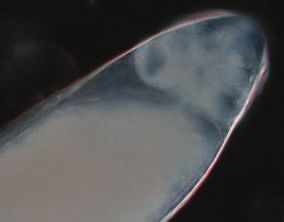
A Protein Inhibitor of Polo

Figure 3. Reducing the Dose of poloþ Suppresses mtrm Defects, and Increasing the Dose of poloþ Partially Mimics the Effects of mtrm
(A) Schematic diagram of the polo gene (black boxes depict the five exons) indicating the insertions sites for the two polo alleles (polo16–1 and
poloKG03033).
(B) Summary of the genetic interaction of mtrm and polo mutants as examined by assaying the frequency of nondisjunction of X and 4th chromosomes.
As shown by Harris et al. [9], mtrm/þ heterozygotes display high levels of nondisjunction for both achiasmate X and 4th chromosomes (42% and 37%,
respectively) when compared to mtrmþ/mtrmþ females. However, simultaneously reducing the dose of polo, as a result of heterozygosity for either the
two P element insertion site mutants or a deficiency that uncovers polo (Df(3L)rdgC-co2) suppresses the meiotic phenotype of mtrm/þ heterozygotes.
(C) Expression of the UASP-poloþ transgene in mtrmþ/mtrmþ females results in a dose-dependent increase in the frequency of achiasmate
nondisjunction for both the X and the 4th chromosomes. However, two weaker alleles of polo, polo01673 and polo1, showed little or no suppression of the
segregational defect (unpublished data). The polo1 mutant, which is the weakest of the known polo mutants (it is viable over a deletion) is the result of a
point mutation at base pair 725, V242E, in the kinase domain. Although polo01673 is recessive lethal, it must retain some degree of function because it
complements at least one other hypomorphic allele of polo, polox8. The results indicate that reduction of poloþ dosage rescues mtrm defects and the
suppressive effect of a given polo mutant correlates with the severity in the reduction of Polo function.
doi:10.1371/journal.pbio.0050323.g003

exchange. (The 4th chromosome is obligately achiasmate.) As                      activity. Two precise excisions of this insertion were
shown in Figure 3B, FM7/X; mtrm/þ females typically show                         generated, and neither was able to suppress the nondisjunc-
frequencies of X and 4th chromosome nondisjunction in the                        tional effects observed in mtrm/þ heterozygotes (unpublished
range of 35%–45%, which is more than 100-fold above                              data). We also demonstrated that the poloKG03033 allele was
control values.                                                                  able to suppress the meiotic defects generated by hetero-
   However, FM7/X; mtrm126/þ females that were simultane-                        zygosity for mtrmexc13, an independently isolated allele of mtrm
ously heterozygous for either a deficiency (Df(3L)rdgC-co2) that                  (unpublished data).
uncovers polo or for either of two strong alleles of polo,                          Heterozygosity for these same loss-of-function alleles of
poloKG03033 and polo16–1 (Figure 3A) displayed greatly reduced                   polo has no detectable effect on meiotic chromosome
levels of meiotic nondisjunction (Figure 3B). The fact that the                  segregation in mtrmþ/mtrmþ females. In females of the
poloKG03033 mutation is due to a P element insertion allowed                     genotypes FM7/X; poloKG03033/þ or FM7/X; polo16–1/þ, the
us to demonstrate that the observed interaction with mtrm                        observed levels of nondisjunction for the X chromosome
was indeed a direct consequence of a reduction in polo                           were 0.2% and 0.4%, respectively. Similarly, the observed

       PLoS Biology | www.plosbiology.org                                 2834                              December 2007 | Volume 5 | Issue 12 | e323
A Protein Inhibitor of Polo
levels of nondisjunction for the 4th chromosome were 0.6% experiment, we used ovary extracts from wild-type females
and 0.5% respectively (n ¼ 1,109 for FM7/X; poloKG03033 /þ and and performed the coimmunoprecipitation using a mono-
n ¼ 1,226 for FM7/X; polo16–1/þ females). These data alone are clonal antibody to Polo [14]. In both experiments, we were
consistent with either a hypothesis in which Mtrm acts to able to show that Mtrm coimmunoprecipitated with Polo
inhibit Polo, excess Polo creates a meiotic defect or a scenario (Figure S2).
in which Polo inhibits Mtrm, and the absence of sufficient In addition, MudPIT mass spectrometry reveals that Mtrm
Mtrm creates the defect. However, as we will show below, our and Polo interact in oocytes with a stoichiometry of
additional data support the model whereby Mtrm inhibits approximately 1:1. We analyzed three independent affinity
Polo. purifications from ovarian extracts expressing a C-terminally
33 FLAG-tagged Mtrm, and we used MudPIT mass spectrom-
Increasing the Dose of poloþ Partially Mimics the Effects of etry [15] to identify interacting proteins. We then compared
mtrm and Enhances the Defects Observed in mtrm/þ the identified proteins to those detected in five control FLAG
Heterozygotes immunoprecipitations from control (w1118) flies. Among the
If reducing the quantity of Polo suppresses the meiotic proteins that showed reproducible and significant p-values (p
defects observed in mtrm/þ females, then over-expression of , 0.001) identified in all three analyses, Polo was detected by
Polo alone should mimic the effects of reducing the dosage of multiple peptides and stands out as the only protein
mtrmþ (i.e., we should see a chromosome segregation defect recovered at levels similar to those of Mtrm, as estimated by
solely in the presence of increased dosage of poloþ, even in normalized spectral abundance factor (NSAF) counts [16,17].
mtrmþ/mtrmþ oocytes). To test this hypothesis, we analyzed Although the NSAF values for Mtrm and Polo vary across the
FM7/X females carrying two doses of a UASP-poloþ transgene three biological replicates analyzed (Figure 4C), the ratio
construct driven by the nanos-GAL4 driver. As shown in between the two proteins remains constant with an average of
Figure 3C, expression of the UASP-poloþ transgene construct 0.96 6 0.11, suggesting one Mtrm molecule binds to one
results in a dosage-dependent increase in the frequency of molecule of Polo.
achiasmate nondisjunction for both the X and the 4th Thus, three lines of evidence demonstrate that Mtrm
chromosomes. Similar observations were made using chro- physically interacts with Polo: the yeast two-hybrid work
mosomal duplications that carry two copies of poloþ (Adelaide [10], our coimmunoprecipitation studies, and our MudPIT
Carpenter, personal communication). Moreover, increasing mass spectrometry experiments presented in this section. The
the dose of Polo in females heterozygous for mtrm126 resulted observation of strong genetic interactions between mutants
in severe meiotic defects. Females carrying a single copy of in these two genes (Figure 3) demonstrates a functional
the UASP-poloþ transgene and which were also heterozygous significance to this interaction.
for mtrm126 were virtually sterile (unpublished data). Thus,
increasing the dosage of Polo enhances the defect observed in Mutation of the First PBD Binding Site of Mtrm Both
mtrm/þ heterozygotes by inducing sterility. Prevents Its Ability to Interact with Polo and Ablates Mtrm
The genetic interaction between Mtrm and Polo during Function
oogenesis is paralleled by their patterns of expression. Mtrm Polo interacts with target proteins via the interaction of its
reaches its maximum level of expression from the end of PBD and the sequences STP or SSP on the target protein. In
stage 10 onward, filling the oocyte during stages 11–12, and both of these PBD-binding sites, the center residues
then diminishes at stage 13. Analysis of Polo expression using (threonine or serine) are phosphorylated to facilitate Polo
an antibody to Polo [13,14] and wild-type oocytes revealed binding [18–20]. Mtrm carries two potential PBD-binding
that Polo is present in the oocyte at low levels (except in the sites: STP with the central threonine at residue 40 and SSP
germarium) until stages 11 or 12 and then rapidly fills the with the central serine at residue 124 (Figure 4A). To
oocyte cytoplasm from stages 12–13 onward (Figure S1). determine whether the interaction between Mtrm and Polo
Taken together, these data support a model in which the is mediated through the interaction of the Polo PBD with
presence of Mtrm inhibits Polo in the early stages of either or both of these two potential PBD-binding sites, we
expression, while permitting the function of Polo at stage created UASP-driven transgenes that carried mutations in
13, when Mtrm is degraded. Data directly demonstrating that either or both of the STP or SSP motifs. In each case, we
assertion are provided below. mutated the central residue of the PBD-binding sites on
Mtrm to the nonphosphorylateable residue alanine. These
Mtrm and Polo Physically Interact In Vivo mutants are denoted as mtrmT(40)A, which disrupts the STP
A large scale yeast two-hybrid screen identified Mtrm as a motif, and mtrmS(124)A, which disrupts the SSP motif. Each of
candidate interactor with Polo [10] and showed that Mtrm these mutant constructs was expressed under the control of
carries two putative PBD binding sites: STP and SSP (Figure the nanos-GAL4 driver in a mtrm null background to insure
4A). To confirm that Mtrm interacts with Polo physically in that they were the only source of Mtrm protein in the oocytes.
vivo, we performed coimmunoprecipitation experiments on Coimmunoprecipitation experiments using antibodies to
wild-type ovary extracts using a polyclonal antibody to Mtrm. Mtrm revealed that MtrmS(124)A protein still interacted with
As shown in lane 1 of Figure 4B, the antibody to Mtrm also Polo (Figure 4B). However, MtrmT(40)A failed to bind to Polo
precipitated Polo. (Figure 4B), indicating that the STP residues define a motif
We used two separate approaches to confirm the inter- that is critical for the Mtrm–Polo interaction. Mutation of
action between Polo and Mtrm. In the first experiment, we both PBD sites also resulted in a version of Mtrm that did not
used ovary extracts from females expressing a Green Fluorescent interact with Polo (unpublished data).
Protein (GFP)-polo transgene [13] and performed the coimmu- Because the interaction of Polo with target proteins via its
noprecipitation using an antibody to GFP. In the second PBD requires the phosphorylation of the center residues
PLoS Biology | www.plosbiology.org 2835 December 2007 | Volume 5 | Issue 12 | e323A Protein Inhibitor of Polo
Figure 4. Mtrm Physically Interacts with Polo with a Stoichiometry of Approximately 1:1
(A) Schematic depiction of the Mtrm protein. Mtrm has two potential PBD-binding sites, STP and SSP, with the central serine/threonine residue at 40
and 124, respectively, and a SAM domain at the C terminus. Two independent transgenes expressing mutated PBD-binding sites were generated:
MtrmT(40)A, which disrupts the STP site and MtrmS(124)A, which disrupts the SSP site.
(B) Mtrm and Polo physically interact as shown by coimmunoprecipitation experiments. An antibody to Mtrm precipitates Polo from wild-type ovary
extracts (lane 1). Expression of the mutated PBD binding site constructs in a mtrm null background reveals that MtrmS(124)A does not ablate the Mtrm-
Polo interaction (lane 2); however, MtrmT(40)A failed to bind Polo (lane 3), indicating that the STP motif is critical for the Mtrm-Polo interaction.
(C) Three independent affinity purifications from ovarian extracts expressing a C-terminally 33 FLAG-tagged Mtrm were used for the MudPIT mass
spectrometry assay. Among the reproducible and significant (p-value , 0.001) proteins identified in all three analyses, Polo was detected by multiple
peptides and stands out as the only protein recovered at levels similar to those of Mtrm, as estimated by NSAF.
(D) Phosphorylated sites detected in Mtrm (blue bars) and Polo (yellow bar) are shown. Modification levels are estimated based on local spectral count
and averaged across the three immunoprecipitations. The underlined numbers in each bar represent the number of times (out of three) the residues
were found modified.
(E) The STP site required for Polo binding is also required for Mtrm function. As noted above, FM7/X; mtrm/þ heterozygotes display approximately 40% X
nondisjunction (ND) and 37% 4th nondisjunction. Although the MtrmS(124)A protein was able to rescue the meiotic defect (3.6% X and 4.4% 4th ND), the
MtrmT(40)A protein displayed similar levels of nondisjunction as mtrm/þ heterozygotes, indicating that the STP motif is critical for Mtrm function. The
finding that only the STP site is required for both Mtrm function and the binding of Mtrm to Polo is consistent with the observation that only the STP
motif is conserved across all twelve sequenced Drosophila genomes, whereas the SSP motif is conserved only within the six species that belong to the
D. melanogaster–D. ananassae clade.
doi:10.1371/journal.pbio.0050323.g004
(threonine or serine) of the STP or SSP motifs [18–20], we binding site, S124 phosphorylation was found less reprodu-
searched the MS/MS dataset for phosphorylated peptides cibly. In addition, Mtrm S48, S52, and S137 were found
derived from Mtrm or Polo. For each of the detected sites, we phosphorylated at reproducibly high levels in two out of
estimated the levels of modification by dividing the number three experiments. We also observed that Polo T182 was
of spectra matching a particular phosphopeptide by the total detected as phosphorylated at high levels (over 80%) in all
spectral count for this peptide (Figure 4D). We were able to three immunoprecipitations, indicating that those Polo
detect phosphorylation on both T40 and S124; although, in proteins that are bound to Mtrm were fully activated [21].
agreement with the second PBD not being the primary Not only is the STP motif important for Polo binding, but
PLoS Biology | www.plosbiology.org 2836 December 2007 | Volume 5 | Issue 12 | e323A Protein Inhibitor of Polo
Figure 5. mtrm Causes Precocious NEB
(A) Representative examples of NEB in stage 11 and 12 egg chambers for wild-type (w1118) and mtrm126 homozygotes. NEB in wild-type oocytes occurs
at stage 13. The nucleus is still present (seen as a dark mass by phase contrast microscopy) at stage 11 and stage 12 in wild-type. mtrm homozygotes
show precocious NEB (absence of the dark mass) that can occur prior to stage 11. Scale, 60 lm.
(B) Summary of NEB in stage 11 and stage 12 egg chambers for wild-type (w1118), mtrm heterozygotes (mtrm126/þ), mtrm homozygotes (mtrm126/
mtrm126), double heterozygotes for both mtrm, polo (mtrm126 þ/þ polo16–1), and over-expression of Polo in mtrm heterozygotes (þ/UASP-polo; mtrm126/þ).
doi:10.1371/journal.pbio.0050323.g005
it is also required for proper Mtrm function (Figure 4E). We Polo interacting site to produce a hyperfunctional Mtrm, not
assayed the frequency of nondisjunction in females express- a nonfunctional protein.
ing either the mtrmS(124)A or the mtrmT(40)A construct in the
germlines of FM7/X; mtrm/þ heterozygotes. Although the As Either a Heterozygote or a Homozygote, mtrm Causes
mtrmS(124)A construct was able to rescue the meiotic defects Precocious NEB
seen in mtrm/þ heterozygotes, the mtrmT(40)A construct failed The early stages of meiosis appear normal in both mtrm/þ
to rescue the mtrm defect and maintained the high non- and mtrm/mtrm oocytes. The germarium and early stages
disjunction frequency seen in FM7/X; mtrm/þ heterozygotes. A appear morphologically normal and, at least in mtrm/þ
similar failure to rescue was observed using a double mutant oocytes, both recombination and SC assembly are indistin-
construct that carried both the mtrmS(124)A and the mtrmT(40)A guishable from normal ([9] and unpublished data). However,
mutations (unpublished data). Based on these observations, following stage 11 (the period during which Mtrm is
we conclude that the STP site is critical for Mtrm function maximally expressed), we observed multiple defects in oocyte
and the T(40)A mutation ablates Mtrm function as a direct maturation in both mtrm/þ and mtrm/mtrm oocytes. Most
consequence of a failure to interact with Polo. critically, we show that a loss-of-function allele of mtrm
induces precocious NEB in a dosage-dependent manner.
Mtrm Functions as an Inhibitor of Polo In wild-type oocytes, NEB usually does not occur until stage
In the previous sections, we presented three separate lines 13; only a single case of NEB at stage 12 was observed among
of evidence that Mtrm acts to inhibit Polo function, and not the 61 stage 11 and 12 wild-type oocytes examined (Figure 5).
vice versa. First, effects of heterozygosity for mtrm can be However, in mtrm126/þ heterozygotes, more than a third of
suppressed by a corresponding reduction in the dose of poloþ. stage 12 egg chambers exhibited NEB. To ensure that the
Second, we observed that the phenotype created by reducing precocious NEB defect is the consequence of reducing the
the dose of mtrmþ can be mimicked by increasing the dose of copy number of mtrmþ, we repeated these experiments using
Polo. Third, and most importantly, the observation that females that are heterozygous for an independently isolated
mutating the STP Polo binding site by a conservative amino allele of mtrm; mtrmexc13. These females also displayed preco-
acid replacement (STP ! SAP) ablates Mtrm function argues cious NEB at stage 12 (data not shown). As is the case for the
strongly that Mtrm functions as an inhibitor of Polo. Were it chromosome segregation defects observed in mtrm/þ oocytes,
the case that Polo inhibits Mtrm, one would expect loss of the the precocious NEB that is seen in mtrm126/þ heterozygotes is
PLoS Biology | www.plosbiology.org 2837 December 2007 | Volume 5 | Issue 12 | e323A Protein Inhibitor of Polo
strongly suppressed by simultaneous heterozygosity for a loss- distinguished but were still physically associated). As dis-
of-function allele of polo (Figure 5B), suggesting that the cussed in the legend to Figure 7, despite this dissociation into
timing of NEB is determined by the relative abundances of individual bivalents, in most oocytes the chromosomes are
Mtrm and Polo. This conclusion is further strengthened by capable of reaggregating into a single mass and eventually
the observation that overexpression of Polo (using a UASP- forming a bipolar spindle.
poloþ transgene driven by the nanos-GAL4 driver) increases the A striking example where all four chromosome pairs can be
frequency of precocious NEB in mtrm126/þ heterozygotes by clearly distinguished is the image taken 26 min after NEB for
nearly 2-fold (from 42% to 77%). FM7/X; mtrm126/mtrmþ oocytes (Figure 7). In those oocytes in
The extent of the precocious NEB defect is even more which bivalent individualization was observed, the two major
evident in mtrm126 homozygotes. As shown in Figure 5, NEB autosomes appeared to be held together by at least two
had already occurred in 32 out of 33 stage 12 oocytes chiasmata (one on each arm), suggesting that sister-chromatid
examined and in six of ten stage 11 oocytes examined. Thus, cohesion along the euchromatic arms of these chromosomes
the loss of Mtrm causes precocious NEB in a dosage- still persists. The two X chromosomes remain physically
dependent fashion. Taken together, these data argue that associated, despite the lack of chiasmata, presumably as a
the presence of Mtrm prevents Polo from inducing NEB until consequence of the maintenance of heterochromatic pairing
stage 13 (see Discussion), and that a reduction or absence of [29,30].
available Mtrm allows the Polo that is synthesized during Because the nondisjunction of achiasmate chromosomes
stages 11 and 12 to initiate NEB. observed in mtrm126/mtrmþ heterozygotes was suppressed by
The precocious breakdown of the nuclear envelope at heterozygosity for loss-of-function alleles of polo, we next
stages 11 to 12 is important because the karyosome undergoes tested whether a polo mutation could also suppress this
dramatic changes in structure during this period [2]. As noted karyosome maintenance defect. As shown in Figure 7 and
above, in stages 9–10, the karyosome expands to the point Video S3, bivalent individualization was only observed in
that individual chromosomes can be detected [22–24]. These three out of 13 (23%) of FM7/X; mtrm126 poloþ/mtrmþ polo16–1
chromosomes recondense into a compact karyosome during oocytes, and thus 77% of the oocytes maintained the
stages 11–12, the exact time at which a reduction in the level karyosome as a single mass throughout the process of spindle
of Mtrm causes precocious NEB. Thus, the early NEB events assembly. These data are consistent with the genetic data
promoted by heterozygosity for mtrm might be expected to presented above: reducing the dose of poloþ strongly
result in the release of incompletely condensed or disordered suppresses the deleterious effects of heterozygosity for mtrm.
karyosomes. To test this hypothesis, we examined karyosome
morphology during the 20 min that preceded NEB in wild- The Defects in Karyosome Maintenance Are Followed by
type, mtrm126/mtrmþ, and mtrm126 poloþ/mtrmþ polo16–1 oocytes. Defective Co-Orientation of Achiasmate Centromeres on
As shown in Figure 6, only two out of 28 (7%) wild-type the Meiotic Spindle
oocytes with incompletely compacted or disordered karyo- Because the karyosomes of mtrm/þ females were poorly
somes were observed. However, 7 out of 27 (26%) mtrm126/ formed before NEB and are usually transiently dissolved to
mtrmþ oocytes displayed a disordered karyosome, an effect individual bivalents shortly after NEB (see above), we also
that was largely suppressed (to 8%) by simultaneous hetero- examined centromere co-orientation on bipolar prometa-
zygosity for polo16–1 (Figure 6). These data support the view phase spindles using FISH probes (see Materials and Methods)
that the precocious NEB that is induced by lowering the level directed against the X and 4th chromosomes (Figure 8) in
of Mtrm results in the release of improperly formed both wild-type and mtrm/þ oocytes.
karyosomes into the cytoplasm and are again consistent with In wild-type oocytes, the vast majority of most X and 4th
the possibility that Mtrm inhibits meiotic progression chromosome centromeres co-oriented properly (Figure 8).
through its effects on Polo. The frequencies of abnormal centromere co-orientation in
oocytes with chiasmate X chromosomes (XX) were only 2%
Mtrm Is Also Required to Maintain Karyosome Structure for the X chromosome and 4% for the 4th chromosome. In
after NEB FM7/X females, where X chromosomal crossingover is
The karyosome plays a critical role in directing the blocked, the frequencies of abnormal co-orientation were
formation of the acentriolar spindle in Drosophila oocytes. still quite low (4% for the X chromosome and 2% for the 4th).
In 8 out of 9 (89%) wild-type oocytes, the karyosome remains However, co-orientation of achiasmate centromeres was
associated even after NEB; it is then surrounded by micro- often aberrant in mtrm/þ heterozygotes, such that the
tubules and forms a bipolar meiotic spindle (Figure 7 and centromeres of both homologs were often oriented toward
Video S1). At metaphase I, chiasmate chromosomes are still the same pole (Figure 8A). In these cases, the two homologs
condensed into a single mass at the metaphase plate in a also occupied different arcs of the meiotic spindle, a feature
tapered bipolar spindle [25–28]. that is rarely, if ever, observed in wild-type oocytes. In
However, in FM7/X; mtrm126/mtrmþ oocytes, the karyosome chiasmate X females, 43% of observed oocyte nuclei
usually dissolved within 10–20 min after NEB, and the displayed an aberrant co-orientation of 4th chromosome
individual bivalents became clearly visible (Figure 7 and centromeres, and 6% of these oocytes displayed aberrant X
Video S2). In 15 out of 17 (88%) FM7/X; mtrm126/ mtrmþ centromere co-orientations (Figure 8B); these oocytes likely
oocytes examined, the chromosomes were individualized reflect the 8%–10% of oocytes that fail to undergo crossing-
during spindle assembly. Indeed, in 14 of these examinations, over even in females bearing structurally normal X chromo-
all three pairs of major chromosomes were physically somes. The defect in 4th chromosome centromere co-
separated at some point during the time course of imaging orientation was fully suppressed by simultaneous hetero-
(in the remaining case, the three bivalents could be zygosity for polo16–1 (Figure 8A and 8B).
PLoS Biology | www.plosbiology.org 2838 December 2007 | Volume 5 | Issue 12 | e323A Protein Inhibitor of Polo
Figure 6. mtrm Is Defective in Karyosome Maturation before NEB
The karyosomes in stage 11–12 oocytes, which have a nuclear envelope, were imaged after the injection of Oli-green and Rhodamine-tubulin until NEB.
NEB was defined as the time when the nuclear envelope seems ruffled and the Rhodamine-tubulin enters the nucleus.
(A) Representative examples of karyosomes 12–16 min before and at NEB are shown for wild type, mtrm126/mtrmþ, and mtrm126 poloþ/mtrmþ polo16–1
with achiasmate X chromosomes (FM7/X). Wild type displays a circular karyosome with a smooth outline for 12–16 min before NEB, whereas mtrm126/
mtrmþ oocytes bear scabrous or bi-lobed karyosomes. The disordered morphology of karyosomes in mtrm126/mtrmþ oocytes was suppressed by
simultaneously reducing the dose of polo. Scale, 5 lm.
(B) Summary of karyosome morphology during the 20 min before NEB.
doi:10.1371/journal.pbio.0050323.g006
As expected, due to the suppression of X chromosomal transition in oogenesis to the role of the karyosome structure
crossingover in FM7/X females, mtrm/þ heterozygotes dis- in facilitating the proper segregation of achiasmate chromo-
played frequent abnormal centromere co-orientation for somes.
both X and 4th chromosomes, i.e., 43% for X chromosomes
and 37% for 4th chromosomes (Figure 8B). These results Polo Plays a Critical Role in Initiating the G2/M Transition
indicate that the mtrm heterozygotes display an obvious defect in Oogenesis by Regulating Cdc25
in centromere co-orientation. However, once again, both the The trigger for the G2/M transition is activation of Cdk1 by
defect in X and 4th chromosome centromere co-orientation Cdc25 (reviewed in [31]), and multiple lines of evidence
was fully suppressed by simultaneous heterozygosity for suggest that Polo can activate Cdc25 [32]. First, in Caeno-
polo16–1. Thus, as was the case with the previously considered rhabditis elegans, RNAi experiments demonstrate that ablation
defects, the deleterious effects of reducing the amount of of Polo prevents NEB [33]. Second, the Xenopus Polo homolog
available Mtrm can be suppressed by a simultaneous Plx1 is activated in vivo during oocyte maturation with the
reduction in the amount of Polo. same kinetics as Cdc25. Additionally, microinjection of Plx1
accelerates the activation of both Cdc25 and cyclinB-Cdk1
[34]. Moreover, microinjection of either an antibody to Plx1
Discussion
or kinase-dead mutant of Plx1 inhibited the activation of
The data presented above argue that Mtrm serves to Cdc25 and its target cyclinB-Cdk1. A later study by Qian et al.
inactivate newly synthesized Polo during the period of demonstrated that injection of a constitutively active form of
meiotic progression that precedes NEB. An excess of func- Plx1 accelerated Cdc25 activation [35]. As pointed out by
tional (unbound) Polo, produced by reducing the amount of these authors, these studies support ‘‘the concept that Plx1 is
available Mtrm, causes the early onset of NEB. This early the ‘trigger’ kinase for the activation of Cdc25 during the G2/
entry into prometaphase releases an immature karyosome M transition.’’ Finally, a small molecule inhibitor of Polo
into the cytoplasm, which then fails to properly align the kinase (BI 2536) also results in extension of prophase [36].
centromeres of achiasmate chromosomes on the prometa- These data are consistent with the view that the presence of
phase spindle. These observations raise a number of ques- functional (unbound) Polo plays a critical role in ending the
tions ranging from the role of Polo in mediating the G2/M extended G2 that is characteristic of oogenesis in most
PLoS Biology | www.plosbiology.org 2839 December 2007 | Volume 5 | Issue 12 | e323A Protein Inhibitor of Polo
Figure 7. mtrm Causes the Individualization of Bivalents after NEB
Stage 12 oocytes were injected with Oli-green to visualize karyosomes. After this injection, we analyzed the change in karyosome structure during NEB
using live imaging. Time frames from NEB (time 0) are shown for: (A) FM7/X; mtrmþ/mtrmþ for control; (B) FM7/X; mtrm126/mtrmþ; and (C) FM7/X;
mtrm126 poloþ/mtrmþ polo16–1 oocytes (see also Videos S1–S3). In control oocytes, the karyosome stays condensed after NEB and then becomes
elongated at about 13 min, presumably as a consequence of the chromosomes establishing proper centromere co-orientation. As noted in the text,
almost all control oocytes (8/9) exhibited a karyosome in which chromosomes are tightly associated. In the remaining case, three bivalents could be
distinguished but were still physically associated. However, in FM7/X; mtrm126/mtrmþ oocytes, the 4th chromosomes are separated from a single mass of
chromatin at 6–8 min after NEB, and then X, 2nd and 3rd chromosomes start to spread out. At approximately 16 min after NEB, they are individualized
into three obvious and fully separate bivalents.
Individualized chromosomes begin to re-condense around 46 min and form a single mass. Indeed, the majority (11/15) of those oocytes that underwent
bivalent individualization eventually formed bipolar spindles with the chiasmate chromosomes properly balanced on the metaphase plate (see also Figure
4 of Harris et al. [9]). Thus the karyosome maintenance defect induced by heterozygosity for mtrm does not permanently impair the progression of
prometaphase. Finally, the karyosome maintenance defect induced by heterozygosity for mtrm was suppressed by reducing the dosage of the poloþ gene.
As noted in the text, 10 of the 13 FM7/X; mtrm126 poloþ/mtrmþ polo16–1 oocytes maintained the karyosome as a single mass throughout the process of
spindle assembly. The three remaining cases may be described as follows: (1) the karyosome dissolved into three clearly distinguishable bivalents, but
this oocyte did not ever succeed in forming a bipolar spindle; (2) the three major bivalents could be distinguished but did not physically separate; and
(3) in an oocyte that may have been leaking or damaged, the bivalents individualized at about 8 min after the initiation of spindle assembly, but their
morphology was abnormally stretched and thread-like. 7 min later, these chromosomes began to fragment into much smaller pieces, which led to the
assembly of a spindle with at least five and possibly more poles. It seems likely that this case reflects simply the fragility of the karyosome, even in
polo16–1/poloþ suppressed oocytes, rather than the defect observed in FM7/X; mtrm126/mtrm oocytes that are wild type for polo.
doi:10.1371/journal.pbio.0050323.g007
animals. We should note by Riparbelli et al. [37] that the activate Cdc25, initiating the chain of events that leads to
careful study of female meiosis in polo1 homozygotes failed to NEB and the initiation of prometaphase. In the absence of a
observe a defect in the timing of NEB. However, as discussed sufficient amount of Mtrm, an excess of Polo causes the
in the legend to Figure 3, polo1, a missense mutant that is precocious activation of Cdc25, and thus an early G2/M
viable even over some deficiencies and does not suppress transition. A model describing this hypothesis is presented in
mtrm, is the weakest of the known polo mutants, and it is thus Figure 9. Based on this model, one can visualize that
reasonable that no defect was observed. decreasing the dose of Mtrm or increasing the dose of Polo
In light of these data, it is tempting to suggest that in wild- will hasten NEB, whereas simultaneous reduction in the dose
type Drosophila oocytes, the large quantity of Mtrm deposited of both proteins should allow for proper timing of NEB.
into the oocyte from stage 10 onward inhibits the Polo that is Two lines of evidence directly support a model in which
either newly synthesized or transported into the oocyte Mtrm exerts its effect on Polo, with respect to preventing
during stages 11–12. However, at stage 13, an excess of precocious NEB, by blocking the ability of Polo to activate
functional Polo is created when the number of Polo proteins Cdc25. First, as shown in Figure 10, mutants in the Drosophila
exceeds the available amount of inhibitory Mtrm proteins. cdc25 homolog twine fail to undergo NEB in stage 13. In
This unencumbered and thus functional Polo then serves to addition, heterozygosity for twine also decreases the frequency
PLoS Biology | www.plosbiology.org 2840 December 2007 | Volume 5 | Issue 12 | e323A Protein Inhibitor of Polo
Figure 8. Heterozygosity for mtrm126 Impairs the Proper Co-Orientation of Achiasmate Centromeres during Prometaphase
(A) FISH analysis using probes homologous to the X and 4th chromosomal heterochromatin [29] were used to assay centromere co-orientation during
meiotic prometaphase. In mtrmþ/ mtrmþ oocytes carrying either chiasmate X chromosomes (XX females) or achiasmate X chromosomes (FM7/X
females), the centromeres of both the X and the 4th are virtually always oriented toward opposite poles (see panels 1 and 4 and (B)). However, in mtrm/þ
heterozygotes, the centromeres of achiasmate bivalents are often oriented towards the same pole (see panels 2 and 5 and (B)). In double heterozygotes
for both mtrm and polo, these defects in achiasmate chromosome centromere co-orientation are greatly suppressed (panels 3 and 6).
(B) Quantitative summary of centromere co-orientation patterns for the various genotypes studied. Although heterozygosity for mtrm126 has a dramatic
effect on 4th chromosome centromere malorientation in both XX and FM7/X females, there is little effect on X chromosome segregation in XX oocytes
when compared with the dramatic effect observed in FM7/X females. This is to be expected based on the genetic studies of Harris et al. [9], who
observed that only achiasmate bivalents nondisjoin in mtrm/þ females.
doi:10.1371/journal.pbio.0050323.g008
of precocious NEB in mtrm126/þ heterozygotes from 42% serves to inhibit Polo. In general, enzymes are usually not
(Figure 5) to less than 10% (7/72). recovered from affinity purifications at levels similar to their
targets. They do not form stable complexes, but rather form
How Might Mtrm Inhibit Polo? transient interactions with their substrates, which is how
Mtrm’s first PBD binding site (T40) is required for its efficient catalysis is achieved. Here, Mtrm is able to sequester
interaction with Polo. Mtrm T40 has to be first phosphory- activated Polo away in a stable binary complex over a long
lated by a priming kinase, such as one of the Cdks or MAPKs, period of time. It is only when this equilibrium is disturbed at
and was indeed detected as phosphorylated in the mass the onset of stage 13 by the production of an excess of Polo
spectrometry dataset. The NetPhosK algorithm [38] predicts (as suggested in Figure 9) or by degradation of Mtrm that Polo
T40 to be a Cdk5 site, and the serines immediately distal to can be released. The molecular determinants of the
T40—S48 and S52—which were also detected as phosphory- Mtrm::Polo sequestration event are not clear, but it would
lated (Figure 4D), are sites for proline-directed kinases such be interesting to test whether the serines found phosphory-
as Cdk or MAPK sites as well. The other prominent lated in the vicinity of Mtrm PBD binding sites play a role in
phosphorylation event occurs at S137, which could be a Polo locking the binary complex into place.
phosphorylation site since it falls within a Polo consensus (D/
E-X-S/T-Ø-X-D/E). Although the combined sequence coverage mtrm Exerts Its Effects on Achiasmate Nondisjunction Via
for Mtrm was 44%, indicating that some phosphorylated sites a Cdc25-Independent Pathway
might have been missed, Mtrm S137 is a suitable binding site Our data demonstrate that a reduction in the levels of
for activated Polo, in agreement with the processive Mtrm results in the release of an incompletely compacted
phosphorylation model [18]. At this point of our studies, karyosome that rapidly dissolves into individual bivalents
Mtrm T40 priming kinase or the kinase responsible for Polo during the early stages of spindle formation. For chiasmate
activating phosphorylation on T182 has not been identified. bivalents, this is apparently not a problem, because they still
The finding that Polo not only is able to bind to Mtrm in co-orient correctly (for example, the chiasmate X chromo-
vivo in a 1:1 ratio, but also is fully phosphorylated on T182 in somes shown in Figure 8 still achieve proper co-orientation in
its activation loop [21] suggests a method by which Mtrm the vast majority of oocytes). However, the nonexchange
PLoS Biology | www.plosbiology.org 2841 December 2007 | Volume 5 | Issue 12 | e323A Protein Inhibitor of Polo
cious NEB from 42% (Figure 5) to less than 10% (7/72), two
alleles of twine tested (twe1 and twek08310) failed to suppress the
levels of meiotic nondisjunction observed in FM7/X; mtrm126/þ
heterozygotes. These data suggest that the effects of excess
Polo on nondisjunction may not be regulated via Cdc25/
Twine, but rather by the effects of excess Polo on some other
as-yet-unidentified Polo target. This suggests that the effects
of Mtrm on the level of Polo might affect multiple Polo-
related processes.
Support for such an idea that Mtrm can inhibit Polo-
regulated proteins that are unrelated to NEB comes from the
observation that the ectopic expression of Drosophila Mtrm in
Schizosaccharomyces pombe blocks karyokinesis, producing long
multi-septate cells with only one or two large nuclei ([39] and
Figure 9. A Model for the Control of NEB by Mtrm-Induced Inhibition of Bruce Edgar, personal communication). This phenotype is
Polo
similar, if not identical to, that exhibited by mutants in the S.
According to this model, in wild-type Drosophila oocytes, the excess of
Mtrm inhibits those Polo proteins that are deposited in the oocyte pombe Polo homolog plo1 (Plo1), which fail in later stages of
during stages 11 to 12. However, by stage 13, an excess of Polo exceeds mitosis due to the role of Plo1 in activating the septation
the available amount of inhibitory Mtrm proteins. This unencumbered initiation network to trigger cytokinesis and cell division.
Polo then serves to activate Cdc25, initiating the chain of events that
lead to NEB and the initiation of prometaphase. In the absence of a
However, Plo1 also plays a role in bipolar spindle assembly
sufficient amount of Mtrm, an excess of functional Polo causes the that might also be inhibited in the Mtrm expressing cells, but
precocious activation of Cdc25 and thus an early G2/M transition. Based this function of Plo1 is less well understood.
on this model, one can visualize that decreasing the dose of Mtrm or Thus the possibility exists that the effect of mtrm mutants
increasing the dosage of Polo will hasten NEB, while simultaneous
reduction in the level of both proteins will normalize the timing of NEB. on meiotic chromosome segregation may well not be the
doi:10.1371/journal.pbio.0050323.g009 direct consequence of early NEB, but rather may be due to
the role of Polo in other meiotic activities, such as spindle
formation or the combined effects of these defects with
bivalents frequently fail to co-orient properly, such that both precocious NEB. Efforts to identify such processes and their
homologs are oriented toward the same pole (but often components are underway in the lab.
occupy two different arcs of the spindle). This initial failure Finally, we should note that while Mtrm is the first known
of proper co-orientation leads to high frequencies of protein that is able to inactivate Polo by physical interaction
nondisjunction as demonstrated by the genetic studies and with Polo itself; there is certainly additional mechanisms of
analysis of metaphase I images presented in [9]. Polo regulation. For example, Archambault et al. [40] have
Although achiasmate homologs are properly co-oriented in described mutants in the gene that encodes Greatwall/Scant
wild-type oocytes [29,30] , we have noted previously such kinase, which have both late meiotic and mitotic defects.
homologs can often vacillate between the poles such that two Although there is no evidence for a physical interaction
achiasmate homologs are often found on the same arc of the between these two kinases, the authors speculate that the
same half-spindle during mid to late prometaphase ([25] and function of the Greatwall kinase serves to antagonize that of
unpublished data). These chromosomes are often observed to Polo. The Scant mutations create a hyperactive form of
be physically associated. This situation is quite different from Greatwall, which might be expected to lower the dosage Polo,
the defect observed in mtrm/þ heterozygotes, where the and thus perhaps partially suppress the defects observed in
homologs are neither physically associated nor on the same mtrm/þ heterozygotes. Indeed, exactly such a suppressive
arc of the spindle. effect has been observed in Scant homozygotes (however, this
It is tempting to suggest that the chromosome segregation suppression is much weaker than that obtained by hetero-
defects we observe in mtrm/þ heterozygotes are simply the zygosity for loss of function alleles of polo).
result of precocious release of an incompletely re-compacted
karyosome. According to this explanation, the defects Summary
observed in meiotic chromosome segregation are solely the The data presented above demonstrate that Mtrm acts as a
consequence of premature NEB. (Implicit in this model is the negative regulator of Polo during the later stages of G2 arrest
assumption that it is the events that occur during karyosome during meiosis. Indeed, both the repression of Polo expres-
re-compaction, at stages 11 and 12, that serve to initially bi- sion until stage 11 and the inactivation of newly synthesized
orient achiasmate chromosomes, and we do not have direct Polo by Mtrm until stage 13 play critical roles in maintaining
evidence to support such a hypothesis.) and properly terminating G2 arrest. Our data suggest a model
Alternatively, Polo plays multiple roles in the meiotic in which the eventual activation of Cdc25 by an excess of Polo
process [7,8], and it is possible that the chromosome at stage 13 triggers NEB and entry into prometaphase.
segregation defects we see represent effects of excess Polo Although our data do shed some light on the mechanism by
that are un-related to the precocious breakdown of the which Mtrm inhibits Polo, it is not entirely clear whether
nuclear envelope. Such a view is supported by two observa- Polo’s ability to phosphorylate targets other than Cdc25
tions. First, as shown in Figure 7, the bivalent individualiza- might be blocked by Mtrm::Polo binding. These issues will
tion observed after NEB in mtrm/þ oocytes does not disrupt need to be addressed in the future studies. Finally, we note
FM7-X pairings. Second, although heterozygosity for twine in that although small molecule inhibitors of Polo have been
mtrm126/þ heterozygotes suppresses the frequency of preco- identified [36], Mtrm represents the first case of a protein
PLoS Biology | www.plosbiology.org 2842 December 2007 | Volume 5 | Issue 12 | e323You can also read