The regulation of acetylation and stability of HMGA2 via the HBXIP-activated Akt-PCAF pathway in promotion of esophageal squamous cell carcinoma ...
←
→
Page content transcription
If your browser does not render page correctly, please read the page content below
4858–4876 Nucleic Acids Research, 2020, Vol. 48, No. 9                                                                  Published online 20 April 2020
doi: 10.1093/nar/gkaa232

The regulation of acetylation and stability of HMGA2
via the HBXIP-activated Akt–PCAF pathway in
promotion of esophageal squamous cell carcinoma
growth
Yue Wu, Xue Wang, Feifei Xu, Lu Zhang, Tianjiao Wang, Xueli Fu, Tianzhi Jin,
Weiying Zhang* and Lihong Ye *

                                                                                                                                                                  Downloaded from https://academic.oup.com/nar/article/48/9/4858/5822968 by guest on 28 November 2020
State Key Laboratory of Medicinal Chemical Biology, Tianjin Key Laboratory of Protein Sciences, Department of
Biochemistry, College of Life Sciences, Nankai University, Tianjin 300071, P.R. China

Received October 30, 2019; Revised March 02, 2020; Editorial Decision March 29, 2020; Accepted April 12, 2020

ABSTRACT                                                                          INTRODUCTION
High-mobility group AT-hook 2 (HMGA2) is an ar-                                   Esophageal cancer (EC) is an aggressive and lethal malig-
chitectural transcription factor that plays essential                             nancy, ranking sixth in terms of mortality and eighth in
roles in embryonic development and cancer pro-                                    terms of incidence among all cancer types (1). EC is pri-
gression. However, the mechanism of HMGA2 reg-                                    marily classified into two pathological subtypes: esophageal
                                                                                  squamous cell carcinoma (ESCC) and esophageal adeno-
ulation remains largely uncharacterized. Here, we
                                                                                  carcinoma (2). ESCC, which is the most severe pathological
demonstrate that HMGA2 can be modulated by hep-                                   subtype of EC, accounts for ∼90% of all esophageal car-
atitis B X-interacting protein (HBXIP), an oncogenic                              cinomas at the time of diagnosis and has a high-incidence
transcriptional coactivator, in esophageal squamous                               in China (2). Considering late-stage tumor detection and
cell carcinoma (ESCC). HMGA2 expression was pos-                                  limited clinical therapeutic strategies, patients with ESCC
itively associated with HBXIP expression in clinical                              show an extremely poor prognosis, with the overall 5-year
ESCC tissues, and their high levels were associ-                                  survival rate of ∼17% (3). Although extensive efforts have
ated with advanced tumor stage and reduced over-                                  been devoted to overcome this disease, information on its
all and disease-free survival. We found that onco-                                molecular drivers remains limited. Therefore, a better un-
genic HBXIP could posttranslationally upregulate                                  derstanding of the molecular foundation of the occurrence
HMGA2 protein level in ESCC cells. HBXIP induced                                  and development of ESCC is urgently required for earlier
                                                                                  diagnosis and more efficient treatment.
HMGA2 acetylation at the lysine 26 (K26), result-
                                                                                     High-mobility group AT-hook 2 (HMGA2), an archi-
ing in HMGA2 protein accumulation. In this process,                               tectural transcription factor, constitutes ∼109 amino acid
HBXIP increased the acetyltransferase p300/CBP-                                   residues and three basic DNA-binding domains called ‘AT-
associated factor (PCAF) phosphorylation and acti-                                hooks’, which bind to the AT-rich regions in DNA (4).
vation via the Akt pathway, then PCAF directly in-                                HMGA2 modulates transcription by inducing structural
teracted with HMGA2, leading to HMGA2 acetyla-                                    changes in the chromatin, enabling the transcriptional ma-
tion in the cells. HMGA2 K26 acetylation enhanced                                 chinery to access the target regions to regulate the expres-
its DNA binding capacity and blocked its ubiqui-                                  sion of many mammalian genes in terms of both activa-
tination and then inhibited proteasome-dependent                                  tion and repression (4). HMGA2 is highly expressed dur-
degradation. Functionally, HBXIP-stabilized HMGA2                                 ing tumorigenesis but rarely in normal adult tissues (4,5).
could promote ESCC cell growth in vitro and in vivo.                              Various studies indicate that high expression of HMGA2
Strikingly, aspirin suppressed ESCC growth by in-                                 is related to poor survival rates in breast cancer (6), col-
                                                                                  orectal cancer (7) and lung cancer patients (8). Further-
hibiting HBXIP and HMGA2. Collectively, our findings                              more, there is evidence that oncogenic HMGA2 partici-
disclose a new mechanism for the posttranslational                                pates in DNA damage repair (9), stem cell self-renewal
regulation of HMGA2 mediated by HBXIP in ESCC.                                    (10), aggressive tumor growth (11) and tumor cell differ-
                                                                                  entiation (12). Importantly, HMGA2 is considered to pro-

* To
   whom correspondence should be addressed. Tel: +86 22 23501385; Fax: +86 22 23501385; Email: yelihong@nankai.edu.cn
Correspondence may also be addressed to Weiying Zhang. Email: zhwybao@nankai.edu.cn

C The Author(s) 2020. Published by Oxford University Press on behalf of Nucleic Acids Research.
This is an Open Access article distributed under the terms of the Creative Commons Attribution Non-Commercial License
(http://creativecommons.org/licenses/by-nc/4.0/), which permits non-commercial re-use, distribution, and reproduction in any medium, provided the original work
is properly cited. For commercial re-use, please contact journals.permissions@oup.com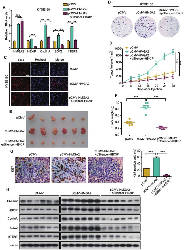
Nucleic Acids Research, 2020, Vol. 48, No. 9 4859
mote tumorigenesis in part through the modulation of hibit HBXIP/HOXB13 axis, overcoming tamoxifen resis-
a group of target genes. For instance, HMGA2 counter- tance in breast cancer (31). Based on these previous find-
acts the repression activity of the transcription repressor ings, we focus on the investigation of the role of aspirin in
p120E4F to induce cyclinA expression, which controls cell HBXIP-associated ESCC.
cycle progression (13). Additionally, HMGA2 stimulates In the present study, we explored the function and reg-
human telomerase reverse transcriptase (hTERT) expres- ulation of HMGA2 in the development of ESCC. HBXIP
sion, preventing the gradual telomere shortening in can- enhances HMGA2 acetylation at the lysine 26 residue (K26)
cer cells (14). Moreover, HMGA2 directly activates the through the Akt pathway-induced PCAF phosphorylation
transcription of pro-metastatic genes, including SNAIL, and activation in ESCC. HMGA2 K26 acetylation func-
SLUG, and CXCR4 (15–17). Equally, much attention has tionally enhances its DNA binding ability on the target
been focused on the regulatory cascades of HMGA2 ex- genes and blocks its ubiquitination and proteasomal degra-
pression during cancer progression. HMGA2 can be pos- dation, thus leading to HMGA2 accumulation and carcino-
itively regulated via the active Wnt/-catenin pathway (18) genesis. Intriguingly, aspirin can suppress ESCC growth
Downloaded from https://academic.oup.com/nar/article/48/9/4858/5822968 by guest on 28 November 2020
and repressed via the ZBRK1/BRCA1/CtIP pathway (19). through repressing HBXIP and HMGA2. Thus, our stud-
Interestingly, posttranslational modifications (PTMs) of ies identify a novel regulatory mechanism of HMGA2 in
HMGA2 confer a profound effect on its biological func- ESCC growth, which probably provides an effective strat-
tions. For example, HMGA2 phosphorylation at the acidic egy for ESCC therapy.
C-terminal tail may affect its DNA-binding properties
(20), and HMGA2 SUMOylation may promote promyelo- MATERIALS AND METHODS
cytic leukemia (PML) protein degradation (21). However,
whether PTM functions in the regulation of HMGA2 ex- Tissue specimens
pression remains largely unknown. The ESCC tissue microarray containing 151 primary ESCC
Mammalian hepatitis B X-interacting protein (HBXIP), tissues and 43 normal esophageal tissues with information
also known as LAMTOR5 (22), is a conserved 18-kDa pro- of patients’ overall survival and disease-free survival was ac-
tein, which was identified initially based on its binding to quired from Shantou University Medical College between
the C-terminus of hepatitis B virus X proteins (23). HBXIP February 2011 and November 2016. The patient records
is expressed in nearly all tissues (24). It can function as are presented in Supplementary Table S1. The other two
a cofactor of survivin to control cell apoptosis and regu- ESCC tissue microarrays (Catalog No.: Es-kx03c and Cat-
late centrosome duplication and cytokinesis to mediate cell alog No.: Es-kx14c) containing 124 cases of human ESCC
growth (24,25). Additionally, HBXIP can serve as a regula- tissues, two cases of human esophagus basal cell carcinoma
tory component required for the activation of mammalian tissues and 10 cases of normal esophagus tissues in total
target of rapamycin complex 1 via amino acids (22). Our were purchased from Aomeibio Company (Xian, China).
group has reported that HBXIP is highly expressed in breast The clinical characteristics are presented in Supplementary
carcinoma and that it acts as an oncogenic transcriptional Tables S5 and S6 respectively. All samples were approved
coactivator of multiple transcription factors, such as c-Myc, by Ethics Committee of Hospital providing tissues. Written
LXR, Sp1 and E2F1 to promote breast cancer growth and informed consent was obtained from patients before sam-
metastasis (26–29). Moreover, it supports the migration of ples were collected. All specimens, including tumor tissues
breast cancer cells through GCN5-mediated modulation of of ESCC patients and normal esophageal tissues, were ob-
microtubule acetylation (30). Our study has revealed that tained during surgery.
HBXIP as an important oncoprotein can regulate PTMs
of some transcription factors. For instance, HBXIP can in-
Cell culture and reagents
duce the acetylation of transcription factor HOXB13 to pre-
vent HOXB13 degradation in the promotion of tamoxifen The ESCC cell lines KYSE2, KYSE180, KYSE450,
resistance of breast cancer (31). In addition, HBXIP can KYSE510 and the human embryonic kidney cell line 293T
increase the phosphorylation levels of c-Fos through acti- (HEK293T) were obtained from the American Type Cul-
vating ERK1/2, which is a benefit for the nuclear localiza- ture Collection (ATCC). ESCC cell lines were cultured in
tion of c-Fos in breast cancer (32). One study found that the RPMI 1640 (Gibco, USA) supplemented with 10% fetal
abnormal expression of HBXIP was associated with poor bovine serum (FBS; Gibco). HEK293T was maintained
prognosis in ESCC (33). Accordingly, in the present study in Dulbecco’s Modified Eagle’s Medium (Gibco) supple-
we are interested in whether HBXIP is involved in HMGA2 mented with 10% FBS. All cells were cultured at 37◦ C in
PTM in ESCC development. a humidified atmosphere with 5% CO2 . Cells were collected
Aspirin (ASA), a nonsteroidal anti-inflammatory drug, and seeded in 6-, 24- or 96-well plates for 24 h and then
displays anti-cancer effect and has been applied in colorec- transfected with plasmids or small interference RNAs (siR-
tal cancer therapy (34). Substantial evidence indicates that NAs) using Lipofectamine 2000 (Invitrogen, Life Technolo-
regular aspirin use is useful for the reduction of incidence, gies, Grand Island, NY, USA), according to the manufac-
mortality and distant metastasis of cancers including breast turer’s protocol. All experiments were conducted in cells
cancer, liver cancer, and colorectal cancer (35–37). Several with ∼80% convergence. The reagents used in this study
epidemiologic studies have proven that the use of aspirin were trichostatin A (TSA), cycloheximide (CHX), the in-
and other nonsteroidal anti-inflammatory drugs protects hibitors of Akt, ERK1/2 and p38, and aspirin (ASA). TSA
against the development of esophageal cancer (38,39). We and CHX were purchased from MedChem Express (USA).
have recently revealed that aspirin can target HBXIP to in- GSK690693 (an inhibitor of Akt), PD98059 (an inhibitor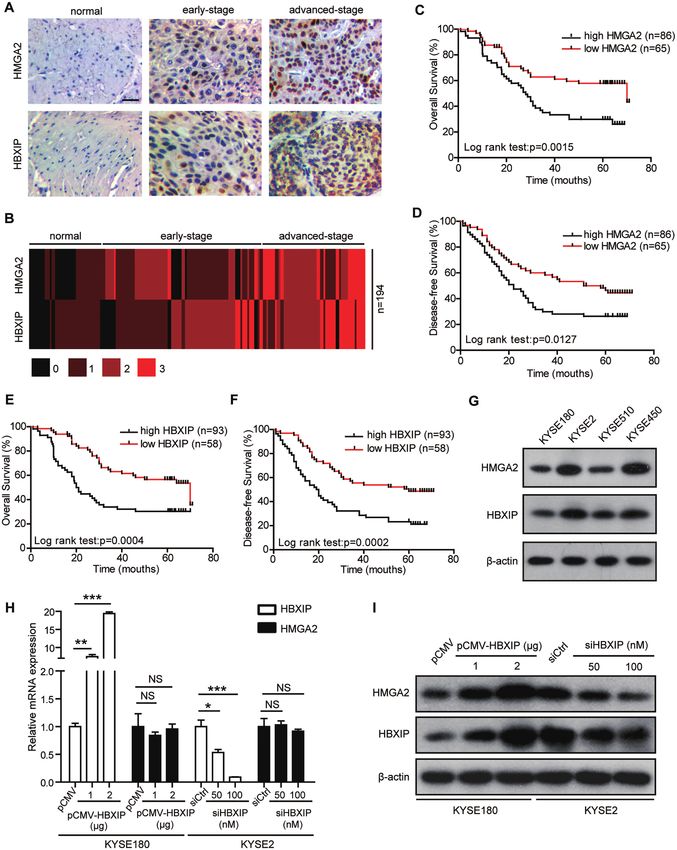
4860 Nucleic Acids Research, 2020, Vol. 48, No. 9
of the upstream kinase of ERK1/2), and SB202190 (an in- 1 = 1–29% stained; 2 = 30–65% stained; 3 = 66–100%
hibitor of p38) were all purchased from MedChem Express stained). A multiplied score (intensity score × percentage
(USA). ASA were purchased from Sigma-Aldrich (USA). score) lower than 1 was considered to be negative staining
(0), 1, 2 and 3 were considered to be weak staining (1), 4
and 6 were considered to be moderate staining (2) and 9 was
Plasmid construction and siRNAs
considered to be intense staining (3). For Ki67 and AcK26-
Plasmids, including pCMV-Tag2B, pCMV-HBXIP, HMGA2 staining in tumor xenograft, tumor slides were
pcDNA3.1(+), pcDNA3.1 (+)-HBXIP, pSilencer 3.1-neo fixed in 4% paraformaldehyde for 2 days. Then, sections
and shRNA construct pSilencer-HBXIP, were kept in were stained using a primary antibody against Ki67 (Santa
our laboratory. The complete human HMGA2 (NCBI Cruz Biotechnology, Santa Cruz, CA, USA) or specific
Reference Sequence: NM 003483.4) cDNA sequence AcK26-HMGA2 antibody. The positive Ki67 and AcK26-
was amplified by polymerase chain reaction (PCR) and HMGA2 staining were identified by Image-Pro Plus soft-
subcloned into the pEGFP-C2 or pCMV-Tag2B vector ware.
Downloaded from https://academic.oup.com/nar/article/48/9/4858/5822968 by guest on 28 November 2020
to generate GFP-HMGA2 or pCMV-HMGA2 (FLAG-
HMGA2). All point or deletion mutants of HMGA2 were
Co-immunoprecipitation (Co-IP) assay
generated by PCR and subcloned into the pCMV-Tag2B
vector and verified by sequencing. The vector expressing Indicated cells were harvested and lysed in a lysis buffer
FLAG-tagged human full-length PCAF was kindly pro- (50 mM Tris–HCl pH 7.5, 150 mM NaCl, 1 mM EDTA,
vided by Prof. Hongquan Zhang (Peking University Health 0.5% Triton X-100, 10% glycerine, 1 mM protease inhibitor
Science Center, Beijing, China). The complete PCAF PMSF). The lysates were incubated with Anti-FLAG M2
cDNA sequence was amplified by PCR and subcloned into affinity gel (Sigma-Aldrich) at 4◦ C for 3 h or incubated with
the pEGFP-C2 to generate GFP-PCAF. All siRNAs and rabbit anti-HMGA2 antibody (Supplementary Table S3) at
related primers are listed in Supplementary Table S2. All 4◦ C for 2 h before incubated with Protein G Magnetic beads
siRNAs were purchased from Riobio Co. (Guangzhou, (Pierce, Waltham, MA, USA) at 4◦ C for 1 h. After extensive
China). washing (3 times), precipitated proteins were eluted from
the gel or beads by 0.1 M glycine–HCl (pH 3.0) buffer and
neutralized with 1 M Tris–HCl (pH 7.5) containing 1.5 M
Western blotting analysis
NaCl. Immunoprecipitated samples were resolved by SDS-
Western blotting analysis was carried out with the stan- PAGE followed by western blotting with appropriate anti-
dard protocol. Tissues or cells were lysed in RIPA buffer bodies (Supplementary Table S3).
(Solarbio, Beijing, China). Equal amounts of total protein
were loaded for western blotting. Following SDS-PAGE,
In vivo ubiquitination assays
resolved proteins were transferred onto PVDF membranes
(Millipore, USA). The membranes were blocked in 5% skim Cells were transfected with the indicated plasmids and
milk for 2 h at room temperature, and then probed with treated with 40 M MG-132 for 6 h before harvest. The cells
primary antibodies (Supplementary Table S3) for 2 h at were washed with cold PBS and then lysed in 200 l de-
room temperature or overnight at 4◦ C. The rabbit poly- naturing buffer (150 mM Tris–HCl pH 7.4, 1% SDS). Af-
clonal antibody recognizing the acetylated HMGA2 at ly- ter incubation at 4◦ C for 10 min, the lysate was sonicated
sine 26 residue was produced with a synthetic acetylated hu- and boiling for 10 min. Lysates were added with lysis buffer
man HMGA2 peptide: APQ(AcK)RGRGRPR (Jia Xuan to 1 ml and incubated with anti-FLAG M2 agarose beads
Zhi Rui, Beijing, China). After incubation with secondary (Sigma-Aldrich, St. Louis, MO, USA) for 3 h at 4◦ C. The
antibody for 1 hour, the membrane was visualized by ECL immunoprecipitates were washed five times with 1× PBS
(Millipore). The Image J software was used to quantify the before being resolved by SDS-PAGE and immunoblotted
intensity in western blotting analysis. with the indicated antibodies (Supplementary Table S3).
Immunohistochemistry staining Immunofluorescence staining and confocal microscopy
Immunohistochemical staining was carried out as described Indicated cells were cultured on acid-treated glass cov-
previously (40). The ESCC tissue samples were incubated erslips. The cells were fixed in 4% paraformaldehyde for
with HBXIP and HMGA2 primary antibodies (Supple- 15 min at room temperature, washed in pre-cooled PBS
mentary Table S3), or incubated with specific AcK26- three times and permeabilized with 0.1% Triton X-100
HMGA2 antibody at 4◦ C for overnight, followed by incu- for 20 min. After blocking non-specific antibody-binding
bation with horseradish peroxidase-conjugated secondary sites with PBS containing 3% BSA (w/v) for 1 h, the
antibody at room temperature for 30 min. Immunostain- cells were stained with primary antibodies such as rabbit
ing was developed by using a diaminobenzidine (DAB) sub- anti-HMGA2 (GeneTex, USA), mouse anti-PCAF (Santa
strate kit (Zhong Shan Jin Qiao, Beijing, China), and coun- Cruz), or rabbit anti-FLAG tag (Santa Cruz) (Supplemen-
terstained with hematoxylin. The staining levels of HBXIP, tary Table S3) at room temperature for 60 min. Follow-
HMGA2 and AcK26-HMGA2 were classified into four ing three times washing with PBS, the secondary antibod-
groups using a modified scoring method based on the inten- ies such as Alexa Fluor 488 goat anti-mouse IgG (Invitro-
sity of staining (0 = negative; 1 = low; 2 = moderate; 3 = gen), and Alexa Fluor 594 goat anti-rabbit IgG (Invitrogen)
high) and the percentage of stained cells (0 = 0% stained; were added at room temperature for 30 min. Nuclei were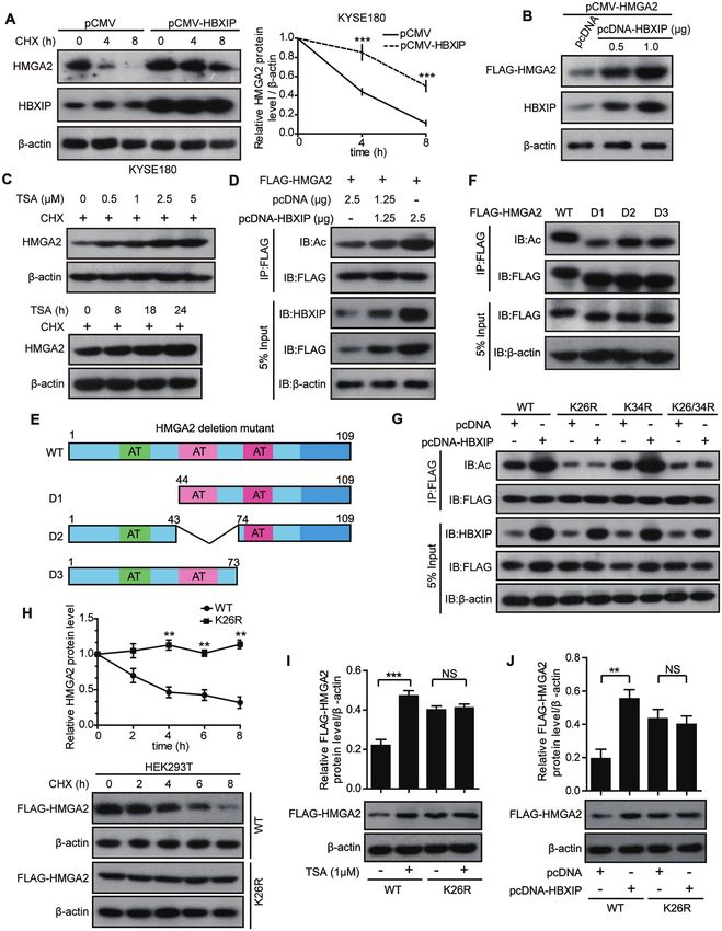
Nucleic Acids Research, 2020, Vol. 48, No. 9 4861

counterstained with DAPI (Sigma-Aldrich, St. Louis, MO,         MTT assays
USA). Fluorescent micrographs were obtained using laser
                                                                Cell viability assays were carried out using 3-(4,5-
scanning confocal microscopy (Leica, Germany).
                                                                dimethylthiazol-2-yl)-2,5 diphenyltetrazolium bromide
                                                                (MTT) reagent (Sigma) as described previously (43).
Chromatin immunoprecipitation (ChIP) assay                      Briefly, transfected cells were trypsinized, counted, and
                                                                plated into 96-well plates at a density of 1000 cells per well.
Chromatin immunoprecipitation (ChIP) assays were per-
                                                                After forming a confluent monolayer, the cells were treated
formed in KYSE180 cells transfected with the indicated
                                                                with aspirin (2.5 mM) for different time points. Then,
plasmids using the EpiQuik™ chromatin immunoprecipita-
                                                                MTT was added directly to each well. Four hours later, the
tion kit from Epigentek Group Inc. (Brooklyn, NY, USA)
                                                                supernatant was removed and dimethyl sulfoxide (DMSO)
as reported previously (41). The protein-DNA complexes
                                                                was added to stop the reaction. Absorbance at 490 nm was
were immunoprecipitated with anti-FLAG antibody (Sup-
                                                                measured using a reader system (Labsystem, Multiskan
plementary Table S3), using normal mouse IgG as a nega-
                                                                Ascent).

                                                                                                                                  Downloaded from https://academic.oup.com/nar/article/48/9/4858/5822968 by guest on 28 November 2020
tive control. DNA purified from these samples was analyzed
by real-time RT-PCR. The primers used are listed in Sup-
plementary Table S2.                                            Monolayer colony formation assay
                                                                The transfected cells were trypsinized and seeded in six-well
Total RNA isolation, reverse transcription-PCR (RT-PCR)         plates. The medium added with corresponding reagents was
and quantitative real-time PCR (qRT-PCR)                        replaced every 3 days. After growth for 2 weeks, the cells
Total RNA was extracted from cells using Trizol reagent         were washed with PBS three times and fixed in methanol for
(Invitrogen, USA) according to manufacturer’s instruc-          20 min at room temperature. Fixed cells were then stained
tions. First-strand cDNA was synthesized with the Prime         with crystal violet for 30 min at room temperature. After
Script reverse transcriptase Kit (TaKaRa Bio, China). RT-       extensive washing and air drying, monoclonal colonies were
PCR and qRT-PCR assays were carried out as previously           photographed. The number of colonies was counted and the
described (40). All primers were listed in Supplementary Ta-    colony forming efficiency was determined with the formula:
ble S2.                                                         colony-forming efficiency = number of colonies counted /
                                                                number of cells plated × 100%.

GST pull-down assays
                                                                Xenograft
The sequences of the primers used to amplify PCAF
cDNA are as follows: forward: 5 -ATGTCCGAGGCTG                 All experimental procedures involving animals were con-
GCGGGGCCGG-3 , reverse: 5 -TCACTTGTCAATTAA                    ducted in accordance with the guidelines of the National In-
TCCAGCTT-3 . The sequences of the primers used to              stitutes of Health Guide for the Care and Use of Laboratory
amplify HMGA2 cDNA are as follows: forward: 5 -G               Animals. Briefly, 5-week-old female BALB/c athymic nude
GAGGCAGGATGAGCGCA-3 , reverse: 5 -CTAGTCC                     mice from Experimental Animal Center of Peking (Beijing,
TCTTCGGCAGACTC-3 . The PCAF cDNA was inserted                  China) were fed and housed. The cells transfected with cor-
into pGEX-4T-1 vector, and HMGA2 cDNA was cloned                responding plasmids were harvested and suspended at a
into pET-28a vector. Proteins were expressed in E. coli         density of 5 × 10⁷ cells/ml in phosphate saline and then sub-
BL21 after induction with 0.5 mM IPTG at 16◦ C overnight        cutaneously injected into the right flank of each mouse (0.2
(∼12 h). The GST-PCAF or His-HMGA2 fusion pro-                  ml of cell suspension). Daily oral administration of saline
teins expressed in bacteria were purified with glutathione      or aspirin at 75 mg/kg was initiated after the tumor size
Sepharose 4B (GE Healthcare, USA) or Ni2+ -NTA agarose          exceeded 100 mm3 ∼10–14 days after injection. Tumor vol-
beads (Qiagen, USA) as described previously (42). In brief,     ume and body weight were monitored every 3 days. Tumor
the beads were washed, and purified His-HMGA2 was               volume (V) was monitored by measuring the length (L) and
added. The binding reaction was performed in binding            width (W) with calipers and was calculated using the follow-
buffer (20 mM Tris, pH 7.5; 150 mM NaCl; 0.1% PMSF).            ing formula: (L × W2 ) × 0.5. The mice were sacrificed when
After the incubation and washes, proteins were eluted by        the tumor size reached ∼1000 mm3 . The tumors were ex-
boiling in SDS-PAGE loading buffer and separated by SDS-        cised and assessed. The investigators who assessed the out-
PAGE. Precipitated HMGA2 was detected by western blot-          come data were blinded to the treatment groups.
ting analysis.
                                                                Statistical analysis
5-Ethynyl-2 -deoxyuridine (EdU) assay
                                                                Each experiment was repeated at least three times. The sta-
The EdU assay was used to measure the cell proliferative        tistical significance of in vitro and in vivo data was assessed
capacity. The cells (5 × 10³ cells/well) were seeded into 96-   by comparing mean values (±SD) using Student’s t-test,
well plates. All operations were performed based on instruc-    and significance was assumed at P < 0.05 (*), P < 0.01
tions of the Cell-Light TM EdU imaging detecting kit (Ri-       (**), P < 0.001 (***) and not significant (NS). The asso-
boBio, Guangzhou, China). The images were acquired and          ciation between HBXIP and HMGA2 expression in ESCC
analyzed under fluorescence microscope (Zeiss Axio Imager       tissue microarray was statistically analyzed by Pearson chi-
Z1, Carl Zeiss, Oberkochen, Germany).                           square independence test using the SPSS software program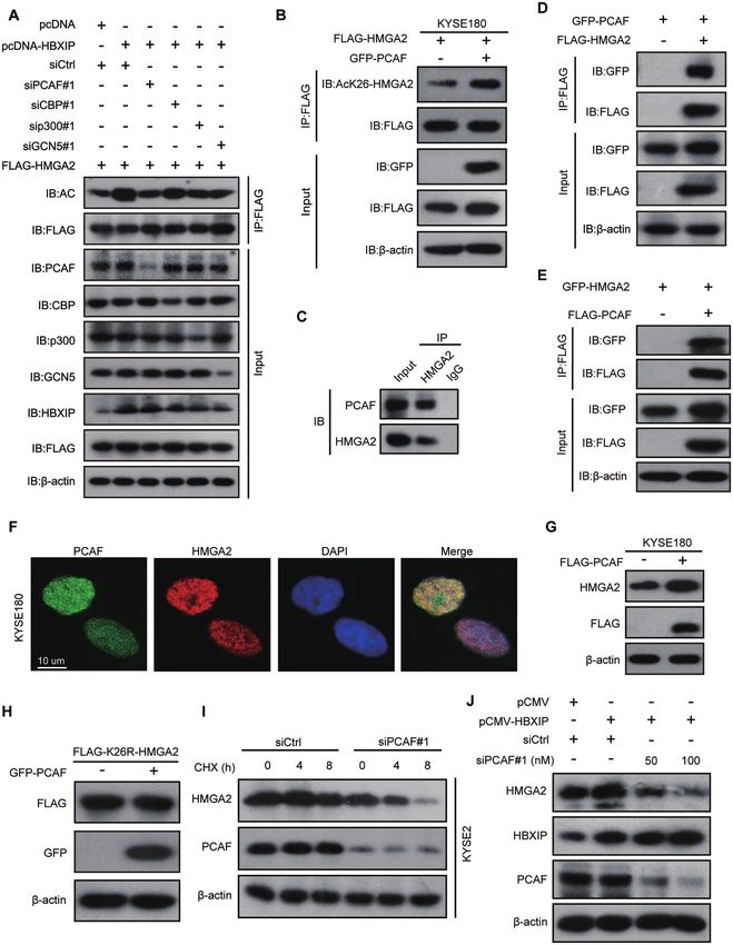
4862 Nucleic Acids Research, 2020, Vol. 48, No. 9
(SPSS, Chicago, USA). Survival rates were calculated us- ESCC tissues, and HMGA2 can be upregulated by HBXIP
ing the Kaplan–Meier method, and differences in survival in ESCC cells.
curves were analyzed by log-rank tests using the GraphPad
Prism 6.0.
HBXIP contributes to HMGA2 stabilization through induc-
ing its K26 acetylation
RESULTS To explore how HBXIP upregulates HMGA2, KYSE180
and KYSE2 cells were treated with CHX (a protein synthe-
HMGA2 is upregulated by HBXIP in human ESCC tissues
sis inhibitor) for different periods to block protein transla-
and cells
tion, and the degradation rates of the existing HMGA2 pro-
HMGA2, an architectural transcription factor, plays a piv- tein were assessed by western blotting. Compared with the
otal role in carcinogenesis (13–15,44). HBXIP emerges as control groups, HBXIP overexpression in KYSE180 cells
an oncogenic protein in many cancers (26–28,31). Either prolonged the half-life of endogenous HMGA2 protein
Downloaded from https://academic.oup.com/nar/article/48/9/4858/5822968 by guest on 28 November 2020
HMGA2 or HBXIP exhibits increased expression and is (Figure 2A), whereas HBXIP knockdown in KYSE2 cells
related to worse prognosis in ESCC (33,45). Therefore, we accelerated HMGA2 degradation (Supplementary Figure
are wondering whether HMGA2 is associated with HBXIP S2A). Moreover, the elevated expression of HBXIP led to
in ESCC development. The immunohistochemical staining dose-dependent increase of exogenous FLAG-HMGA2 at
of HMGA2 and HBXIP in human tissue microarray (Sup- the protein level (Figure 2B), supporting that HBXIP could
plementary Table S1) containing 43 normal esophageal tis- stabilize HMGA2 at the posttranslational level. Emerging
sues and 151 primary ESCC tissues revealed that HMGA2 studies demonstrated that lysine acetylation as a frequent
and HBXIP were both overexpressed in clinical human posttranslational modification (PTM) is critical for mod-
ESCC tisses (Figure 1A and B). Furthermore, in 43 nor- ulating protein stability and function (46–48). Thus, we
mal esophageal tissues and 151 primary ESCC tissues (Sup- are wondering whether HBXIP could stabilize HMGA2
plementary Table S1) a positive correlation was revealed through modulating its acetylation. We first examined
between HMGA2 and HBXIP expression (Pearson chi- whether HMGA2 could be acetylated. The Co-IP assay
square independence test, 2 = 25.05, P < 0.01. Supple- showed that both endogenously and exogenously expressed
mentary Table S4). In addition, the Kaplan–Meier sur- HMGA2 could be acetylated in vivo, and its acetylation dra-
vival analysis of 151 ESCC patients (Supplementary Table matically increased upon treatment with TSA, an inhibitor
S1) for 80 months demonstrated that high HMGA2 and of histone deacetylases (Supplementary Figure S2B and C).
HBXIP expressions were associated with advanced tumor To determine whether HMGA2 acetylation affects its sta-
stage (Supplementary Figure S1A and B) and reduced over- bility, we treated KYSE180 cells with TSA and/or CHX.
all and disease-free survival (Figure 1C–F). To further ex- Then, we found an increase in endogenous HMGA2 pro-
plore the relevance of HBXIP and HMGA2, we evaluated tein levels (Figure 2C and Supplementary Figure S2D), but
HBXIP and HMGA2 expressions in four ESCC cell lines HMGA2 mRNA levels had no significant changes in the
including KYSE180, KYSE2, KYSE510, and KYSE450. presence of TSA (Supplementary Figure S2E and F), sug-
Western blotting analysis presented a close connection be- gesting that HMGA2 remains stable upon its acetylation.
tween HMGA2 and HBXIP in these four ESCC cell lines Next, we ascertained the role of HBXIP in acetylation
(Figure 1G). We then analyzed the effect of HBXIP on modulation of HMGA2 protein. As expected, Co-IP assay
the protein levels of HMGA1, the other member of the showed that HBXIP dose-dependently enhanced the acety-
HMGA family. The data showed that the protein levels lation level of HMGA2 in HEK293T cells (Figure 2D), sug-
of HMGA1 were not changed by HBXIP (Supplementary gesting that HBXIP is involved in HMGA2 acetylation. To
Figure S1C). Taken a step further, HBXIP overexpression identify the acetylation sites of HMGA2, the acetylation
dose-dependently enhanced HMGA2 protein level with- levels of a series of deletion mutants of HMGA2 were eval-
out altering HMGA2 mRNA level in both KYSE180 and uated (Figure 2E). Compared with other mutants including
KYSE510 cells (Figure 1H, I and Supplementary Figure D2 and D3, the D1 mutant lacking 43 amino acids (1–43
S1D, E). Conversely, HBXIP knockdown by siRNA dose- aa) showed obviously decreased HMGA2 acetylation, indi-
dependently inhibited HMGA2 protein level, whereas had cating that the main acetylation residues of HMGA2 might
no effect on HMGA2 mRNA level in either KYSE2 or be located in this region (Figure 2F). The fragment from 1
KYSE450 cells (Figure 1H, I and Supplementary Figure to 43 aa contains two lysine residues (K26 and K34) that
S1F, G), implying that HMGA2 could be modulated by might be modified by acetylation. To further confirm the
oncogenic HBXIP through a posttranscriptional mecha- acetylation site, the K26 and K34 residues were mutated
nism. We also tested the effect of HBXIP overexpression to arginine (R, mimics of acetylation-deficient HMGA2)
and HBXIP knockdown on the protein levels of HMGA2 or glutamine (Q, mimics of hyperacetylated HMGA2), ei-
in a liver cancer cell line Hep3B and a breast cancer cell line ther alone (K26R and K34R or K26Q and K34Q) or to-
MCF-7. The protein levels of HMGA2 were obviously in- gether (K26/34R or K26/34Q). The acetylation level of
creased by HBXIP overexpression, while the knockdown of HMGA2 were no longer elevated by HBXIP in the K26R,
HBXIP significantly reduced the protein levels of HMGA2 K26/34R, K26Q, and K26/34Q mutants (Figure 2G and
in Hep3B cells (Supplementary Figure S1H). However, the Supplementary Figure S2G), suggesting that K26 could be
HMGA2 protein levels were not changed by HBXIP in the major acetylation site of HMGA2 mediated by HBXIP.
MCF-7 cells (data not shown). Collectively, HMGA2 and Subsequently, we compared the amino acid sequence of
HBXIP expression exhibit a positive relationship in clinical HMGA2 aligned with the other five species. Interestingly,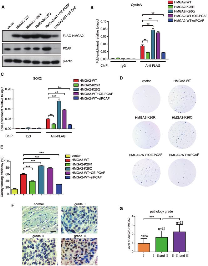
Nucleic Acids Research, 2020, Vol. 48, No. 9 4863
Downloaded from https://academic.oup.com/nar/article/48/9/4858/5822968 by guest on 28 November 2020
Figure 1. HMGA2 is upregulated by HBXIP in human ESCC tissues and cells. (A) HMGA2 and HBXIP expressions were assessed by immunohisto-
chemical staining in an esophageal tissue microarray containing 43 normal esophageal tissues and 151 ESCC tissues from Shantou University Medical
College between February 2011 and November 2016. Scale bars, 50 m. (B) Heatmap view of HMGA2 and HBXIP expressions of the esophageal tissue
microarray used in (A). Numbers 0, 1, 2 and 3 represent negative, week, moderate, and intense staining, respectively. (C and D) Kaplan–Meier plots of
the overall survival (C) and disease-free survival (D) rates of 151 ESCC patients stratified according to HMGA2 expression from the esophageal tissue
microarray containing 43 normal esophageal tissues and 151 ESCC tissues used in (A). (E and F) Kaplan–Meier plots of the overall survival (E) and
disease-free survival (F) rates of 151 ESCC patients stratified according to HBXIP expression from the esophageal tissue microarray containing 43 normal
esophageal tissues and 151 ESCC tissues used in (A). (G) HMGA2 and HBXIP expressions were measured by western blotting in four ESCC cell lines. (H)
Real-time PCR analysis of HBXIP and HMGA2 mRNA levels in KYSE180 cells transfected with the pCMV or pCMV-HBXIP plasmids and in KYSE2
cells transfected with control siRNA (siCtrl) or HBXIP siRNA (siHBXIP). Each bar shows the means ± SD (n = 3). (I) The HBXIP and HMGA2 protein
levels were measured by western blotting in KYSE180 cells transiently transfected with pCMV or pCMV-HBXIP plasmids and KYSE2 cells transfected
with siRNA (siCtrl) or HBXIP siRNA (siHBXIP). Statistically significant differences are indicated: *P < 0.05, **P < 0.01 and ***P < 0.001; NS, not
significant; Student’s t-test.4864 Nucleic Acids Research, 2020, Vol. 48, No. 9
Downloaded from https://academic.oup.com/nar/article/48/9/4858/5822968 by guest on 28 November 2020
Figure 2. HBXIP contributes to HMGA2 stabilization through inducing its K26 acetylation. (A) Degradation of HMGA2 protein was measured in
KYSE180 cells treated with 100 g/ml CHX for the indicated periods after transient transfection with pCMV or pCMV-HBXIP vectors. The HMGA2
intensity normalized to -actin was plotted. Each bar shows the means ± SD (n = 3). (B) Exogenous FLAG-HMGA2 was measured by western blotting
in HEK293T cells transiently transfected with pCMV-HMGA2 co-transfected with pcDNA or pcDNA-HBXIP plasmids. The FLAG-HMGA2 protein
level was detected using the anti-FLAG antibody. (C) Endogenous HMGA2 in KYSE180 cells were measured by western blotting after treatment with
100 g/ml CHX along with different concentrations of TSA for 18 h or 1 M TSA for the indicated periods. (D) Exogenous FLAG-HMGA2 was
immunoprecipitated with FLAG beads in HEK293T cells, and then the acetylation of HMGA2 protein in precipitation was measured by western blotting
with an anti-acetylated-lysine antibody. The cells were transiently transfected with the indicated plasmids. (E) A Schematic of FLAG-tagged full-length or
serial deletion mutant HMGA2. (F) HEK293T were transfected with FLAG-tagged full-length HMGA2 expression vectors (WT) or serial deletion mutant
HMGA2 expression vectors (D1, D2 or D3). Cell lysates were immunoprecipitated with FLAG beads, followed by western blotting with anti-FLAG and
anti-acetylated-lysine antibodies. (G) FLAG-tagged WT, K26R, K34R, or K26/34R of HMGA2 along with pcDNA or pcDNA-HBXIP plasmids was
transfected into HEK293T cells. Exogenous FLAG-tagged HMGA2 was immunoprecipitated with FLAG beads, and then acetylation levels of HMGA2
protein in precipitation were tested by western blotting with an anti-acetylated-lysine antibody. (H) The stabilities of WT and K26R mutant HMGA2
were measured by western blotting. HEK293T cells expressing FLAG-tagged WT or K26R mutant HMGA2 were treated with CHX for the indicated
periods. Each bar shows the means ± SD (n = 3). (I) Ectopically expressed FLAG-tagged WT and K26R mutant HMGA2 levels were measured after
the treatment of 1 M TSA for 18 h in KYSE180 cells by western blotting with an anti-FLAG antibody. The upper panel is the quantification of the
intensity relative to -actin. Each bar shows the means ± SD (n = 3). (J) Ectopically expressed FLAG-tagged WT and K26R mutant HMGA2 levels were
measured in KYSE180 cells transfected with pcDNA or pcDNA-HBXIP vectors by western blotting with an anti-FLAG antibody. The upper panel is the
quantification of the intensity relative to -actin. Each bar shows the means ± SD (n = 3). All experiments were repeated at least three times. Statistically
significant differences are indicated: **P < 0.01 and ***P < 0.001; NS not significant; Student’s t-test.Nucleic Acids Research, 2020, Vol. 48, No. 9 4865
we found that HMGA2-K26 was highly conserved in mul- bind to His-HMGA2 directly (Supplementary Figure
tiple species during evolution (Supplementary Figure S2H). S3I). Additionally, confocal microscopic analysis showed
Based on these findings, we hypothesized that the acety- that endogenous HMGA2 and PCAF (or exogenously
lation of K26 in HMGA2 is important for its stability. overexpressed EGFP-HMGA2 and FLAG-PCAF) were
Accordingly, we transfected HEK293T cells with differ- colocalized in the nucleus (Figure 3F and Supplemen-
ent vectors of HMGA2 including wild-type (WT), K26R tary Figure S3J). Intriguingly, PCAF overexpression
mutant and K26Q mutant. The K26R and K26Q mutant increased endogenous HMGA2 protein levels (Figure 3G),
HMGA2 were more stable than WT HMGA2 (Figure 2H whereas in K26R mutant-overexpressed cells PCAF lost
and Supplementary Figure S2I). Moreover, the K26R and the capacity of increasing HMGA2 protein levels (Figure
K26Q mutations abolished the TSA-induced increase in 3H), suggesting that K26 in HMGA2 is required for
HMGA2 and abrogated the HBXIP-induced stability of PCAF-induced HMGA2 acetylation. Furthermore, PCAF
HMGA2 (Figure 2I, J and Supplementary Figure S2J, K). knockdown destabilized HMGA2 (Figure 3I). HBXIP-
Thus, we conclude that HBXIP posttranslationally stabi- mediated stabilization of HMGA2 was largely abrogated
Downloaded from https://academic.oup.com/nar/article/48/9/4858/5822968 by guest on 28 November 2020
lizes HMGA2 via acetylation modification at K26 in ESCC by PCAF knockdown in KYSE180 cells (Figure 3J and
cells. Supplementary Figure S3K). Altogether, these findings
demonstrate that PCAF is required for HBXIP-induced
HMGA2 acetylation at K26 in ESCC.
The acetylase PCAF is responsible for HMGA2 acetylation
at K26 by HBXIP
To investigate the acetyltransferase responsible for
The Akt pathway activates the acetylase PCAF to promote
HMGA2 acetylation, we evaluated the effect of different
HMGA2 acetylation mediated by HBXIP
acetyltransferases including PCAF, CBP, p300 and GCN5
on HMGA2 acetylation. Notably, the knockdown of To clarify how HBXIP regulates the HMGA2 acety-
PCAF effectively abolished the HBXIP-mediated increase lation by PCAF, we assessed whether HBXIP modu-
in HMGA2 acetylation in KYSE2 cells, whereas the lated the binding affinity between HMGA2 and PCAF
silencing of other acetyltransferases, including CBP, p300 by Co-IP assays. HEK293T cells were transiently trans-
and GCN5, showed little effect on HMGA2 acetylation fected with GFP-HMGA2 in the presence or absence of
(Figure 3A and Supplementary Figure S3A-D). Based on FLAG-PCAF and/or HBXIP, followed by Co-IP assays
the same amount of FLAG-tagged HMGA2 in IP samples, with an anti-FLAG antibody. Indeed, the levels of GFP-
we found that the acetylation levels of FLAG-HMGA2 HMGA2 co-immunoprecipitated with the same amount
were increased upon PCAF overexpression in IP samples of FLAG-PCAF were increased upon HBXIP overexpres-
immunoprecipitated with the same amount of anti-FLAG- sion, demonstrating that HBXIP can enhance the HMGA2-
coated agarose beads, demonstrating that PCAF can PCAF interaction (Figure 4A). It was shown previously
enhance the HMGA2 acetylation (Supplementary Fig- that the histone acetyltransferase (HAT) activity of PCAF
ure S3E). To further confirm whether K26 of HMGA2 could be enhanced by its phosphorylation (49). There-
can be acetylated by PCAF, we entrusted a company fore, we subsequently examined whether PCAF activity
(Jia Xuan Zhi Rui, Beijing, China) to generate a rabbit was regulated in response to HBXIP overexpression. PCAF
polyclonal antibody specifically recognizing the acetylated phosphorylation level was measured after HBXIP overex-
HMGA2-K26 (AcK26-HMGA2). The specificity of the pression or silencing. As a result, HBXIP overexpression
AcK26-HMGA2 antibody was verified as it recognized the dramatically promoted PCAF phosphorylation, whereas
K26-acetylated peptide, but not the unacetylated HMGA2 HBXIP silencing efficiently prevented PCAF phosphoryla-
peptide (Supplementary Figure S3F). Furthermore, the tion (Figure 4B and C). It has been reported that HBXIP
acetylation signal was blocked by preincubation of the can activate the Akt signaling in hepatocellular carcinoma,
antibody with antigen peptides (Supplementary Figure and activate ERK1/2 or p38 signaling pathways in breast
S3G), indicating the specificity of the AcK26-HMGA2 cancer to promote tumor cell proliferation and migration
antibody for the recognition of HMGA2 acetylation at (50–52). To screen the kinases responsible for PCAF phos-
K26. Further data showed that the AcK26-HMGA2 phorylation, we treated KYSE180 cells with three kinase
antibody recognized acetylated HMGA2-WT at K26, but inhibitors, including GSK690693 (an inhibitor of Akt),
did not recognize K26Q or K26R mutations (Supplemen- PD98059 (an inhibitor of the upstream kinase of ERK1/2),
tary Figure S3H). Using this antibody, we found that the and SB202190 (an inhibitor of p38). We found that the
acetylation level of HMGA2 at K26 was clearly increased Akt inhibitor markedly reduced HBXIP-enhanced PCAF
after PCAF was overexpressed in KYSE180 cells (Figure phosphorylation, whereas the ERK1/2 and p38 inhibitors
3B). Given that PCAF might serve as the acetyltransferase showed little effects on PCAF phosphorylation (Figure
of HMGA2, we wondered whether PCAF could interact 4D). Moreover, exclusive treatment with the Akt inhibitor
with HMGA2. The data revealed endogenous interaction reduced PCAF phosphorylation and suppressed the inter-
of HMGA2 with PCAF in KYSE180 cells (Figure 3C). actions between HMGA2 and PCAF, even upon HBXIP
Furthermore, exogenously expressed HMGA2 and PCAF overexpression (Figure 4E). Furthermore, treatment with
could be co-immunoprecipitated by each other using the Akt inhibitor dramatically inhibited HBXIP-induced
different tag antibodies, implying that HMGA2 interacts HMGA2 acetylation (Figure 4F). These results were con-
with PCAF in the cells (Figure 3D and E). Moreover, GST firmed using siRNA-mediated Akt knockdown (Supple-
pull-down assays in vitro revealed that GST-PCAF could mentary Figure S4A and B). Thus, we conclude that the Akt4866 Nucleic Acids Research, 2020, Vol. 48, No. 9 Figure 3. The acetylase PCAF is responsible for HMGA2 acetylation at K26 by HBXIP. (A) FLAG-HMGA2 vectors accompanied by the indicated Downloaded from https://academic.oup.com/nar/article/48/9/4858/5822968 by guest on 28 November 2020 plasmids or siRNAs against a variety of acetyltransferases were transfected into KYSE2 cells. Exogenous FLAG-HMGA2 was immunoprecipitated with FLAG beads, and then the acetylation of HMGA2 protein in precipitation was tested by western blotting with an anti-acetylated-lysine antibody. (B) FLAG-HMGA2 vectors were cotransfected with or without GFP-PCAF plasmids into KYSE180 cells. Exogenous FLAG-HMGA2 was immunopre- cipitated with FLAG beads, and then the acetylation of HMGA2 protein in precipitation was tested by western blotting with the anti-AcK26-HMGA2 antibody. (C) Co-IP assays were performed to detect the interaction of endogenous PCAF with HMGA2 in KYSE180 cells with anti-HMGA2 antibody or control IgG. (D) GFP-PCAF and FLAG-HMGA2 plasmids were co-transfected into HEK293T cells, immunoprecipitated with FLAG beads followed by western blotting with anti-GFP and anti-FLAG antibodies. (E) HEK293T cells were co-transfected with FLAG-PCAF and GFP-HMGA2 plasmids, immunoprecipitated with FLAG beads followed by western blotting with anti-GFP and anti-FLAG antibodies. (F) The co-localization of endogenous PCAF and HMGA2 in KYSE180 cells was examined by confocal microscopy. KYSE180 cells were stained with an anti-PCAF monoclonal antibody (green) and an anti-HMGA2 polyclonal antibody (red). Nuclei were stained with DAPI (blue), followed by visualization with confocal microscopy. Scale bars, 10 m. (G) Endogenous HMGA2 was determined by western blotting in KYSE180 cells transfected with the indicated plasmids. (H) FLAG-K26R mutant HMGA2 vector was co-transfected with or without GFP-PCAF into KYSE180 cells. The levels of FLAG-K26R mutant HMGA2 were measured by western blotting with an anti-FLAG antibody. (I) Western blotting analysis of endogenous HMGA2 protein in KYSE2 cells. The cells were treated with CHX for the indicated periods after transfection with control siRNA (siCtrl) or PCAF siRNA#1 (siPCAF#1). (J) Endogenous HMGA2 protein levels were detected by western blotting in KYSE180 cells cotransfected with pCMV-HBXIP plasmids and/or PCAF siRNA#1. All experiments were repeated at least three times.
Nucleic Acids Research, 2020, Vol. 48, No. 9 4867
Downloaded from https://academic.oup.com/nar/article/48/9/4858/5822968 by guest on 28 November 2020
Figure 4. The Akt pathway activates the acetylase PCAF to promote HMGA2 acetylation mediated by HBXIP. (A) Co-IP assays were performed to exam-
ine the interaction of exogenous FLAG-PCAF with GFP-HMGA2 in HEK293T cells. GFP-HMGA2 and FLAG-PCAF along with pcDNA or pcDNA-
HBXIP plasmids were coexpressed into HEK293T, immunoprecipitated with FLAG beads, and detected with indicated antibodies. (B) KYSE180 cells
were transiently transfected with FLAG-PCAF along with pcDNA or pcDNA-HBXIP plasmids. Exogenous FLAG-PCAF was immunoprecipitated with
FLAG beads from cell lysates, and then the phosphorylation of PCAF protein in precipitation was tested by western blotting with an anti-phospho-serine
antibody. (C) KYSE2 cells were transiently transfected with FLAG-PCAF along with control siRNA (siCtrl) or HBXIP siRNA (siHBXIP). Exogenous
FLAG-PCAF was immunoprecipitated with FLAG beads from cell lysates, and then the phosphorylation of PCAF protein in precipitation was examined
by western blotting using an anti-phospho-serine antibody. (D) KYSE180 cells were transiently transfected with FLAG-PCAF along with pcDNA or
pcDNA-HBXIP plasmids and treated with AKTi (GSK690693, 10 M), ERKi (PD98059, 10 M) or p38i (SB202190, 10 M) for 6 h. Exogenous FLAG-
PCAF was immunoprecipitated with FLAG beads from cell lysates, and then the phosphorylation of PCAF protein in precipitation was examined by
western blotting with an anti-phospho-serine antibody. (E) KYSE180 cells were transiently transfected with FLAG-PCAF along with pcDNA or pcDNA-
HBXIP plasmids and treated with or without AKTi (GSK690693, 10 M) for 6 h. The interaction of HMGA2 with PCAF and the phosphorylation level
of PCAF were detected by a Co-IP assay using FLAG beads followed by western blotting with anti-HMGA2 and anti-phospho-serine antibodies. (F)
KYSE180 cells were transiently transfected with FLAG-HMGA2 along with the indicated plasmids and treated with or without AKTi (GSK690693, 10
M) for 6 h. Exogenous FLAG-HMGA2 was immunoprecipitated with FLAG beads from cell lysates, and then the acetylation of HMGA2 protein in
precipitation was tested by western blotting with an anti-acetylated-lysine antibody. All experiments were repeated at least three times.4868 Nucleic Acids Research, 2020, Vol. 48, No. 9
pathway is activated by HBXIP to increase PCAF phospho- HMGA2 acetylation by HBXIP inhibits its ubiquitination to
rylation and sequentially enhance HMGA2 acetylation. stabilize HMGA2
The acetylation of specific lysines can increase the sta-
bility of a protein through preventing ubiquitination of
HMGA2 K26 acetylation enhances its DNA binding ability the same lysine residues (47,54,55). To probe the potential
and predicts a poor prognosis of ESCC patients mechanism by which HBXIP-mediated acetylation regu-
lates HMGA2 protein stability, we first examined HMGA2
To uncover the physiological function of HMGA2 acety-
ubiquitination and found that it was actively ubiquitinated
lation at K26, we investigated the effect of HMGA2 K26
in HEK293T cells (Figure 6A). Moreover, HMGA2 pro-
acetylation on its DNA binding ability. We firstly trans-
tein was accumulated in cells treated with the proteasome
fected KYSE180 cells with plasmids expressing FLAG-
inhibitor MG-132 at different time points, indicating that
HMGA2-WT, FLAG-HMGA2-K26R, FLAG-HMGA2-
HMGA2 stability could be regulated by the ubiquitin–
K26Q, FLAG-HMGA2-WT with PCAF overexpression,
proteasome pathway (Figure 6B). According to Phospho-
Downloaded from https://academic.oup.com/nar/article/48/9/4858/5822968 by guest on 28 November 2020
and FLAG-HMGA2-WT with PCAF knockdown sepa-
SitePlus (http://www.phosphosite.org/homeAction.action),
rately (Figure 5A). Two common HMGA2 target genes in-
there existed an ubiquitination modification of HMGA2
cluding cyclin A and SOX2 were selected to investigate the
at K26. Therefore, we assessed the ubiquitination level
effect of HMGA2 acetylation at K26 on its DNA bind-
of the K26R HMGA2 mutant in cells. Interestingly, the
ing ability. It has been reported that HMGA2 binds di-
K26R mutation dramatically reduced HMGA2 ubiquitina-
rectly to the cyclin A and SOX2 promoter (13,53). Results
tion, indicating that K26 might also be the target residue
showed that HMGA2-K26Q occupied the promoter re-
for ubiquitination (Figure 6C). Importantly, the inhibi-
gions of the target genes to a high extent than the HMGA2-
tion of deacetylases with TSA decreased the ubiquitina-
WT as determined by ChIP assays (Figure 5B and C). How-
tion of WT but not of the K26R or K26Q HMGA2 mu-
ever, HMGA2-K26R decreased the DNA binding capac-
tants (Figure 6D and E). These results indicate a competi-
ity of HMGA2 to the target gene promoters compared to
tive interaction between the acetylation and ubiquitination
HMGA2-WT in ChIP assays (Figure 5B and C). Notably,
of HMGA2 at K26. We subsequently investigated whether
PCAF overexpression enhanced the HMGA2-WT occu-
HMGA2 ubiquitination was regulated by HBXIP, which
pancy to the target gene promoters by ChIP assays (Fig-
stimulates HMGA2 acetylation and stabilizes HMGA2.
ure 5B and C). In contrast, knockdown of PCAF decreased
We found that the proteasome inhibitor MG132 blocked
the DNA binding capacity of HMGA2-WT to the target
the HBXIP silencing-induced loss of endogenous HMGA2
gene promoters (Figure 5B and C). These data suggest that
(Figure 6F and G), and this was associated with the accu-
HMGA2 K26 acetylation enhances its DNA binding ability
mulation of polyubiquitinated HMGA2 (Figure 6H). Con-
to the target genes.
sistently, HBXIP overexpression upregulated the acetyla-
We next investigated the effect of HMGA2 K26 acety-
tion and downregulated the ubiquitination of WT HMGA2
lation on ESCC cell proliferation. The expression of
but showed no effects on the K26R or K26Q HMGA2 mu-
HMGA2-K26Q obviously increased its ability in pro-
tants (Figure 6I). These results indicate that the acetyla-
moting the proliferation of KYSE180 cells compared to
tion and ubiquitination of HMGA2 at K26 occur mutu-
HMGA2-WT (Figure 5D and E). However, compared to
ally exclusively and that HBXIP-induced acetylation at K26
the HMGA2-WT, the cell proliferation of HMGA2-K26R-
blocks HMGA2 ubiquitination and proteasomal degrada-
expressed group was significantly decreased (Figure 5D and
tion.
E). Moreover, cells ectopically expressing HMGA2-WT
with PCAF overexpression in order to increase the acetyla-
Stable accumulation of oncogenic HMGA2 mediated by
tion of HMGA2 showed a similar effect to HMGA2-K26Q
HBXIP promotes its target gene expression and ESCC
in promoting the cell proliferation. However, knockdown
growth
of PCAF decreased the ability of HMGA2-WT in promot-
ing the proliferation of KYSE180 cells (Figure 5D and E). Previous studies have shown that HMGA2 can modulate
These results demonstrate that the acetylation at K26 en- the transcription of cyclin A, SOX2, and hTERT to promote
hances the ability of HMGA2 in promoting ESCC cell pro- tumorigenesis (13,14,56). As expected, shRNA-mediated
liferation. HBXIP silencing disrupted the HMGA2-enhanced cyclin
To further show the clinical relevance of HMGA2 K26 A, SOX2 and hTERT expression at the mRNA level in
acetylation, we examined the levels of HMGA2 K26 acety- KYSE180 and KYSE510 cells (Figure 7A and Supple-
lation in two human ESCC tissue microarrays (Supplemen- mentary Figure S5A), suggesting that HBXIP contributes
tary Tables S5 and S6) containing 10 normal esophageal tis- to HMGA2-mediated transcription. We then investigated
sues and 124 ESCC tissues by immunohistochemistry stain- the effect of HMGA2 accumulation on ESCC growth in
ing analysis using the anti-AcK26-HMGA2 specific anti- vitro and in vivo. EdU and colony formation assays re-
body. Importantly, we found that the levels of HMGA2 K26 vealed that ectopic HMGA2 expression significantly pro-
acetylation were significantly higher in ESCC tissues than moted KYSE180 and KYSE510 cell proliferation. How-
that in normal esophageal tissues and HMGA2 K26 acety- ever, this promotion was dramatically abrogated by HBXIP
lation was gradually increased along with the cancer patho- knockdown in the cells (Figure 7B, C and Supplemen-
logical grade (Figure 5F and G). These findings indicate tary Figure S5B–G). Moreover, the growth of KYSE180
that elevated HMGA2 K26 acetylation may predict a poor xenografts in mice was markedly reinforced by HMGA2
outcome for ESCC patients. overexpression, while this stimulation of tumor xenograftNucleic Acids Research, 2020, Vol. 48, No. 9 4869
Downloaded from https://academic.oup.com/nar/article/48/9/4858/5822968 by guest on 28 November 2020
Figure 5. HMGA2 K26 acetylation enhances its DNA binding ability and predicts a poor prognosis of ESCC patients. (A) KYSE180 cells were transiently
transfected with FLAG-HMGA2-WT, FLAG-HMGA2-K26R, FLAG-HMGA2-K26Q, FLAG-HMGA2-WT along with FLAG-PCAF, or FLAG-
HMGA2-WT along with PCAF small interfering RNA (siRNA). Cell lysates from these treatments were prepared and subjected to western blotting
with indicated antibodies. (B) KYSE180 cells transfected with the indicated plasmids were subjected to ChIP assays with control IgG or an anti-FLAG
antibody. The occupancy of HMGA2 in the promoters of cyclin A was examined by qRT-PCR. Each bar shows the means ± SD (n = 3). (C) KYSE180
cells transfected with the indicated plasmids were subjected to ChIP assays with control IgG or an anti-FLAG antibody. The occupancy of HMGA2 in
the promoters of SOX2 was examined by qRT-PCR. Each bar shows the means ± SD (n = 3). (D) Monolayer colony-formation assay of KYSE180 cells
transfected with the indicated plasmids. (E) Colony forming efficiency of KYSE180 cells expressing the indicated vectors. Each bar shows the means ± SD
(n = 3). (F) HMGA2 K26 acetylation was assessed by immunohistochemical staining in 10 normal esophageal tissues and 124 ESCC tissues from human
ESCC tissue microarrays. Scale bars, 50 m. (G) The acetylation level of HMGA2-K26 was analyzed based on pathological grade. Statistically significant
differences are indicated: **P < 0.01, and ***P < 0.001; Student’s t-test.4870 Nucleic Acids Research, 2020, Vol. 48, No. 9
Downloaded from https://academic.oup.com/nar/article/48/9/4858/5822968 by guest on 28 November 2020
Figure 6. HMGA2 acetylation by HBXIP inhibits its ubiquitination to stabilize HMGA2. (A) HEK293T cells were transfected with FLAG-HMGA2
and treated with 40 M proteasome inhibitor MG-132 for 6 h. Exogenous FLAG-HMGA2 was immunoprecipitated with FLAG beads in HEK293T
cells, and then the ubiquitylation level of HMGA2 protein in precipitation was tested by western blotting with an anti-ubiquitin antibody. (B) Endogenous
HMGA2 level was detected by western blotting in KYSE180 cells treated with or without 40 M proteasome inhibitor MG-132 for the indicated periods.
(C) Exogenous FLAG-tagged WT and K26R mutant HMGA2 were immunoprecipitated with FLAG beads in HEK293T cells pretreated with 40 M
proteasome inhibitor MG-132 for 6 h, and then the ubiquitylation level of HMGA2 protein in precipitation was tested by western blotting with an anti-
ubiquitin antibody. (D) Exogenous FLAG-tagged WT, K26R, or K26Q mutant HMGA2 was immunoprecipitated with FLAG beads in HEK293T cells,
and then the ubiquitylation level of HMGA2 protein in precipitation was tested by Western blotting with an anti-ubiquitin antibody. The cells were treated
with or without TSA for 12 h and then treated with 40 M proteasome inhibitor MG-132 for additional 6 h. (E) The quantification of the polyubiquitination
intensity relative to immunoprecipitated FLAG-HMGA2 in Figure 6D. Each bar shows the means ± SD (n = 3). (F and G) Endogenous HMGA2 level
was detected by western blotting in KYSE2 cells (F) and KYSE450 cells (G) transfected with control siRNA (siCtrl) or HBXIP siRNA (siHBXIP) for
40 h and then treated with 40 M proteasome inhibitor MG-132 for additional 6 h. (H) KYSE2 cells were transfected with control siRNA (siCtrl) or
HBXIP siRNA (siHBXIP) for 40 h and then treated with 40 M proteasome inhibitor MG-132 for additional 6 h. Exogenous FLAG-HMGA2 was
immunoprecipitated with FLAG beads from cell lysates, and then the ubiquitylation level of HMGA2 protein in precipitation was tested by western
blotting with an anti-ubiquitin antibody. (I) Exogenous FLAG-tagged WT, K26R or K26Q mutant HMGA2 were immunoprecipitated with FLAG beads
in HEK293T cells transfected with or without pcDNA-HBXIP plasmids, and then the ubiquitylation level and acetylation level of HMGA2 protein in
precipitation were tested by western blotting with anti-ubiquitin and anti-acetylated lysine antibodies, respectively. All experiments were repeated at least
three times. Statistically significant differences are indicated: **P < 0.01; NS, not significant; Student’s t-test.Nucleic Acids Research, 2020, Vol. 48, No. 9 4871
Downloaded from https://academic.oup.com/nar/article/48/9/4858/5822968 by guest on 28 November 2020
Figure 7. Stable accumulation of oncogenic HMGA2 mediated by HBXIP promotes target gene expression and ESCC growth. (A) Real-time PCR analysis
of HMGA2, HBXIP, cyclin A, SOX2, and hTERT mRNA levels in KYSE180 cells separately transfected with pCMV vectors, pCMV-HMGA2 vectors,
or cotransfected with pCMV-HMGA2 and pSilencer-HBXIP vectors. Each bar shows the means ± SD (n = 3). (B) Monolayer colony-formation assay
of KYSE180 cells transfected with the indicated plasmids. (C) EdU incorporation assays were used to assess EdU-positive cells among KYSE180 cells
expressing the indicated vectors. Scale bars, 100 m. (D) The curves of tumor growth in nude mice transplanted with KYSE180 cells is shown. Each bar
shows the means ± SD (each group, n = 6). (E) Imaging of the tumors derived from nude mice transplanted with KYSE180 cells pretreated with indicated
plasmids. (F) Weights of tumors in each group were shown. Each bar shows the means ± SD (each group, n = 6). (G) The expression levels of Ki67 and the
statistics of Ki67-positive cells from above tumor tissues were examined by immunohistochemical assay. Scale bars, 50 m. Each bar shows the means ±
SD (n = 3). (H) The levels of HMGA2, HBXIP, CyclinA, SOX2 and hTERT from above tumor tissues were detected by western blotting. All experiments
were repeated at least three times. Statistically significant differences are indicated: *P < 0.05, **P < 0.01 and ***P < 0.001; Student’s t-test.You can also read