Origin-Independent Assembly of Kaposi's Sarcoma-Associated Herpesvirus DNA Replication Compartments in Transient Cotransfection Assays and ...
←
→
Page content transcription
If your browser does not render page correctly, please read the page content below
JOURNAL OF VIROLOGY, Feb. 2001, p. 1487–1506                                                                                      Vol. 75, No. 3
0022-538X/01/$04.00⫹0 DOI: 10.1128/JVI.75.3.1487–1506.2001
Copyright © 2001, American Society for Microbiology. All Rights Reserved.

        Origin-Independent Assembly of Kaposi’s Sarcoma-Associated
         Herpesvirus DNA Replication Compartments in Transient
          Cotransfection Assays and Association with the ORF-K8
                         Protein and Cellular PML
                FREDERICK Y. WU,1,2 JIN-HYUN AHN,2 DONALD J. ALCENDOR,1 WON-JONG JANG,1
                     JINSONG XIAO,1 S. DIANE HAYWARD,1,2 AND GARY S. HAYWARD1,2*
       Molecular Virology Laboratories, Department of Pharmacology and Molecular Sciences,1 and Viral Oncology Program,
            Department of Oncology,2 Johns Hopkins University School of Medicine, Baltimore, Maryland 21231-1000

                                                                                                                                                   Downloaded from http://jvi.asm.org/ on January 25, 2021 by guest
                                         Received 22 September 2000/Accepted 7 November 2000

             Six predicted Kaposi’s sarcoma virus herpesvirus (KSHV) proteins have homology with other well-charac-
          terized herpesvirus core DNA replication proteins and are expected to be essential for viral DNA synthesis.
          Intact Flag-tagged protein products from all six were produced from genomic expression vectors, although the
          ORF40/41 transcript encoding a primase-helicase component proved to be spliced with a 127-bp intron. The
          intracellular localization of these six KSHV replication proteins and the mechanism of their nuclear translo-
          cation were investigated. SSB (single-stranded DNA binding protein, ORF6) and PPF (polymerase processivity
          factor, ORF59) were found to be intrinsic nuclear proteins, whereas POL (polymerase, ORF9), which localized
          in the cytoplasm on its own, was translocated to the nucleus when cotransfected with PPF. PAF (primase-
          associated factor, ORF40/41), a component of the primase-helicase tripartite subcomplex together with PRI
          (primase, ORF56) and HEL (helicase, ORF44), required the presence of all five other replication proteins for
          efficient nuclear translocation. Surprisingly, even in the absence of a lytic cycle replication origin (ori-Lyt) and
          any known initiator or origin binding protein, the protein products of all six KSHV core replication genes
          cooperated in a transient cotransfection assay to form large globular shaped pseudo-replication compartments
          (pseudo-RC), which excluded cellular DNA. These pseudo-RC structures were confirmed to include POL, SSB,
          PRI, and PAF but did not contain any newly synthesized DNA. Similar to the human cytomegalovirus system,
          the peripheries of these KSHV pre-RC were also found to be surrounded by punctate PML oncogenic domains
          (PODs). Furthermore, by transient cotransfection, the six KSHV core replication machinery proteins success-
          fully replicated a plasmid containing EBV ori-Lyt in the presence of the Epstein-Barr virus-encoded DNA
          binding initiator protein, ZTA. The KSHV-encoded K8 (ORF-K8) protein, which is a distant evolutionary
          homologue to ZTA, was incorporated into pseudo-RC structures formed by transient cotransfection with the
          six core KSHV replication genes. However, unlike ZTA, K8 displayed a punctate nuclear pattern both in
          transfected cells and at early stages of lytic infection and colocalized with the cellular PML proteins in PODs.
          Finally, K8 was also found to accumulate in functional viral RC, detected by incorporation of pulse-labeled
          bromodeoxyuridine into newly synthesized DNA in both tetradecanoyl phorbol acetate-induced JSC-1 primary
          effusion lymphoblasts and in KSHV lytically infected endothelial cells.

   Kaposi’s sarcoma (KS)-associated herpesvirus (KSHV), also                 of KSHV in PEL cells can be induced by various chemicals,
called human herpesvirus 8, is a gamma-2 class herpesvirus                   such as tetradecanoyl phorbol acetate (TPA) and n-butyrate
that is distantly related to herpesvirus saimiri (HVS) and Ep-               (48, 57). In EBV, a critical event in the reactivation of the lytic
stein-Barr virus (EBV) but contains several novel loci (9, 12,               cycle is the expression of both the ZTA (BZLF1) and RTA
50, 58). KSHV DNA is present in virtually all tumor samples of               (BRLF1) proteins, both known DNA binding transcriptional
classical, endemic, and AIDS-associated forms of KS (12), as                 activators, whose mRNAs are among the first viral mRNAs
well as in peripheral blood mononuclear cells in up to 50% of                expressed after reactivation in latently infected B cells (17, 29,
homosexual AIDS patients with KS (72). KSHV is also present                  36, 39, 41, 75). Reactivation of the whole EBV lytic cycle
in a limited subset of AIDS-associated lymphoproliferative                   process including the expression from the delayed-early EBV
disorders referred to as primary effusion lymphomas (PELs),
                                                                             promoters required for viral DNA replication can be activated
as well as in a large proportion of cases of multicentric Castle-
                                                                             by either ZTA or RTA (15, 30, 33, 55, 75).
man’s disease, but is rare in normal blood donors except in
                                                                                In EBV, lytic cycle DNA replication depends on EBV-en-
Africa (7, 8, 65).
                                                                             coded core replication proteins, whereas latent state DNA
   Gammaherpesviruses characteristically establish latent in-
fections in lymphoid cells. The initiation of the viral lytic cycle          replication requires the cellular DNA replication machinery
                                                                             (74). Unlike controlled plasmid-state EBV latent replication,
                                                                             which depends on EBNA-1 binding to multiple binding sites in
                                                                             ori-P (56), in the lytic cycle, multiple rounds of DNA replica-
   * Corresponding author. Mailing address: CRB-3M08, 1650 Orleans
St., Baltimore, MD 21231-1000. Phone: (410) 955-8684. Fax: (410)             tion are initiated from within a different origin, ori-Lyt (origin
955-8685. E-mail: ghayward@jhmi.edu.                                         of lytic DNA replication), and require many viral gene prod-

                                                                      1487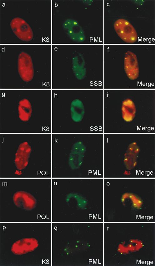
1488 WU ET AL. J. VIROL.
TABLE 1. KSHV replication genes and expression plasmids used in this study
Expression vectora Nucleotide positionb
KSHV gene Protein Function EBV gened
pSG5 pSG5-Flag ATG Stop
Core machinery
ORF6 SSB Single-stranded DNA binding pJX3 3210 6611 BALF2
ORF9 POL DNA polymerase pJX1 pJX8 11363 14401 BMRF1
ORF40/41 PAF Primase associated pJX5 pJX12 60308 62444 BBLF2/3
ORF44 HEL Helicase subunit pJX7 pJX14 64892 67258 BBLF4
ORF56 PRI Primase subunit pJX4 pJX11 79436 81967 BSLF1
ORF59 PPF Polymerase processivity pJX2 pJX9 96739 95549 BMRF1
Associated components
ORF57 MTA Posttranscriptional pJX6 pJX13 82717 83544 BMLF1
ORF-K8 K8 DNA replication (?) pCJC581 74850 75569 BZLF1
pFYW1c
Downloaded from http://jvi.asm.org/ on January 25, 2021 by guest
a
pJX8 to pJX14 are Flag-tagged pSG5 expression plasmids, and pJX1 to pJX7 are nontagged versions.
b
Genomic locations of the translated start and stop sites for the individual open reading frames of the KSHV replication loci were subcloned into an expression vector
(pSG5) containing SV40 enhancer, -globin intron, and SV40 polyadenylation signals as described in Materials and Methods.
c
pSG5-Myc expression plasmid for ORF-K8.
d
KSHV homologue.
ucts (28). In a cotransfection-replication assay first developed UL29) as well as HSV ori-S DNA and the specific ori-S and
for herpes simplex virus (HSV) (10, 73), EBV ori-Lyt was used ori-L DNA binding protein OBP (UL9) in transient DNA
to demonstrate the functional requirement for six EBV-en- cotransfection assays with bromodeoxyuridine (BrdU) incor-
coded core lytic replication proteins, POL (polymerase; poration (45, 76). The isolated genes for these same seven
BALF5), PPF (polymerase processivity factor; BMRF1), SSB proteins had already been shown to be essential and sufficient
(single-stranded DNA binding protein; BALF2), PRI (pri- for the specific amplification of cotransfected bacterial plasmid
mase; BSLF1), HEL (helicase; BBLF4), and PAF (primase- DNA containing the HSV origin (ori-S) as assayed by DpnI
associated factor; BBLF2/3) (22), as well as ZTA (21). The resistance and Southern blot hybridization (10, 73). Similar
EBV-encoded MTA (BMLF1) and RTA (BRLF1) proteins cotransfection studies have also been carried out to detect
also acted as nonessential accessory proteins in these assays. phosphonoacetic acid (PAA)-sensitive HCMV ori-Lyt-depen-
ZTA binds as a homodimer to both AP-1 sites and related dent DNA replication and assembly of RC in the presence of
sequences called ZTA response elements (ZREs) that are the six HCMV core replication proteins and a set of auxiliary
present in both ori-Lyt and key lytic cycle promoters (6, 13, 20, proteins (53, 61).
39, 41, 68) and stabilizes the formation of a DNA-bound com- KSHV also encodes a set of six genes which have various
plex containing the basal transcription factors TFIID and levels of homology to the equivalent EBV, HSV, and HCMV
TFIIA (16, 40). In addition to transcriptional regulation, ZTA replication genes. These include ORF9 (POL), ORF59 (PPF),
also has a role in lytic cycle reactivation, serving as an essential ORF6 (SSB), ORF56 (PRI), ORF40/41 (PAF), and ORF44
initiatior protein for replication of ori-Lyt (21, 60, 62). In fact, (HEL), which are expected to represent the six core replication
ZTA is implicated as an origin binding protein (OBP) because proteins (Table 1). However, an OBP and the ori-Lyt of KSHV
it is indispensable for the ori-Lyt-dependent replication in are still unidentified. In the immediate-early (IE) phase of the
transient cotransfection-replication assays (21), and deletion of KSHV lytic cycle, a 3.6-kb mRNA encompassing both RTA
specific ZRE motifs within ori-Lyt abolishes its ability to rep- (ORF50) and K8 (ORF-K8) is transcribed (42, 77). A separate
licate (62, 63). 1.0-kb spliced K8 mRNA encodes a 237-amino-acid (aa) pro-
Except for ZTA, the other essential EBV replication protein tein with a leucine zipper domain near its C terminus, which is
genes have sequence and positional homology with the known distantly related to the ZTA (BZLF1) protein of EBV and
core DNA replication protein genes of HSV and human cyto- Jun/Fos family proteins (42). Although K8 exhibits certain
megalovirus (HCMV) (14, 18, 47, 53). Their functions are properties suitable for an OBP because of analogous genome
sufficiently conserved that the six core proteins of HSV can location and similar splicing pattern with respect to EBV ZTA,
replicate EBV ori-Lyt in the presence of ZTA (21) and those there is no evidence that K8 itself is either a DNA binding
of EBV can replicate HCMV ori-Lyt in the presence of the protein or a transcription factor. Furthermore, RTA but not
UL84 initiator protein (61). K8 can reactivate the KSHV lytic cycle from latency (26, 66).
In all herpesviruses, infected cells at late stages of the lytic Recent studies have revealed that the cellular promyelocytic
cycle display large globular or kidney-shaped subnuclear do- leukemia protein (PML)-associated nuclear bodies known as
mains which exclude the nucleolus and are filled with a specific PML oncogenic domains (PODs) or nuclear domain 10 are the
set of viral replication proteins and display active viral progeny sites for input viral DNA accumulation in adenovirus, simian
genome DNA synthesis. These herpesvirus subnuclear do- virus 40 (SV40), HSV, and HCMV infections, as well as for IE
mains are known as viral DNA replication compartments transcription in HCMV (31, 32). PODs consist of 20 to 30
(RC). In HSV, the assembly of complete and functionally spherical 0.3- to 0.5-m structures that are present in most
active viral RC was detected in cells receiving all six HSV core cells and are thought to be associated with the nuclear matrix.
DNA replication genes (UL5, UL8, UL42, UL52, UL30, and Herpesvirus DNA replication proteins seem to target PODs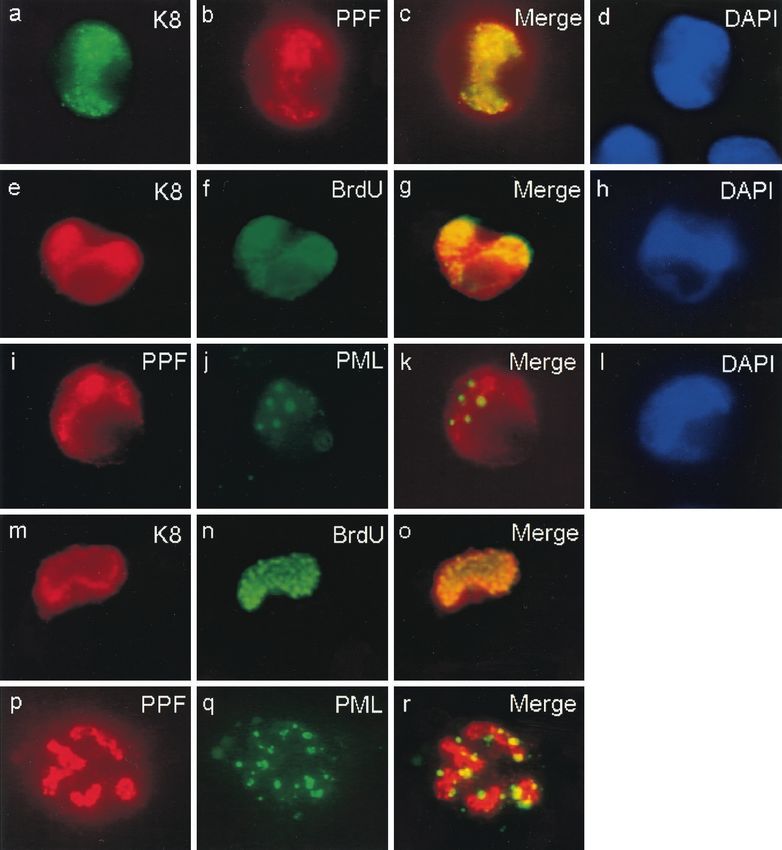
VOL. 75, 2001 ASSEMBLY OF KSHV DNA REPLICATION COMPARTMENTS 1489
TABLE 2. Oligonucleotide primers designed for KSHV replication and replication-associated genes
Target gene
Primer Genomic position Sequence (5⬘-3⬘)
(product)
LGH2485 3210–3240 CAG TGG ATC CAT GGC GCT AAA GGG ACC 5⬘ ORF6 (SSB)
LGH2486 6585–6611 TGA CAG ATC TCT ACA AAT CCA GGT CAG 3⬘ ORF6
LGH2481 11363–11389 CAG TGG ATC CAT GGA TTT TTT CAA TCC 5⬘ ORF9 (POL)
LGH2482 14375–14401 TGC AGG ATC CCT AGG GCG TGG CAA AAG 3⬘ ORF9
LGH2489 60308–60333 CAG TAG ATC TAT GGC AAC GAG CGA AG 5⬘ ORF40/41 (PAF)
LGH2490 62412–62444 TGA CAG ATC TTC AAA ATA AAG ATA AAA GCC TGG 3⬘ ORF40/41
LGH2493 64892–64918 CAG TAG ATC TAT GGA CAG CTC GGA AGG 5⬘ ORF44 (HEL)
LGH2446 67228–67258 CAG TAG ATC TTC AGT AGA TCA GAG TAG TCT T 3⬘ ORF44
LGH2487 79436–79462 CAG TAG ATC TAT GGA GAC GAC ATA CCG 5⬘ ORF56 (PRI)
LGH2488 81942–81967 TGA CAG ATC TTT AAC TGG CCA GTC CC 3⬘ ORF56
LGH2491 82717–82745 CAG TGG ATC CAT GGT ACA AGC AAT GAT AG 5⬘ ORF57 (MTA)
LGH2492 83516–83544 TGA CAG ATC TTT AAG AAA GTG GAT AAA AG 3⬘ ORF57
LGH2483 96739–96712 CAG TGG ATC CAT GCC TGT GGA TTT TCA 5⬘ ORF59 (PPF)
Downloaded from http://jvi.asm.org/ on January 25, 2021 by guest
LGH2484 95578–95549 TGA CAG ATC TTC AAA TCA GGG GGT TAA ATG 3⬘ ORF59
for immediate access to viral genomes deposited there; this was 3.0-kb intact ORF9 gene fragment with BamHI cohesive ends was obtained by
further implied when HCMV RC were seen growing from the PCR with primers LGH2481 and LGH2482. Plasmid pJX2 contains a 1.19-kb
ORF59 (PPF) gene fragment with BamHI and BglII cohesive ends obtained with
PODs into kidney-shaped RC structures surrounded by PODs primers LGH2483 and LGH2484 inserted at the same BamHI site of pSG5.
(2). The PML domains are targeted by several known herpes- Plasmid pJX3 contains a 3.4-kb ORF6 (SSB) gene fragment with EcoRI cohesive
virus regulatory proteins such as HSV IE110 (ICP0) and ends obtained with primers LGH2485 and LGH2486 inserted at the EcoRI site
HCMV IE1 and IE2, which colocalize with or adjacent to in pSG5. Plasmid pJX4 contains a 2.5-kb ORF56 (PRI) gene fragment with BglII
PODs, and in the case of IE2 later also associate with viral cohesive ends obtained with primers LGH2487 and LGH2488 inserted at the
BglII site of pSG5. Plasmid pJX5 contains a 2.13-kb ORF40/41 (PAF) gene
DNA RC (2). In addition, other key proteins involved in her- genomic fragment with BglII cohesive ends obtained with primers LGH2489 and
pesvirus DNA replication that are not known to target to LGH2490 inserted at the BglII site in pSG5. Plasmid pJX6 contains a 1.58-kb
PODs, including HSV IE175 (ICP4), HSV OBP (UL9), and ORF57 (MTA) genomic DNA fragment with BamHI cohesive ends obtained
EBV ZTA, also efficiently colocalize with their homologous with primers LGH2491 and LGH2492 inserted at the BglII sites in pSG5. Plas-
mid pJX 7 contains a 2.36-kb ORF44 gene fragment with BglII cohesive ends
functionally active RC.
obtained with primers LGH2493 and LGH2446 inserted at the BamHI and BglII
Very little is known about the lytic DNA replication of sites in pSG5. The oligonucleotide PCR primers used to clone the pJX plasmid
KSHV. In this study, we cloned the six putative KSHV core series are listed in Table 2.
replication genes based on homology to EBV and HSV and Empty vector plasmid pYW51 carries an oligonucleotide Flag epitope tag
confirmed that they expressed protein products of the expected bounded by BglII and BamHI cohesive ends and inserted at the BamHI site of
the pSG5 vector. Plasmid pJX8 carries the gene encoding POL (ORF9) from
sizes in transfected Vero cells. We also showed that the six
pJX1 with an added in-frame 5⬘ Flag epitope from plasmid pYW51. Similarly,
functional KSHV core replication proteins could substitute for plasmid pJX9 carries the PPF (ORF59) gene from pJX2 with an in-frame 5⬘ Flag
their EBV counterparts in the replication of EBV ori-Lyt. We epitope, plasmid pJX11 contains the PRI (ORF56) gene from JX4 with an added
then investigated (i) the intracellular localization of these pro- in-frame Flag epitope, plasmid pJX12 contains the PAF (ORF40/41) genomic
teins, as well as their complex interactive requirements for DNA fragment from pJX5 with an added 5⬘ Flag epitope, plasmid pJX13 carries
the MTA (ORF57) genomic DNA fragment with an added in-frame 5⬘ Flag
nuclear translocation; (ii) the requirements for formation of
epitope, and plasmid pJX14 carries the HEL (ORF44) gene with an added
subnuclear RC-like viral domains in a transient transfection in-frame 5⬘ Flag epitope.
system when all six core genes were cotransfected together; Plasmid pDY048 contains a polylinker insert bounded by EcoRI and BamHI
and (iii) the association of ORF-K8 and cellular PODs with the sites added to pSG5 with an oligonucleotide sequence encoding a Myc epitope
transiently assembled RC-like structures. Finally, we con- tag with NotI and SrfI cohesive ends inserted within the polylinker. Plasmid
pCJC565 contains an intact multiply spliced 714-bp KSHV K8 cDNA clone
firmed that functional KSHV RC in lytic cycle-induced PEL
flanked by BamHI sites in a pUC18 background (C. J. Chiou, F. Y. Wu, D. M.
cells and KSHV-infected dermal microvascular endothelial Ciufo, D. Alcendor, S. J. Kim, J. C. Zong, and G. S. Hayward, submitted for
cells (DMVEC) also colocalized with K8 and PODs. publication). To construct a Myc-K8 expression plasmid, the K8 cDNA was
inserted into the BamHI site of pDY048 in frame with the 5⬘ Myc epitope tag.
Plasmid pYNC100 carries an intact 735-bp EBV transactivator ZTA cDNA
MATERIALS AND METHODS
clone placed behind the leader plus ATG region from a black beetle virus for
Cells and virus. Vero cells were grown in Dulbecco’s modified Eagle’s medium efficient in vitro translation (13). Plasmid pCJC514 carries the K8 cDNA clone
containing 10% fetal bovine serum (FBS) in humidified 5% CO2 in a 37°C with both BamHI cohesive ends inserted into the BglII site of plasmid pGH255,
incubator. Vero cells were seeded at 8 ⫻ 104 cells per well in two-well slide which contains the same black beetle virus leader region as pYNC100. Plasmid
chambers for transfection. JSC-1 PEL cells (5a) were grown in RPMI medium pEF52 is a pBR322 vector that carries a 5.4-kb BamHI fragment of EBV ori-Lyt
containing 5% FBS in a humidified 5% CO2 incubator. Harvesting of the KSHV from genomic coordinates 48848 to 54858 (21, 22). Plasmid pRTS21 is an pSG5
supernatant virons from PELs and infection of human primary dermal micro- expression vector for EBV ZTA (60). pSG5 plasmids containing the six EBV
vascular endothelial cells (DMVEC) were all performed as described elsewhere core lytic replication genes, pRTS13, -14, -11, -28, -25, and -12, expressing EBV
(D. J. Ciufo, J. Cannon, L. Poole, F. Y. Wu, J. Orenstein, R. Ambinder, and G. S. POL, PPF, PRI, HEL, PAF, and SSB, respectively, were also used (23).
Hayward, submitted for publication). Transient DNA transfection. Transient DNA transfection assays for immuno-
Expression plasmids. Plasmid pJX1 carries the unspliced 3.0-kb KSHV ORF9 fluorescence assay (IFA) were carried out with 8 ⫻ 104 Vero cells in two-well
(POL) gene driven by the SV40 enhancer-promoter inserted at the BamHI site slide chambers. Different combinations of up to eight DNA plasmids (0.3 g of
downstream from the SV40 promoter in the pSG5 eukaryotic expression vector each) carrying tagged and/or nontagged versions of the gene encoding KSHV
(Stratagene). Using KSHV(BCBL-R) phage lambda clones as templates (50), a ORF9 (POL), ORF59 (PPF), ORF56 (PRI), ORF40/41 (PAF), ORF44 (HEL),1490 WU ET AL. J. VIROL.
ORF6 (SSB), ORF57 (MTA), or K8 driven by the SV40 enhancer region were cells were incubated with 4 N HCl for 10 min at room temperature and then
transfected into each well to study the intracellular localization of the protein washed in PBS for three times with 5-min intervals. The primary mouse mono-
products into RC-like structures. The CsC1-purified plasmid DNAs were co- clonal antibody (MAb) and rabbit polyclonal antibody (PAb) were diluted to-
transfected by the calcium phosphate precipitation procedure in BBS buffer (49). gether in PBS with 2% goat serum for double-label assays or diluted separately
Empty vector plasmid pSG5 DNA was used as a carrier to normalize the total for single-label assays. Primary antibodies were incubated for 1 h at 37°C fol-
amount of transfected DNA. Transfected cells were incubated in Dulbecco lowed by incubation with the appropriate combination of fluorescein isothiocya-
modified Eagle medium supplemented with 10% FBS in a 3% CO2 incubator at nate (FITC)-conjugated and rhodamine-conjugated anti-mouse and anti-rabbit
35°C overnight. The medium was changed 18 h after transfection, and the slides secondary antibodies at 1:100 dilution for 30 min at 37°C for double-label assays.
were placed into a 5% CO2 incubator at 37°C. Cells were fixed 48 h after Rhodamine-conjugated anti-mouse secondary antibody was diluted at 1:100 for
transfection for IFA. BrdU was added to the culture medium at a final concen- single labeling. To visualize cellular chromatin, a drop (20 l) of 4⬘, 6-diamidino-
tration of 10 M for 30 min before fixation when appropriate. 2-phenylindole (DAPI)-containing antifade slide mounting solution (Vector
Transient transfection replication assay. Vero cells were seeded at 106 cells Shield) was added to the slide prior to microscopy. Antibodies used included
per 100-mm-diameter dish and were transfected by the procedures described mouse anti-BrdU MAb (Becton Dickinson), rabbit anti-ORF6 PAb (SSB),
above. Approximately 10 g of total plasmid DNA containing either the EBV mouse anti-ORF59 MAb (PPF), rabbit polyclonal anti-K8, rabbit polyclonal
replication genes including ZTA or the KSHV replication genes plus ZTA were anti-PML(C), directed against amino acid positions 484 to 498 of the human
cotransfected with 5.0 g of EBV viral origin DNA (pEF52). For harvesting at 90-kDa PML isoform (1), mouse MAb and rabbit PAb anti-Flag (Sigma), and
80 h after the posttransfection medium change, the cell monolayer was washed mouse MAb and rabbit PAb anti-Myc (Santa Cruz). Rabbit anti-ORF K8 pep-
Downloaded from http://jvi.asm.org/ on January 25, 2021 by guest
twice with phosphate-buffered saline (PBS) and subsequently scraped into 4 ml tide PAb was generated by immunization with the N-terminal peptide (16-
of 40 mM Tris-hydrochloride (pH 7.5)–1 mM EDTA–150 mM NaCl. The cells DNSEKDEAVIEED-28) bounded by N-Y and C-CS residues. Rabbit anti-
were then pelleted and lysed in 2 ml of lysis buffer (10 mM Tris-HCl [pH 8.0], 10 ORF6 (SSB) peptide PAb was generated by immunization with C-terminal
mM EDTA, 2% sodium dodecyl sulfate [SDS], 100 g of proteinase K per ml). peptide (1116-GKKRKIASLLSDL-1128). Anti-ORF59 (PPF) MAb was a gift
Following overnight incubation at 37°C, the samples were diluted to 4 ml with from Bala Chandran (University of Kansas) (11).
Tris-EDTA (pH 8.0), extracted with phenol, phenol-chloroform, and chloro- Slides were screened and photographed with 40⫻, 63⫻, or 100⫻ oil immersion
form, and ethanol precipitated after the addition of sodium acetate (pH 5.2) to objectives on a Leitz Dialux 20EB epifluorescence microscope with Image-Pro
a final concentration of 0.3 M. The DNA pellets were resuspended in 450 l of software (Media Cybernetics, Silver Spring, Md.) and appropriate narrow-band
distilled H2O (dH2O), treated with 100 g of RNase A per ml, ethanol precip- FITC or rhodamine filters. For confocal microscopy, a Noran OZ CLSM con-
itated, and resuspended in 300 l of dH2O. Approximately 10 g of extracted focal microscope system with Intervision software (Noran Inc., Madison, Wis.)
cellular DNA was digested with 30 U of BamHI in a 100-l reaction volume for was used.
5 at 37°C. The reaction digest containing BamHI was then inactivated at 80°C for Western blot analysis. Vero cells were washed with PBS while still attached to
20 min and cooled on ice for 30 min. The cooled reaction sample was then
100-mm-diameter tissue culture plates and harvested with 10 ml of fresh PBS
incubated overnight with 30 U of DpnI at 37°C. To monitor DpnI activity, 5 l of
using a cell scraper. Cells were then pelleted at 6,000 rpm for 5 min and washed
the DpnI reaction digest was removed and incubated simultaneously with 500 ng
twice with 5 ml of PBS. The cell pellet was lysed with 0.4 ml of ice-cold lysis buffer
of pUC19 DNA overnight at 37°C. Complete cleavage of the pUC19 DNA
(50 mM Tris-HCl [pH 7.4], 150 mM NaCl, 1 mM EDTA, 0.5% Triton X-100, 1.5
indicated that the experimental DNA was also completely digested. The cellular
mM Na3VO4, 50 mM NaF, 10 mM sodium pyrophosphate, 10 mM glycerol
DNA was resolved by electrophoresis on a 0.8% agarose gel at 35 V for 20 h,
phosphate, 1 mM phenylmethylsulfonyl fluoride). Clarified cell extracts from the
transferred to a Nytran membrane (Schleicher & Schuell) after treatment of the
equivalent of 104 cells were separated by electrophoresis on SDS–10% polyacryl-
gel at 20°C for 10 min in 200 mM HCl, and then incubated in 0.4 M NaOH–0.6
amide gels followed by electroblotting onto nitrocellulose. The filter sheets were
M NaCl for 20 min. The agarose gel was then vacuum transferred to the nylon
blocked by incubation for 1 h at 20°C in PBS plus 0.1% Tween 20 containing 5%
membrane in the presence of 10⫻ SSC (1.5 M NaCl, 0.15 M sodium citrate) for
nonfat dry milk, then washed twice with PBS-Tween 20 for 15 min, and incubated
30 min. After the membrane was dried completely at 20°C, the DNA was
with appropriate MAb at a dilution of 1:3,000 for 1 h at 20°C. After three 10-min
irreversibly cross-linked by UV radiation (Stratalinker; Stratagene). For hybrid-
washes with PBS-Tween 20, the filter sheet were incubated with horseradish
ization, the membrane was incubated overnight at 60°C in 25 ml of buffer
peroxidase-conjugated goat anti-mouse immunoglobulin G (IgG; Bio-Rad) for
consisting of 1% SDS, 0.5 mg of heparin per ml, and 5⫻ SSC (750 mM NaCl, 50
1 h at 20°C and then washed three times, and the reacting protein bands were
mM Na2HPO4, 5 mM Na2EDTA). Approximately 100 ng of a gel-purified
detected with an enhanced chemiluminescence (ECL) system (Amersham ECL
BamHI ori-Lyt fragment was radiolabeled with [␣-32P]dCTP by random priming
to a specific activity of 108 cpm/g (Boehringer Mannheim-Roche kit). The RP2106) using Kodak XAR film.
membrane was then incubated at 60°C with 106 cpm of denatured radiolabeled RT-PCR and sequencing. Total RNAs (DNase treated) from both TPA-in-
ori-Lyt probe DNA per ml and fresh hybridization buffer. Following hybridiza- duced (48 h) and uninduced BCBL-1 cells were used for reverse transcriptase-
tion for 8 h to 12 h, the membrane was washed twice in 0.1⫻ SSC–0.1% SDS at mediated PCR (RT-PCR). First, 3 g of total RNA suspended in 100% ethanol
65°C for 45 min and exposed to Kodak XAR5 film for 24 h at ⫺80°C using an was centrifuged at 4°C for 30 min. The RNA pellet was air dried and resus-
intensifying screen. The resulting autoradiograms were quantified with a Kontes pended in 10 l of diethyl pyrocarbonate-treated dH2O. Second, to produce
fiber optic scanner. cDNA from total RNA, 10 l of resuspended RNA was added to a reaction
TPA induction and BrdU incorporation in PEL cells. Starting with an 80% mixture containing 1 l of 40 mM oligo(dT) primer; the mixture was heated at
viable suspension culture of the cell line JSC-1, cells were pelleted at 3,000 rpm 70°C for 10 min and quickly chilled on ice. To the chilled mixture, 1 l of RNA
and 5 ⫻ 105 were seeded into each well of a standard six-well tissue culture dish. inhibitor (Promega), 4 l of first-strand buffer, 2 l of 0.1 M dithiothreitol, 1 l
After initial pelleting, half of the original cell medium was replaced with fresh of 10 mM deoxynucleoside triphosphate, and 1 l of Superscript II reverse
RPMI medium with 5% FBS warmed to 37°C to a final volume of 2 ml. Cells transcriptase (GIBCO BRL) were added. The final mixture was incubated at
were pelleted and transferred to the each of the six wells. Then cells were 50°C for 1 h to generate cDNA. Third, 2 l of cDNA was used as template for
incubated at 37°C with 5% CO2 for 10 min. TPA was added to each well at a final Taq DNA polymerase, and two sets of oligonucleotides were used as PCR
concentration of 20 ng/ml. After 12, 48, or 72 h of incubation, the viability of the primers: first, LGH2489 and LGH2490 (described above), covering the 5⬘ and 3⬘
suspension culture was determined again, and 106 cells were pelleted and resus- ends of the full genomic coding region encompassing both ORF40 and ORF41;
pended in 1 ml of fresh prewarmed medium containing 10 M BrdU. The caps and second, LGH3752 (5⬘-GAA GAT CTC CAT CCG GTC TGG TGG CCG
of the Eppendorf tubes were punctured with sterile needles to allow air diffusion. TG-3⬘) and LGH3753 (5⬘-GAA GAT CTC CCC ATT TCC CTC AGT GTC
The cells were incubated at 37°C in a 5% CO2 incubator for 45 min. The cells TGG-3⬘), flanking the 127-bp putative intron of the ORF40/41 mRNA transcript.
were pelleted again and washed once with fresh 1 ml 1⫻ PBS. The cell pellet was Total DNA were isolated from BCBL-1 PEL cells as described for the transient
suspended in 200 l of fresh PBS and plated it onto the polylysine-loaded glass transfection replication assay, and 1 l of total DNA was used as the PCR
slides for 20 min or until the cells become fully adherent to the slides. Extra template. PCR amplification was conducted in a Thermal Cycler (Eppendorf)
liquid was drained, and the slides were dried completely. The slides were either with the following program: 94°C for 3 min; 40 cycles of 94°C for 30 s, 55°C for
stored at ⫺20°C or used for immunofluorescence staining immediately. 30 s, and 72°C 130 s; 72°C for 3 min. PCR products were resolved by electro-
IFA. Infected, induced, or transfected cells were washed in 1⫻ Tris-saline (100 phoresis on a 1% agarose gel, gel purified, and cloned into TA cloning vector
mM NaCl, 10 mM Tris-HCl [pH 7.5]), fixed with 2% paraformaldehyde in PBS pCR2.1 (Invitrogen). The cloned inserts from RT-PCR were sequenced with a
for 10 min at room temperature, and then permeabilized in 0.2% Triton X-100 T7 primer from the flanking 5⬘ end of the TA cloning site of pCR2.1 using an
in PBS for 20 min on ice. To expose incorporated BrdU residues, pulse-labeled automated model 310 Genetic Analyzer (ABI Prism).VOL. 75, 2001 ASSEMBLY OF KSHV DNA REPLICATION COMPARTMENTS 1491
RESULTS
Cloning and expression of isolated KSHV core DNA repli-
cation and replication-associated proteins. The genomic posi-
tions of KSHV ORFs encoding homologues of the six core
EBV DNA replication proteins were predicted from analysis
of the primary DNA sequence (Tables 1 and 2). The KSHV
genes referred to as KSHV ORF9 (POL), ORF59 (PPF),
ORF6 (SSB), ORF56 (PRI), ORF40/41 (PAF), and ORF44
(HEL) were predicted to encode proteins of 1,012, 396, 1,130,
843, 669, and 788 aa, respectively. Genomic DNA fragments
encompassing each of these ORFs were amplified as PCR
products derived from the KSHV (BCBL-R) PEL cell line
DNA as template and cloned into both Flag-tagged and un-
Downloaded from http://jvi.asm.org/ on January 25, 2021 by guest
tagged versions of the pSG5 expression plasmid driven by the
SV40 early region promoter-enhancer. The synthesis of each
protein with the expected approximate molecular weight from
these expression plasmids was confirmed in DNA-transfected
Vero cells by Western blot analysis after incubating the mem-
branes with either a mouse MAb directed against the Flag
epitope (Fig. 1a, lanes 1 to 3, and c) or a rabbit PAb specific for
KSHV SSB (Fig. 1b).
By analogy to EBV, the KSHV ORF57 (MTA) and ORF-K8
nuclear regulatory proteins were also suspected to be either
directly or indirectly associated with the viral DNA replication
process (Table 1). The structures of intact cDNAs for these
two genes have been analyzed previously (Chiou et al., sub-
mitted). The Flag-tagged ORF-K8 expression plasmid was
generated from an isolated intact spliced cDNA clone that
encodes the putative leucine zipper motif, whereas the Flag-
tagged MTA version represents a genomic DNA version.
ORF57 and ORF-K8 were predicted to encode proteins of 275
and 237 aa, which was confirmed by Western blot analysis with
MAb Flag antibody (Fig. 1a, lanes 4 and 5). FIG. 1. Western blot analysis of proteins expressed by eight KSHV
Evidence that ORF40 and ORF41 are transcribed as a sin- DNA replication and replication-associated genes in transient DNA
gle spliced mRNA species encoding PAF. ORF40 and ORF41 transfection assays. (a) Protein products from Flag-tagged expression
plasmids encoding HEL, POL, PPF, MTA, and K8 were transfected
are homologous to the EBV BBLF2 and BBLF3 ORFs, which into Vero cells. After 48 h, whole-cell protein extracts were electro-
are transcribed as a single mRNA transcript with an intron of phoretically fractionated on SDS– 10% polyacrylamide gels, and West-
128 bp that encodes PAF (21). The KSHV PAF mRNA tran- ern blot analysis was performed by incubating the membrane with a
script was also believed to span two ORFs, ORF40 and specific MAb or PAb followed by ECL color development. (b) Protein
ORF41. Consequently, the construction of the PAF genomic products of untagged pSG5 plasmid vector encoding KSHV SSB de-
tected with rabbit anti-SSB PAb, (c) Protein products of Flag-tagged
DNA expression plasmid in this case encompassed both the expression plasmids encoding PAF and PRI detected with mouse anti-
ORF40 and ORF41 genomic DNA regions to permit appro- Flag MAb.
priate cellular splicing (pJX5). The Flag-tagged version of
ORF40/41 was constructed by attaching a Flag epitope-encod-
ing sequence adjacent to the 5⬘ ATG start codon of ORF40/41
(pJX12). To confirm the presence of a spliced ORF40/41 joint the induced total RNA (Fig. 2b, lane 5). Sequencing of the
transcript, RT-PCR was conducted on total RNA extracted 475-bp RT-PCR product confirmed that the lytic cycle
from TPA-induced BCBL-1 PEL cells. A set of primers that ORF40/41 RNA encompassed a 127-bp intron spliced out
flanks the entire 2.0-kb ORF40/41 coding sequence, from the from 1,350 bp downstream from the initiation codon ATG of
initiation codon of ORF40 (5⬘) to the termination codon of ORF40 (nucleotide position 60308), which also removed the
ORF41 (3⬘), was designed and used to confirm the total length termination codon for ORF40. The splice donor site located at
of the ORF40/41 mRNA transcript. We designed a second set nucleotide position 61657 in ORF40 proved to be fused in
of flanking oligonucleotide primers encompassing the putative frame to the predicted acceptor site at nucleotide position
127-nucleotide intron (Fig. 2a) and spanning 603 bp (Fig. 2b, 61784 in ORF41, resulting in an intact cDNA sequence of
lane 3) between genomic nucleotide positions 61409 to 62011. 2,009 bp. Sequence alignment showed that the KSHV PAF
The results obtained from PCR with the intron flanking prim- protein encoded by the spliced form of ORF40/41 cDNA
ers showed that a 475-bp product was obtained from the 48- shares over 20% amino acid identity throughout its length with
h-TPA-induced total RNA (Fig. 2b, lane 2). With the full EBV BBLF2/3 (PAF) protein and up to 35% amino acid iden-
sequence primers, a 2.0-kb RT-PCR product was obtained in tity with equivalent predicted spliced PAF proteins from the1492 WU ET AL. J. VIROL.
Downloaded from http://jvi.asm.org/ on January 25, 2021 by guest
FIG. 2. A spliced ORF40 and ORF41 transcript is induced in TPA-treated BCBL-1 PEL cells. (a) Diagram of the genomic organization of
ORF40 and ORF41 between coordinates 60308 to 62444 in KSHV. The locations and orientations of the PCR primers used (3) and the sizes of
PCR products from spliced and unspliced cDNA templates are shown. (b) Photograph of an ethidium bromide-stained 1% agarose gel showing
the separated RT-PCR products. Lanes 1 and 4, no RT-PCR product detected from DNase-treated total RNA isolated from BCBL-1 PEL cell
line before TPA induction; lanes 2 and 5, RT-PCR products from RNA isolated 48 h after TPA induction; lanes 3 and 6, PCR products from the
total DNA isolated from BCBL-1 cells. For lanes 1 to 3, PCR primers covering the intron region were used; for lanes 4 to 6, PCR primers covering
the entire coding region of ORF40/41 were used. (c) Amino acid sequence alignment of KSHV ORF40/41, rhesus rhadinovirus (RRV) ORF40/41,
HVS ORF40/41, and EBV BBLF2/3.
rhesus rhadinovirus (RRV) and HVS gamma-2 rhadinoviruses locate POL to the nucleus, the two were cotransfected in a 1:1
(Fig. 2c). ratio (Fig. 4, bottom image). The results revealed that the
Intracellular localization of each KSHV core DNA replica- cellular localization of POL was efficiently altered from cyto-
tion and replication-associated protein in transfected mam- plasmic to nuclear in the presence of PPF, with 78% of the
malian cells. To investigate the intracellular localization pat- positive cells showing a nuclear diffuse POL pattern and 16%
terns of the eight KSHV presumed replication-associated showing a predominantly nuclear pattern with some cytoplas-
proteins, Vero cells were first transfected with individual ex- mic background (Table 3). Again, 6% of the positive cells still
pression plasmids encoding Flag-tagged POL, PPF, PRI, PAF, showed completely cytoplasmic POL localization, which can
HEL, or MTA or the untagged SSB or K8 protein. The results presumably be explained by the lack of expression of PPF in
of single-label IFA carried out at 48 h showed that POL, HEL, those cells. As negative controls, differential pairing with each
and PRI were each localized in the cytoplasm, whereas PAF of the other four core proteins in cotransfection assays did not
gave a mixed, primarily cytoplasmic staining pattern (Fig. 3). In affect the cytoplasmic localization pattern of POL (Fig. 4, top
contrast, SSB, PPF, MTA, and K8 were all transported in the and middle images).
nucleus as individual isolated proteins. SSB and PPF gave a All six core replication proteins are required for nuclear
diffuse nuclear pattern, whereas most of the MTA- and K8- translocation of the KSHV PAF (ORF40/41). In EBV, PRI,
positive cells also formed nuclear punctate patterns within a HEL, and PAF are all intrinsically cytoplasmic on their own
nuclear diffuse background (Fig. 3). As shown elsewhere, these but can form a tripartite subcomplex after cotransfection, with
punctate domains colocalize with SC35 spliceosomes and PML HEL (BBLF4) and PRI (BSLF1) being sufficient to completely
nuclear bodies, respectively (Chiou et al., submitted). translocate PAF (BBLF2/3) into the nucleus (23). However,
Cotransfection with PPF is sufficient to translocate POL the KSHV requirements for PAF (ORF40/41) nuclear trans-
into the nucleus. The localization pattern of KSHV POL alone location proved to be more complex than the EBV require-
was intrinsically cytoplasmic. However, cotransfection of the ments.
POL plasmid with all five of the other core viral replication Our initial IFA showed that the KSHV PAF (ORF40/41)
protein genes together allowed efficient nuclear translocation protein on its own also produced a mixed pattern with pre-
of POL; 71% of the transfected cells now showed a nuclear dominantly cytoplasmic staining (85%) in transfected Vero
staining pattern for POL, but with 29% remaining cytoplasmic cells (Table 4; Fig. 5k and m). To further differentiate between
(presumably because of inefficient cotransfection) (Table 3). partial nuclear states and cytoplasmic localization of PAF,
To determine which factor might be responsible for POL trans- three additional subclasses of staining were defined under the
location into the nucleus, each core protein was omitted in turn categories of mixed nuclear staining (Table 4): predominantly
from the cotransfection mixture described above. Interestingly, nuclear (PN) (Fig. 5c and d), nuclear more than cytoplasmic
POL remained localized in the cytoplasm only when PPF was (N⬎C) (Fig. 5e and g), and whole-cell diffuse (Fig. 5i and j).
omitted, whereas sequential omission of any single viral pro- Efficient nuclear localization of PAF was achieved only when
tein other than PPF in the cotransfection mixtures still pro- all six KSHV core replication proteins (POL, PPF, PRI, PAF,
duced up to 70% nuclear translocation of POL (data not HEL, and SSB) were present; 55% of the PAF-positive cells
shown). To test whether PPF on its own is sufficient to trans- showed completely nuclear staining (Fig. 5a and b), 21%VOL. 75, 2001 ASSEMBLY OF KSHV DNA REPLICATION COMPARTMENTS 1493
Downloaded from http://jvi.asm.org/ on January 25, 2021 by guest
FIG. 2—Continued.
showed a mixed pattern, and 22% retained a completely cyto- the positive cells showed a PN staining pattern (Fig. 5c and d);
plasmic localization (Table 4). Complete nuclear staining pat- when both POL and PPF were omitted, 43% of the positive
terns were never observed for PAF when one or more of the six cells showed PN staining pattern (Fig. 5e and f); and when
core genes were omitted. When only POL was omitted, 56% of POL, PPF, and SSB were all omitted, leaving only the PRI-1494 WU ET AL. J. VIROL.
Downloaded from http://jvi.asm.org/ on January 25, 2021 by guest
FIG. 3. Intracellular localization of single transfected KSHV DNA
replication and replication-associated proteins detected with IFA.
Equal samples of plasmid DNA (0.2 g) were used for each gene in
FIG. 4. Contribution of PPF but not PAF, PRI, HEL, or SSB to
transient transfection assays in Vero cells. All except SSB and K8, were
nuclear translocation of POL. The Flag-tagged POL expression plas-
detected with mouse anti-Flag MAb and donkey FITC or rhodamine-
mid was paired with other untagged replication genes, cotransfected
labeled anti-mouse IgG. SSB and K8 were detected with SSB or K8-
into Vero cells, and then detected with mouse anti-Flag and rhodam-
specific rabbit PAb and donkey FITC or rhodamine-labeled anti-rabbit
ine-labeled anti-mouse IgG. POL alone shows cytoplasmic staining but
IgG.
was efficiently translocated into the nucleus in the presence of PPF.
HEL-PAF tripartite complex present, the PN staining pattern
dropped to 12%, although 51% still gave an N⬎C staining significantly altering the PAF localization pattern (Table 4).
pattern (Fig. 5g and h). Cotransfection of PAF with SSB elevated the number of cells
Differential pairing of PAF with each of the other core displaying a PN staining pattern from 3% to 64% compared to
proteins was done to determine which of them was involved in the basal localization state of PAF alone. In addition, cotrans-
fection of PPF with PAF also raised the PN state of PAF from
the basal 3% to 24%. Cotransfection of PRI and HEL with
TABLE 3. KSHV ORF9 (POL) nuclear translocation in Flag-tagged PAF, the analogous components of a presumed
cotransfection assaysa KSHV triparte primase-helicase subcomplex, significantly ele-
vated the number of cells showing an N⬎C staining pattern of
Distribution (%)
PAF (from 10% to 51%). Repeating the experiment with PRI
Total no. of
Protein
Mixed
positive as the Flag-tagged version also increased the number of posi-
Nuclear Whole-cell Cytoplasmic cells counted tive cells showing an N⬎C pattern for PRI, whereas for a
PN N⬎C
diffuse negative control, cotransfection of PAF with PRI alone did not
All 71 ⬎1 ⬎1 ⬎1 29 88 significantly increase the proportion of PN or N⬎C patterns
POL ⬎1 ⬎1 ⬎1 ⬎1 ⬍99 76 for PAF (Fig. 5i and j; Table 4). Cotransfection of POL with
POL ⫹ PPF 78 14 2 ⬎1 6 181 PAF also did not alter the cytoplasmic localization of PAF
a
Pairing with other replication genes other than PPF did not alter the cyto- (data not shown). Therefore, although formation of the tripar-
plasmic localization of POL. tite helicase-primase complex contributed, more efficientVOL. 75, 2001 ASSEMBLY OF KSHV DNA REPLICATION COMPARTMENTS 1495
TABLE 4. KSHV ORF40/41 (PAF) nuclear translocation in cotransfection assaysa
Distribution (%)
Total no. of positive
Protein Mixed
Nuclear Cytoplasmic cells counted
PN N⬎C Whole-cell diffuse
All 55 10 4 7 22 92
All ⫺ POL ⬍1 56 24 ⬍1 20 34
All ⫺ PPF, POL ⬍1 43 24 2 31 42
All ⫺ SSB, PPF, POL ⬍1 12 51 9 26 136
PAF ⫹ SSB ⬍1 64 32 1 3 31
PAF ⫹ PPF ⬍1 24 23 3 50 94
PAF ⫹ PRI ⬍1 4 29 21 46 24
PAF ⬍1 3 10 2 85 296
PAF ⫹ MTA ⬍1 ⬍1 40 15 45 20
PAF ⫹ K8 ⬍1 47 39 5 9 64
Downloaded from http://jvi.asm.org/ on January 25, 2021 by guest
a
ORF40/41 was Flag tagged; Flag MAb and rhodamine-labeled anti-mouse IgG were used for IFA visualization.
translocation of PAF to the nucleus required additional inter- were observed for both SSB and POL (data not shown). A
actions with SSB and PPF. similar result was obtained for PAF when POL was omitted
In addition, cotransfection of KSHV MTA with PAF did not from the mixture (Fig. 5c and d).
significantly increase the number of PN cells, but it did dra- Evidently the formation of these nuclear structures, which
matically elevate the N⬎C cell number up to 40% compared to closely resembled the complete mature RC seen in other her-
the basal level of 10% (Table 4). Clearly, KSHV MTA is not pesvirus systems, required and incorporated all six of the
required for full nuclear translocation of the spliced cDNA KSHV core DNA replication proteins. However, obviously we
PAF, whereas the six core replication proteins themselves were had to consider these as only RC-like instead of complete
sufficient for stable nuclear accumulation of the PAF protein. functionally active RC on the presumption that in the absence
Furthermore, cotransfection of K8 with PAF alone signifi- of ori-Lyt, they did not synthesize any viral DNA. To exclude
cantly increased the efficiency of PAF nuclear translocation, the possibility that those structures were synthesizing viral or
with the PN state of PAF being elevated from the basal value cellular DNA, we pulse-labeled DNA-transfected cells with
of 3% up to 47% (Table 4). Therefore, in the case of KSHV, BrdU prior to fixation and detected no BrdU incorporation in
the collective effort of all six core replication proteins as well as any viral protein-positive Vero cells, including those with the
MTA and K8 appears to be required for maximal nuclear large RC-like structures (data not shown). Furthermore, by
accumulation of the primase-helicase subcomplex. double-label techniques with the appropriate fluorescence fil-
Assembly of KSHV RC-like structures in transient cotrans- ters, we were able to visualize the distribution of cellular chro-
fection assays. When carrying out preliminary assays to at- matin by including DAPI in the mounting solution and found
tempt to identify the lytic cycle replication origin and the that nuclear DNA proved to be excluded from the large viral
putative OBP or initiation protein of KSHV, we were surprised RC-like structures (Fig. 7). Therefore, the absence of cellular
to find that DNA replication-related structures were formed in DNA from these domains appeared to confirm their existence
the negative control cells receiving the set of plasmids express- as distinct structures or bodies that could physically displace
ing just the six core KSHV replication proteins. This was also cellular chromosomal DNA to the margins of the nucleus.
evident in the positive control panels from the PAF transloca- Because of the apparent absence of progeny viral DNA also,
tion experiment described above (Fig. 5a and b). we have elected to refer to those structures as pseudo-RC to
To examine whether SSB colocalized with the other core avoid confusion with the authentic but much smaller viral
proteins in these structures, cell cultures were cotransfected DNA-negative pre-RC structures observed in herpesvirus-in-
with various combinations of plasmids expressing five non- fected cells in the presence of PAA or before viral DNA
tagged replication genes and a sixth plasmid expressing either synthesis initiates.
Flag-tagged POL, PRI, or PAF. At 48 h after transfection, the KSHV core replication proteins replicate EBV ori-Lyt in the
cells were fixed and examined by double-label IFA. Interest- presence of the EBV ZTA protein. We wished to examine
ingly, 20 to 30% of the positive cells showed large irregularly whether the KSHV RC-like domains could in fact represent
shaped RC-like nuclear bodies detected with anti-SSB PAb, functional precursors to the typical mature herpesvirus RC
and these same cells also showed identical patterns for Flag- that actively synthesize viral DNA. Because of the lack of a
tagged POL, PRI, or PAF (Fig. 6a to f). Some cells contained known KSHV ori-Lyt, we used instead EBV ori-Lyt, which can
small punctate SSB patterns (see below), but the rest of the be efficiently replicated by HSV core replication proteins plus
SSB-positive cells generally showed a nuclear diffuse pattern the EBV ZTA initiation protein in transient assays (21). The
for SSB, probably because of the lack of at least one of the six core replication genes of KSHV (POL, PPF, PRI, PAF,
other five proteins (Fig. 6d). As negative controls, when we HEL, and SSB) were cotransfected with a target pBR322 plas-
included only POL, PPF, and SSB in the cotransfection assay mid containing a 5.4-kb EBV ori-Lyt (pEF52) as well as an
or sequentially omitted any one of the individual core DNA expression plasmid encoding the multifunctional EBV OBP
replication genes, none of the cells displayed any large or small ZTA (pRTS21). KSHV MTA (ORF57) was included in the
RC-like bodies in the nucleus, and only nuclear diffuse patterns cotransfection mixture to potentially enhance the expression1496 WU ET AL. J. VIROL.
ments were characteristic of the assembled herpesvirus DNA
RC seen in both the HCMV and HSV transient assembly assay
systems (61, 76) and were not detected in a negative control
experiment when EBV ori-Lyt and/or EBV ZTA were omitted
(data not shown).
To further verify this finding, a demethylation hybridization
assay for transient replication (10) was conducted by cotrans-
fecting the six KSHV core replication genes (POL, PPF, PRI,
PAF, HEL, and SSB) with KSHV MTA, EBV ZTA, and EBV
ori-Lyt. Total Vero cell DNA recovered from this experiment
was analyzed by Southern blot hybridization with an ori-Lyt
BamHI fragment probe which revealed the presence of newly
synthesized EBV ori-Lyt plasmid DNA that was resistant to
DnpI digestion (Fig. 8, lane 7). Therefore, we infered that the
Downloaded from http://jvi.asm.org/ on January 25, 2021 by guest
input methylated bacterial EBV ori-Lyt plasmid was indeed
replicated to produce unmethylated progeny copies by the
KSHV core replication genes. When KSHV POL was omitted
in the negative control experiment (Fig. 8, lane 8), no repli-
cated EBV ori-Lyt DNA was detected. Therefore, the KSHV
pseudo-RC are clearly capable of replicating herpesvirus DNA
when an appropriate origin plasmid and OBP are included in
the assay.
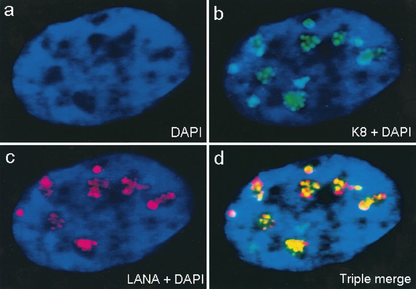
K8 targets to PODs and accumulates in the pseudo-RC that
are formed by the six core proteins in cotransfection assays.
Although there is no significant residual amino acid homology,
the K8 protein of KSHV is thought to be evolutionarily equiv-
alent to the ZTA protein of EBV, based on its colinear
genomic localization and similar C-terminal splicing pattern
producing a leucine zipper motif (42; Chiou et al., submitted).
Given the role of ZTA in EBV DNA replication, we investi-
gated whether K8 also accumulates in the pseudo-RC formed
in transient cotransfection assays. As shown elsewhere (Chiou
et al., submitted), when Vero cells were transfected with a
plasmid expressing K8 alone, the K8 protein produced numer-
ous small nuclear punctate bodies, which perfectly colocalized
with similar punctate nuclear bodies detected with PAb against
the PML proto-oncogene in double-label IFA experiments
FIG. 5. All six viral replication gene products are required for full (Fig. 9a to c). We have shown previously that in HCMV in-
nuclear translocation of PAF in cotransfected Vero cells. Flag-tagged fection, two of the accessory regulatory proteins needed for
PAF plasmids were used along with various combinations of the other efficient DNA replication, IE2 and UL112–113, initially target
untagged replication genes, and mouse anti-Flag and rhodamine-la-
beled anti-mouse IgG were used to visualize the PAF intracellular
to the PODs and subsequently accumulate into the functionally
localization. (a and b) Two separate single-label frames showing cells active RC (2). Therefore, we wished to test whether K8 was
cotransfected with the whole set of plasmids encoding POL, PPF, PRI, also directly incorporated into the KSHV RC.
PAF, HEL, and SSB; (e to j) omission experiments showing cotrans- To permit greater flexibility in double-label IFA transient
fection of all plasmids except those encoding POL (c and d), PPF and assembly assays, a Myc epitope-tagged version of the ORF-K8
POL (e and f), SSB, POL, and PPF (g and h), and HEL, SSB, PPF, and
POL (i and j). Sequential omission of the other replication genes mammalian expression plasmid (pFW1) was used. To examine
reverts PAF to a cytoplasmic localization similar to that obtained by whether the K8 protein colocalized with POL and PAF, Flag-
transfection with PAF alone (k and m). tagged versions of these two core replication genes (pJX8 and
pJX12, respectively) were used separately in combination with
the Myc-tagged K8 and the five other nontagged core replica-
level of the KSHV genes, based on previous evidence that the tion genes. Anti-SSB PAb was used to detect SSB in the same
EBV and HCMV versions enhance the efficiency of such assays cotransfection assays when needed. The results revealed that
(21, 61). Prior to fixation, the cells were pulse-labeled with when cotransfected with the six core replication genes, Myc-K8
BrdU and subsequently subjected to double-label IFA with was indeed incorporated into large irregularly shaped RC-like
KSHV SSB-specific PAb and a BrdU-specific MAb. In a small structures similar to those previously described for the repli-
proportion (0.5 to 1%) of cotransfected cells, the SSB protein cation core proteins. Furthermore, using anti-Myc MAb and
displayed large RC-like structures similar to those described anti-SSB PAb in double-label IFA, K8 was observed to pre-
above, and these exactly colocalized with large subnuclear do- cisely colocalize with SSB in the large viral pseudo-RC do-
mains that actively incorporated BrdU, indicating the presence mains (Fig. 9g to i). In similar experiments carried out with
of ongoing new DNA synthesis (Fig. 6g to j). These compart- Flag-tagged POL and PAF (data not shown), 18, 23, and 16%VOL. 75, 2001 ASSEMBLY OF KSHV DNA REPLICATION COMPARTMENTS 1497
of the double-positive cells contained RC-like structures for
POL and K8, for PAF and K8, and for SSB and K8, respec-
tively. Among these cells, the K8 protein colocalized with POL,
PAF, and SSB in the pseudo-RC with over 80% efficiency but
still occasionally remained nuclear diffuse (20%) in some cells
with positive pseudo-RC. We emphasize that K8 itself was not
required for the formation of those core replication structures,
because the six replication genes alone were sufficient for gen-
erating the pseudo-RC. However, if any of the replication
genes were intentionally omitted or not expressed in the cells,
RC were not formed and K8 displayed either a nuclear diffuse
or nuclear punctate pattern.
The pre-RC of KSHV initially form in association with
PODs and finally become surrounded by PODs. As previously
Downloaded from http://jvi.asm.org/ on January 25, 2021 by guest
described, the peripheries of the PODs are known to be sites of
initiation of RC formation in both HSV and HCMV (31, 32).
Therefore, we examined whether PODs were associated with
the formation of KSHV pre-RC in a similar manner. Double-
label IFA experiments with POL, SSB, and PML as markers
were carried out in cotrasfection assays receiving all six KSHV
replication genes and K8. We found both typical large irregu-
larly shaped pseudo-RC formed by POL as well as less fre-
quently occurring small globular or nuclear punctate struc-
tures. Double staining with anti-PML(C) PAb and anti-flag
MAb showed that several PML punctate domains precisely
bounded the periphery of each of the large pseudo-RC struc-
tures containing Flag-tagged POL (Fig. 9m to o). A similar
assay carried out with Flag-tagged PAF yielded the same re-
sults (data not shown). A Myc-tagged K8 expression plasmid
was also included in the transfection mixture; after double-
label IFA with anti-Myc MAb and anti-PML PAb, we found
that K8 also accumulated in large RC-like structures that were
surrounded by punctate PML bodies (Fig. 9p to r).
In addition to the large pseudo-RC, a small number of the
transfected cells contained five to eight much smaller POD-
like nuclear punctate structures detected with anti-SSB PAb,
which perfectly colocalized with K8 (and presumably PML)
(Fig. 9d to f). As a negative control, when SSB or PPF was
cotransfected with K8, SSB and PPF remained nuclear diffuse
whereas K8 was still nuclear punctate. Clearly, in the absence
of all six core replication proteins, SSB or PPF did not asso-
ciate with K8 or PODs, which confirmed that neither SSB nor
PPF intrinsically targeted PODs on their own. In contrast, the
viral punctate structures were formed in association with
PODs and K8 only when all six core replication proteins were
FIG. 6. All six core KSHV DNA replication proteins are required present. These viral nuclear punctate bodies closely resembled
for the assembly of complete RC-like structures in contransfected the pre-replication initiation foci (pre-RF) found in HSV-in-
Vero cells, and active DNA synthesis occurs within KSHV RC assem-
bled in the presence of EBV ori-Lyt and ZTA. (a to f) Double-label
IFA demonstrating colocalization of POL, PRI, and PAF with SSB in
large pseudo-RC in transient assembly assays. SSB was detected by
IFA with FITC-labeled anti-SSB rabbit PAb, whereas POL, PRI, and
PAF Flag-tagged fusion proteins were detected with rhodamine-la- plasmids (POL, PPF, PRI, PAF, HEL, and SSB) plus EBV ori-Lyt and
beled anti-Flag mouse MAb. (a and b) Flag-tagged POL cotransfected the ZTA DNA binding protein. Cells were pulse labeled for 30 min
with the untagged plasmids encoding PPF, PRI, PAF, HEL, and SSB; with BrdU prior to double-label screening for RC formation with an
(c and d) Flag-tagged PRI cotransfected with all five other untagged anti-SSB PAb and for active DNA synthesis with an anti-BrdU MAb.
plasmids; (e and f) Flag-tagged PAF cotransfected with all five other (g and i) Anti-KSHV SSB antibody to identify intranuclear RC con-
untagged plasmids. The incompletely transfected cell at the upper taining core viral DNA replication proteins. (h and j) Anti-BrdU
right in panels c and d displays nuclear diffuse SSB and cytoplasmic antibody to identify sites of ongoing DNA synthesis in the same cells
PRI. Deliberate omission of any of the six replication genes also (arrows indicate structures that resemble complete viral DNA RC).
disrupted RC formation (not shown). (g to j) Evidence for newly The anti-BrdU antibody also detected typical background speckled
synthesized DNA within KSHV RC formed in Vero cells cotransfected BrdU incorporation patterns found in the 25% of untransfected cells
with the complete set of untagged KSHV core replication expression in S phase.You can also read