RSC Chemical Biology REVIEW

Page created by Fernando Moore
 
CONTINUE READING
RSC Chemical Biology REVIEW
RSC
                                                                                                                Chemical Biology
            This article is licensed under a Creative Commons Attribution-NonCommercial 3.0 Unported Licence.

                                                                                                                                                                                                                                                   View Article Online
                                                                                                                    REVIEW                                                                                                                         View Journal | View Issue

                                                                                                                                                         DNA folds threaten genetic stability and can be
                                                                                                                                                         leveraged for chemotherapy
Open Access Article. Published on 30 September 2020. Downloaded on 04/19/2021 08:23:18.

                                                                                                                Cite this: RSC Chem. Biol., 2021,
                                                                                                                2, 47                                    Joanna Zell,a Francesco Rota Sperti,a Sébastien Britton                            *bc and
                                                                                                                                                         David Monchaud *a

                                                                                                                                                         Damaging DNA is a current and efficient strategy to fight against cancer cell proliferation. Numerous
                                                                                                                                                         mechanisms exist to counteract DNA damage, collectively referred to as the DNA damage response
                                                                                                                                                         (DDR) and which are commonly dysregulated in cancer cells. Precise knowledge of these mechanisms is
                                                                                                                                                         necessary to optimise chemotherapeutic DNA targeting. New research on DDR has uncovered a series
                                                                                                                                                         of promising therapeutic targets, proteins and nucleic acids, with application notably via an approach
                                                                                                                                                         referred to as combination therapy or combinatorial synthetic lethality. In this review, we summarise the
                                                                                                                                                         cornerstone discoveries which gave way to the DNA being considered as an anticancer target, and the
                                                                                                                                                         manipulation of DDR pathways as a valuable anticancer strategy. We describe in detail the DDR
                                                                                                                                                         signalling and repair pathways activated in response to DNA damage. We then summarise the current
                                                                                                                                                         understanding of non-B DNA folds, such as G-quadruplexes and DNA junctions, when they are formed
                                                                                                                                                         and why they can offer a more specific therapeutic target compared to that of canonical B-DNA. Finally,
                                                                                                                                                         we merge these subjects to depict the new and highly promising chemotherapeutic strategy which
                                                                                                                Received 17th August 2020,               combines enhanced-specificity DNA damaging and DDR targeting agents. This review thus highlights
                                                                                                                Accepted 20th September 2020             how chemical biology has given rise to significant scientific advances thanks to resolutely
                                                                                                                DOI: 10.1039/d0cb00151a                  multidisciplinary research efforts combining molecular and cell biology, chemistry and biophysics. We
                                                                                                                                                         aim to provide the non-specialist reader a gateway into this exciting field and the specialist reader with a
                                                                                                                rsc.li/rsc-chembio                       new perspective on the latest results achieved and strategies devised.

                                                                                                                a
                                                                                                                  Institut de Chimie Moléculaire de l’Université de Bourgogne, ICMUB CNRS UMR 6302, UBFC Dijon, France. E-mail: david.monchaud@cnrs.fr
                                                                                                                b
                                                                                                                  Institut de Pharmacologie et de Biologie Structurale, IPBS, Université de Toulouse, CNRS, UPS, Toulouse, France
                                                                                                                c
                                                                                                                  Équipe Labellisée la Ligue Contre le Cancer 2018, Toulouse, France. E-mail: Sebastien.Britton@ipbs.fr

                                                                                                                                                    Dr Joanna Zell obtained her MChem                                            Francesco Rota Sperti obtained
                                                                                                                                                    degree from the University of                                                his BSc degree in chemistry from
                                                                                                                                                    Sheffield (UK) supervised by Dr                                                the Universita di Pavia (Italia).
                                                                                                                                                    David M. Williams. She moved to                                              He then moved to Orsay
                                                                                                                                                    Paris (France) for her PhD at the                                            (France) to complete his MSc
                                                                                                                                                    Institut Curie under the supervision                                         degree in the group of Dr
                                                                                                                                                    of Drs Ludger Johannes and Frederic                                          Daniela Verga at the Institut
                                                                                                                                                    Schmidt, developing copper-free                                              Curie, dealing with the use
                                                                                                                                                    clickable tools to reconstitute                                              of c-exNDI derivatives as
                                                                                                                                                    glycolipids into live cells, defended                                        G-quadruplex sensors. He joined
                                                                                                                                                    in 2018. She joined the ICMUB                                                the ICMUB Dijon in Oct. 2019
                                                                                                                                                    Dijon (France) in 2019 to study the                                          to complete his PhD on
                                                                                                                           Joanna Zell              therapeutic relevance of three-way                Francesco Rota Sperti      quadruplexes, being in charge of
                                                                                                                                                    DNA junctions as anticancer                                                  the design, the synthesis and the
                                                                                                                targets. In parallel she is an active member of the Chimistes sans                 study of ever smarter template-assembled synthetic G-quartet
                                                                                                                Frontières and the Franco-British Council Local Leaders.                          (TASQ)-based biomimetic G-quadruplex ligands.

                                                                                                                    2021 The Author(s). Published by the Royal Society of Chemistry                                                       RSC Chem. Biol., 2021, 2, 4776 | 47
RSC Chemical Biology REVIEW
View Article Online

                                                                                                                Review                                                                                                            RSC Chemical Biology

                                                                                                                Introduction                                                           proliferating cells. The present review aims at gathering the
                                                                                                                                                                                       most recent results obtained in this new and promising
                                                                                                                High levels of genetic instability and mutations is a general          approach, to demonstrate the relevance of targeting higher-
                                                                                                                enabling hallmark of cancer, as defined by Douglas Hanahan             order DNA structures to treat cancer.
            This article is licensed under a Creative Commons Attribution-NonCommercial 3.0 Unported Licence.

                                                                                                                and Robert A. Weinberg in their cornerstone reviews.1,2 This is
                                                                                                                likely a result of the need for tumour progression to acquire
                                                                                                                multiple mutations, e.g. in oncogenes and tumour-suppressor            1. The discovery of DNA as a chemotherapeutic target
                                                                                                                genes, for successful tumour initiation, and potentially meta-            1a. Discovering the vulnerability of DNA. It was known that
                                                                                                                stasis. The genome maintenance systems limit such genetic              organisms could be mutated or damaged through radiative and
                                                                                                                defects in healthy cells but this is often impaired in cancer          chemical attack before what we now call the blueprint of life,
Open Access Article. Published on 30 September 2020. Downloaded on 04/19/2021 08:23:18.

                                                                                                                cells, making the accumulation of DNA lesions a common                 the DNA, was discovered.19 Ionising (IR) and ultraviolet (UV)
                                                                                                                feature in cancer. Cancer cells’ reduced ability to repair DNA         radiation were known to induce lethal and non-lethal mutation
                                                                                                                damage, driving their genomic instability, provides an impor-          in cells from the 1930s,20–22 in a U.S. American push to under-
                                                                                                                tant vulnerability that is exploited therapeutically.3–5               stand radiation biology for military applications, proceeding
                                                                                                                    Recent advances in genetics, genomics and proteomics have          the fabrication of atomic weapons. Repair mechanisms in
                                                                                                                provided a better understanding of the complex interplay               irradiated cells were first described in bacteria independently
                                                                                                                between DNA lesions and repair mechanisms, and what is                 by two United States laboratories in 1949.23,24 The mechanistic
                                                                                                                therapeutically relevant to halt cancer cell proliferation.6 These     basis of this repair process, referred to as enzymatic
                                                                                                                advances have generated momentum for designing more selec-             photoreactivation,25 was elucidated only several years later in
                                                                                                                tive and efficient anticancer strategies, often relying on combi-        light of the discovery of DNA structure in 1953,26–28 as well as
                                                                                                                nations of DNA damaging agents and DNA repair                          the description of irradiation-promoted formation of cyclo-
                                                                                                                inhibitors.5,7,8 Recent advances in our understanding of the           butane pyrimidine dimers (CPD) in vitro29 and the discovery
                                                                                                                complex aspects of DNA/RNA secondary structure9–12 provide             of the DNA photolyase that directly repairs CPD formed by
                                                                                                                brand new opportunities to exploit DNA damage in a more                UV-light.30,31
                                                                                                                precise manner. Alternative nucleic acid structures are defined            DNA is constantly exposed to damaging agents – ‘‘tens of
                                                                                                                as structures that deviate from the canonical Watson–Crick             thousands of DNA lesions per day’’3 – of both endogenous and
                                                                                                                DNA double helix, B-DNA or duplex-DNA. From a molecular                exogenous origins.32,33 Beyond irradiation and cross-linking
                                                                                                                viewpoint, the selective small-molecule targeting of such DNA          chemicals, different types of lesion have now been unravelled,
                                                                                                                structures holds potential over gene sequence targeting, given         which include chemical modifications (e.g. oxidations, deami-
                                                                                                                that they present well-defined 3D-structures,13,14 in a manner         nations), DNA replication errors (e.g. mismatched Watson–
                                                                                                                reminiscent of modern pharmaceutical targeting of proteins             Crick base pairs) and DNA transaction impediments (e.g. non-
                                                                                                                and enzymes.15 Furthermore, alternative DNA structure for-             B helix structured DNA, sometimes called ‘difficult-to-replicate’
                                                                                                                mation is coupled with DNA transactions (replication, tran-            sequences34,35) which can stall replication and transcription
                                                                                                                scription) due to transient strand separation and local DNA            (Fig. 1). The plurality of DNA damage explains why cells have
                                                                                                                deformation.16–18 Such structures thus offer the added advan-          evolved such a highly sophisticated and efficient network of
                                                                                                                tage of creating replication/transcription-associated DNA              surveillance, signalling and repair pathways, collectively
                                                                                                                damage whose repercussions will be felt primarily in highly            referred to as the DNA damage response (DDR).3,4,33,36,37 The

                                                                                                                                                 Dr Sébastien Britton received his                                      Dr David Monchaud received his
                                                                                                                                                 PhD in 2009 (University of                                              PhD in chemistry 2002 at the
                                                                                                                                                 Toulouse, France) on the study of                                       University of Geneva (Switzerland)
                                                                                                                                                 DNA repair. Then he joined Prof.                                        under the supervision of Prof. Jérôme
                                                                                                                                                 Steve      Jackson’s     laboratory                                     Lacour. After two post-docs in Paris
                                                                                                                                                 (Cambridge, UK) where he was                                            (France), he was appointed as a
                                                                                                                                                 initiated to chemical biology. In                                       CNRS researcher in 2005 in the
                                                                                                                                                 2013, he moved to Dr Patrick                                            laboratory of Prof. Jean-Marie Lehn
                                                                                                                                                 Calsou’s lab (Toulouse, France) to                                      (College de France, Paris) under the
                                                                                                                                                 characterise the interplay between                                      supervision of Dr Marie-Paule
                                                                                                                                                 DNA repair mechanisms, and the                                          Teulade-Fichou, before moving to
                                                                                                                                                 mechanism of action of small                                            Institut Curie (Orsay) in 2007 with
                                                                                                                      Sébastien Britton         molecules. In 2015, he was                   David Monchaud             Dr Teulade-Fichou, and to the
                                                                                                                                                 appointed as a CNRS researcher                                          Institut de Chimie Moléculaire de
                                                                                                                and in 2018 he was awarded the CNRS Bronze Medal for his               l’Université de Bourgogne (ICMUB, Dijon, France) in 2009, to develop
                                                                                                                discoveries. Starting in 2021, he will lead the ‘‘Deciphering and      chemical genetics programs on DNA/RNA secondary structures, smart
                                                                                                                Drugging DNA Repair’’ team at IPBS (Toulouse, France).                 ligands and probes.

                                                                                                                48 | RSC Chem. Biol., 2021, 2, 4776                                          2021 The Author(s). Published by the Royal Society of Chemistry
RSC Chemical Biology REVIEW
View Article Online

                                                                                                                RSC Chemical Biology                                                                                                                        Review
            This article is licensed under a Creative Commons Attribution-NonCommercial 3.0 Unported Licence.
Open Access Article. Published on 30 September 2020. Downloaded on 04/19/2021 08:23:18.

                                                                                                                Fig. 1 Examples of DNA-damaging agents, the lesion which they induce, and the downstream consequence. Temozolomide (TMZ), ionising radiation
                                                                                                                (IR), camptothecin (CPT), chlorambucil (Chl), cisplatin (cisPt), doxorubicin (Dox), single-strand break (SSB), double strand break (DSB), cyclobutane
                                                                                                                pyrimidine dimer (CPD), interstrand cross-link (ICL), base excision repair (BER), mismatch repair (MMR) and global genome nucleotide excision repair
                                                                                                                (GG-NER), transcription-coupled NER (TC-NER). Adapted from ref. 70, created with BioRender.

                                                                                                                molecular complexity of the DDR is just starting to be understood,          to an extended study that was communicated after the end of
                                                                                                                because of the multiplicity of pathways involved, including excision        WWII.41,47,48 Elucidation of the cross-linking mechanism,49 again
                                                                                                                (BER, NER, MMR), recombination (HR) and joining (NHEJ) path-                driven by military interests to conceive possible antidotes to
                                                                                                                ways (see Sections 2a and b) and their constant and intricate cross-        chemical weapons, fuelled numerous studies that built the founda-
                                                                                                                talk. DDR ultimately aims at controlling genomic instability in             tions for modern chemotherapy of new DNA repair mechanisms.
                                                                                                                normal cells, whilst from a therapeutic viewpoint, the DDR presents            These results gave new impetus to the quest for small
                                                                                                                a key strategic Achille’s heel of cancer cells, which can be targeted to    molecule therapy. Similarly to Gilman and Goodman, Sidney
                                                                                                                impede their anarchic proliferation.                                        Farber made the link between the high lymphocyte levels
                                                                                                                    1b. Damaging DNA to treat cancer. Cancer has likely                     observed in leukaemia patients’ blood and potentially leuko-
                                                                                                                existed since the beginning of mankind but ageing and the constant          penic vitamins antifolates, who began pioneering trials on last-
                                                                                                                modification of our lifestyles has dramatically increased its occur-        chance child leukaemia patients with synthetic antifolates in
                                                                                                                rence. Treatment consisted solely of surgery up until radiotherapy          1947.48,50,51 These research efforts led to the first example of
                                                                                                                treatments were introduced, with the first uses of X-rays in 1897 and       targeted therapy with the use of 5-fluorouracil (5-FU) in 1957,52
                                                                                                                of radium in 1904, following the discoveries of physicists Wilhelm C.       which specifically targets the abnormal uracil dependence of
                                                                                                                Rontgen and Marie Skłodowska Curie respectively.38,39 Unknown at            hepatomas.53 The benchmark example of targeted therapy,
                                                                                                                the time, radiotherapy exploits the principle of inducing DNA               imatinib (Gleevec) was reported in 1996,54 being the first
                                                                                                                damage, including DNA double strand breaks (DSBs) that show                 approved anticancer drug molecularly designed for its specific
                                                                                                                moderate selectively in killing rapidly dividing cells. The first           protein target, the constitutively active BCR-ABL tyrosine kinase
                                                                                                                chemotherapy approaches again targeted DNA, yet still in a some-            produced by the Philadelphia chromosome in chronic myelo-
                                                                                                                what empirical manner due to the lack of mechanistic details on             genous leukaemia.55
                                                                                                                how DNA damage is created and repaired. The chemical arms-race                 The most defining characteristic of cancer cells is their
                                                                                                                headed by the Germans from the beginning of WWI (1914) led to the           uncontrolled growth, and this is exploited by antiproliferative
                                                                                                                military use of mustard gas (bis(2-chloroethyl)sulfide),40,41 along with    agents. This class of chemicals encompasses the oldest and still
                                                                                                                a wide insight into DNA-targeting agents.42,43 DNA cross-linking            most popular strategy of chemotherapy. Such agents now
                                                                                                                molecules such as mustard gas exert cytotoxic effects that resembled         include several major subgroups (Fig. 1): chemicals which
                                                                                                                those triggered by irradiations.44 Shrouded in secrecy, Alfred Gilman       directly alkylate the DNA such as bifunctional cross-linking
                                                                                                                and Louis S. Goodman at Yale University made the link between the           agents (nitrogen mustards including chlorambucil (Chl) and
                                                                                                                leukopenic effects (drop in white blood cell count) of low doses of          platinum drugs including cisplatin (cisPt), carboplatin and
                                                                                                                mustard gas and the potential for lymphosarcoma treatment, which            oxaliplatin) which cause intrastrand cross-links (and less fre-
                                                                                                                was transposed to a single undocumented clinical trial in 1942.45           quently interstrand and DNA–protein cross-links)56,57 and
                                                                                                                These crude results from ‘personal recollection’ of a ‘misplaced            monofunctional alkylators such as temozolomide (TMZ); topo-
                                                                                                                chart’ provided the first chemotherapy proof-of-concept,46 giving way       isomerase poisons such as etoposide, which intercalates at the

                                                                                                                  2021 The Author(s). Published by the Royal Society of Chemistry                                              RSC Chem. Biol., 2021, 2, 4776 | 49
RSC Chemical Biology REVIEW
View Article Online

                                                                                                                Review                                                                                                           RSC Chemical Biology

                                                                                                                breakage site formed as DNA topoisomerase 2 (TOP2) induces a            induce a robust response that is initiated by the detection of the
                                                                                                                DSB to disentangle DNA, thus trapping the DNA–TOP2 cleavage             lesion by PIKKs and/or PARPs (see Section 2b) and that leads to
                                                                                                                complex,58–60 and camptothecin (CPT) and derivatives (topotecan,        (1) activation of cell cycle checkpoints that blocks or slow down
                                                                                                                irinotecan), which similarly trap the DNA–topoisomerase 1 (TOP1)        cell cycle progression at specific boundaries; (2) DSB repair that
            This article is licensed under a Creative Commons Attribution-NonCommercial 3.0 Unported Licence.

                                                                                                                complex;61,62 antimetabolites (inhibitors of nucleotide metabolism      is principally mediated by HR and NHEJ, and (3) activation of a
                                                                                                                such as folate antagonist methotrexate, and 5-FU);63 and antimitotic    gene expression program that dictates long term response. The
                                                                                                                agents such paclitaxel that impedes progression through mitosis by      DDR can result in cell survival when the amount and type of
                                                                                                                targeting tubulin.                                                      DSBs is manageable, or in senescence or cell death when
                                                                                                                    It is widely accepted that cytotoxic agents can attack normal       damage is too severe (Fig. 2).3,71,72 DNA repair mechanisms
                                                                                                                cells as well. In order to treat patients whilst reducing extreme       can be faithful or instead fix mutations in the genome. While
Open Access Article. Published on 30 September 2020. Downloaded on 04/19/2021 08:23:18.

                                                                                                                side-effects, drug combinations are often used, accompanied by           DSB-induced genetic variations can be deleterious, with for
                                                                                                                surgery and radiotherapy of solid tumours. The first combi-             example the formation of translocations that can be responsible
                                                                                                                nation of DNA targeting agents associated several cytotoxic             for secondary cancers, they can also be a desired outcome of
                                                                                                                drugs (e.g. etoposide and cisPt for small-cell lung carcinoma           several physiological processes. This is the case for antibody
                                                                                                                or FOLFIRINOX protocol for metastatic pancreatic cancers).              diversification by variable (diversity) joining (V(D)J) recombination
                                                                                                                    Another hallmark of cancer cells is their genetic instability,      and class-switch recombination (CSR) in which recombination-
                                                                                                                notably caused by an imbalance in DDR mechanisms.2,64                   activating gene (RAG) nuclease for V(D)J, and activation-induced
                                                                                                                Among the most documented examples of oncogenic dysregu-                cytidine deaminase (AID) for CSR promote sites-specific DSBs,
                                                                                                                lations, BRCA1/2 and TP53 mutations rank highly. Mutations in           and for meiotic recombination which relies on SPO11 for genome-
                                                                                                                Breast cancer susceptibility genes BRCA1 and BRCA2 are asso-            wide DSBs formation to promote recombination between homo-
                                                                                                                ciated with breast and ovarian cancers.65,66 BRCA1/2 play a             logous chromosomes.73–75 The DDR can be activated by bona
                                                                                                                critical role in DSB repair by homologous recombination (HR), a         fide DSBs (leading to ATM and DNA-PKcs activation), by
                                                                                                                key DDR pathway (see Section 2a2). TP53, the gene encoding the          accumulation of single-stranded DNA (leading to ATR activation),
                                                                                                                apoptosis- and DNA-damage-checkpoint-regulating protein p53, is         by conversion of a DNA lesion into one these structures as a
                                                                                                                the most frequently mutated gene in cancer, at approximately half       result of defective DNA repair, or by conversion of stalled DNA
                                                                                                                of all cancers.67,68 It is classically proposed that these genetic      replication or transcription complexes.
                                                                                                                dysregulations are advantageous to tumour progression,2 and in a            2a. DNA repair mechanisms
                                                                                                                recent study, most cancers analysed from The Cancer Genome                  2a1. Single-strand breaks and damaged/mismatched nucleo-
                                                                                                                Atlas were enriched in mutations coding for DDR proteins.69 For         tides repair. Tomas Lindahl, Paul L. Modrich and Aziz Sancar
                                                                                                                this reason, modern therapeutic strategies that target DDR, and         were awarded the Nobel prize in chemistry in 201576–78 for their
                                                                                                                combinations of DNA-damaging agents and DDR inhibitors, are             pioneering work describing DNA repair mechanisms, including
                                                                                                                evolving rapidly (discussed in Section 2d).                             base excision repair (BER), mismatch repair (MMR) and nucleo-
                                                                                                                                                                                        tide excision repair (NER). Multiple repair mechanisms exist for
                                                                                                                2. The DNA damage response (DDR)                                        all types of DNA damage (Fig. 1),3,33,79,80 and a large degree of
                                                                                                                The DNA damage response (DDR) is a broad term that desig-               overlap exists within the multiple DNA repair pathways and
                                                                                                                nates the mechanisms involved in the response to all DNA                with double-strand break (DSB) repair pathways.69 Single-
                                                                                                                lesions. However it is frequently used in a restrictive manner to       strand breaks (SSBs) are the most common DNA damage
                                                                                                                designate the mechanisms responding to DNA double-strand                occurring throughout a cell’s lifetime, with a large amount
                                                                                                                breaks (DSB). Indeed, owing to their high cytotoxicity, DSBs            being generated endogenously by reactive oxygen species (ROS)

                                                                                                                Fig. 2 DNA damage response (DDR) is activated to varying degrees depending on the extremity of DNA damage. Adapted from ref. 72, created with
                                                                                                                BioRender.

                                                                                                                50 | RSC Chem. Biol., 2021, 2, 4776                                          2021 The Author(s). Published by the Royal Society of Chemistry
RSC Chemical Biology REVIEW
View Article Online

                                                                                                                RSC Chemical Biology                                                                                                              Review

                                                                                                                generating SSB directly or resulting in DNA lesions processed         group E (XPE) coupled with the damage DNA binding protein 2
                                                                                                                by DNA glycosylases and apurinic/apyrimidinic endonucleases           (DDB2). These distinct sensors recruit the same downstream
                                                                                                                (APEs). We provide bellow a brief description of the SSB repair       NER mediators: first, a ternary complex that includes the
                                                                                                                pathways:                                                             transcription factor IIH (TFIIH) and two helicases, xeroderma
            This article is licensed under a Creative Commons Attribution-NonCommercial 3.0 Unported Licence.

                                                                                                                    BER: small nucleobase lesions such as alkylations, oxida-         pigmentosum group B and group D (XPB and XPD) that unwind
                                                                                                                tions and deaminations are repaired by BER, in which a single         the damaged DNA and create a B30 nucleotide bubble; then, an
                                                                                                                nucleobase is removed in a concerted dual excision by a DNA           endonuclease complex comprising the xeroderma pigmentosum
                                                                                                                glycosylase (e.g. 8-oxoguanine DNA glycosylase 1, OGG1) that          group F (XPF) and excision repair cross-complementation
                                                                                                                specifically recognises the base adduct, and an endonuclease          group 1 (ERCC1) proteins incises on the 5 0 end of the bubble
                                                                                                                recruited to the abasic site (e.g. APE1). The SSB thereby gener-      while the xeroderma pigmentosum group G (XPG) endonuclease
Open Access Article. Published on 30 September 2020. Downloaded on 04/19/2021 08:23:18.

                                                                                                                ated is recognised by the crucial mediator protein PARP-1 that        incises at the 3 0 end, releasing an oligonucleotide carrying the
                                                                                                                promotes the assembly of several proteins, including SSB repair       DNA lesion. Like in MMR, the gap is then filled by DNA Pole
                                                                                                                enzymes, through the formation of poly-ADP ribose chains on           and Pold, the leading and lagging strand polymerases respectively87
                                                                                                                local DNA-associated proteins (see Section 2b). Depending on          and ligated (Lig1).78,80,88 GG-NER is not constitutive like TC-NER,
                                                                                                                the DNA lesion, short- or long-patch BER will process the             but is induced during DNA-damage repair signalling, and is less
                                                                                                                damage. In short-patch BER, in which a single nucleotide is           sensitive to certain DNA adducts than TC-NER.70
                                                                                                                repaired, the abasic site is filled by DNA polymerase b and              Direct repair: certain common methylation adducts are reversed
                                                                                                                sealed by DNA ligase 3 (Lig3) in complex with the X-ray repair        in a single step, such as methyl group transfer (O6-methylguanine)
                                                                                                                cross-complementing protein 1 (XRCC1).32 In long-patch BER,           or oxidative demethylation (1-methyladenine and 3-methylcytosine),
                                                                                                                where 2–12 nucleotides must be replaced due to a bulkier DNA          by O6-meG-DNA methyltransferase (MGMT) and 1meA/3meC-DNA
                                                                                                                lesion, DNA polymerases (DNA Pold, e or b) act to fill the gap in     dioxygenase respectively (AlkB homologues).89
                                                                                                                concert with the action of flap endonuclease 1 (FEN1) that
                                                                                                                processes the resulting flap intermediate and DNA ligase I                2a2. Double-strand breaks (DSBs) repair. As compared to
                                                                                                                (Lig1) that seals the nick.80                                         SSBs, DSBs pose a more serious threat to genetic integrity since
                                                                                                                    MMR: the mismatch recognition protein complex Mutator S           they result in the loss of chromosomal integrity which can lead
                                                                                                                alpha (MutSa), heterodimer of mutS homologues 2 and 6                 to the loss of chromosome arms, translocations and cell death.
                                                                                                                (MSH2/MSH6), senses Watson–Crick base mismatch protru-                DSBs are highly toxic and a single DSB can result in cell cycle
                                                                                                                sions formed from imperfect DNA replication. It moves with            arrest90 and cell death.3 DSBs can be generated directly, such as
                                                                                                                DNA replication machinery including the proliferating cell            those generated by TOP2 poisoning, ionising radiation, or as a
                                                                                                                nuclear antigen (PCNA) and DNA Pold, although it may or               result of the conversion of a primary lesion or blocking struc-
                                                                                                                may not be coupled directly, scanning the newly synthesised           ture by transcription or DNA replication.91,92 DSBs are repaired
                                                                                                                DNA for mismatches in leading and lagging strands.81,82 At a          by two principal mechanisms (Fig. 3): the non-homologous end
                                                                                                                point of damage, the endonuclease complex MutLa, hetero-              joining (NHEJ) pathway, which joins two DNA ends without a
                                                                                                                dimer of MutL homologue 1 (MLH1) and the post-meiotic                 DNA template; and the homologous recombination (HR), in
                                                                                                                segregation increased 2 (PMS2) proteins are recruited and             which nucleases strip away a stretch of nucleotides at broken
                                                                                                                forms a nick on either the 3 0 or the 5 0 side of the damage.         ends, exposing a 3 0 overhang of ssDNA in a process called DNA
                                                                                                                From this nick, exonucleases, primarily Exo1, remove a stretch        end resection, then use the corresponding sister chromatid as a
                                                                                                                of 1 to 2 kilobases (kb) from the damaged strand. The exposed         template to repair the damaged strand.6,37,93
                                                                                                                single-stranded DNA (ssDNA) is stabilised by Replication Pro-             NHEJ: NHEJ is initiated when the DNA ends are recognised
                                                                                                                tein A (RPA) and the gap is filled by DNA replication machinery       by the Ku complex, the heterodimer of Ku70 and Ku80.33 Ku
                                                                                                                (PCNA-DNA Pold) and ligated by Lig1. New mediators and                shields the DNA ends from exonuclease activities and is a hub
                                                                                                                damage substrates of this pathway are still being discovered,         for recruiting the other NHEJ factors, including the catalytic
                                                                                                                and this pathway partly overlaps with NER, for which reason           subunit of the DNA-dependent protein kinase (DNA-PKcs).
                                                                                                                Sancar once warned against the rigid, over-simplified one-            Multiple DNA processing enzymes are involved in cleaning
                                                                                                                damage-one-pathway classification.83                                  the DNA ends for repair by NHEJ, including Artemis (DNA
                                                                                                                    NER: bulkier, helix-distorting lesions such as CPD, intrastrand   cross-link repair 1C, DCLRE1C), the X polymerases Polm and
                                                                                                                cross-links (covalent linkages between two bases of the same          Poll, and the polynucleotide kinase/phosphatase (PNKP).
                                                                                                                strand) and small DNA–protein cross-links (o8–10 kDa)61,84,85 are     Finally, the DNA ends are sealed by the action of the ligation
                                                                                                                repaired by NER, which is further categorised into transcription-     complex composed of XLF–XRCC4–DNA ligase 4 (Lig4). Since
                                                                                                                coupled (TC-NER) and global genome (GG-NER) repair pathways,          NHEJ does not rely on nucleotide sequence matching and it
                                                                                                                differing in when and where in the cell cycle they occur and in the   allows short deletions or insertions upon repair, it is consid-
                                                                                                                initial sensor proteins they involve. TC-NER occurs in rhythm with    ered an error-prone repair mechanism. And yet, sequence
                                                                                                                the advancing RNA polymerase (RNAP) and is initiated when the         fidelity is mostly maintained with little loss in genetic material
                                                                                                                transcription complex is blocked by other lesions.86 Unlike TC-NER,   or chromatin rearrangement, likely thanks to fast recognition
                                                                                                                GG-NER operates throughout the genome after recognition by            of blunt ends of DSB by highly abundant sensor proteins such
                                                                                                                the protein sensors xeroderma pigmentosum group C (XPC) or            as Ku, which keeps breaks in close proximity to be repaired.94

                                                                                                                  2021 The Author(s). Published by the Royal Society of Chemistry                                     RSC Chem. Biol., 2021, 2, 4776 | 51
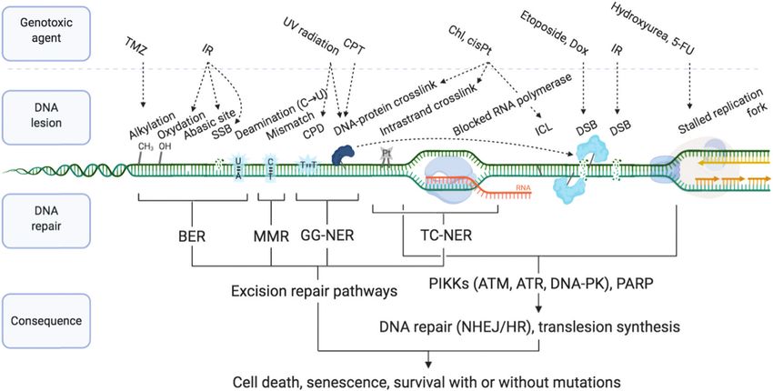
RSC Chemical Biology REVIEW
View Article Online

                                                                                                                Review                                                                                                            RSC Chemical Biology
            This article is licensed under a Creative Commons Attribution-NonCommercial 3.0 Unported Licence.
Open Access Article. Published on 30 September 2020. Downloaded on 04/19/2021 08:23:18.

                                                                                                                Fig. 3 Single-strand breaks (SSB) and double-strand breaks (DSB) repair pathways. Dotted arrows indicate that PARP1 can be activated by DSB and
                                                                                                                stimulate DSB repair by both HR and NHEJ.33,109 Adapted from ref. 37, created with BioRender.

                                                                                                                NHEJ occurs throughout the cell cycle and is the most prevalent          (S) phase, when two sister chromatids are present, and enables
                                                                                                                DSB repair mechanism in non-dividing cells.                              faithful DNA replication (Section 2b).
                                                                                                                    HR: HR is initiated by the generation of a 3 0 ended ssDNA
                                                                                                                extension produced by exonucleolytic processing of the DNA                  2a3. Interstrand cross-links (ICLs) repair. ICLs are covalent
                                                                                                                ends. This process, called DNA end resection, is controlled by           linkages between two different DNA strands, as opposed to
                                                                                                                the nuclease activity of MRE11 (Fig. 3). MRE11, as part of the           intrastrand cross-links that occur on the same strand and
                                                                                                                MRE11–RAD50–NBS1 (MRN complex) associates to the chro-                   which are primarily repaired by NER. ICLs are detected and
                                                                                                                matin flanking each DSB, through ATP-dependent RAD50 DNA                 repaired during DNA replication by the Fanconi Anemia (FA)
                                                                                                                clamping activity. CtIP (C-terminal binding protein 1 (CtBP)-            pathway, named after the disease resulting from genetic muta-
                                                                                                                interacting protein, also known as retinoblastoma-binding                tions of key components of this pathway. The FA pathway
                                                                                                                protein 8, RBBP8), an interaction partner of MRN, controls               requires activation by ATR kinase, a master mediator of DDR
                                                                                                                MRE11 endonuclease activity which is stimulated when CtIP is             signal transduction (see Section 2b), which simultaneously
                                                                                                                phosphorylated by CDKs and ATM/ATR (see Section 2b).95 This              reduces replication fork speed.99–101 Currently, 22 genes have
                                                                                                                triggers the formation of a nick which is the initiation site for        been described as FA genes, some of them being implicated in
                                                                                                                bidirectional resection performed in the 3 0 –5 0 direction by the       other DNA repair mechanisms: FANC-A, -B, -C, -D1 (BRCA2),
                                                                                                                exonuclease activity of MRE11 and in the 5 0 –3 0 direction by the       -D2, -E, -F, -G (XRCC9), -I, -J (BRIP1), -L (PHF9), -M, -N (PALB2),
                                                                                                                exonuclease activity of EXO1 and/or DNA2 in complex with                 -O (RAD51C), -P (SLX4), -Q (ERCC4/XPF), -R (RAD51), -S
                                                                                                                the Bloom syndrome helicase (BLM). Following end resection,              (BRCA1), -T (UBE2T), -U (XRCC2), -V (REV7/MAD2L2) and -W
                                                                                                                the ssDNA is stabilised by RPA, which comprises the hetero-              (RFWD3).102 The FANC-A, -D, -C, -E, -F, -G, -L and -M proteins
                                                                                                                trimer RPA1/2/3 known to rapidly bind to and stabilise ssDNA             form the core complex which monoubiquitinates FANCD2/
                                                                                                                via its oligonucleotide/oligosaccharide-binding fold (OB-fold)           FANCI upon ICL recognition, while the other proteins mediate
                                                                                                                domains during DNA replication and repair96 (Fig. 3). RPA is             the lesion incision or ‘‘unhooking’’ forming a DSB whose repair
                                                                                                                replaced by Rad51 (homologue of bacterial recombination                  involves components of translesion synthesis (TLS, DNA poly-
                                                                                                                protein A, recA), responsible for scanning the sister chromatid for      merase k) and HR pathways. The FA complex recognises the
                                                                                                                the homologous strand, aided by BRCA2 and BRCA1 in association           ICL, often at the collision site of one or two stalled replication
                                                                                                                with BRCA1-associated RING domain 1 (BARD1).97,98 Homologous             forks, leading to incision by endonucleases (the structure-
                                                                                                                strand invasion requires stabilisation of the non-template sister        specific endonucleases SLX1, MUS81 (also known as SLX3),
                                                                                                                strand by Rad51, whilst DNA Pold fills in the invading strand using      XPF and ERCC1) in one strand on either side of the ICL thus
                                                                                                                the template strand (Fig. 3). HR occurs during and after synthesis       ‘‘unhooking’’ the ICL lesion and forming a DSB. Replicative

                                                                                                                52 | RSC Chem. Biol., 2021, 2, 4776                                           2021 The Author(s). Published by the Royal Society of Chemistry
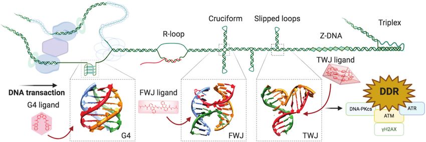
RSC Chemical Biology REVIEW
View Article Online

                                                                                                                RSC Chemical Biology                                                                                                                              Review

                                                                                                                polymerases act on the intact strand to fill the template whilst               The role of PARP3 in DNA repair is more poorly understood,
                                                                                                                retaining the incised strand in proximity. Finally the HR machin-              although has been shown to have more protein PARylation
                                                                                                                ery repairs the DSB in the incised strand from the template                    targets than PARP1 and 2 with minor overlap.110 PARP3 has a
                                                                                                                double strand.103 ICLs can also be repaired by a replication-                  different DNA-binding domain, it is described to recognise DSB
            This article is licensed under a Creative Commons Attribution-NonCommercial 3.0 Unported Licence.

                                                                                                                independent mechanism involving incisions on the ICL flanks                    only111 and it has been shown to promote the recruitment of
                                                                                                                by the NER machinery and lesion bypass by Polk.99,104,105                      the NHEJ ligation machinery, the XRCC4/Lig4 complex.112 Of
                                                                                                                   2b. The DDR mediators. The key mediators in the repair of                   interest, PARP3 was found to promote chromosome rearrange-
                                                                                                                DNA lesions resulting from SSB, DSB and arrested transcription                 ments and its genetic knockout in lung adenocarcinoma cells
                                                                                                                and replication forks are the poly(ADP-ribose) polymerases                     showed extreme sensitivity to small molecules which induce
                                                                                                                (PARPs) and the phosphatidylinositol 3-kinase-related kinases                  DSB113 or stabilise the secondary DNA structure G-quadruplexes
Open Access Article. Published on 30 September 2020. Downloaded on 04/19/2021 08:23:18.

                                                                                                                (PIKKs, i.e. DNA-PK, ATM and ATR, Fig. 3).                                     (G4).114
                                                                                                                   PARP1 and PARP2 (the latter accounting for 5–10% of total                      The PIKKs coordinate the central alarm system to DSB and
                                                                                                                PARP activity)106 are considered a major first line of defence in              stalled forks, often during NHEJ/HR (see Section 2a2), through
                                                                                                                the DDR response.4,33,37 PARP activation, PARylation (synthesis                activation by (auto-)phosphorylation and recruitment of a vast
                                                                                                                of branched and linear chains of poly(ADP-ribose) (PAR) on                     range of signalling molecules.33,115 These signal transducers
                                                                                                                proteins) and auto-PARylation (Fig. 4a) occurs rapidly at sites of             have significant structure and function similarity, however are
                                                                                                                SSB, DSB and stalled replication forks.80,107 PAR attachments                  activated by distinct sensor proteins which recognise the DNA
                                                                                                                on PARP and on nearby histones act as a platform promoting                     damage. PIKKs comprise two families of effectors, DNA depen-
                                                                                                                the recruitment of DDR factors including XRCC1/Lig3 and                        dent protein kinase (DNA-PK) on one hand, and ataxia-
                                                                                                                inducing chromatin remodelling.108 Importantly, PARP1 is also                  telangiectasia mutated (ATM) and ataxia-telangiectasia and
                                                                                                                activated by DSBs where it promotes the recruitment of NHEJ                    Rad3-related (ATR) on the other hand, which are recruited to
                                                                                                                and HR factors.33,109 Repair of SSB can either be processed in a               DNA damage sites through molecular interactions with analo-
                                                                                                                short- or long-patch DNA gap filling mechanism similar to that                 gous C-terminal motifs of Ku80, Nibrin (or Nijmegen) breakage
                                                                                                                of BER,107 mediated by nucleases such as FEN1 and DNA Pold,                    syndrome gene (NBS1) and ATR-interacting protein (ATRIP)
                                                                                                                the lagging strand polymerase capable of strand displacement.87                respectively (Fig. 3):37,116

                                                                                                                Fig. 4 (a) PARP recruitment to SSB and synthesis of poly(ADP-ribose) (PAR) to activate SSB repair. Inhibition of repair by PARP-trapping at the break site.
                                                                                                                (b) Examples of inhibitors (with their protein target) and the substrate they mimic (blue). (a) Created with BioRender.

                                                                                                                  2021 The Author(s). Published by the Royal Society of Chemistry                                                  RSC Chem. Biol., 2021, 2, 4776 | 53
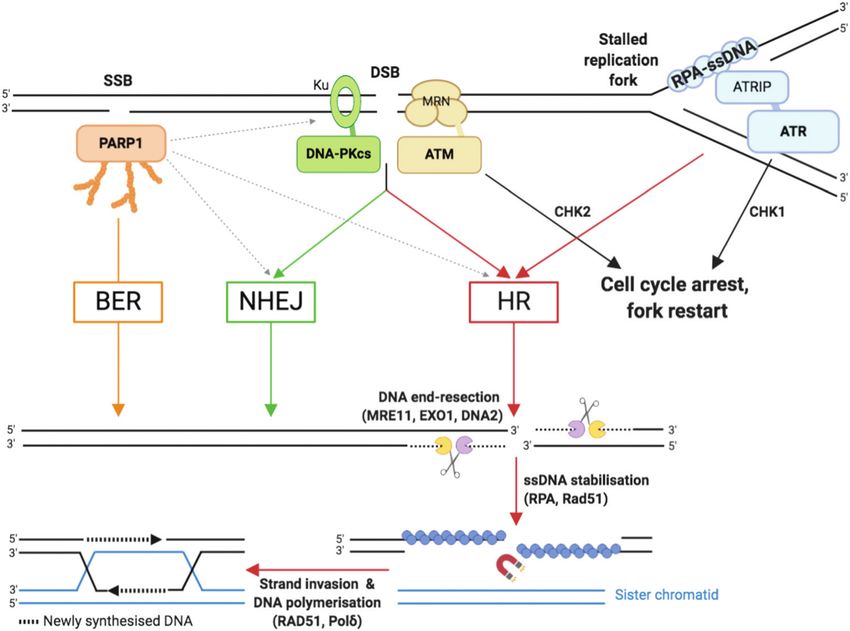
RSC Chemical Biology REVIEW
View Article Online

                                                                                                                Review                                                                                                       RSC Chemical Biology

                                                                                                                    – DNA-PKcs (also known as DNA dependent protein kinase          as XRCC4, XRCC4-like factor (XLF) and DNA-PKcs.134–137 ATM
                                                                                                                catalytic subunit) is involved in DSB repair by NHEJ. The Ku        also plays a crucial role in enforcing the cell cycle checkpoints
                                                                                                                heterodimer comprising Ku70/Ku80 (also known as X-ray repair        through activation by phosphorylation of CHK2, which in turn
                                                                                                                cross-complementing protein 6 and 5, XRCC6 and XRCC5,               activates p53, a key apoptosis regulator.68 53BP1 is recruited to
            This article is licensed under a Creative Commons Attribution-NonCommercial 3.0 Unported Licence.

                                                                                                                respectively) binds to free DSB ends (amongst other DNA             nucleosomes with specific histone modifications: Ring Finger
                                                                                                                structures) and recruits DNA-PKcs. The Ku–DNA-PKcs–DNA              Protein 168 (RNF168)-ubiquitylated H2AK15 and dimethylated
                                                                                                                complex forms the active DNA-PK holoenzyme which is a               H4K20;138 where it functions as a central determinant in the
                                                                                                                serine/threonine kinase able to phosphorylate several sub-          repair pathway choice made at DSB by promoting the recruit-
                                                                                                                strates including itself. Autophosphorylation of DNA-PKcs reg-      ment of the shieldin complex,139 thereby limiting DNA end
                                                                                                                ulates DNA end processing.                                          resection by BRCA1/BARD1, which exposes ssDNA to be
Open Access Article. Published on 30 September 2020. Downloaded on 04/19/2021 08:23:18.

                                                                                                                    – ATM, through a specific amplification loop, plays an apical   repaired by HR. As such 53BP1 inhibits HR and supports NHEJ
                                                                                                                role in DSB signalling and cell cycle checkpoint activation, in     repair in the G1-phase of the cell cycle.6,140 PARP3, in inter-
                                                                                                                addition to its function in promoting DSB repair via HR.6,71 The    action with Ku, similarly is described to favour DSB repair to
                                                                                                                MRN complex associates to the flank of each DSB through the         NHEJ by limiting end resection.113
                                                                                                                ATP-dependent RAD50 clamp which recognises the DNA                      – ATR is the key damage signalling mediator at replication
                                                                                                                damage. The MRN complex mediates the recruitment of ATM             forks and in dividing cells. ATR binds through ATRIP to the
                                                                                                                by direct interaction between ATM and the NBS1 C-terminus.117       RPA:ssDNA complex,141,142 which forms rapidly at DNA replica-
                                                                                                                However, in contrast to Ku whose recruitment is limited to one      tion fork blockages during the S phase of the cell cycle (Fig. 2
                                                                                                                or two Ku proteins per DSB end,118 multiple MRN–ATM com-            and 3) and stabilises ssDNA during DNA replication. Upon DNA
                                                                                                                plexes are recruited to chromatin thanks to an amplification        damage, ATR induces a signalling cascade, phosphorylating a
                                                                                                                loop relying on ATM kinase activity. Indeed, ATM is responsible     long list of cell cycle-related substrates, notably checkpoint
                                                                                                                for the phosphorylation of a large number of proteins including     kinase 1 (CHK1) and apoptosis and replication stress regulator
                                                                                                                the C-terminus of the histone variant H2AX, generating gH2AX,       p53.4,143 ATR activation stabilises the stalled fork (preventing
                                                                                                                the S139-phosphorylated form of H2AX. This initial phosphor-        fork collapse which can otherwise lead to DSB formation)144
                                                                                                                ylation recruits MDC1 (mediator of DNA damage checkpoint            and prevents proximal replication origin restart.142,145,146
                                                                                                                protein 1), via its BRCA1 C terminus (BRCT) repeat                  Stalled replication forks can be restarted by an HR-dependent
                                                                                                                domains,119–122 which is itself phosphorylated by casein kinase     mechanism. Unlike ATM and DNA-PKcs, ATR is necessary for
                                                                                                                2 (CK2). The phosphorylated form of MDC1 is recognised by the       cell growth and embryonic development,147,148 meaning
                                                                                                                forkhead-associated (FHA) and BRCT domains of NBS1 which            mechanistic studies of ATR were greatly facilitated by the recent
                                                                                                                drives the secondary recruitment of the MRN–ATM complex             development of ATR-inhibiting chemicals. Cross-talk exists
                                                                                                                that subsequently phosphorylates other more distant sub-            between the ATM and ATR kinases since ATR can activate
                                                                                                                strates including H2AX.123–126 This amplification loop leads        ATM in some instances,149 the two kinases having many
                                                                                                                the gH2AX signal to spread from kilo- to megabases from the         substrates in common and their cellular roles strongly over-
                                                                                                                DSB, making the detection of the gH2AX signal a sensitive and       lapping. However, most of the details regarding the intricate
                                                                                                                widespread approach to monitor the induction of DNA damage          genetic regulations in which ATM and/or ATR remain to be
                                                                                                                in cells (examples in Section 3c). The rapid spreading of gH2AX     discovered.
                                                                                                                from DSB sites is controlled by chromatin domain boundaries             2c. DNA damage checkpoints. Healthy cell cycle progres-
                                                                                                                delimited by DNA-binding cohesin protein complexes,127,128          sion is driven by the cyclin-dependent kinases (CDKs) and
                                                                                                                which also repress transcription of damaged genes.129 In addi-      CDK–cyclin complexes150 and exit is controlled by p53 and p21
                                                                                                                tion to NBS1, MDC1 recruits other proteins that govern cell         signalling.68,151 In addition to regulation by cell mass and
                                                                                                                cycle checkpoints, such as checkpoint kinase 2 (CHK2), and          proliferative signals, cell cycle progression can be interrupted
                                                                                                                mechanisms of DNA repair pathway choice including 53BP1             upon DNA damage at specific cell cycle boundaries, called the
                                                                                                                (promoting NHEJ) and BRCA1 (promoting HR).130,131 As well as        cell cycle checkpoints.152 Three DNA damage checkpoints exist:
                                                                                                                recruiting DNA repair proteins (MRN/ATM), gH2AX recruits            the G1/S, G2/M and intra-S phase checkpoints (Fig. 2). The G1/S
                                                                                                                chromatin remodelling machinery to aid damage repair.122,132        checkpoint blocks S-phase entry of cells carrying DNA damage,
                                                                                                                It is noteworthy that ATM can also be directly activated by         allowing repair and preventing the conversion of simple DNA
                                                                                                                oxidative stress via oxidation of some of its cysteines.71 In       lesions into more complex ones during the replication process.
                                                                                                                parallel to its signaling functions, ATM also stimulates HR         It is enforced by activation of the ATM–CHK2–p53 axis, which
                                                                                                                repair (Fig. 3) by phosphorylating several proteins including the   results in the accumulation of the CDK–cyclin inhibitor p21.153
                                                                                                                MRN interacting protein CtIP. CtIP phosphorylations promotes        As a result of its dependence on p53, this checkpoint is
                                                                                                                MRE11 endonuclease activity, responsible for nicking the            frequently lost in cancer cells, providing an explanation for
                                                                                                                side(s) of the DSB to initiate end resection. The ATM-              the high level of genomic instability in cancer cells. The intra-S
                                                                                                                dependent CtIP phosphorylations promote Ku eviction from            checkpoint relies on both ATM and ATR-CHK1154 and consists
                                                                                                                the DSB end, thereby directing DSB repair towards HR.133 In         of blocking the initiation of new replication origins once DNA
                                                                                                                addition, ATM stimulates HR by phosphorylating several HR           damage is detected. Activation of the intra-S phase checkpoint
                                                                                                                factors, such as BRCA2, EXO1 and BLM, and NHEJ factors such         delays the progression through S-phase although it does not

                                                                                                                54 | RSC Chem. Biol., 2021, 2, 4776                                      2021 The Author(s). Published by the Royal Society of Chemistry
RSC Chemical Biology REVIEW
View Article Online

                                                                                                                RSC Chemical Biology                                                                                                               Review

                                                                                                                inhibit it completely. Finally, the G2/M checkpoint relies on the   increasing the combined tolerated dose and decreasing the
                                                                                                                activation of ATM-CHK2 and ATR-CHK1 and prevents damaged            effective dose.8 The first-in-class example of a synthetic lethality
                                                                                                                cells from initiating mitosis.155 Progression through mitosis       strategy applied to cancer is the PARP inhibitor olaparib
                                                                                                                could result in generating two daughter cells with incomplete       (Fig. 4b),8,108 which was approved for treatment of several
            This article is licensed under a Creative Commons Attribution-NonCommercial 3.0 Unported Licence.

                                                                                                                or abnormal genomes or in failure to partition DNA leading to       BRCA-deficient cancer types by the US Food and Drug Admin-
                                                                                                                mitotic catastrophe, a type of cell death that is frequently        istration (FDA) and European Medicines Agency (EMA) in 2014.
                                                                                                                observed when treating cancer cells with DNA damaging               More recently (in 2018), another PARP inhibitor talazoparib was
                                                                                                                agents. Key enforcers of the DNA damage checkpoints are the         approved as it displays a higher cytotoxicity in BRCA-deficient
                                                                                                                CHK2 and CHK1 serine/threonine kinases that are activated by        tumours, likely due to its stronger PARP–DNA trapping affinities
                                                                                                                ATM and ATR respectively, and can phosphorylate various             (Fig. 4a).166 PARP inhibitors have proved efficacious in multiple
Open Access Article. Published on 30 September 2020. Downloaded on 04/19/2021 08:23:18.

                                                                                                                substrates,141 including the cell division cycle 25 (CDC25)         cancers with deficient DDR pathways, as well as in maintenance
                                                                                                                phosphatases A, B and C. CDC25 dual-specificity phosphatases        therapy for cancers which have previously responded to antipro-
                                                                                                                normally remove inhibitory phosphorylations of the                  liferative agents.5,160 However, various resistance mechanisms to
                                                                                                                CDKs,156,157 a rate-limiting step in CDK activation which allows    PARP inhibitors have been described, including via the restora-
                                                                                                                cell cycle progression.158 Once phosphorylated by CHK1/CHK2,        tion of HR, for example via inactivation of 53BP1, or by increased
                                                                                                                CDC25 phosphatases are degraded, inactivated or sequestered         replication fork stability, for example via inactivation of the
                                                                                                                into the cytoplasm by association with the 14-3-3 proteins,         junction endonuclease MUS81 or the N-methyltransferase enhan-
                                                                                                                thereby blocking cell cycle progression. Considering that can-      cer of zeste homolog 2 (EZH2).109,166,167
                                                                                                                cer cells display high genomic instability, due to increased            Other DDR inhibitors (see Section 2c) have shown potential
                                                                                                                levels of replication, mitosis and metabolism and/or dysregula-     in the clinic, including inhibitors of specific DNA repair path-
                                                                                                                tion of the DDR machinery, it has been envisioned that they         ways such as DNA-PK inhibitors (M3814 and CC-115, currently
                                                                                                                should be more dependent on DNA damage checkpoints than             in phase I trials168,169) which block NHEJ-dependent DSB
                                                                                                                normal cells. This idea led to a race to develop DNA damage         repair, and MGMT inhibitors (lomeguatrib, Fig. 4b) which
                                                                                                                checkpoint inhibitors, which could induce selective cell death      block the removal of alkylated bases. Also in clinical testing
                                                                                                                of cancer cells as a standalone treatment, or in combination        are the inhibitors of cell cycle checkpoint mediators ATR, ATM,
                                                                                                                could increase the selectivity and activity of current DNA          WEE1 and CHK1, such as M6620, M3541, AZD1775, LY2606368
                                                                                                                damaging anticancer treatments (see Sections 1 and 2d).4,159        respectively, for both single-agent and combination therapies.8,170
                                                                                                                   2d. Small molecule targeting of DDR. Impairments in the          Despite promising, the therapeutic indexes of these molecules
                                                                                                                mechanisms responsible for the surveillance and repair of           (toxic dose/therapeutic dose) are often limiting, as demonstrated
                                                                                                                genetic material throughout cell division are unsurprisingly        in the cases of lomeguatrib160 and UCN-01 (first specific CHK1
                                                                                                                causative of oncogenetic dysregulations. Because cancer cells       inhibitor developed from the broad-spectrum kinase inhibitor
                                                                                                                are often DDR-deficient, current strategies aim at fostering the    staurosporine155,171). PARP inhibitors had initially been trialled in
                                                                                                                genetic instability of cancers by further deactivating these        combination with DNA damaging agent temozolomide and failed
                                                                                                                surveillance and repair systems.5,8,160 There are in fact several   due to excessive toxicity, but following these studies, its synthetic
                                                                                                                rationales justifying the use of DDR-disruptive agents in cancer    lethality properties in BRCA-deficient cancers were uncovered.160
                                                                                                                treatment. Small molecule inhibitors of DNA repair proteins are     This exemplifies the advantage of synthetic lethality strategies, in
                                                                                                                able to increase the level of DNA damage in cancer cells (see       which a single agent can be applied without incurring unacceptable
                                                                                                                below), whereas inhibitors of checkpoint regulators, such as        collateral damage, by taking advantage of a known weakness in the
                                                                                                                WEE1,155 a crucial regulator of the G2/M checkpoint which           patient’s cancer. With the aid of modern high-throughput and
                                                                                                                stops cells entering into mitosis on activation by CHK1, func-      genome-wide sequencing techniques,172 the body of knowledge on
                                                                                                                tion by metaphorically speeding up the cell cycle to an             genetic biomarkers is being established for each cancer type,
                                                                                                                uncontrollable speed, causing it to ‘derail’ under the pressure     opening up the development of specific small molecules suited
                                                                                                                of unsustainable genetic aberrations.161 DDR inhibition can be      to personalised cancer therapy.4,162 Thus, our understanding of the
                                                                                                                used in combination with a DNA damaging agent, or crucially         cancer genotypes that are likely to be sensitive to selected treatment
                                                                                                                can be used in cancers which are already deficient in a DDR         is growing.
                                                                                                                pathway, a strategy known as synthetic lethality.                       A common trend in the design of small molecule inhibitors is
                                                                                                                   Synthetic lethality describes the relationship between two       to structurally mimic the biological substrate of the target protein:
                                                                                                                gene deactivations which cause cell lethality when both genes       O6-benzylguanine and lomeguatrib (O6-(4-bromothenyl)guanine)
                                                                                                                are silenced (through endogenous mutation, exogenous inhibi-        both mimic O6-methylated guanine (O6-MeG), the DNA base lesion
                                                                                                                tion or knockdown) but cells are viable when only one is            repaired by MGMT (O6-methylguanine–DNA methyltransferase,
                                                                                                                deactivated.7,162–165 As discussed above, DDR pathways func-        Fig. 4b); olaparib mimics NAD+, the building block used by PARP
                                                                                                                tion with significant redundancy, yet with one pathway miss-        for PARylation signalling (Fig. 4a); and palbociclib fits into the ATP-
                                                                                                                ing, cancer cells become more dependent on the remaining            pocket of checkpoint kinase CDK4/6173 (Fig. 4b), whereas other
                                                                                                                pathways. A myriad of clinical trials are in place to assess the    kinase inhibitors can mimic the target protein. Some DDR inhibi-
                                                                                                                benefit of inhibiting multiple DDR pathways concomitantly,          tors, such as NU6027174 and UCN-01175 (ATR and CHK1 inhibitors
                                                                                                                with the hope of being able to widen the therapeutic window by      respectively, Fig. 4b), were initially designed to target CDKs, and the

                                                                                                                  2021 The Author(s). Published by the Royal Society of Chemistry                                     RSC Chem. Biol., 2021, 2, 4776 | 55
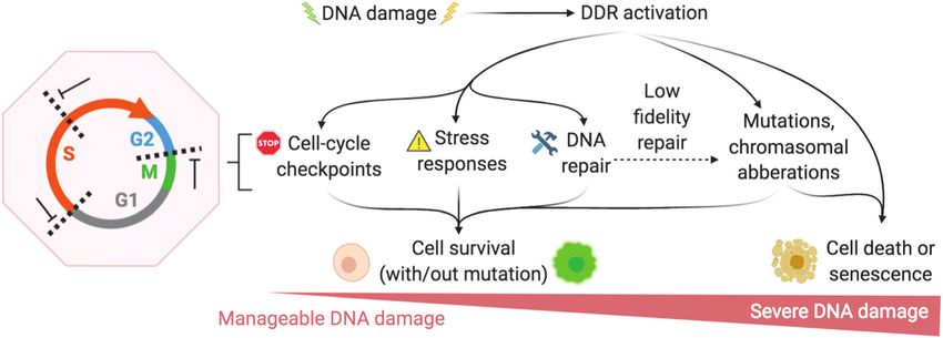
RSC Chemical Biology REVIEW
View Article Online

                                                                                                                Review                                                                                                                         RSC Chemical Biology

                                                                                                                marginal specificity for DDR checkpoint kinases over another kinase             guanine-rich ssDNA displays the motif GZ3NxGZ3NxGZ3NxGZ3
                                                                                                                was developed by often lengthy medicinal chemistry methods: trial-              (where G is guanine, and N any intervening nucleobase, and
                                                                                                                and-error synthesis, structure–activity relationship (SAR) studies and          x ranges from 1 to 420), the sequence can give rise to a G4
                                                                                                                enzyme assays, since many active site crystal structures of these               (Fig. 5).179,180 The basic building block of G4s is a G-quartet181
            This article is licensed under a Creative Commons Attribution-NonCommercial 3.0 Unported Licence.

                                                                                                                targets have not yet been resolved.                                             resulting from the self-association of 4 Gs in a square planar
                                                                                                                    In the next chapter, we discuss another approach, which could               arrangement, and was first reported in vitro in 1962.182 Contiguous
                                                                                                                bypass certain toxic and resistance-incurring pitfalls of DDR, or               G-stretches within a strand of ssDNA come together into contiguous
                                                                                                                prove synergistic. Inspired by a strategy at the very foundation of             planar G-quartets, which stack on top of each other through
                                                                                                                chemotherapy, we look again to triggering the DDR by drugs                      p-system interactions, with each G-quartet being stabilised by a
                                                                                                                targeting the DNA itself. Accumulating evidence now supports the                central physiological cation (K+, Na+) to form a G4 structure
Open Access Article. Published on 30 September 2020. Downloaded on 04/19/2021 08:23:18.

                                                                                                                existence of non-B DNA structures in cells that fold as a result of             that can be classified as antiparallel-, hybrid- or parallel-type G4,
                                                                                                                cellular activity. The resulting higher-order structures are closer to          depending on the polarity of the strands (Fig. 5). Of note, this
                                                                                                                protein than B DNA in terms of 3D structure and offer more                       topological diversity leads to a variety of intervening loops, which can
                                                                                                                structurally defined binding sites for small molecules (e.g. the                be diagonal, lateral or reversal loops (Fig. 5). Initially only thought of
                                                                                                                accessible G-quartets of G-quadruplexes, the central cavity of DNA              as an in vitro oddity, G4s are now considered key players in cellular
                                                                                                                junctions, see Sections 3a and 4a) than major/minor groove inter-               processes: recent sequencing-based methods have demonstrated
                                                                                                                action and/or intercalation in between two successive base pairs of B           that thousands of quadruplex-forming sequences (QFS) are present
                                                                                                                DNA. This offers the possibility of targeting higher-order nucleic acid          in our genome, 4700 000 by G4-seq.183 These sequences are pre-
                                                                                                                structures with a better degree of selectivity, thus re-establishing            dominantly maintained in an unfolded state, as exemplified by the
                                                                                                                DNA in its many forms as a promising chemotherapeutic target with               detection in live cells of only 410 000 G4s by G4 ChIP-seq184 and ca.
                                                                                                                unique and structurally defined small molecule binding sites. In the            3000 G4s by the fluorophore SiR-PyPDS (see Section 3c),185 with a
                                                                                                                following sections, we will describe the new trend that is emerging             strong correlation between individual G4 formation and the
                                                                                                                based on the stabilisation of non-B DNA structures with specifically            transcriptional activity of the gene they fold from. This tran-
                                                                                                                designed small molecules (ligands), as a way to damage cancer cells’            siency originates from the various mechanisms the cell has
                                                                                                                DNA in a more specific manner.                                                  evolved to regulate G4 formation, among which the G4 helicases
                                                                                                                                                                                                are being actively studied (see Section 3b). Interestingly, the QFS
                                                                                                                3. G-Quadruplexes (G4s) as targets to induce DNA damage                         distribution is not random as they are significantly enriched in
                                                                                                                  3a. The prevalence of G4 in the human genome. The non-B                       key regulatory regions including gene promoters, replication
                                                                                                                DNA structure which has been most studied for triggering                        origins and telomeres.10,186
                                                                                                                DNA damage is undoubtedly the four-stranded structure                              G4s in gene promoters: the occurrence of QFS in gene
                                                                                                                named G-quadruplex-DNA, or G4-DNA. If a transiently formed                      promoters (defined as 1 kb upstream of the transcription start

                                                                                                                Fig. 5 Schematic representation of a G-rich sequence that folds into a G4 structure (upper panel), highlighting the structure of a guanine (G, upper
                                                                                                                panel, left) and a G-quartet (right). Topological diversity of G4s that can adopt parallel, hybrid and antiparallel conformation (lower panel, arrows indicate
                                                                                                                the polarity of the DNA strands), as elucidated by either NMR (PDB IDs 143D176 and 2GKU177) or X-ray structure analysis (PDB ID 1KF1178).

                                                                                                                56 | RSC Chem. Biol., 2021, 2, 4776                                                    2021 The Author(s). Published by the Royal Society of Chemistry
View Article Online

                                                                                                                RSC Chemical Biology                                                                                                                     Review

                                                                                                                site, TSS) is significantly high in mammals (42000),187–189               represents a constant impediment to the advancing transcription/
                                                                                                                inferring a regulatory role of G4s in gene expression.190 The             replication complexes along the DNA. Cells have evolved an
                                                                                                                genesis of G4 structures is linked to a high transcriptional              enzymatic machinery to tackle this threat, the helicases, some of
                                                                                                                activity, likely due to physical requirements for the DNA duplex          which have now been shown to interact with and resolve G4s.209,210
            This article is licensed under a Creative Commons Attribution-NonCommercial 3.0 Unported Licence.

                                                                                                                to open and for chromatin structure to be relaxed (euchroma-              Helicases are increasingly being studied for their role in
                                                                                                                tin, as opposed to tightly-bound heterochromatin) thus allow-             diseases,211,212 notably because their genetic silencing is involved
                                                                                                                ing G4s to fold, but this link also indicates a possible causative        in severe dysregulations that confer premature ageing and cancer
                                                                                                                role of G4s in the recruitment of transcription factors to                susceptibility. To date, 95 different helicases are known, 31 for DNA
                                                                                                                euchromatin.184 The observation that G4 sequences are signifi-            and 64 for RNA.211 The two most studied families are (1) the RecQ
                                                                                                                cantly enriched in oncogenes and regions predisposed to                   helicases, including Bloom (BLM) and Werner (WRN) helicases,
Open Access Article. Published on 30 September 2020. Downloaded on 04/19/2021 08:23:18.

                                                                                                                amplification in cancers explains and warrants the active                 along with RecQ-like helicases 1, 4 and 5 (RECQL1, RECQL4 and
                                                                                                                search for chemicals (small molecule ligands) that can interact           RECQL5, respectively), and (2) the iron–sulfur (Fe–S) helicases,
                                                                                                                specifically with promoter G4s,191 in order to gain control of            including XPD, Fanconi anaemia complementation group J (FANCJ,
                                                                                                                processes underlying cancer onset and progression. The text-              or BRIP1), regulator of telomere length helicase 1 (RTEL1) and
                                                                                                                book example is c-MYC, a transcription factor and proto-oncogene          DEAD/H-Box helicase 11 (DDX11).211,213
                                                                                                                whose protein was considered undruggable owing to its lack of                 These enzymes are deeply involved in genome surveillance
                                                                                                                catalytic activity, but for which gene expression can be down-            and DNA damage repair (DDR). They are known to resolve
                                                                                                                regulated by ligand-mediated stabilisation of its G4-containing           higher-order DNA structures formed during DNA transactions
                                                                                                                promoter region.43                                                        (e.g. the 5 0 flap intermediate resulting from the processing of
                                                                                                                     G4s in replication origins: in homeostatic conditions, DNA           Okazaki’s fragments during replication, or the DNA:RNA
                                                                                                                replication origins are regulated by two triggers, transcription          hybrids referred to as R-loops formed during transcription)
                                                                                                                and G4 formation.192 Genome mapping showed a correlation                  and structures formed during DDR (e.g. the four-way junction
                                                                                                                between replication origins and G4s in metazoans: 470% of                 known as the Holliday junction that is the central intermediate
                                                                                                                initiation sequences are followed by a QFS, also known as                 of HR). Some examples have already been discussed above:
                                                                                                                Origin G-rich Repeated Element (OGRE).193 Furthermore, a                  BLM is involved in HR-mediated DSB repair (see Section 2b);
                                                                                                                genome-wide CRISPR-mediated gene editing study showed that                XPD mediates NER when embedded in the ternary complex
                                                                                                                by inserting or deleting QFSs, replication origin activity could          TFIIH (see Section 2a1). Of particular interest here is the
                                                                                                                be increased or decreased respectively,194 demonstrating one of           prevalence of reports that helicases are inhibited by G4 ligands,
                                                                                                                the multiple regulatory roles of quadruplexes in healthy divid-           likely indirectly through DNA blockages (Table 1).
                                                                                                                ing cells. However, the detailed mechanism by which G4s act at                More recently, certain helicases have been shown to unwind G4s
                                                                                                                replication origins is not yet fully understood.                          in vitro and evidence is now accumulating that they do so in vivo
                                                                                                                     G4s in telomeres: mammalian telomeres are formed by                  (Table 1).212,214 For instance, BLM localises at telomeres where it
                                                                                                                                                        0           0 0          0
                                                                                                                thousands of non-coding repeats (5 TTAGGG3 /5 CCCTAA3 ) and               allows for proper replication to occur, resolving telomeric G4, an
                                                                                                                a single-stranded G-rich 3 0 -tail, known as the 3 0 overhang.195,196     action that is retarded by G4 ligands (PhenDC3).240 Similarly, BLM-
                                                                                                                                                                        0          0
                                                                                                                The telomeric G4 that folds from the G-rich (5 TTAGGG3 ) 3 0 -            deficiency leads to genomic instability caused by elevated levels of
                                                                                                                overhang is undoubtedly the most intensively studied G4 structure,        recombination by sister chromatid exchange (SCE) events at G4 sites
                                                                                                                given that it was the first discovered, and that its 3 TTA loops are      in transcribed regions of the genome.241 WRN is also found at
                                                                                                                flexible enough to give rise to different G4 topologies, as exemplified    telomeres where it resolves telomeric G4 to repress chromosomal
                                                                                                                by the 3 structures seen in Fig. 5 (lower panel), formed in either Na+-   instability, and interact with the shelterin complex to regulate T-loop
                                                                                                                (PDB ID 143D176) or K+-rich conditions (PDD IDs 2GKU177 and               formation.242 A pioneering study conducted with Dog-1, the
                                                                                                                1KF1178). Telomeres act as a cap at the end of chromosomes,               Caenorhabditis elegans homologue of FANCJ, showed that deletions
                                                                                                                protecting them from enzymatic degradation. Repeated units are            in Dog-1 helicase promotes genetic instability by allowing G4s
                                                                                                                removed after each round of DNA replication and cell division (the        to act as replication barriers.243 In eukaryotic cells, FANCJ-
                                                                                                                so-called ‘end replication problem’),197,198 making telomeric regions     knockdown (FANCJKD) cell growth is strongly inhibited upon
                                                                                                                the ‘mitotic clock’ of the cell, limiting the number of cell cycle        treatment with G4 ligand telomestatin (TMS).235 Similarly,
                                                                                                                divisions before the onset of senescence.196 Telomeres are thus           FANCJ-null cells show replication fork slowing upon TMS treat-
                                                                                                                implicated in both cancer and the ageing process.199,200 It is largely    ment.244 Genome-wide analysis performed with S. cerevisiae by
                                                                                                                accepted that G4s form in telomere ends,201,202 and the earliest          Pif1-ChIP-qPCR showed an enrichment of G4 motifs at Pif1-
                                                                                                                evidence of biological G4 formation was obtained at telomeres. Even       binding sites, which are responsible for stalling replication forks
                                                                                                                if telomeric G4s can either protect telomeres against exonucleases203     and are prone to DNA breakage in Pif1-deficient cells.245 This was
                                                                                                                or jeopardise telomere organisation by preventing telomeric loop (T-      further substantiated by the demonstration that Pif1-deficient
                                                                                                                loop) formation and telomerase recognition,204–207 they have been         S. cerevisiae displayed enhanced genetic instability triggered by G4
                                                                                                                mostly studied as targets for fostering chromosomal fragility in          motifs of different (and controllable) stability, and chemical G4
                                                                                                                cancers.208                                                               stabilisation via G4 ligand treatment (PhenDC3).246
                                                                                                                    3b. G4 unwinding enzymes – G4 helicases. Given the wide-                  Beyond their biological roles, G4-helicases have been imple-
                                                                                                                spread distribution of QFSs in our genome, the formation of G4s           mented as biomolecular tools to study and/or modulate G4

                                                                                                                  2021 The Author(s). Published by the Royal Society of Chemistry                                           RSC Chem. Biol., 2021, 2, 4776 | 57
You can also read