DNA damage repair: historical perspectives, mechanistic pathways and clinical translation for targeted cancer therapy - Nature
←
→
Page content transcription
If your browser does not render page correctly, please read the page content below
Signal Transduction and Targeted Therapy                                                                                              www.nature.com/sigtrans

                  REVIEW ARTICLE                        OPEN

                  DNA damage repair: historical perspectives, mechanistic
                  pathways and clinical translation for targeted cancer therapy
                  Ruixue Huang1 and Ping-Kun Zhou               2

                     Genomic instability is the hallmark of various cancers with the increasing accumulation of DNA damage. The application of
                     radiotherapy and chemotherapy in cancer treatment is typically based on this property of cancers. However, the adverse effects
                     including normal tissues injury are also accompanied by the radiotherapy and chemotherapy. Targeted cancer therapy has the
                     potential to suppress cancer cells’ DNA damage response through tailoring therapy to cancer patients lacking specific DNA damage
                     response functions. Obviously, understanding the broader role of DNA damage repair in cancers has became a basic and attractive
                     strategy for targeted cancer therapy, in particular, raising novel hypothesis or theory in this field on the basis of previous scientists’
                     findings would be important for future promising druggable emerging targets. In this review, we first illustrate the timeline steps for
                     the understanding the roles of DNA damage repair in the promotion of cancer and cancer therapy developed, then we summarize
                     the mechanisms regarding DNA damage repair associated with targeted cancer therapy, highlighting the specific proteins behind
                     targeting DNA damage repair that initiate functioning abnormally duo to extrinsic harm by environmental DNA damage factors,
                     also, the DNA damage baseline drift leads to the harmful intrinsic targeted cancer therapy. In addition, clinical therapeutic drugs for
1234567890();,:

                     DNA damage and repair including therapeutic effects, as well as the strategy and scheme of relative clinical trials were intensive
                     discussed. Based on this background, we suggest two hypotheses, namely “environmental gear selection” to describe DNA damage
                     repair pathway evolution, and “DNA damage baseline drift”, which may play a magnified role in mediating repair during cancer
                     treatment. This two new hypothesis would shed new light on targeted cancer therapy, provide a much better or more
                     comprehensive holistic view and also promote the development of new research direction and new overcoming strategies for
                     patients.

                     Signal Transduction and Targeted Therapy (2021)6:254                                ; https://doi.org/10.1038/s41392-021-00648-7

                  INTRODUCTION                                                                        explored and found to be associated with DNA sequences and
                  The Journey of DNA repair machinery system discovery                                genomic profiles.3–6 In Fig. 1, we illustrate the brief history of the
                  In recent years, the availability of high-quality data on DNA from                  DNA and DNA damage repair discovery journey. Some important
                  in vivo and in vitro research reported in the literature, as well as                moments and scientists should be noted for their groundbreaking
                  international conferences, funding, and collaboration among                         contributions to this journey. In 1927, the landmark discovery of
                  scientific communities has increased. In addition, new technolo-                     gene mutation induced by X-ray was claimed by Gager and
                  gies related to translational research, targets for clinical therapy,               Blakeslee, Miller.7One year later, the genetic transformation of
                  and expertize among scientists have developed rapidly, indicating                   bacteria was reported by Frederick Griffith.8 In 1946, Hermann J.
                  that DNA damage repair and genomic stability research has                           Muller was awarded the Nobel Prize in Physiology or Medicine for
                  entered a new era after a century of steady progress. This field                     his contribution in discovery of genetic mutations in fruit flies and
                  continuous to offer unprecedented opportunities for exploring                       revealed that higher the dose of X-ray and other ionizing radiation
                  further the secret of our genomic DNA structure integrity and                       exposed, the greater the number mutations that occurred.9,10 In
                  function harmonization while also promoting clinical disease                        1944, Oswald Avery, Colin MacLeod and Maclyn McCarty provided
                  prevention and therapeutic options, especially boosting the                         robust evidence demonstrating that DNA was our genetic
                  precise cancer therapy. The secret veil of DNA was uncovered                        material.11 Shortly thereafter, Watson and Crick published the
                  70 years ago, since the famous “Photo 51”was published1 along                       structure of DNA and announced that “we have discovered the
                  with the ground breaking report entitled “Molecular Structure of                    secret of life” in 19532. In 1964, the keyword “DNA repair” was
                  Nucleic Acids: A Structure for Deoxyribose Nucleic Acid” by James                   formally introduced with the discovery of “Dark Repair” and photo
                  Watson and Francis Crick2 in 1953. In this landmark study, DNA                      reactivating “repair-replication” of UV light-induced E. Coli DNA
                  was illustrated as a double helix, resembling a ladder twisted                      injury by excision of damaged areas containing thymine
                  along its length.. Over the following decades, many distinct                        dimers.12–15Since then, the DNA damage repair research rapidly
                  biological topics such as DNA damage repair, genomic instability,                   spreads into the area of photobiology, radiobiology and cancer
                  cancer therapy and control of genetic diseases have been                            biology, etc. We understand presently that the term of “DNA

                  1
                   Department of Occupational and Environmental Health, Xiangya School of Public Health, Central South University, Changsha, Hunan, China and 2Department of Radiation
                  Biology, Beijing Key Laboratory for Radiobiology, Beijing Institute of Radiation Medicine, AMMS, Beijing, China
                  Correspondence: Ping-Kun Zhou (zhoupk@bmi.ac.cn)

                  Received: 24 January 2021 Revised: 28 April 2021 Accepted: 13 May 2021

                  © The Author(s) 2021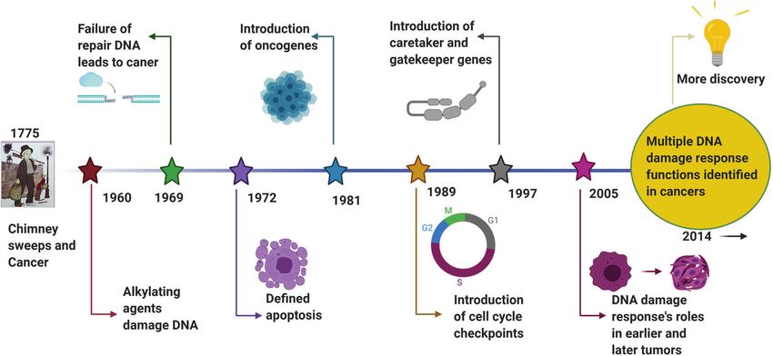
DNA damage repair: historical perspectives, mechanistic pathways and. . .
                                                           Huang and Zhou
2

    Fig. 1   The journey of DNA discovery

    repair” defines the biochemical and molecular biological pro-                and diversity of diseases. Future efforts are needed to elucidate
    cesses of DNA damage removing and genomic integrity restoring,              further the secrets of life using DNA and to introduce novel
    which including DNA damage sensing and signaling, repair                    concepts and hypotheses as powerful and compelling as the
    machinery proteins recruited onto the damage sites, functioning             discovery of the DNA double helix and DNA repair-based genomic
    and released step by step to restore the genomic integrity. The             integrity maintaining mechanisms.22–25
    first evidence of the direct association between DNA repair
    deficiency and human disease and cancer predisposition was                   A historical perspective of linking the genomic distortion with
    demonstrated in Xeroderma pigmentosum.16,17 In 2015, the Nobel              cancer
    Prize in Chemistry has been awarded to Tomas Lindahl, Paul                  The origin of cancer medicine is associated with a clinical
    Modrich and Aziz Sancar for their pioneering and fundamental                discovery based on medical record analysis and an epidemiolo-
    contributions of mapping, at a molecular level, how cells repair            gical survey. Percival Pott, known as the father of epidemiology,
    damaged DNA and safeguard the genomic stability. Their work                 observed a high prevalence of scrotal cancer among the boys who
    has provided fundamental knowledge of how a living cell                     were employed as chimney sweeps, and attributed the cancer to
    functions and is, especially, used for the development of new               soot exposure.26 This was the first evidence of occupational
    cancer treatments.                                                          exposure to hazard factors associated with cancer development.
       Now, it is well known that mammalian cells have evolved                  Prior to the discovery of the structure of DNA, Dr. Theodor Boveri
    multiple and diverse machineries for repairing every type of                proposed in 1914 the remarkable theory that the origin of
    spontaneously occurring as well as exogenous factors-induced                malignant tumors was from cancer cells, and that cancer cells
    DNA damage.                                                                 formed through alteration of normal cells.27 He expounded on this
       Following these critical discoveries and foundational works, the         theory by suggesting that tiny microscopic bodies called
    Human Genome Project was initiated in 2001, which provided a                chromosomes might be abnormally distributed in tumor cells.28,29
    deep understanding of the evolution of the human population as              In the late 1920s, Hermann Muller, the principal discoverer of gene
    well as relationships between human health and diseases.18,19               mutations mentioned above, reported that exposing Drosophila
    Other projects exploring the topics of genome organization and              melanogaster to ionizing radiation from X-rays could result in the
    function, such as the Encyclopedia of DNA Elements(ENCODE),                 “transmutation” of a gene, contributing to aberration of the
    have provided significant conclusions based on extensive                     chromosome.9,10 In the 1930s, it was observed that, compared to
    sequencing results.20,21 Over the past century, understanding of            the normal human cells with 46 chromosomes, the number of
    the DNA double helixintegrityled to extremely significant                    chromosomes in cancer cells typically varies and frequently
    advances in our appreciation of biological processes including              exceeds 46.30,31 Meanwhile, scientists noted that cancer cells
    gene transcription, replication and protein expression. Notably,            have more rapid and stronger growth ability than normal
    clinical medicine has become more broadly based on genetics and             cells.32–34 By the 1950s, shortly after the DNA structure was
    DNA function,22,23 the increasing knowledge of DNA repair                   described, it was shown that exposure to chemical mutagens such
    opened up a new irreplaceable path of precisely targeted cancer             as the chemical benzene could produce chromosome breakage
    therapy. Over the next century, many more promising advance-                and increase DNA mutation rates.35,36 In the 1980s, the process of
    ments will undoubtedly occur in DNA-related research. Despite               carcinogenesis was described, with necessary conditions of DNA
    these significant past discoveries, the underlying molecular                 mutations generated due to environmental mutagen insult and
    mechanisms and functions of DNA have remained unclear in                    the occurrence of DNA damage without effective repair.37,38 Thus,
    recent decades. Clinical applications for disease therapy necessa-          the critical role of DNA damage response(DDR) was determined.
    rily lag behind research into DNA damage repair-based medical               In the following years, extensive evidence obtained using many
    diagnosis and therapeutic targets. Nevertheless, DNA damage                 new methods developed from the study of DDR processes
    repair research is leading to better appreciation of the complexity         indicated that DNA repair,39 DNA damage signaling and repair

                                                                                  Signal Transduction and Targeted Therapy (2021)6:254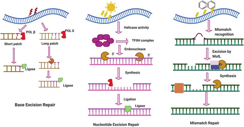
DNA damage repair: historical perspectives, mechanistic pathways and. . .
Huang and Zhou
3
pathways,40,41 cell cycle checkpoints,42,43 apoptosis,44–46 fidelity of DNA damage repair functions and processes. For example, in
replication,47,48 DNA re-replication49 and telomeres50,51are all many cancer cell lines, such as mantle cell lymphoma(MCL), ATM
closely associated with cancer.51 Based on these studies of the is recurrently mutated in around thirty to almost fifty percent of
DNA molecule integrity and the process of genetic mutation, the cases.72 These mutations may be linked with cancer chemother-
linkage between DNA mutation and cell carcinogenesis became apy resistance.73 Furthermore, cell cycle machinery-related genes
increasingly clear. Phil Lawley, a pioneering researcher of DNA play critical roles in driving avoidance of chemotherapy and
damage and carcinogenesis, found that some alkylating agents, radiotherapy treatment effects by cancer cells.74 Most measures
such as butadiene dioxide,52–54 could interact with DNA, forming developed to kill cancer cells involve: (i) stimulating G1 phase
harmful adducts and eventually disrupting the normal role of DNA aberrant homologous recombination in cancer cells; (ii) inducing
as a molecule template.55 The hypothesis that certain cancer mitotic catastrophe in cancer cells; or (iii) deleting the cell cycle
genes are susceptible to such agents was proposed and checkpoint.70,75,76 Despite data showing that genomic instability
extensively studied over the past few decades. Since then, may be associated with ROS(reactive oxygen species),77,78 in this
chemotherapeutic agents and radiotherapy have been found to review, we focus on DNA damage repair, as it is a major clinical
treat various cancers effectively through DNA damage induction. target of cancer chemotherapy and radiotherapy.
In the war against cancer, numerous agents have been developed
and novel technical strategies have also been explored. However, DNA damage
many challenges and unsolved issues remain that require further DNA damage and cancer. It is critical for maintaining genomic
study, such as: (i) the detailed molecular mechanisms underlying DNA stability due to its role as the template for replication and
the cancer cell DNA response to chemotherapeutic agents and transcription.79 As described above, damage to DNA from
radiotherapy; (ii) how cancer cells become resistant to chemother- environmental hazards insult as well as endogenous toxic agents
apeutic agents and radiotherapy; (iii) possible new and promising such as free radicals can compromise genome stability and cause
biomarkers for investigation as novel inhibitors or therapy agents; or promote many diseases, particularly cancer.37,80,81 As the DNA
and (iv) most importantly, the basic biological mechanisms molecule is the basic genetic material, it is vital for ensuring the
underlying the DDR. With such information, effective cancer integrity of DNA structure and function to support normal life
therapies could be developed to target DDR and ultimately activities and stable species characteristics.82,83 Indeed, when
prevent or cure cancer. experiencing either endogenous or exogenous stresses, cells can
generate various types of DNA damage, including base pair
DNA damage, cellular response, repair and cancer alterations, DNA replication errors84 and distortion and breakage
Genome stability. To support survival and reproduction, main- of the DNA double helix strands.85 Common exogenous factors,
taining genome stability is a critical priority of all cells.56 Any especially certain environmental hazards such as toxic heavy
abnormal alterations of the genetic base sequence can disrupt metals and ionizing radiation, have been intensively studied and
cellular biological processes, hampering cellular functions and found to cause serious DNA damage.86–90 Endogenous materials
possibly inducing carcinogenesis or even cell death.57 Specifically, are often released during the metabolism of exogenous materials
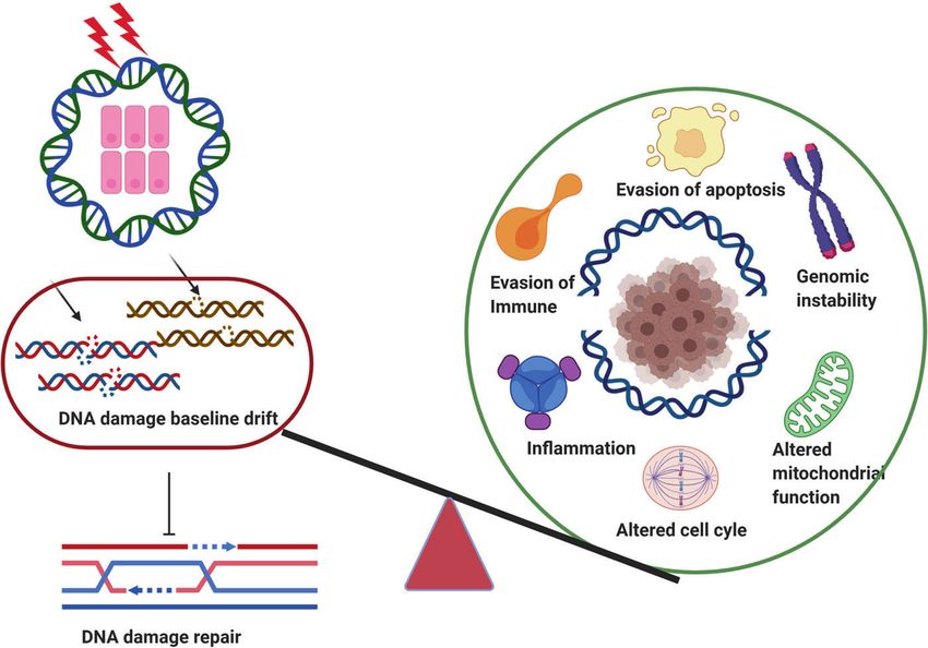
strong evidence has indicated that genomic instability promotes in the body or after cell damage and the loss of cell membrane
cancer pathogenesis through a cascade response involving a integrity.91 DNA damage can occur through two pathways, namely
series of proto-oncogenes that are continuously triggered or anti- direct effects and indirect effects. In the direct pathway,
oncogenes that are suppressed.58–60 In this context, the EGFR endogenous or exogenous materials directly contact DNA, leading
(epidermal growth factor receptor), MYC and RAS families have to the breakage of chemical bonds in DNA molecules, and thereby
been commonly recognized as proto-oncogenes,61 whereas TP53 changing the structure and activity of DNA.92,93 In the review by
is a well known tumor suppressor gene.62,63 Accordingly, to Anthony T et al., endogenous stresses including gene transcription
reduce the possibility of genetic dysregulation of genome stability, and replication in cancer cells are noted to cause genomic
cells have evolved a range of genome stability-related signal instability.79 In the indirect pathway, endogenous or exogenous
pathways and post-translational modifications,64 which assess the materials activate products such as free radicals94,95 that can
accuracy of DNA metabolism and prevent accumulation of DNA damage DNA.96
damage.65 For example, multiple families including ATM (ATM Several types of DNA damage have been reported previously, as
serine/protein kinase), ATR (ATR serine/threonine kinase), and follows: (i) single-strand breaks; (ii) double-strand breaks (DSBs);97
DNA-PKcs (DNA-dependent protein kinase catalytic subunit) can (iii) base damage; (iv) sugar damage; (iv) DNA cross-linking and (v)
initiate the signaling cascade in mammalian cells.66 A recent clustered damaged sites,98 of which the most deleterious lesion
review by Monique PCM et al. summarized the advances of and the most severe threat to cells is the DSB. DSBs that occur
ubiquitination research and noted that ubiquitination performs without effective repair or error-prone repair can cause carcino-
vital roles in regulating cellular homeostasis through numerous genesis or cell death.99 Lindahl et al. reported that, each day, our
enzymes67 and proteins. The complex functions of this compound cells may be subject to around 70,000 instances of DNA
have become known as the “ubiquitin code” in the scientific damage.100 Most of these lesions are single-strand breaks, and
community.56 Genomic instability is a common characteristic of only a few are DSBs, which are less frequent. Numerous studies
most cancer cells.68 For example, a high ratio of chromosomal have illustrated that DNA subject to oxidative stress exhibits a
instability is associated with mitotic spindle checkpoint deficiency large number of base and sugar lesions,101 such as guanine
in most breast cancer cell lines.69 The molecular mechanisms modification or 7,8-dihydro-8-oxo-2′-deoxyguanosine (8-OH-dG).
through which cells maintain genome stability and the repercus- Base lesions are usually caused indirectly by ROS generated due to
sions of genomic instability are essential emerging issues relevant oxidative stresses such as radiolysis of water molecule induced by
to clinical cancer avoidance. ionizing radiation.102 Sugar damage, such as 8,5′-cyclopurine-2′-
Accumulating evidence has shown that a DNA double-strand deoxynucleosides, can be caused by free radical insult to the sugar
break(DSB) is typically the most harmful type of DNA damage, and moiety.103,104 DNA cross-linking is often attributed to exposure to
that it compromises genome stability.70 In mammalian cells, a chemical cross-linking agents, e.g. cis-platinum, or free radical-
number of vital DNA repair functions and processes against generating ionizing radiation.105 With this type of damage, DNA
various DNA damage have evolved. For example, the mismatch repair-related proteins are trapped with DNA, causing the proteins
repair pathway, base excision repair pathway and nucleotide to adhere to the 5′ or 3′ end of the DNA strand break.106
excision pathways have been well characterized.71 However, Furthermore, DNA cross-linking can hinder the activities of some
cancer cells have frequently evolved in relation to abnormal vital enzymes such as DNA helicases and polymerases.107,108
Signal Transduction and Targeted Therapy (2021)6:254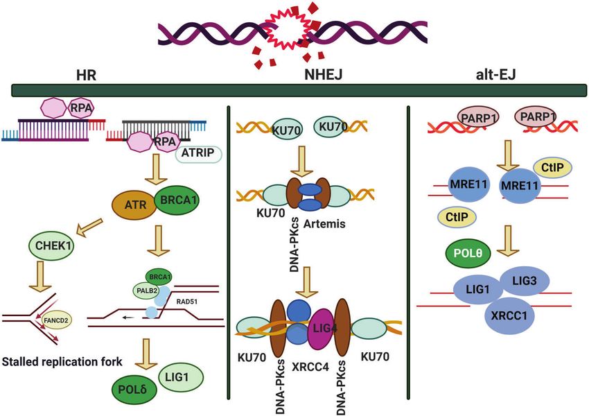
DNA damage repair: historical perspectives, mechanistic pathways and. . .
                                                           Huang and Zhou
4

    Fig. 2 Main types of DNA damage along with differential definitions of double-strand breakage-based and non-double-strand breakage-
    based clustered DNA damage

    A review described the formation of DNA cross-links due to                  been identified as the DNA damage response (DDR). Generally,
    exposure to various endogenous, environmental and chemother-                DDR mechanisms involve feedback signals from damage sites and
    apeutic agents.106 However, elucidation of how this process is              movement of repair factors to cluster at damage sites. In our
    regulated and its full biological functions in mammalian cells and          previous published review, we used ionizing radiation to explore
    cancer cells require further research. Clustered DNA damage,                how the cell’s DNA damage sensors and signaling transducers
    sometimes described as multiple local damage sites, refers to               interact in the DDR. We focused on the critical issue of recognizing
    damage in which at least 20 base pairs are separated.109 Clustered          and identifying DNA damage signals to activate the subsequent
    DNA damage usually consists of multiple lesions such as base                biological response cascade.6 Therefore, in this review, we focus
    damage, a basic site damage and single-strand breakage.110                  on the association between DDR and cancer. Due to the
    However, in contrast to DSBs, the multiple lesions of clustered             characteristic genomic instability of cancer cells, mutations and
    DNA damage may be present on the same DNA strand or on                      tumor heterogeneity are common and widespread.116 These
    opposing strands within a tiny range. Figure 2 illustrates the main         features suggest that cancer cells are prone to enhanced
    types of DNA damage along with differential definitions of                   proliferation, growth and tumorigenes is due to dysregulation of
    double-strand breakage-based and non-double-strand breakage-                DDR-related mechanisms.117 The acquisition of specific mutations
    based clustered DNA damage. In general, clustered DNA damage                in cancer cells might, in turn, increase the dependence on other
    results in enhanced mutation frequency,111 cancer, and cell death.          DDR factors for survival.118 The development of cancer requires
    The mechanism of clustered damage has been described as a                   both mutagenic and non-mutagenic events. Cells exposed to
    base obtaining a single electron, after which multiple electron             endogenous and exogenous factors that act as mutagenic agents
    pathways are activated.111 However, whether a beneficial result              show impacts throughout the process of cell oncogenesis, but
    (friend) or non-beneficial result (foe) is obtained from clustered           these effects are stronger in cancer cells with mutated or deficient
    DNA damage in cancer cells or normal cells requires further                 DDR genes.119,120 Alteration of DDR genes has been demonstrated
    study.112 As cells face a tremendous amount of DNA damage                   in various cancers, including breast cancer and prostate
    arising from various exogenous and endogenous stressors, such as            cancer.40,118 For example, BRCA1 or BRCA2 inactivating mutations
    ionizing radiation or ROS,112,113 recognition of how DNA damage             were found in ninety out of almost six hundred breast cancer
    occurs requires deeper investigation. Many scientific issues remain          patients.121 Moreover, DNA mismatch repair-dependent DDR
    to be addressed in future research, such as: (i) excluding the              pathways, such as loss of non-canonical mismatch repair gene
    currently known DNA damage types, other novel DNA damage                    functions, contribute to improved treatment outcomes of color-
    styles may exist that have not yet been discovered; (ii) methods to         ectal cancer.122
    evaluate and measure DNA damage types and degrees, or                          Here, as a few DNA damage sensors such as γH2AX, Mre11-
    visualization techniques for DNA damage; (iii) monitoring                   RAD50-NBS1 complex, Ku70/Ku80, MDC1 and 53BP1 can initiate
    processes for DNA damage and identification of effective                     the damage signaling thus trigger the DDR,123 In a study,γH2AX
    biomarkers for early detection of DNA damage; and (iv) obtaining            may be expressed not only to detect genetic effects caused by
    reference values for the exogenous and endogenous stressors that            various toxic substances but also to monitor the clinical efficacy of
    drive DNA damage. Investigating these issues may help to                    chemotherapy and radiotherapy and the sensitivity alternations of
    standardize DNA damage caused by various insults. Importantly,              cancer cells to anticancer agents.124 Another study assessed DDR
    innovative technologies and unique theoretical models would be              processes after hepatocellular cancer therapy and found that
    developed while exploring these interesting issues.                         γH2AX expression increased.125 Screening for H2AX variant
                                                                                functions and targeting of H2AX have been proposed as cancer
    DNA damage response                                                         treatments.126 Ku70/Ku80 expression also exhibited a significant
    Sensors in the DNA damage response. To avoid DNA damage, cells              increase in rectal cancer patients after chemotherapy and
    have evolved numerous interacting mechanisms for ensuring                   radiotherapy treatment, and further study showed that the
    genomic stability or have even used DNA damage to produce new               increase in Ku70/Ku80 expression was correlated with chemo-
    opportunities for natural selection.114,115 These mechanisms have           and radio-resistance in various cancers.127 Ku70/Ku80 expression

                                                                                  Signal Transduction and Targeted Therapy (2021)6:254
DNA damage repair: historical perspectives, mechanistic pathways and. . .
                                                                        Huang and Zhou
                                                                                                                                                    5

                                              CH3
                                          O                                                          O

                                      N           N                                          HN              N
                                                            Alkytransferase                                  N
                               H2N        N       N                                     H 2N         N

                                  NH2         NH2                                                NH2             NH2

                              N                   N                                          N                       N

                          O       N           N       O         Photolyase               O       N               N       O

                                              NH2                                                NH2
                                                      CH3
                                                  N                                                  N

                                              N       O         Dioxygenase                      N       O

Fig. 3 Timeline of DDR-related findings and concepts related to cancer, highlighting the scientists who worked to provide a deeper
understanding of the roles of DDR in cancer

can be used as a molecular cluster for predicting the susceptibility    defined, which is a programmed cell death pathway occurring in
of rectal cancer to chemoradiotherapy.128 In contrast to oxygen         cells during the normal tissue development or encountering
sensors, which have been extensively investigated, these DDR            exogenous stresses, especially DNA damage.139,140 In 1981, the
sensors are still in the early stages of molecular characterization,    concept of oncogenes was introduced,141 followed by the concept
and their roles in sensing DNA damage and signaling, cancer             of tumor suppressors three years later.142 In 1989, cell cycle
progression and therapy require further study.                          checkpoints were proposed and in 1990, p53 was reported to be
                                                                        mutated in various cancers.143 Then, in 1997, caretaker and
DNA damage response and cancer. A review literature high-               gatekeeper genes were proposed based on the research
lighted the various concepts behind targeting of DDR in cancer,129      discoveries on DNA repair genes BRCA1/2, RAD51.144–146 In 2002,
which were summarized that (i) DDR can be used as a target of           ROS production and DNA damage attributable to deregulated
anticancer drug treatments; (ii) as most cancer cells have a            metabolism       induced       by    oncogene      expression   were
deficiency of some DDR pathways’ ability, inhibition targets can be      reported.147,148 In 2005, DDR was described as an anticancer
explored in the remaining pathways; (iii) DDR inhibition can be         barrier in early-stage tumorigenesis, but the genes showing DDR
used to investigate cancer replication stress; and finally (iv) the      mutations were absent from later-stage tumors.149 To date, a large
author considered using DDR inhibitors in specific DDR-lacking           number of DDR genes have been identified in various can-
backgrounds initially to promote exploration of DDR-based agents        cers.150,151 Figure 3 illustrates the timeline of DDR-related findings
for cancer treatment in the future. The first description on the         and concepts related to cancer, highlighting the scientists who
association of cancer with occupational exposures was presented         worked to provide a deeper understanding of the roles of DDR in
in 1775 by the British surgeon Percivall Pott,130 who first showed       cancer.
the link between the occupational exposure of chimney sweeps               Alongside the DDR processes described above, including cell
and scrotal cancer. In 1946, the X-ray induced recessive lethal of      cycle checkpoints and apoptosis, we present DDR signaling by
Drosophila was first reported to be related to the chromosomal           way of a brief introduction to how DDR pathways can affect
breakage.131,132 Later, the discovery of the helical structure of DNA   cancer development. First, a healthy cell affected by environ-
led to the introduction of the concept of DNA as the hereditary         mental hazards, viral or bacterial infection, or ROS may initiate
material.133,134 Soon thereafter, repair of X-ray damage to DNA         DNA damage and mutations, increasing oncogene activation,
was reported in bacteria in 1966135 and in eucaryotic cell in           tumor suppressor inactivation and replicative and oxidative
1967.136 Significant research showed that defective DNA repair           stresses. The oncogene activation might occur directly or
resulted in many diseases and, in particular, cancer propensity,        indirectly. As a result, DNA replication fork fidelity and replication
during the period from 1969 to 2015.137,138 In 1972, apoptosis was      recovery are compromised.152–154 Hence, increased DNA damage

Signal Transduction and Targeted Therapy (2021)6:254
DNA damage repair: historical perspectives, mechanistic pathways and. . .
                                                           Huang and Zhou
6
    and mutations in normal cells would hamper genomic stability. At            on observations, as described above.177 In 1940, American
    this stage, the damaged cells still exhibit a range of responses,           biologist Albert Kelner identified photo reactivation, which is an
    including activation of checkpoint arrest and triggering of                 enzyme-catalyzed reaction, as the enlightenment DNA repair
    increased p53 expression to protect cells against further damage.           mechanism.178 Then, in 1964, Setlow RB and Carrier WL reported
    However, downregulation of DDR processes should disturb the                 an error-correcting mechanism in which intrastrand thymine
    proliferation of pre-cancerous cells.155–157 General DDR pathway            dimers formed after UV radiation of DNA accounted for a large
    research in relation to cancer development provides important               fraction of the observed biological damage to DNA.15,179 In the
    information that may be useful for the design of targeted cancer            same year, the term “DNA repair” was formally founded with the
    therapies.158,159 More importantly, understanding DDR can also              discovery of “Dark repair” and “Repair-replication” or “unscheduled
    help elucidate why targeted clinical therapy strategies often               DNA synthesis” of ultraviolet injury to the DNA in Escherichia
    fail.160,161 Debjani P et al. assessed the performance of cancer cells      coli.12,13,180 These studies showed that one strand of damaged
    escaping targeted lung cancer therapy, found that the key event             DNA could be excised, and the resulting gap could be repaired,
    was activation of the TGF-β signaling pathway in some cancer cells          using the intact complementary strand as a template.12,181 This
    after targeted therapy.162 TGF-β activation can inhibit the                 repair pathway is known as nucleotide excision repair. In 1968, J R
    expression of DDR-related genes, resulting in decreased DNA                 Cleaver et al. validated it as a repair replication mechanism
    repair ability and, thus, accumulation of mutations. Other                  through observation of UV-induced lesions to HeLa cell DNA.182
    advances have shown that tumor heterogeneity may influence                   Soon thereafter, polynucleotide ligase activity was discovered in
    the outcomes of targeted cancer therapy.163,164 For example,                the cell-free extracts from E. Coli by Gellert M in a study showing
    some scientists have reported that ALK-targeted therapy differs             that E. coli extracts could convert lambda DNA to covalent
    among cancer patients, with many patients treated with ALK-                 circles.183 In other words, polynucleotide ligase can combine a
    targeted therapy developing therapy resistance, which results in            newly synthesized patch with the contiguous parental DNA
    cancer progression.165 Targeted therapies against other cancers,            strand.
    such as non-small cell lung cancer (NSCLS), also face challenges               In the mid-1970s, the excision repair processes of base excision
    related to tumor heterogeneity, which impact acquired and                   repair and mismatch repair were described. Lindahl revealed that
    inherent drug resistance.166                                                an N-glycosidase was active in DNA repair based on its ability to
       New technologies, such as cancer genome profiling using deep              deaminate dCMP residues into an easily repairable form.184
    sequencing and microarrays and single-cell sequencing offer more            Meanwhile, Wagner Jr. and Meselson used E.coli to identify repair
    information about which DDR-related genes are mutated or mis-               tracts originating at mismatches. The repair process propagatesin
    regulated.62,167–170 However, a better understanding of DDR                 the direction of 5′ to 3′ and can cover approximately three
    pathways and discovery of new and valuable ideas for improving              thousand nucleotides.185 In terms of excision repair, the optimal
    cancer treatment are still urgently needed.                                 outcome is that the DNA can be replicated normally after excision
                                                                                and repair, but it is also possible for an advancing replication fork
    DNA damage repair                                                           to encounter the damage site after excision and prior to the
    A historical perspective of DNA damage repair. Many human                   completion of repair, which is known as synthetic death.174
    pathologies such as tumors and chronic metabolic diseases can be            Compared to the former repair type, the latter is more
    clearly attributed to DNA damage induction.171 Indeed, although             complicated and more likely to be lethal. In 1975, an interesting
    DNA damage is common and its occurrence is very frequent, such              hypothesis was raised by Radman, who suggested that E. coli
    damage must be repaired immediately and correctly to ensure the             possesses an inducible DNA repair system, called “SOS repair”.186
    exact transfer of genetic information during cell division.172,173          The main components of this hypothesis were: (i) DNA damage
    Without appropriate DDR capacity after insult from environmental            initiated the “SOS repair” process; (ii) de novo protein synthesis is
    or endogenous stressors, negative effects may occur in normal               involved in the repair process; and (iii) physiological and genetic
    cells, as follows: (i) increasing opportunities for genomic defects;        status requirements must be met for further prophage induc-
    (ii) possible genomic instability and malignant transformation; (iii)       tion.186 This hypothesis was confirmed through many later
    enhanced development of cancer; and (iv) further injury to cellular         experiments, and some new concepts have been incorporated
    DNA repair ability, as DNA damage signaling and inappropriate               into this model. For example, a study showed that cells treated
    repair processing in cancer cells would benefit cancer cell growth           with rifampin to eliminate their ROS repair ability exhibited
    and proliferation while disrupting the outcomes of cancer chemo-            reduced repair efficacy of global cyclobutane pyrimidine dimer
    and radiotherapy. Over the long period of around 4 billion years of         (CPD) formation due to UV radiation exposure.187 These findings
    evolution, it is unsurprising that cells have prioritized minimizing        provided the insights into the enzymes responsible for DNA
    mutagenesis and protecting genomic replication through effective            damage detection, and showed that they may attack undamaged
    and quick repair of DNA damage.174 In recent years, numerous                DNA, with deleterious consequences. In other words, these
    studies have reported evidence of the importance of DNA damage              sensitive DNA repair enzymes perform dual roles depending on
    repair: (i) a few types of DNA damage have been illustrated over            their concentrations. At low concentration, these enzymes are
    recent decades, of which DNA double-strand breaks (DSBs)                    kept in check until needed for repairing specific DNA damage.
    represent the greatest risk for causing genomic instability;173 (ii)        Since these studies, the concepts of transcription-coupled repair
    some components of DNA repair pathways are so important that                sub-pathway and global genome repair sub-pathway of nucleo-
    life would not be sustained without them, such as ATR, which is             tide excision repair (NER) have been supported by numerous
    critical for early embryonic development and its deficiency results          studies.188,189 The DNA damage in the active transcription gene or
    in chromosomal fragmentation and early embryonic lethality;175              the transcribing strand is preferentially repair through the
    (iii) many hereditary disorders have been attributed to DNA repair          transcription-coupled repair sub-pathway.188,190 Global genome
    deficiencies, such as the observation by Jim Cleaver that patients           repair is another sub-pathway of nucleotide excision repair,
    with the rare autosomal-recessive cancer predisposition syn-                through which the DNA damage in whole genome is repaired
    drome, xeroderma pigmentosum, lacked the ability to perform                 with equal efficiency. The mechanistic difference between
    DNA repair after damage due to UV exposure;138 and (iv) defective           transcription-coupled repair and global genome repair is mainly
    DNA repair pathways are associated with cancer initiation, as               that, in the former process, the stalling of RNA polymerase at
    shown by a study in the 1990s, which found Lynch syndrome was               transcriptionally active genes favors the recruitment of Cockayne
    related to mutation of the DNA repair proteins MutS and MutL.176            syndrome proteins A and B, whereas in the latter process, helix-
        From a historical perspective, early research into DDR focused          distorting damage is recognized by XPC and its partners RAD23B

                                                                                  Signal Transduction and Targeted Therapy (2021)6:254
DNA damage repair: historical perspectives, mechanistic pathways and. . .
                                                                          Huang and Zhou
                                                                                                                                                      7
(Rad 23 homolog B) and CETN2 (centrin 2).191 However, compared            as alkylation, inter/intra-strand cross-link. In E. coli, the mechanism
with global genome repair, transcription-coupled repair is more           of the direct DNA damage reversal reaction was described as a
constitutive and is evidently not inducible as a DDR response.            “flip-out”process.209 Briefly, enzymes first form a long loop, then
Excluding excision repair, several other repair pathways that             DNA photolyase binds to duplex DNA, goes through a series of
support improved replication to overcome the obstruction of               energy transfer, single electron transfer and enzymatic catalysis
replication caused by lesions without their removal have been             steps, and forms a flip-out helix structure to skip the break site for
reported; they are known as tolerance pathways. These pathways            direct reversal.209–211 This form of direct DNA damage reversal is
require the function of specialized DNA polymerases.93,192,193 At         considered to be beneficial to cells, as it is a highly effective and
this point, a “collapsed replication fork” had been defined and its        simple method to address an important and necessary issue.
role in the loss of DNA synthesis capacity was known.194 In this             Unlike other molecules, which can be replaced, DNA cannot be
process, the fork collapse contributes to genomic instability or          replaced after being damaged, and must instead be repaired.
even death.194,195 In general, after reviewing DNA damage repair          Three classical DNA damage direct reversal mechanisms have
from a historical perspective,196 concerns remain, which can be           been described, namely, repair of O-alkylated DNA damage by
summarized as follows: (i) Is the previous DNA damage repair              alkyltransferases and dioxygenases, repair of photolesions caused
definition sufficient to represent fully the process and its                by ultraviolet (UV) radiation through the work of spore photo-
significance? Based on the rapid development of DNA damage                 product lyases and photolyases,212,213 and reversal of N-alkylated
research and a deeper appreciation of DNA damage repair, the              base adducts by AlkB family dioxygenases.214 DNA is constantly
definition should be expanded to include exogenous and                     subjected to numerous environmental insults. Among such
endogenous insults, genomic early and later responses, DNA                hazards, alkylating chemicals, which are often applied as cancer
repair-related enzymes, and early events associated with later            chemotherapy agents, can cause DNA damage in the form of
outcomes. (ii) Most of previous researches aimed to uncover new           alkylation.215,216 Endogenous products, including metabolites
targeted proteins and enzymes rather than considering the                 such as adenosylmethionine arising from many biological
interactions among multiple DNA repair pathways. Sometimes,               processes, may also damage DNA.217,218 After damage from these
various DNA repair pathways can handle the same damage sites in           agents, some typical damage response molecules such as
competing ways, but how this interaction occurs remains unclear.          methylguanine and methyladenine are formed on double-
Moreover, the processes that occur at each step of multiple repair        stranded DNA.219,220 These materials can increase the cell’s
routines require further investigation. (iii) The threshold level is an   mutagenic and carcinogenic potentials, for example, by increasing
essential concern for initiation of DNA damage repair, but leads to       the chance of base misincorporation.221 Alkyltransferases are
low-level lesions often being overlooked. However, multiple long-         associated with direct removal of DNA alkylation damage. Studies
term low-level lesions may lead to DNA repair via some novel              have shown that this enzyme is responsible for the removal of
pathway or mechanism, which requires further validation and               mutagenic alkyl adducts on the bases of the O6 atom of 2′-
testing. (iv) Finally, basic information about DNA repair is lacking,     deoxyguanosine and the O4 atom of thymidine.222,223 In cells, the
including how damage to bases and other structures of DNA is              importance of O6-alkylguanine DNA alkyltransferase-based direct
sensed in cells, what roles the sensing machinery plays in the            reversal is greater than that of nucleotide excision repair or base
cellular response to DNA damage, and how the cells perform cell           excision repair, suggesting the critical role of alkyltransferases.224
cycle arrest in response to DNA damage in normal cells and cancer         The potential role of this protein in DNA repair was reported 40
cells. Furthermore, in the context of cell mutagenesis or lethality,      years ago. Its main roles include prevention of mutations,
more information is needed about how DNA damage repair-                   cytotoxicity, and cancer development.225 Alkyltransferases have
related enzymes and proteins regulate downstream events in                been identified in many living things.226 However, whether other
combination with other factors after the recognition of an                co-factors or energy resources interact with alkyltransferases to
aberration. These questions are very basic, but have yet to be            perform direct DNA damage reversal remains unknown.225 Mean-
answered fully and clearly. In addition, it is important to learn from    while, although multitudinous studies have been conducted to
previous research and apply these discoveries in the clinic setting       investigate the activities of alkyltransferases, how their poly-
in the future. The greatest value of DDR research is that a deeper        morphisms relate to health, and specifically cancer therapy,
understanding of the secrets of life will allow us to face the            remain unclear at present.
challenges that arise from environmental, social and technological           Photolyases, which are 50–55kD single-chain flavoproteins, are
issues more effectively.                                                  damage-specific binding proteins active in the response to the
                                                                          formation of UV-induced cyclobutane pyrimidine dimers
DNA damage repair pathways. Several repair pathways exist,                (CPDs)227,228 and 6–4 photoproducts.229 From the perspective of
including direct reversal, base excision repair, nucleotide excision      LO Essen et al., photolyases are highly effective light-driven DNA
repair, mismatch repair, single-strand break repair and DSB               repair enzymes, which function specifically in the reversal of
repair.197–199 Direct repair generally refers to the repair of            genomic lesions induced by UV radiation.230 The mechanism of
pyrimidine dimers formed due to UV exposure or other factors              photolyase-related reversal is generally similar to that following
or the repair of alkylated bases. Nucleotide excision repair refers to    induction of DNA lesions by UV insult; specifically, an electron is
repair of DNA replication lesions or bulky adducts arising from           injected at the lesion site, activating cleavage of cyclobutane-
distortions of the DNA structure.3,200 Mismatch repair refers to          pyrimidine dimers or 6–4 photoproducts inside the duplex DNA
adjustment of mismatched base pairs in double-stranded DNA, as            structure.230 This reversal method is highly effective and simple, as
well as repair of some insertions or deletions of less than 4             an electron is shuttled to the lesion site for direct destabiliza-
nt.201,202 Double-strand break repair refers to repair of DSB             tion.231 The energy to drive this reversal reaction comes from
lesions.203–205                                                           chromophores excited through intake of a photon.232–234 With the
                                                                          advancement of this research field, other mechanisms, including
Direct reversal repair                                                    an exclusive bifurcating-electron-transfer method with a cyclic
As described above, induced DNA damage often refers to damage             radical mechanism, have been continuously reported. For dimer
caused by alkylation, oxidation, UV and cross-linking agents.206          repair, six steps have been identified, typically including three
Direct reversal of the base lesion rather than excision is the one        electron transferences and two bindings to lesions.229 Through
simplest step error-free and most economical DNA repair                   dynamic analysis, new discoveries such as a unique electron-
mechanism to have evolved.206–208Cells have also developed                tunneling pathway and essential residues at repair trigger sites
direct reversal mechanisms for several types of DNA damage, such          have been revealed.229,235,236 Importantly, recent crystal structure

Signal Transduction and Targeted Therapy (2021)6:254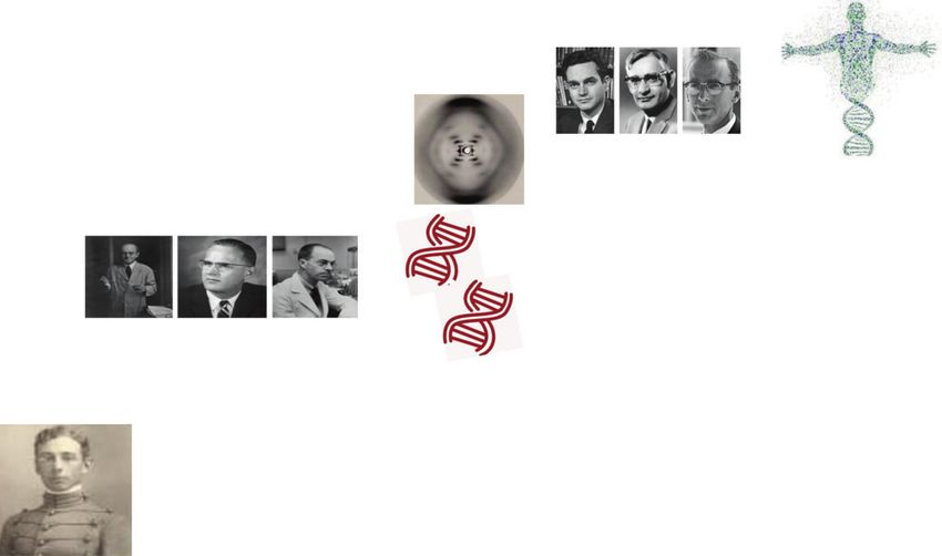
DNA damage repair: historical perspectives, mechanistic pathways and. . .
                                                           Huang and Zhou
8

    Fig. 4 Three types of direct DNA damage reversal including representative substrates, repair proteins, cofactors, and the corresponding repair
    products

    determination of photolyases has provided new direct insights               human diseases, including xeroderma pigmentosum and other
    into the relationship between photolyase structure and its roles in         neurological diseases.200 The repair process has been elucidated
    DNA damage repair,237 supporting further comparison between                 through research over previous decades. In TCR-NER, the
    DNA photolyases and spore photoproduct lyases.237–239                       hampered RNA polymerases by the damage constitutes the initial
       In recent years, the AlkB family of demethylases has attracted           step for recognizing DNA lesions, then recruits the CSB/ERCC6,
    increasing attention for its regulatory role in oxidative DNA repair.       which in turn recruits CSA/ERCC8 complex. In GGR-NER, the
    In 2002, studies by Pal F et al.240and Sarah CT et al.241 revealed a        protein complex XPC/RAD23B/CETN2 can sense and recognize
    third type of direct reversal mechanism for DNA damage,                     DNA distortion and recruit helicase TFIIH to form a XPC-RAD23B-
    reporting that AlkB protein in E. coli can repair cytotoxic damage          TFIIH complex to unwind the DNA helix. Once the pre-incision
    due to 1-methyladenine and 3-methylcytosine in DNA, and that                complex is ready, the endonuclease XPG and XPF/ERCC1 are
    this reaction by AlkB is dependent on oxygen and α-ketoglutarate.           recruited, which can cut the strand at the 3′ and 5′ flanks of
    Figure 4 lists the three types of direct DNA damage reversal                damage site, respectively, to ensure that of a piece of damage-
    described above, including representative substrates, repair                containing nucleotides are removed. Then, the proteins respon-
    proteins, cofactors, and the corresponding repair products.                 sible for synthesizing the missing nucleotides are recruited
                                                                                and, finally, DNA ligase fills the gap to complete the repair
    Base excision repair                                                        process.256–258
    Exposure to ionizing radiation produces radicals.242,243 Radicals
    may cause base lesions and thus, base excision repair may be                Mismatch repair
    initiated to address these lesions.244,245 Key enzymes such as              The primary purpose of mismatch repair is to counteract
    OGG1 can sense damaged bases and are responsible for the                    replication errors and thus improve the fidelity of replication.259
    recognition and removal of 8-oxoguanine.246,247 The result of base          This repair pathway is mainly used to resolve single nucleotide
    excision repair is that 1 to 10 nucleotides, but no more, can be            mismatches and small insertion loops generated by DNA
    replaced by short to long patches.248 The cell’s status and                 polymerase.119,260 The mismatch repair pathway consists of three
    background, lesion style, and levels of exogenous and endogen-              steps. First, protein complexes such as the MSH2-MSH6 hetero-
    ous materials affect the selection between short patch and long             duplex sense and recognize the mismatch and identify the site of
    patch base excision repair.249–251 The importance of base excision          the insertion-deletion loop.261,262 These protein complexes will
    repair is not only for ensuring genomic stability but also that its         quickly move to the mismatch lesion site and bind to the DNA
    dysregulation would lead to increased risks of cancer, aging-               molecule to form a sliding clamp. At this point, many proteins
    related diseases and other serious disorders.252 However, base              gather to perform various functions. For example, exonuclease 1
    excision repair does not simply serve as an isolated repair                 (EXO1) has been reported to carry out excision of nucleotides in
    pathway, it is one component of the larger DNA damage repair                the 5′- > 3′ direction.263 Another protein, replication protein A
    machinery. It forms a network in combination with other                     (RPA) serves as a binding function of single-strand DNA produced
    pathways and may in turn be regulated by other pathways via a               by EXO1 to prevent further DNA degradation.264 MLH1, a subunit
    feedback mechanism.253                                                      of MutLα, whose defect is responsible for ~50% of MMR defected
                                                                                cancers, may restrained DNA excessive excision by EXO1.265
    Nucleotide excision repair                                                  Immediately following the recognition step, the removal step
    Compared to base excision repair, nucleotide excision repair is             occurs. The mismatched bases are removed, and then the
    more complex, as it is responsible for addressing complicated               replacement DNA is synthesized by DNA polymerase δ while
    lesions including bulky adducts and cross-linking lesions,254,255           DNA ligase ligates the remaining nick.259,266,267 Certain proteins
    and there are two sub-pathways of TCR-NER and global genome                 are also necessary for this synthesis step. Proliferating cell nuclear
    repair (GGR)-NER as mentioned above. The source of this type of             antigen (PCNA) functions not only in the mismatch recognition
    DNA damage is alkylating or cross-linking agents. Deficiency of              step but also in the processing of DNA polymerase during the final
    nucleotide excision repair is typically associated with several             synthesis step.268,269 As reported by Kira CB et al., PCNA can

                                                                                  Signal Transduction and Targeted Therapy (2021)6:254
DNA damage repair: historical perspectives, mechanistic pathways and. . .
Huang and Zhou
9
Fig. 5 Main characteristics of base excision repair, nucleotide excision repair and mismatch pathways and the main differences in their lesion
sensors, mediator proteins and effector proteins
trigger other proteins to cut the error-containing strand, leading Double-strand break repair
to more rapid and effective excision and synthesis.270 Due to their Two main types of DSB repair pathways have been reported,
critical roles in DNA damage repair and, in particular, mismatch namely, homologous recombination (HR)280 and non-homologous
repair, loss of expression of essential proteins such as MSH, EXO end joining (NHEJ).281 Compared to the NHEJ pathway, HR is more
and PCNA is closely and significantly linked to increased conservative and error-free due to its dependence on the
predisposition to a number of diseases, including various existence of sister chromatids.282,283 However, this property limits
cancers202 and other metabolic pathologies.201,202,271–273 Recent the HR pathway repair to the cell cycle S/G2 phase when sister
reports indicate a broader spectrum of non-canonical roles of chromatids exist, whereas the NHEJ pathway can repair DSBs
mismatch repair. These roles include the responses to oxidative throughout the cell cycle.284–286
DNA lesions, helix-distorting nucleotide lesions and environmental
chemical toxicants such as benzo(α)pyrene-induced cellular Homologous recombination (HR). The HR pathway is comprised
senescence, as well as regulation of the cell cycle.269,274–276 of three processes: (i) double-strand break recognizing, DSBR; (ii)
Undoubtedly due to its importance in maintaining genomic synthesis-dependent strand annealing, SDSA; and iii) break-
stability, mismatch repair deficiency leads to increased DNA induced replication, BIR. After a DSB occurs, a single-strand is cut
mutations. The opportunity for secondary mutations increases by from the DSB’s end to form two single-strand ends, of which the
100 or even 1000 times in mismatch repair-deficient cells.201,277 3-terminal ends pair with the homologous templates to form a
With the development of high-throughput sequencing technol- D-loop structure. The single-stranded DNA (ssDNA) that breaks
ogy and compound screening strategies, many novel functions after this step is synthesized using homologous DNA as a
and mechanisms of mismatch repair have been identified, and template. After D-loop formation, the repair modes differ among
further research should aim to clarify the genes and proteins of the three pathways. The resected 3′ ssDNA end(s) of the DSB
the mismatch repair pathway in depth and elucidate how each sense, bind with, and insert a homologous DNA sequence to
gene or protein may differentially function in each step. Moreover, prime the synthesis of repaired DNA. Somatic cells use the sister
non-canonical roles of the mismatch repair pathway should be chromatid rather than the homologous chromosome as the
further investigated to provide new insights into DNA damage repair template. In the DSBR pathway, the D-loop structure
repair and identify potential new fields for targeted cancer becomes stable through capture of another 3′ terminus, forming
therapy or improvement of chemo-and radio-therapy outcomes in a double Holliday junction (dHJ) structure. In the SDSA pathway,
the future. the 3′ end is extended and released from the template, and then
Figure 5 illustrates the main characteristics of base excision matches with another 3′ end to continue the DNA repair process.
repair, nucleotide excision repair and mismatch pathways and the In the BIR pathway, the D-loop forms a replication fork to ensure
main differences in their lesion sensors, mediator proteins and synthesis of the following chain and leading chain. Within the HR
effector proteins.278 Generally, in base excision repair, APE1 and pathway, many proteins must combine with each other to
DNA glycosylases are the main DNA damage sensors. Meanwhile, function. After the formation of DSBs, under the action of specific
the damage sensors in nucleotide excision repair are XPC and CSA; nucleases (e.g., the Mre11-Rad50-Nbs1, or MRN, complex), the 5′
the mediator proteins are XPA, XPF and RPA; and the effect or ends of DSBs are excised to form a 3-terminal single-stranded
proteins are XPG, ERCC1, and POLD1. In mismatch repair, the roles DNA (ssDNA). Subsequently, the ssDNA is encapsulated by RPA,
of synthesis proteins and protein complexes differ. which is replaced by Rad51 to form the nuclear fiber structure of
Single-strand break repair can be conducted through the base Rad51. Mediators such as RAD52 and BRCA2 participate in this
excision repair, nucleotide excision repair and mismatch repair process. Subsequently, with the help of PALB2 and rad51ap1, the
pathways.279 In this section, we focus on the DSB repair Rad51 nuclear fiber combines with the homologous double-
pathway, as this damage type is most deleterious to genomic stranded DNA to form a D-loop structure. After this association,
stability. the D-loop dissociates under the action of FANCM to form a
Signal Transduction and Targeted Therapy (2021)6:254DNA damage repair: historical perspectives, mechanistic pathways and. . .
Huang and Zhou
10
product without cross exchange. The dHJ structure formed in the series of heat repeat sequences (huntingtin, elongation factor 3, a
DSBR repair pathway can also be dissolved by a helicase subunit of protein phosphate 2a and tor1, heat). The C-terminus
topoisomerase complex (BLM-TopoIII) to form a product without contains a FAT (FRAP, ATM, TRRAP) domain. The PIKK regulation
cross exchange. domain (PRD) may be located between the kinase domain and the
Rad51 plays an important role as the core molecule in the HR FAT domain.303 Cells lacking DNA-PKcs showed high radio
pathway.287 Rad51 in mammalian cells is similar to Rad51 in sensitivity. Moreover, mouse experiments showed that mice
yeast cells and RecA in bacteria, with specific functions before, lacking DNA-PKcs might suffer from severe comprehensive
during, and after HR association.288 First, Rad51 interacts with immunodeficiency (SCID).304 The binding region between DNA-
DNA to form the Rad51 nuclear fiber structure, which effectively PKcs and Ku is located in the C-terminal region of DNA-PKcs.
elongatesss DNA and is therefore conducive to ssDNA encoun- When the Ku dimer is on DNA, Ku recruits DNA-PKcs to the DNA
tering its homologous DNA template.289 Rad51 can promote the break terminus, and two DNA-PKcs molecules interact with the
combination of ssDNA with homologous DNA templates and, DSB site to form a synaptic complex. The DNA-PKcs/Ku/DSB
thereby, promote formation of the D-loop. After association, complex can fix the ends of DSBs, thereby protecting the DSB site
Rad51 breaks away from the leading strand of DNA during DNA from nuclease digestion.
synthesis, exposing its 3-terminal sequence, which is used as the After the end of DNA is processed by Artemis and other end-
primer for DNA synthesis.290 processing molecules, the subsequent repair process must
RPA is a single-stranded DNA-binding protein comprised of a connect the disconnected DNA, and LIG4 executes the DNA
trimer of RPA1, RPA2 and RPA3.291 RPA1 has four domains in the connection. XRCC4 has no known enzymatic activity, but can
trimer, which play roles in DNA synthesis.292 The N-terminus of function as a scaffold protein, aiding the recruitment of other
RPA1 has a protein-binding domain, DBD-F, along with three NHEJ pathway molecules. In structure, the best binding ligand of
domains that bind to ssDNA, DBD-A, DBD-B and DBD-C. The XRCC4 is LIG4.305 The C-terminus of LIG4 contains two BRCT
second large component of the trimer, RPA2, has a central domains, with a connecting region between the two domains. The
structure domain, DBD-D. RPA3 has only one domain, DBD-E.293 highly stable complex XRCC4-LIG4 can be formed through
CtIP (CtBP-interacting protein) plays important roles in cell interaction with the helical region at the C-terminus of XRCC4.
cycle regulation and DNA damage repair.294,295 It contains a XRCC4 can stabilize LIG4 and promote its activity. The XRCC4-LIG4
dimerization domain (40–165 amino acids) at the N-terminus, complex can interact with Ku, PNK, APLF, XLF and DNA.306 XRCC4
which has the same amino acid sequence and binding site as the can be highly phosphorylated, and DNA damage can promote its
RB family. CtIP possesses a central domain, which interacts with self-phosphorylation. DNA-dependent protein kinase (DNA-PK)is
CtBP. The C-terminal sae2/ctp1-like domain of CtIP is conserved necessary for the phosphorylation of XRCC4 induced by DNA
between human and yeast. The phosphorylation of the CtIP damage, and promotes the binding of XRCC4-LIG4 to DSBs.307 The
S327 site promotes its binding to BRCA1, which then binds to SUMO modification of XRCC4 is essential to its nuclear localization
itself, and then is ubiquitinated by BRCA1 and recruited to the and DSB repair function.
damage site.296 The DNA-binding domain of CtIP is located 53BP1 (p53-binding protein 1) is a very important molecule in
between amino acids 515–557, which is conducive to the the DSB repair pathway, functioning as an intermediary molecule
recruitment of CtIP to DSB sites. The two lysine sites in this or effector.308 It can promote the terminal junction of DNA after
domain, K513 and K515, are crucial to the interaction between DSB occurrence. To be recruited to DNA, 53BP1 must directly
CtIP and DNA. The N-terminus and C-terminus of CtIP contain recognize the specific histone structure produced by the DSB.
structural domain segments that interact with MRN. The Moreover, 53BP1 can promote the NHEJ pathway and inhibit the
T847 site of CtIP can be phosphorylated by CDK, which helps HR repair pathway. The N-terminus of 53BP1 contains 28 serine/
CtIP to activate the nuclease activity of MRN and, thus, to threonine glutamine sites (s/t-q), which are the target sites of ATM.
promote the single-strand excision of DNA DSB ends.297 When the N-terminus of 53BP1 is phosphorylated by ATM, the
interaction of 53BP1 with Rif1 (Rap1-interacting factor 1) and PTIP
Non-homologous end joining (NHEJ). In the classical NHEJ path- (Pax activation domain-interacting protein) is promoted.309,310 The
way, the heterodimer of Ku70 and Ku80 first binds to the broken C-terminus of 53BP1 contains a BRCT domain, which interacts with
DNA ends and then recruits DNA-PKcs (DNA-dependent protein p53 and EXPAND1. The minimal focal region of 53BP1 contains an
kinase catalytic subunit). DNA-PKcs is a member of the OD (oligomerization domain), a glycine- and arginine-rich (GAR)
phosphatidylinositol 3-kinase (PIKK)kinase family that can pull motif, and a ubiquitination-dependent recruitment (UDR) domain.
two broken DNA ends together and recruit processing-related It can be dimethylated atlysine 20 within its GAR motif, and the
enzymes, such as Artemis, PNKP (polynucleotide kinase/ phos- UDR domain can interact with ubiquitinated H2AK15.311
phatase), APE1 (AP endonuclease 1) and Tdp1 (tyrosyl DNA
phosphatase 1), and then recruit the XRCC4-XLF-LIG4 complex.298 Alternative end joining. While the c-NHEJ and HR pathways are
Ku70 and Ku80 are subunits of the first protein complex to be primarily responsible for repairing DSBs of DNA, alternative end
recruited to the damage site, both of which have a central domain joining (alt-EJ) was considered responsible for residual DSBs that
(Ku core) that binds to DNA. An acid domain, serine 6, is present in c-NHEJ and HR are unable to repair.312,313 However, it is unsure
the N-terminus of Ku70 that can be phosphorylated by DNA-PKcs. whether alt-EJ represents a standing pathway or only the end-
SAP (SAF-A/B, Acinus and PIAS) possesses a C-terminal domain.299 joining components of the pathway usually serving in dsDNA
There is a linking region between SAP and the Ku core of about processing of other functions, such as in replication, recombina-
536–560 amino acids. Both SAP and this linking region can bind to tion or repair. Alt-EJ is also called microhomology-mediated end
DNA, so SAP may anchor the Ku dimer to chromatin. The C- joining(MMEJ).314 Alt-EJ refers to repair of DSB damage indepen-
terminal region of Ku80 interacts with the Ku core through a dently of classical NHEJ factors such as Ku70, DNA-PKcs and
highly flexible linking region.300 At the end is a 12-amino-acid lIG4.315 Although this process appears similar to c-NHEJ, alt-EJ is
region that can directly interact with DNA-PKcs. The Ku dimer can Ku-independent, depending instead on regions of microhomology
recruit DNA-PKcs, XRCC4 and XLF to a damage site. When the Ku on each side of the breakage site.315 Specific proteins including
dimer binds to DNA, Ku70 is directed toward DSBs, while Ku80 is PARP-1[Poly(ADP-ribose) polymerase] are critical for facilitating
directed away from DSBs.301 the alt-EJ pathway.316 As reported by Huang YJ et al., PARP-1 is
DNA-PKcs, as a member of the PIKK family of serine/threonine vital to DSB repair in breast cancer cells, and the alt-EJ pathway is
protein kinases,302 contains a leucine-rich domain (LRR) at the N- triggered by radiomimetic agents.317 Other studies have shown
terminus, which may play an important role in DNA binding, and a that PARP-1 and DNA ligases are required for chromosomal
Signal Transduction and Targeted Therapy (2021)6:254You can also read