Targeting DNA Double-Strand Break Repair Enhances Radiosensitivity of HPV-Positive and HPV-Negative Head and Neck Squamous Cell Carcinoma to ...
←
→
Page content transcription
If your browser does not render page correctly, please read the page content below
cancers
Article
Targeting DNA Double-Strand Break Repair
Enhances Radiosensitivity of HPV-Positive and
HPV-Negative Head and Neck Squamous Cell
Carcinoma to Photons and Protons
Eirini Terpsi Vitti 1 , Andrzej Kacperek 2 and Jason L. Parsons 1,2, *
 1    Cancer Research Centre, Department of Molecular and Clinical Cancer Medicine, University of Liverpool,
      200 London Road, Liverpool L3 9TA, UK; E.Vitti@liverpool.ac.uk
 2    Clatterbridge Cancer Centre NHS Foundation Trust, Clatterbridge Road, Bebington CH63 4JY, UK;
      andrzej.kacperek@nhs.net
 *    Correspondence: j.parsons@liverpool.ac.uk; Tel.: +44-151-794-8848

 Received: 16 April 2020; Accepted: 3 June 2020; Published: 7 June 2020

 Abstract: The response of head and neck squamous cell carcinoma (HNSCC) to radiotherapy
 depends on human papillomavirus type 16 (HPV) status, and where improved outcome and survival
 is observed in HPV-positive disease. However, strategies to further radiosensitise the tumours,
 particularly relatively radioresistant HPV-negative HNSCC, are actively being sought. The impact of
 targeting the major protein kinases involved in the signaling of DNA double-strand break (DSB) repair,
 namely ataxia telangiectasia-mutated (ATM), ataxia telangiectasia and Rad3-related (ATR), and the
 catalytic subunit of DNA-dependent protein kinase (DNA-Pkcs), on the radiosensitisation of HNSCC
 cells was examined. The response to both conventional photon radiotherapy, but also proton beam
 therapy, was analysed by clonogenic assays and 3D spheroid growth. We observed that inhibition
 of ATM, ATR, and particularly DNA-Pkcs, caused a significant reduction in HNSCC cell survival
 post-irradiation with both photons and protons, with less of an impact on the most radiosensitive
 HPV-positive cell line. The inhibition of DNA-Pkcs and, to a lesser extent ATM, in combination with
 radiation was also more effective at inhibiting the growth of 3D spheroids derived from relatively
 radioresistant HPV-negative HNSCC. Similar effects of the inhibitors were observed comparing
 photon and proton irradiation, demonstrating the potential for targeting DSB repair as an effective
 combination treatment for HNSCC.

 Keywords: ATM; ATR; DNA-PKcs; DNA repair; ionising radiation; proton beam therapy

1. Introduction
     The incidence of head and neck squamous cell carcinoma (HNSCC) has been reported to be
~800,000 cases per year, and linked with this is the increased rise in oropharyngeal tumours associated
with human papillomavirus type 16 (HPV) infection [1–3]. It has been clearly demonstrated that
patients with HPV-positive squamous cell carcinoma of the oropharynx display improved outcomes
and survival rates in comparison to patients with HPV-negative disease [4–7], which is largely due to
the increased responsiveness of HPV-positive tumours to radiotherapy and chemotherapy. Indeed,
this difference in radiotherapy response between HPV-positive and HPV-negative HNSCC has been
observed in cultured cells derived from patients [8–10]. Several studies have indicated that this is
caused by defects in the signaling and repair of DNA double-strand breaks (DSBs) in HPV-positive
HNSCC cells, largely through the measurement of the DNA damage by neutral comet assays,
but also through analysis of surrogate markers, including γH2AX, 53BP1 and RAD51 foci [9,11,12].

Cancers 2020, 12, 1490; doi:10.3390/cancers12061490                              www.mdpi.com/journal/cancers
Cancers 2020, 12, 1490                                                                              2 of 14

However, there are some discrepancies in relation to the specific DSB repair defect, as the reduced
expression of proteins involved in both non-homologous end joining (NHEJ; 53BP1 and DNA-Pkcs)
and homologous recombination (HR; BRCA2 and RAD51) have been observed. We also recently
reported that HPV-positive HNSCC cells have upregulated levels of enzymes involved in the base
excision repair (BER) pathway, including XRCC1 and PARP-1 [12]. Furthermore, studies conducted at
the genomic level have identified significant genome instability in HPV-positive HNSCC cells and
tissues, including alterations in DNA repair genes [13–15].
      Given that HPV-positive HNSCC cells display an altered capacity for DNA repair, this has revealed
that targeting the DNA damage response, particularly in relatively radioresistant HPV-negative HNSCC
that display proficient DNA repair mechanisms, may be an effective strategy for the radiosensitisation
of the tumour [16]. Specifically, the major protein kinases that co-ordinate the repair of DNA
DSBs through NHEJ and HR, namely ataxia telangiectasia-mutated (ATM), ataxia telangiectasia
and Rad3-related (ATR), and the catalytic subunit of DNA-dependent protein kinase (DNA-Pkcs),
are increasingly being investigated as targets for inhibitors to increase cellular radiosensitisation,
principally in response to conventional (photon) radiotherapy. For example, the DNA-Pkcs inhibitors
KU0060648 [17] and IC87361 [18], and the ATM inhibitor GSK635416A [19] have been demonstrated
to increase radiosensitivity of HNSCC cell lines. A number of studies have also focused on ATR
as a target to radiosensitise HNSCC cells, through the inhibitors VE821 [20] and AZD6738 [17,21].
Whilst the majority of these studies have focused on utilising clonogenic assays as an end-point,
the ATR inhibitor AZD6738 was shown to impede the growth of 3D spheroids of hypopharyngeal
(FaDu) cells in combination with radiation, which are more representative of the original tumour
in vivo [21]. Cumulatively, these data would suggest that targeting the DSB repair pathway can be an
effective approach for increasing the (photon) radiosensitivity of HNSCC cells.
      In addition to conventional (photon) radiotherapy, proton beam therapy is increasingly being
utilised for HNSCC treatment [22]. This is due to precise delivery of the radiation dose to the tumour
via this radiotherapy technique, resulting in sparing of the normal tissues and organs at risk. However,
there is still significant uncertainty regarding the biological impact of protons versus photons, which is
important in defining potential combinatorial strategies using targeted drugs to optimise tumour
cell radiosensitivity (reviewed in [23]). Specifically, and given that DNA DSBs are the major lesion
contributing to ionising radiation-induced cell killing, there are contrasting studies suggesting a
dependence on either NHEJ or HR for DNA DSB repair in response to protons. For example, it has
been suggested that HR is the major pathway for the repair of DNA DSBs induced in response to
protons in A549 lung cancer and glioblastoma cell lines, which would indicate that targeting ATR
may be a successful radiosensitisation strategy [24]. However, studies analysing the comparative
response of HPV-positive and HPV-negative HNSCC cells to photons versus protons, and the impact
of targeting the major kinases involved in DSB repair has not been reported previously. Additionally,
utilising HNSCC cells grown as monolayers, but also as 3D spheroids that more accurately reflect the
structure and environment of the original tumour, is necessary.
      Herein, we have characterised the impact of ATM, ATR and DNA-Pkcs inhibition on the response
of HPV-positive and HPV-negative HNSCC cells from the oropharynx to both photons and protons,
through the utilisation of clonogenic survival assays and 3D spheroid growth assays. Given that the
HPV-negative HNSCC cells are relatively radioresistant compared to their HPV-positive counterparts,
we also expanded the results using cells derived from the hypopharynx and oral cavity focusing on
3D spheroid growth, which is more representative of the original tumour and its treatment in vivo.
We report that the clonogenic survival and growth of 3D spheroids of cells derived from HPV-positive
and HPV-negative HNSCC can be significantly reduced using inhibitors targeting ATM, ATR, and
particularly DNA-Pkcs, in combination with both photon and proton irradiation. This suggests that
these potential therapeutic strategies could be exploited for the effective treatment of HNSCC, and
particularly for relatively radioresistant HPV-negative tumours.
Cancers 2020, 12, 1490 3 of 14
2. Results
2.1. HPV-Positive HNSCC Cells Are More Radiosensitive than HPV-Negative HNSCC Cells to Photons
and Protons
We, and others, have previously demonstrated that there is increased radiosensitivity of cells
Cancers 2020, 12, x FOR PEER REVIEW 3 of 15
derived from HPV-positive HNSCC in comparison to HPV-negative HNSCC, which reproduces
2. Results
the effects observed following irradiation of the respective tumours [9,10,12]. To expand on these
observations, we 2.1. used two HNSCC
HPV-Positive cell lines derived
Cells Are from each
More Radiosensitive thantumour type,
HPV-Negative HNSCC where
Cells tothe expression
Photons and of E6 and
E7 oncogenes was Protonsconfirmed by p16 expression (Figure 1A and Figure S1). Similar to previous data,
we were indeed able We, toandreproduce the difference
others, have previously in radiosensitivity
demonstrated that there is increased ofradiosensitivity
two HPV-positive of cells HNSCC cell
derived from HPV-positive HNSCC in comparison to HPV-negative HNSCC, which reproduces the
lines (UMSCC47 and UPCI-SCC090) in comparison to two HPV-negative HNSCC cell lines (UMSCC6
effects observed following irradiation of the respective tumours [9,10,12]. To expand on these
and UMSCC74A; Figurewe
observations, 1B,C)
used twoin cell
response to from
lines derived photon irradiation
each tumour bythe
type, where clonogenic
expression of E6 assays.
and It should be
E7 oncogenes was confirmed by p16 expression (Figures 1A and S1). Similar to previous data, we
noted that the colony size was variable between the cell lines, but that colony
were indeed able to reproduce the difference in radiosensitivity of two HPV-positive HNSCC cell
counting settings were
optimised for each cell lineand
lines (UMSCC47 and the sameinsettings
UPCI-SCC090) comparisonusedto two across the HNSCC
HPV-negative various celltreatments
lines (UMSCC6 for consistency.
We also analysed and the
UMSCC74A;
survival Figure
of 1B,C) in response
the same to photon
cells following irradiation by clonogenic
proton assays. Itand
irradiation should be
demonstrated that,
noted that the colony size was variable between the cell lines, but that colony counting settings were
similar to results observed
optimised for eachfollowing photons,
cell line and the theused
same settings two most
across radiosensitive
the various treatments forwere from HPV-positive
consistency.
HNSCC (Figure We1D,E). Thethe
also analysed radiosensitivity of the
survival of the same cells cell proton
following lines irradiation
was generally in thethat,
and demonstrated order UMSCC6 >
similar to results observed following photons, the two most radiosensitive were from HPV-positive
UMSCC74A > HNSCC UMSCC47 > UPCI-SCC090, and statistical analysis reveals the
(Figure 1D,E). The radiosensitivity of the cell lines was generally in the order UMSCC6 > significantly increased
radiosensitivityUMSCC74A
of UPCI-SCC090 > UMSCC47 >in comparison
UPCI-SCC090, to UMSCC6
and statistical analysis and UMSCC74A
reveals the significantly (see also Figure S2A,B
increased
for linear scale radiosensitivity
graphs and of UPCI-SCC090 in comparison to UMSCC6 and UMSCC74A (see also Figure S2A,B
data fitting).
for linear scale graphs and data fitting).
Figure 1. Comparative radiosensitivity
Figure 1. Comparative ofhuman
radiosensitivity of human papillomavirus
papillomavirus type 16and
type 16 (HPV)-negative (HPV)-negative
HPV- and
HPV-positive headpositiveand
head neck
and neck squamous cellcell
squamous carcinoma (HNSCC) cells
carcinoma in response
(HNSCC) to photons
cells and protons.to photons and
in response
(A) Whole cell extracts from HNSCC cells were prepared and analysed by immunoblotting with the
protons. (A) Whole cell
indicated extracts
antibodies. from HNSCC
Clonogenic cells were
survival of HNSCC prepared
cells following andwith
treatment analysed
increasingby immunoblotting
doses
with the indicated antibodies.
of (B,C) x-rays or Clonogenic
(D,E) protons wassurvival of from
analysed HNSCC three cells
to fourfollowing
biologically treatment
independent with increasing
doses of (B,C) x-rays or (D,E) protons was analysed from three to four biologically independent
experiments. (B,D) Shown is the surviving fraction ± S.E. (C,E) Representative images of colonies in
non-irradiated and irradiated plates (the latter were seeded with four times and eight times the number
of cells, accordingly). Statistical analysis using a one sample t-test of surviving fractions at a 2 Gy dose
of x-rays reveals significant differences of p < 0.03 (UMSCC6 vs. UPCI-SCC090), p < 0.005 (UMSCC74A
vs. UPCI-SCC090); and at a 4 Gy dose of protons of p < 0.04 (UMSCC6 vs. UPCI-SCC090), p < 0.04
(UMSCC74A vs. UPCI-SCC090). The uncropped blots and molecular weight markers of Figure 1 are
shown in Figure S1.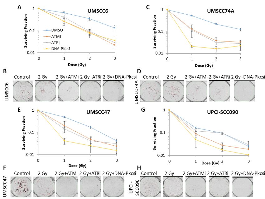
Cancers 2020, 12, 1490                                                                             4 of 14

2.2. Survival of HNSCC Cells Following by Photon and Proton Irradiation Can Be Reduced by Targeting ATM,
ATR and DNA-Pkcs
      Using clonogenic assays, we first analysed the impact of targeting the major protein kinases
involved in DNA DSB repair using specific and characterised inhibitors (ATMi, KU-55933; ATRi, VE-821;
DNA-Pkcsi, KU-57788) on the survival of HPV-positive and HPV-negative HNSCC incubated with the
inhibitors for 24 h in the absence of radiation, versus a vehicle-only control (DMSO). This demonstrated
a varied response dependent on the cell line utilised (Figure S3), although ATRi significantly decreased
cell survival by 41–54% in all HNSCC cell lines, ATMi by 22–44% in three cell lines (UMSCC6,
UMSCC74A and UMSCC47), and DNA-Pkcsi had a significant impact on survival of only one of the
four cell lines (UMSCC47) by ~56%. We then analysed the impact of the inhibitors on HNSCC cell
survival post-irradiation. As a starting point, we demonstrated that the respective inhibitors, following
a 1 h pre-incubation of the cells prior to irradiation, were functional in suppressing ATM, ATR and
DNA-Pk phosphorylation, and therefore DSB signaling, in response to photons (Figure S4) and protons
(Figure S5). In combination with photon irradiation, we demonstrate that there was a significant
impact in reducing cell survival of HPV-negative HNSCC cells in the presence of either ATMi, ATRi or
DNA-Pkcsi (1 h pre-incubation, followed by a further treatment for 24 h post-irradiation) versus the
DMSO control (Figure 2A–D; see also Figure S6A–D for linear scale graphs and data fitting), with dose
enhancement ratios (DER) of 1.91–2.39 (Table 1). The significantly enhanced radiosensitivity of only
one HPV-positive HNSCC cell line (UMSCC47) was also seen (Figure 2E–H), although the DER values
of 1.36–1.69 were notably lower than those observed in the HPV-negative cells (Table 1). The cell
survival of the most inherently radiosensitive HPV-positive cell line (UPCI-SCC090) only appeared to
be dramatically decreased in the presence of DNA-Pkcsi (DER of 1.36). These data are supported by
statistical analysis (Table S1) and, in general, DNA-Pkcsi appeared the most potent radiosensitiser of
all the HNSCC cell lines.

      Table 1. Dose enhancement ratios calculated at 50% cell survival (DER) following ATM, ATR and
      DNA-Pkcs inhibition versus DMSO controls in HNSCC cells in response to photons.

          Inhibitor         UMSCC6           UMSCC74A            UMSCC47            UPCI-SCC090
           ATM                2.06               1.91               1.38                1.15
           ATR                1.91               2.01               1.36                1.02
         DNA-Pkcs             1.93               2.39               1.69                1.36

     Following proton irradiation, and similar to photons, we again observed that ATMi and DNA-Pkcsi
significantly enhanced the radiosensitisation of both HPV-negative HNSCC cell lines (Figure 3A–D
and Table S2; see also Figure S7A–D for linear scale graphs and data fitting) with DER values of
1.52–2.01 (Table 2). HPV-positive HNSCC cell lines were also radiosensitised, with DER values of
1.24–1.49 (Table 2), following proton irradiation in combination with inhibition of ATM and DNA-Pkcs
(Figure 3E–H). However, radiosensitisation was only significantly enhanced in UMSCC47, and not
UPCI-SCC090 cell lines (Table S2). ATRi appeared in general less effective at radiosensitising the
HNSCC cells in response to protons (DER values of 1.25–1.48; Table 2).

      Table 2. Dose enhancement ratios calculated at 50% cell survival (DER) following ATM, ATR and
      DNA-Pkcs inhibition versus DMSO controls in HNSCC cells in response to protons.

          Inhibitor         UMSCC6           UMSCC74A            UMSCC47            UPCI-SCC090
           ATM                1.62               1.52               1.49                1.24
           ATR                1.25               1.42               1.28                1.30
         DNA-Pkcs             2.01               1.64               1.38                1.32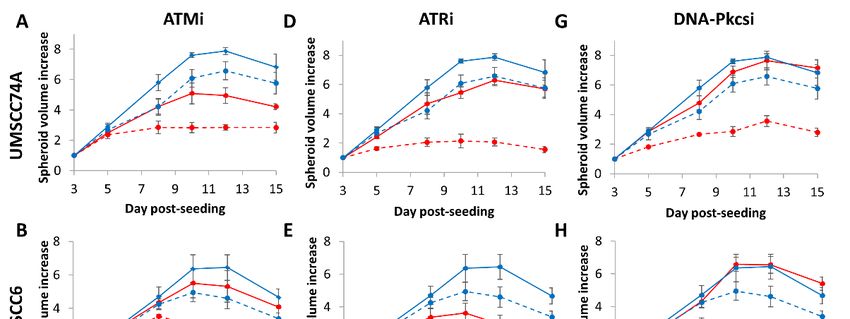
Cancers 2020, 12, 1490                                                                                                              5 of 14
            Cancers 2020, 12, x FOR PEER REVIEW                                                                        5 of 15

      Figure 2. Figure    2. Inhibition
                 Inhibition              of ataxia
                                  of ataxia        telangiectasia-mutated (ATM),
                                               telangiectasia-mutated        (ATM), ataxia telangiectasia
                                                                                        ataxia            and Rad3-related
                                                                                                telangiectasia     and Rad3-related
                (ATR) and DNA-dependent protein kinase (DNA-Pkcs) can enhance sensitivity of HNSCC cells to
      (ATR) and DNA-dependent protein kinase (DNA-Pkcs) can enhance sensitivity of HNSCC cells to
                photon irradiation. Clonogenic survival of HNSCC cells following treatment with increasing doses of
      photon irradiation.
                x-rays in theClonogenic
                                 presence of DMSOsurvival   of HNSCC
                                                      (Control), ATMi (10cells
                                                                            µM),following
                                                                                  ATRi (1 µM)treatment     with(1increasing
                                                                                               and DNA-Pkcsi       µM) was doses of
      x-rays in the   presence      of   DMSO     (Control),    ATMi   (10   µM),   ATRi    (1 µM)   and   DNA-Pkcsi
                analysed from three biologically independent experiments. (A, C, E and G) Shown is the surviving           (1 µM) was
                fraction   ± S.E. (B, D, F and H) representative  images of colonies in non-irradiated and
      analysed from three biologically independent experiments. (A,C,E,G) Shown is the surviving fraction  irradiated plates
                (the latter of which were seeded with four times the number of cells).
      ± S.E. (B,D,F,H)     representative images of colonies in non-irradiated and irradiated plates (the latter of
           Cancers 2020, 12, x FOR PEER REVIEW                                                                              6 of 15
      which were    seeded
                Table 1. Dose   with   four times
                                   enhancement        thecalculated
                                                  ratios  numberatof50% cells).
                                                                            cell survival (DER) following ATM, ATR and
                  DNA-Pkcs inhibition versus DMSO controls in HNSCC cells in response to photons.

                                                                                                     UPCI-
                            Inhibitor         UMSCC6          UMSCC74A          UMSCC47
                                                                                                    SCC090

                              ATM                 2.06             1.91             1.38              1.15
                              ATR                 1.91             2.01             1.36              1.02
                           DNA-Pkcs               1.93             2.39             1.69              1.36

                 Following proton irradiation, and similar to photons, we again observed that ATMi and DNA-
            Pkcsi significantly enhanced the radiosensitisation of both HPV-negative HNSCC cell lines (Figure
            3A–D and Table S2; see also Figure S7A–D for linear scale graphs and data fitting) with DER values
            of 1.52–2.01 (Table 2). HPV-positive HNSCC cell lines were also radiosensitised, with DER values of
            1.24–1.49 (Table 2), following proton irradiation in combination with inhibition of ATM and DNA-
            Pkcs (Figure 3E–H). However, radiosensitisation was only significantly enhanced in UMSCC47, and
            not UPCI-SCC090 cell lines (Table S2). ATRi appeared in general less effective at radiosensitising the
            HNSCC cells in response to protons (DER values of 1.25–1.48; Table 2).

      Figure 3. Figure  3. Inhibition
                Inhibition     of ATM,of ATM,
                                           ATRATRandand  DNA-Pkcs can
                                                      DNA-Pkcs      canenhance
                                                                         enhance sensitivity of HNSCC
                                                                                     sensitivity         cells to proton
                                                                                                     of HNSCC       cells to proton
                irradiation. Clonogenic survival of HNSCC cells following treatment with increasing doses of protons
      irradiation. Clonogenic survival of HNSCC cells following treatment with increasing doses of protons
                in the presence of DMSO (Control), ATMi (10 µM), ATRi (1 µM) and DNA-Pkcsi (1 µM) was analysed
      in the presence   of DMSO
                from four            (Control),
                            biologically          ATMi
                                         independent      (10 µM),(A,
                                                      experiments.  ATRi
                                                                      C, E (1
                                                                            andµM)   and DNA-Pkcsi
                                                                                 G) Shown                  (1 µM)
                                                                                             is the surviving         was
                                                                                                               fraction ± analysed
      from fourS.E.
                  biologically
                     (B, D, F andindependent        experiments.
                                    H) representative                (A,C,E,G)
                                                      images of colonies            Shown and
                                                                         in non-irradiated    is the  surviving
                                                                                                  irradiated platesfraction
                                                                                                                     (the     ± S.E.
      (B,D,F,H) latter of which were
                 representative       seeded with
                                    images         four times
                                              of colonies   inthe number of cells).and irradiated plates (the latter of which
                                                               non-irradiated
      were seeded with four times the number of cells).
                  Table 2. Dose enhancement ratios calculated at 50% cell survival (DER) following ATM, ATR and
                  DNA-Pkcs inhibition versus DMSO controls in HNSCC cells in response to protons.

                                                                                                      UPCI-
                           Inhibitor         UMSCC6           UMSCC74A           UMSCC47
                                                                                                     SCC090

                             ATM                  1.62             1.52              1.49              1.24
                              ATR                 1.25             1.42              1.28              1.30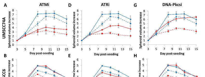
Cancers 2020, 12, 1490 6 of 14
2.3. 3D Spheroid Growth of HNSCC Cells Following by Photon and Proton Irradiation Can Be Inhibited by
Targeting ATM, ATR and DNA-Pkcs
We subsequently analysed the impact of DNA DSB repair inhibitors on the radiosensitivity of
HNSCC cells utilising 3D spheroids, which more accurately reflect the structure and environment of
the original tumour. Of the cells used, unfortunately one HPV-positive cell line (UMSCC47) did not
form 3D spheroids that grew during the 15-day analysis period. It was also noted that spheroids from
both HPV-negative HNSCC grew significantly faster (peaking at days 8–10 post-seeding) than the one
remaining HPV-positive HNSCC (the increase in growth largely occurred at days 7–15 post-seeding).
All spheroids grew ~5–8-fold in volume in the absence of any treatments over the analysis period
(Figure 4A–I and Figure S8). We demonstrate that ATMi alone caused a significant ~1.7-fold delay
inCancers
the growth
2020, 12, xof only
FOR PEERHPV-negative
REVIEW HNSCC (UMSCC74A) spheroids, and that the combination 7 of 15
of ATMi plus photon irradiation was effective in suppressing the growth of these spheroids by
fold compared
~2.0-fold compared to radiation alone,
to radiation but not
alone, but of
notthe
ofother two spheroid
the other models
two spheroid (Figure
models 4A–C4A–C
(Figure and Table
and
3). In contrast, ATRi alone caused a statistically significant ~1.5–1.6-fold growth delay
Table 3). In contrast, ATRi alone caused a statistically significant ~1.5–1.6-fold growth delay in all in all spheroid
models. models.
spheroid The inhibitor significantly
The inhibitor exacerbated
significantly the effects
exacerbated the of photon
effects irradiation,
of photon by ~1.3-fold
irradiation, (UPCI-
by ~1.3-fold
SCC090) to 2.3-fold (UMSCC74A) (Figure 4D–F and Table 3). DNA-Pkcsi alone
(UPCI-SCC090) to 2.3-fold (UMSCC74A) (Figure 4D–F and Table 3). DNA-Pkcsi alone was, interestingly,was, interestingly,
ineffectiveinininhibiting
ineffective inhibitingspheroid
spheroidgrowth,
growth,although
althoughthethecombination
combinationofofDNA-Pkcsi
DNA-Pkcsiwithwithphotons
photonswas was
effectiveininsuppressing
effective suppressingthe thegrowth
growthofofHPV-negative
HPV-negativeHNSCC HNSCCspheroids
spheroids~1.4-fold
~1.4-fold(UMSCC6)
(UMSCC6)and and
~1.6-fold (UMSCC74A) compared to the radiation alone
~1.6-fold (UMSCC74A) compared to the radiation alone (Figure 4G–I). (Figure 4G–I).
Figure4.4.Inhibition
Figure InhibitionofofATM,
ATM,ATR ATRandandDNA-Pkcs
DNA-Pkcs in in
combination
combination with photons
with cancan
photons decrease growth
decrease of
growth
HNSCC
of HNSCC 3D spheroids. Spheroids
3D spheroids. were allowed
Spheroids to develop
were allowed for 48 h, for
to develop pretreated with DMSO
48 h, pretreated (Control),
with DMSO
ATMi (10 µM),
(Control), ATMiATRi(10(1µM), and DNA-Pkcsi
µM)ATRi (1 µM) and(1DNA-Pkcsi
µM), and irradiated
(1 µM), and withirradiated
a single dose
with(1a Gy) of x-rays.
single dose (1
Spheroid growth
Gy) of x-rays. of (A,D,G)
Spheroid UMSCC74A,
growth of (A, D and(B,E,H) UMSCC6 and
G) UMSCC74A, (B,(C,F,I)
E andUPCI-SCC090
H) UMSCC6 and was(C,
measured
F, and I)
by microscopy and
UPCI-SCC090 was analysed
measuredfrombythree biologically
microscopy independent
and analysed experiments. Solid blue line
from three biologically is DMSO
independent
only, dashed blue
experiments. lines
Solid areline
blue DMSO plus 1only,
is DMSO Gy x-rays,
dashedsolid red
blue lineare
lines is inhibitor
DMSO plusonly,1dashed red lines
Gy x-rays, solidare
red
inhibitors plus 1 Gy
line is inhibitor x-rays.
only, dashedShown is theare
red lines spheroid plus 1±Gy
volume
inhibitors S.E.x-rays. Shown is the spheroid volume
± S.E.
Table 3. Targeting of ATM, ATR and DNA-Pkcs alone and in combination with photons and protons
to decrease 3D HPV-positive and HPV-negative HNSCC spheroid growth.
Inhibitor UMSCC74A UMSCC6 UPCI-SCC090
ATM p < 0.0002 p = 0.60 p = 0.34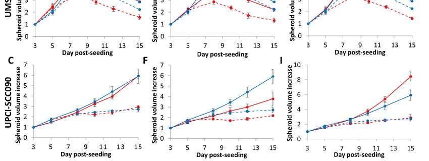
Cancers 2020, 12, 1490 7 of 14
Table 3. Targeting of ATM, ATR and DNA-Pkcs alone and in combination with photons and protons to
decrease 3D HPV-positive and HPV-negative HNSCC spheroid growth.
Inhibitor UMSCC74A UMSCC6 UPCI-SCC090
ATM p < 0.0002 p = 0.60 p = 0.34
ATR p < 0.003 p < 0.002 p < 0.006
DNA-Pkcs p = 0.59 p = 0.89 p = 0.54
ATM + photons p < 0.004 p = 0.18 p = 0.76
ATR + photons p < 0.0005 p < 0.02 p < 0.03
DNA-Pkcs + photons p < 0.02 p < 0.05 p = 0.08
ATM + protons p < 0.02 p = 0.06 p = 0.24
ATR + protons p < 0.0002 p < 0.003 p < 0.0008
DNA-Pkcs + protons p < 0.03 p < 0.02 p = 0.18
Cancers 2020, 12, x FOR PEER REVIEW 8 of 15
Statistical analysis was performed on all the dataset across the 15-day growth period using a one-way ANOVA,
comparing the growth of inhibitor
DNA-Pkcs treated spheroids
+ protons against the appropriate
p < 0.03 p < 0.02 DMSO control
p = 0.18(± radiation).
Statistical analysis was performed on all the dataset across the 15-day growth period using a one-way
ANOVA, comparing the growth of inhibitor treated spheroids against the appropriate DMSO control
We observed very similar results in HPV-negative HNSCC spheroids following proton irradiation
(± radiation).
(Table 3). Here, the combination of protons with ATMi (Figure 5A–C) was significantly effective
in only one spheroid model
We observed (UMSCC74A)
very similar resultsas inobserved
HPV-negativeby the ~2-fold
HNSCC growth
spheroids inhibition
following protonversus the
irradiation (Table 3). Here, the combination of protons with ATMi (Figure 5A–C)
radiation alone, whereas ATRi (Figure 5D–F) and DNA-Pkcsi (Figure 5G–I) had a significant impact was significantly
effective in only one spheroid model (UMSCC74A) as observed by the ~2-fold growth inhibition
on delaying the growth of both spheroid models by ~2.5-fold and ~1.9-fold, respectively (see also
versus the radiation alone, whereas ATRi (Figure 5D–F) and DNA-Pkcsi (Figure 5G–I) had a
Figure S9).significant
HPV-positive
impact HNSCC
on delaying(UPCI-SCC090)
the growth of both spheroids
spheroidwere
modelsonly significantly
by ~2.5-fold radiosensitised,
and ~1.9-fold,
by ~1.6-fold, with protons
respectively in the
(see also presence
Figure of ATRi (Figure
S9). HPV-positive HNSCC 5F). Notably, following
(UPCI-SCC090) bothonly
spheroids were photon and
significantly
proton irradiation of radiosensitised,
the HPV-positiveby ~1.6-fold, with protonsspheroids,
UPCI-SCC090 in the presence of ATRi
there was(Figure 5F). Notably,
a reduced impact of the
following both photon and proton irradiation of the HPV-positive UPCI-SCC090 spheroids, there
inhibitors compared to the radiation alone, which is consistent with this being the most radiosensitive
was a reduced impact of the inhibitors compared to the radiation alone, which is consistent with this
cell line, asbeing
observed byradiosensitive
the most clonogeniccell assays
line, as(Figure
observed1B,D).
by clonogenic assays (Figure 1B,D).
Figure 5. Inhibition of ATM,
Figure 5. Inhibition of ATR
ATM,andATR DNA-Pkcs
and DNA-Pkcs inincombination with
combination with protons
protons can can decrease
decrease growthgrowth of
HNSCC 3Dofspheroids.
HNSCC 3D Spheroids
spheroids. Spheroids were allowed
were allowed to develop
to develop for 48forh,48pretreated
h, pretreated with
with DMSO(Control),
DMSO
(Control), ATMi (10 µM), ATRi (1 µM) and DNA-Pkcsi (1 µM), and irradiated with a single dose (2
ATMi (10 µM), ATRi (1 µM) and DNA-Pkcsi (1 µM), and irradiated with a single dose (2 Gy) of protons.
Gy) of protons. Spheroid growth of (A, D and G) UMSCC74A, (B, E and H) UMSCC6 and (C, F, and
Spheroid growth of (A,D,G)
I) UPCI-SCC090 UMSCC74A,
was measured (B,E,H) and
by microscopy UMSCC6analysedand
from(C,F,I) UPCI-SCC090
three biologically was measured
independent
by microscopy and analysed
experiments. from
Solid blue three
line is DMSObiologically
only, dashedindependent experiments.
blue lines are DMSO Solid blue
plus 2 Gy protons, solidline
red is DMSO
lineblue
only, dashed is inhibitor
lines only, dashed red
are DMSO lines
plus 2 are
Gyinhibitors
protons,plus 2 Gyred
solid protons.
line Shown is the spheroid
is inhibitor volume red lines
only, dashed
± S.E.
are inhibitors plus 2 Gy protons. Shown is the spheroid volume ± S.E.
We extended our observations of the effectiveness of inhibitors targeting ATM, ATR and DNA-
Pkcs in radiosensitising oropharyngeal HNSCC cells by utilising additional spheroid models from
HPV-negative HNSCC, which are relatively more radioresistant than HPV-positive HNSCC. These
were designed to gain further evidence that DNA DSB repair inhibition can enhance the impact of
photons and protons in preventing spheroid growth, which are more representative of the original
tumour and its treatment in vivo. We therefore used spheroids from FaDu and A253 cell lines that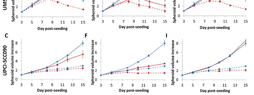
Cancers 2020, 12, 1490 8 of 14
We extended our observations of the effectiveness of inhibitors targeting ATM, ATR and
DNA-Pkcs in radiosensitising oropharyngeal HNSCC cells by utilising additional spheroid models
from HPV-negative HNSCC, which are relatively more radioresistant than HPV-positive HNSCC.
These were designed to gain further evidence that DNA DSB repair inhibition can enhance the
impact of photons and protons in preventing spheroid growth, which are more representative of
the original tumour and its treatment in vivo. We therefore used spheroids from FaDu and A253
cell lines that originate from the hypopharynx and oral cavity, respectively, of which we observed
that these increased dramatically in volume (by ~50-fold and ~15-fold, respectively) over a period
Cancers 2020, 12, x FOR PEER REVIEW 9 of 15
of 15 days post-seeding (Figure 6A–L and Figure S10). FaDu spheroids were particularly resistant to
ATMi, ATRi and DNA-Pkcsi
originate alone, as observed
from the hypopharynx by the
and oral cavity, lack of impact
respectively, of whichonwe spheroid
observedgrowth.
that theseThe A253
increased dramatically in volume (by ~50-fold and ~15-fold, respectively)
spheroids appeared to display some delayed growth in the presence of the inhibitors alone, over a period of 15 days
particularly
post-seeding (Figures 6A–L and S10). FaDu spheroids were particularly resistant to ATMi, ATRi and
at the 12- and 15-day time points, although this was not statistically significant
DNA-Pkcsi alone, as observed by the lack of impact on spheroid growth. The A253 spheroids
across the whole time
course (Table 4). The combination of photons with either of the inhibitors significantly
appeared to display some delayed growth in the presence of the inhibitors alone, particularly at the suppressed the
growth of A253
12- andspheroids, whichalthough
15-day time points, was markedly enhanced
this was not by
statistically ~2.8–3.2-fold
significant versus
across the whole the
timeradiation
course alone
(Figure 6A–F(Table
and4).Table
The combination of photons were
4). FaDu spheroids with either
only of the inhibitorsradiosensitised
significantly significantly suppressed
in the the
presence of
growth of A253 spheroids, which was markedly enhanced by ~2.8–3.2-fold versus the radiation alone
DNA-Pkcsi(Figure
following photon irradiation, through a dramatic ~4.6-fold decrease in spheroid growth.
6A–F and Table 4). FaDu spheroids were only significantly radiosensitised in the presence of
In responseDNA-Pkcsi
to proton irradiation,
following ATRi wasthrough
photon irradiation, not significantly effective
a dramatic ~4.6-fold at radiosensitising
decrease in spheroid growth. the cells,
but the combination
In response toofprotonATMi with protons
irradiation, ATRi waswasnotable to suppress
significantly effectivegrowth of both the
at radiosensitising A253cells,and FaDu
but the combination of ATMi with protons was able to suppress growth
spheroids by ~3.7-fold. Furthermore, DNA-Pkcsi was particularly effective in combination with of both A253 and FaDu
spheroids by ~3.7-fold. Furthermore, DNA-Pkcsi was particularly effective in combination with
protons as observed by the ~3.6-fold and ~7.6-fold decrease in the spheroid growth of A253 and FaDu
protons as observed by the ~3.6-fold and ~7.6-fold decrease in the spheroid growth of A253 and FaDu
cells, respectively, in comparison
cells, respectively, to radiation
in comparison alone
to radiation alone(Figure 6G–L
(Figure 6G–L andand Table
Table 4). 4).
Figure 6. Inhibition
Figure 6. Inhibition of ATM, of ATM,
ATRATR andand DNA-Pkcs in
DNA-Pkcs in combination
combination withwith
photons and protons
photons can
and protons can
decrease growth of HPV-negative HNSCC 3D spheroids. Spheroids were allowed to develop for 48
decrease growth of HPV-negative HNSCC 3D spheroids. Spheroids were allowed to develop for 48 h,
h, pretreated with DMSO (Control), ATMi (10 µM), ATRi (1 µM) and DNA-Pkcsi (1 µM), and
pretreated with DMSO
irradiated with (Control),
a single doseATMi
of (A–F)(10 µM),
x-rays at 1ATRi
Gy or (1 µM)
(G-L) and at
protons DNA-Pkcsi
2 Gy. Spheroid(1 growth
µM), and
of (A,irradiated
with a single dose
C, E, G, Iof
and(A–F) x-rays at 1(FaDu)
K) hypopharynx Gy orand (G–L)
(B, D,protons atL)2 A253
F, H, J and Gy. Spheroid
was measured growth of (A,C,E,G,I,K)
by microscopy
hypopharynxand (FaDu)
analysed and
from (B,
threeD,
biologically
F, H, J andindependent
L) A253experiments.
was measured Solid blue line is DMSO only,
by microscopy anddashed
analysed from
blue lines (A–F) are DMSO plus 1 Gy x-rays or (G–L) 2 Gy protons, solid red lines are inhibitor only,
three biologically independent experiments. Solid blue line is DMSO only, dashed blue lines (A–F) are
dashed red lines are inhibitor plus (A–F) 1 Gy x-rays or (G–L) 2 Gy protons. Shown is the spheroid
DMSO plusvolume
1 Gy ±x-rays
S.E. or (G–L) 2 Gy protons, solid red lines are inhibitor only, dashed red lines are
inhibitor plus (A–F) 1 Gy x-rays or (G–L) 2 Gy protons. Shown is the spheroid volume ± S.E.
Table 4. Targeting of ATM, ATR and DNA-Pkcs alone and in combination with photons and protons
to decrease 3D HPV-negative HNSCC spheroid growth.
Inhibitor FaDu A253
ATM p = 0.69 p = 0.49Cancers 2020, 12, 1490 9 of 14
Table 4. Targeting of ATM, ATR and DNA-Pkcs alone and in combination with photons and protons to
decrease 3D HPV-negative HNSCC spheroid growth.
Inhibitor FaDu A253
ATM p = 0.69 p = 0.49
ATR p = 0.89 p = 0.72
DNA-Pkcs p = 0.82 p = 0.88
ATM + photons p = 0.09 p < 0.002
ATR + photons p = 0.28 p < 0.003
DNA-Pkcs + photons p < 0.003 p < 0.002
ATM + protons p < 0.03 p < 0.006
ATR + protons p = 0.24 p = 0.11
DNA-Pkcs + protons p < 0.005 p < 0.002
Statistical analysis was performed on the dataset across the 15-day growth period using a one-way ANOVA,
comparing the growth of inhibitor treated spheroids against the appropriate DMSO control (± radiation).
3. Discussion
Accumulating evidence has suggested that the increased response of patients with HPV-positive
versus HPV-negative HNSCC to radiotherapy, and thus the improved survival rates, is caused by
defects in the repair of DNA DSBs [9,11,12]. Therefore, targeting key enzymes involved in DNA
DSB repair, particularly the protein kinases ATM, ATR and DNA-Pkcs, in relatively radioresistant
HPV-negative HNSCC that are DSB repair-proficient is considered to be an approach to sensitise
these tumours to radiotherapy. Indeed, there is evidence of at least one clinical trial utilising either
ATRi or DNA-Pkcsi in combination with conventional radiotherapy that is currently underway [16].
Additionally, while there is an increasing use of proton beam therapy for the treatment of HNSCC, there
is no preclinical evidence to date examining the impact of DNA DSB repair inhibitors in combination
with protons, and whether there is any substantial difference compared to that observed following
photon irradiation. In this study, we have now analysed the effect of ATMi, ATRi and DNA-Pkcsi on
both monolayer and 3D spheroid models of HPV-positive and HPV-negative HNSCC in combination
with photons and protons.
Interestingly, we discovered that targeting either ATM, ATR or DNA-Pkcs can decrease the
clonogenic survival of HNSCC cells in response to photons and protons. DNA-Pkcsi appeared
particularly effective in all cell lines in combination with radiation. This would correlate with studies
in HPV-negative HNSCC cells describing downregulation of DNA-Pkcs using siRNA in UTSCC15
and UTSCC45 cells [25], as well as the DNA-Pkcs inhibitors KU0060648 in HN4 and HN5 cells [17],
and IC87361 in UTSCC54, UTSCC74B and UTSCC76B cells [18], which were shown to enhance
radiosensitisation. Only a single study has examined ATM inhibition (GSK635416A) in HNSCC
cells [19], although this demonstrated increased radiosensitivity in five HPV-negative HNSCC cell
lines (UTSCC2, UTSCC8, UTSCC24A, UTSCC36 and UTSCC40), which is comparable with our data.
However, there are a number of studies that have focused on ATR as a target, including using siRNA
in UPCI-SCC029B, UPCI-SCC040 and UPCI-SCC131 cells [26]. Additionally, the ATR inhibitor VE821
displayed improved radiosensitivity in SQ20B cells [20], and an alternative inhibitor, AZD6738, showed
the same phenotype in Cal27, FaDu, HN4 and HN5 cells [17,21]. In our experiments utilising clonogenic
assays, ATRi appeared to be less effective at radiosensitising cells following proton irradiation. We also
observed less of an impact of DNA DSB repair inhibition in combination with radiation in HPV-positive
HNSCC cells, particularly the UPCI-SCC090 cell line largely as this is the most inherently radiosensitive
as shown here, and in our previous study [12].
Utilising 3D spheroid models that more accurately replicate the structure and environment of
the original tumour, we further demonstrated the effectiveness of DNA-Pkcsi in combination with
both photons and protons in inhibiting growth of all the HPV-negative HNSCC spheroids analysed.
Interestingly though, inhibition of DNA-Pkcs alone did not appear to have any impact on the growth
of 3D spheroids of both HPV-positive and HPV-negative HNSCC (which was largely supportedCancers 2020, 12, 1490 10 of 14
by utilising clonogenic survival assays). This suggests that DNA-Pkcs is not essential for HNSCC
cell growth and survival in the absence of ionising radiation-induced stress. Nevertheless, and
similar to clonogenic assay results, the combination strategy of DSB inhibition (particularly ATMi and
DNA-Pkcsi) did not significantly enhance the effect of radiation on the HPV-positive HNSCC spheroids
(UPCI-SCC090), due to these cells being the most radiosensitive. The inhibition of ATR displayed
some effectiveness in combination with photons and protons in preventing spheroid growth. However,
less of an impact was observed on the relatively radioresistant HPV-negative HNSCC spheroid models,
FaDu and A253, that displayed significant spheroid growth over the time period post-irradiation.
This observation is similar to previous data utilising the ATR inhibitor AZD6738 with photons only,
which demonstrated that this combination did not impede growth of 3D spheroids of FaDu cells [21].
Noteworthily, as a monotherapy, the inhibition of ATR alone in the absence of radiation was effective
in inhibiting clonogenic survival, but also the growth of HNSCC spheroids (apart from FaDu and
A253), which was comparable to the impact caused by a single dose of radiation alone.
Cumulatively, our results suggest that targeting DNA DSB repair via NHEJ (ATM and DNA-Pkcs)
or HR (ATR) can exacerbate the impact of photons in radiosensitising HNSCC cell models, and
that the combination of DNA-Pkcsi with photons in HPV-negative HNSCC cells that are relatively
radioresistant was particularly effective. This adds to the growing preclinical evidence [17–21,25,26]
that this is an effective combination for the treatment of HNSCC that should be investigated further,
particularly using more advanced 3D models (e.g., patient-derived organoids) and appropriate in vivo
experiments. However, we now also demonstrate that DSB repair inhibition, particularly DNA-Pkcsi
and to a lesser extent ATMi, are efficient in reducing the survival and spheroid growth of HNSCC
cells in response to protons. In fact in general, relatively similar results were observed comparing
photons and protons, although the DER values derived from clonogenic assay results were much lower
with ATRi following protons than with photons. This would contradict some very limited evidence
suggesting a greater dependence on the HR pathway mediated by ATR for repairing DNA DSBs
induced by protons, which was obtained using RAD51 siRNA in A549 lung cancer cells [24]. In fact
other studies, largely conducted in Chinese hamster ovary cells, reflect that NHEJ, coordinated by
ATM and DNA-Pkcs, is the major DSB repair pathway employed following proton irradiation [27,28].
This is in agreement with our results. Consequently, we would advocate that inhibition of NHEJ
through DNA-Pkcs is the most promising strategy in optimising the radiosensitisation of HNSCC cells
with either photons or protons. Nevertheless, it should be noted that our study utilised low linear
energy transfer (LET) protons at the entrance dose of a pristine beam, and that different results may be
obtained with cells irradiated at or around the Bragg peak where the LET increases. This is due to the
increased amount of complex DNA damage, where multiple lesions are generated in close proximity,
and therefore the potential for the generation of complex DNA DSBs that could have a different
requirement for either NHEJ or HR [23]. We are also acutely aware of the availability of more potent
and selective inhibitors than the ones used in the current study, specifically those targeting ATM (e.g.,
AZD1390), ATR (e.g., AZD6738) and DNA-Pkcs (e.g., AZD7648), which require examination of their
potential to radiosensitise HNSCC cell models following photon and proton irradiation. These points
are consequently the subject of our ongoing and future studies.
4. Materials and Methods
4.1. Cell Lines and Culture Conditions
Oropharyngeal squamous cell carcinoma cells (UMSCC6, UMSCC74 and UMSCC47) were kindly
provided by Prof T. Carey, University of Michigan, USA. Cells from the hypopharynx (FaDu) and
submaxillary gland (A253) originated from ATCC (Teddington, UK). HPV-positive oropharyngeal
squamous cell carcinoma cells (UPCI-SCC090) were kindly provided by Dr S. Gollin from the University
of Pittsburgh. All cells, apart from UPCI-SCC090 and FaDu (which were cultured in Minimal Essential
Medium (MEM)), were routinely cultured as monolayers in Dulbecco’s Modified Eagle MediumCancers 2020, 12, 1490 11 of 14
(DMEM) supplemented with 10% fetal bovine serum, 2 mM L-glutamine, 1× penicillin-streptomycin
and 1× non-essential amino acids. All cells were cultured under standard conditions in 5% CO2 at
37 ◦ C, and were authenticated in our laboratory by short tandem repeat (STR) profiling.
4.2. Clonogenic Assays
Cells were harvested and a defined number seeded in triplicate into 6-well plates or 35 mm dishes
before incubation overnight in 5% CO2 at 37 ◦ C to allow the cells to attach. Plating efficiencies for the
cells were as followed: UMSCC6 (~10%), UMSCC74A (~10%), UMSCC47 (~10%) and UPCI-SCC090
(~2%). For inhibition experiments, cells were pretreated with DMSO (as a vehicle only control), 10 µM
ATM inhibitor (ATMi; KU-55933), 1 µM ATR inhibitor (ATRi; VE-821) or 1 µM DNA-Pkcs inhibitor
(DNA-Pkcsi; KU-57788; Selleck Chemicals, Munich, Germany) for 1 h prior to irradiation. Cells were
then irradiated using a CellRad x-ray irradiator (Faxitron Bioptics, Tucson, AZ, USA) or with a passive
scattered horizontal proton beam line of 60 MeV maximal energy, as previously described [29,30].
Higher doses of protons were comparatively used due to cells being positioned at the entrance dose of
a pristine (unmodulated) beam (~1 keV/µm). Following irradiation, fresh media containing inhibitors
was added to the cells for 24 h, which was then replaced with fresh media alone and colonies allowed
to grow for 7–12 days, prior to fixing and staining with 6% glutaraldehyde and 0.5% crystal violet
for 30 min. Dishes were washed, left to air dry overnight and colonies counted using the GelCount
colony analyser (Oxford Optronics, Oxford, UK). Colony counting settings were optimised for each
cell line, based on inclusion of distinct colonies of specific size and intensity, although the same settings
were used across the various treatments. Relative colony formation (surviving fraction) was expressed
as colonies per treatment level versus colonies that appeared in the untreated control, and data was
derived from at least three individual biological replicates.
4.3. Spheroid Growth Assays
Cells (500–1000/well) were seeded in triplicate in 100 µL Advanced MEM media (Life
Technologies, Paisley, UK) containing 1% B27 supplement, 0.5% N-2 supplement, 2 mM L-glutamine,
1× penicillin-streptomycin, 5 µg/mL heparin, 20 ng/µL epidermal growth factor and 10 ng/µL fibroblast
growth factor into 96-well ultra-low attachment plates (Corning B.V. Life Sciences, Amsterdam,
The Netherlands) and spheroids of ~200 µm in diameter allowed to form for 48 h (Day 3). DMSO,
ATMi, ATRi and DNA-Pkcsi were added 1 h prior to irradiation. Post-irradiation, 50 µL media was
removed and replaced with 50 µL fresh media containing DMSO or inhibitors for 24 h, and then 50 µL
media removed and replaced by 100 µL with fresh media alone. Images of spheroids were captured
up to 15 days post-seeding using an EVOS M5000 Imaging System (Life Technologies, Paisley, UK).
The diameter of the spheroids was analysed using ImageJ, and used to calculate spheroid volume
using the formula 4/3 × π × (d/2)3 .
4.4. Statistical Analysis
Dose enhancement ratios (DER) were used to assess the significance of the clonogenic assay results.
DER values are derived from the ratio of the dose (Gy) required for a surviving fraction of 0.5 in the
vehicle (DMSO) treated cells (D50DMSO ), over the dose (Gy) required for the same surviving fraction in
the inhibitor treated cells (D50inhibitor ) [DER = D50DMSO /D50inhibitor ]. D50 values were calculated using
a linear quadratic fitting on each curve. Statistical analysis of spheroid growth data was performed
on the dataset across the 15-day growth period using a one-way ANOVA. For this, the effect of each
inhibitor on the spheroid growth was compared against the vehicle (DMSO) for a given radiation dose
and radiation type. p-values ofCancers 2020, 12, 1490 12 of 14
5. Conclusions
We have demonstrated that the inhibition of DNA DSB repair can effectively act in combination
with both conventional (photon) radiotherapy and proton beam therapy in radiosensitising in vitro
models of HNSCC. DNA-Pkcsi was shown to be particularly effective in preventing clonogenic survival
and 3D spheroid growth of HNSCC, and specifically models of relatively radioresistant HPV-negative
HNSCC. Our data suggest that targeting DNA-Pkcs in combination with radiotherapy can be an
effective strategy for the treatment of HNSCC.
Supplementary Materials: The following are available online at http://www.mdpi.com/2072-6694/12/6/1490/s1,
Figure S1: Expression of p16 in HPV-negative and HPV-positive HNSCC cells; Figure S2. Increased radiosensitivity
of HPV-positive HNSCC cells in comparison to HPV-negative cells in response to photons and protons. Figure S3:
Comparison of cell survival in HNSCC cell lines in the presence of DNA DSB repair inhibitors; Figure S4: Inhibition
of DSB repair signaling following photon irradiation; Figure S5: Inhibition of DSB repair signaling following
proton irradiation; Figure S6. Inhibition of ATM, ATR and DNA-Pkcs increases radiosensitivity of HNSCC cells to
photon irradiation; Figure S7. Inhibition of ATM, ATR and DNA-Pkcs increases radiosensitivity of HNSCC cells
to proton irradiation. Figure S8: Inhibition of HNSCC spheroid growth using DSB repair inhibitors in the absence
and presence of photon irradiation; Figure S9: Inhibition of HNSCC spheroid growth using DSB repair inhibitors
in the absence and presence of proton irradiation; Figure S10: Inhibition of HPV-negative HNSCC spheroid growth
using DSB repair inhibitors in the absence and presence of photon and proton irradiation; Table S1: Inhibition of
ATM, ATR and DNA-Pkcs decreases cell survival in response to photon irradiation; Table S2: Inhibition of ATM,
ATR and DNA-Pkcs decreases cell survival in response to proton irradiation
Author Contributions: Conceptualization, J.L.P.; methodology, J.L.P., A.K. and E.T.V.; validation, J.L.P. and E.T.V.;
formal analysis, J.L.P. and E.T.V.; writing—original draft preparation, E.T.V.; writing—review & editing, J.L.P.
and E.T.V.; supervision, J.L.P.; project administration, J.L.P.; funding acquisition, J.L.P. All authors have read and
agreed to the published version of the manuscript.
Funding: Supported by a grant to J.L.P. from North West Cancer Research (CR CC7).
Acknowledgments: The authors thank T. Carey and S. Gollin for providing the HNSCC cells. We also thank Brian
Marsland and Ian Taylor at the Clatterbridge Cancer Centre for technical assistance with proton irradiation of cells.
Conflicts of Interest: The authors declare no conflict of interest.
References
1. Chaturvedi, A.K.; Anderson, W.F.; Lortet-Tieulent, J.; Curado, M.P.; Ferlay, J.; Franceschi, S.; Rosenberg, P.S.;
Bray, F.; Gillison, M.L. Worldwide trends in incidence rates for oral cavity and oropharyngeal cancers. J. Clin.
Oncol. 2013, 31, 4550–4559. [CrossRef] [PubMed]
2. Marur, S.; D‘Souza, G.; Westra, W.H.; Forastiere, A.A. HPV-associated head and neck cancer: A virus-related
cancer epidemic. Lancet Oncol. 2010, 11, 781–789. [CrossRef]
3. Bray, F.; Ferlay, J.; Soerjomataram, I.; Siegel, R.L.; Torre, L.A.; Jemal, A. Global cancer statistics 2018:
GLOBOCAN estimates of incidence and mortality worldwide for 36 cancers in 185 countries. CA Cancer J.
Clin. 2018, 68, 394–424. [CrossRef] [PubMed]
4. Ang, K.K.; Harris, J.; Wheeler, R.; Weber, R.; Rosenthal, D.I.; Nguyen-Tan, P.F.; Westra, W.H.; Chung, C.H.;
Jordan, R.C.; Lu, C.; et al. Human papillomavirus and survival of patients with oropharyngeal cancer.
N. Engl. J. Med. 2010, 363, 24–35. [CrossRef] [PubMed]
5. Fakhry, C.; Westra, W.H.; Li, S.; Cmelak, A.; Ridge, J.A.; Pinto, H.; Forastiere, A.; Gillison, M.L. Improved
survival of patients with human papillomavirus-positive head and neck squamous cell carcinoma in a
prospective clinical trial. J. Natl. Cancer Inst. 2008, 100, 261–269. [CrossRef] [PubMed]
6. Kumar, B.; Cordell, K.G.; Lee, J.S.; Worden, F.P.; Prince, M.E.; Tran, H.H.; Wolf, G.T.; Urba, S.G.; Chepeha, D.B.;
Teknos, T.N.; et al. EGFR, p16, HPV Titer, Bcl-xL and p53, sex, and smoking as indicators of response to
therapy and survival in oropharyngeal cancer. J. Clin. Oncol. 2008, 26, 3128–3137. [CrossRef]
7. Lassen, P.; Eriksen, J.G.; Hamilton-Dutoit, S.; Tramm, T.; Alsner, J.; Overgaard, J. Effect of HPV-associated
p16INK4A expression on response to radiotherapy and survival in squamous cell carcinoma of the head and
neck. J. Clin. Oncol. 2009, 27, 1992–1998. [CrossRef]
8. Gupta, A.K.; Lee, J.H.; Wilke, W.W.; Quon, H.; Smith, G.; Maity, A.; Buatti, J.M.; Spitz, D.R. Radiation
response in two HPV-infected head-and-neck cancer cell lines in comparison to a non-HPV-infected cell line
and relationship to signaling through AKT. Int. J. Radiat. Oncol. Biol. Phys. 2009, 74, 928–933. [CrossRef]Cancers 2020, 12, 1490 13 of 14
9. Rieckmann, T.; Tribius, S.; Grob, T.J.; Meyer, F.; Busch, C.J.; Petersen, C.; Dikomey, E.; Kriegs, M. HNSCC
cell lines positive for HPV and p16 possess higher cellular radiosensitivity due to an impaired DSB repair
capacity. Radiother. Oncol. 2013, 107, 242–246. [CrossRef]
10. Kimple, R.J.; Smith, M.A.; Blitzer, G.C.; Torres, A.D.; Martin, J.A.; Yang, R.Z.; Peet, C.R.; Lorenz, L.D.;
Nickel, K.P.; Klingelhutz, A.J.; et al. Enhanced radiation sensitivity in HPV-positive head and neck cancer.
Cancer Res. 2013, 73, 4791–4800. [CrossRef]
11. Weaver, A.N.; Cooper, T.S.; Rodriguez, M.; Trummell, H.Q.; Bonner, J.A.; Rosenthal, E.L.; Yang, E.S. DNA
double strand break repair defect and sensitivity to poly ADP-ribose polymerase (PARP) inhibition in
human papillomavirus 16-positive head and neck squamous cell carcinoma. Oncotarget 2015, 6, 26995–27007.
[CrossRef] [PubMed]
12. Nickson, C.M.; Moori, P.; Carter, R.J.; Rubbi, C.P.; Parsons, J.L. Misregulation of DNA damage repair
pathways in HPV-positive head and neck squamous cell carcinoma contributes to cellular radiosensitivity.
Oncotarget 2017, 8, 29963–29975. [CrossRef] [PubMed]
13. Gillison, M.L.; Akagi, K.; Xiao, W.; Jiang, B.; Pickard, R.K.L.; Li, J.; Swanson, B.J.; Agrawal, A.D.; Zucker, M.;
Stache-Crain, B.; et al. Human papillomavirus and the landscape of secondary genetic alterations in oral
cancers. Genome Res. 2019, 29, 1–17. [CrossRef] [PubMed]
14. Akagi, K.; Li, J.; Broutian, T.R.; Padilla-Nash, H.; Xiao, W.; Jiang, B.; Rocco, J.W.; Teknos, T.N.; Kumar, B.;
Wangsa, D.; et al. Genome-wide analysis of HPV integration in human cancers reveals recurrent, focal
genomic instability. Genome Res. 2014, 24, 185–199. [CrossRef] [PubMed]
15. Rusan, M.; Li, Y.Y.; Hammerman, P.S. Genomic landscape of human papillomavirus-associated cancers.
Clin. Cancer Res. 2015, 21, 2009–2019. [CrossRef]
16. Glorieux, M.; Dok, R.; Nuyts, S. Novel DNA targeted therapies for head and neck cancers: Clinical potential
and biomarkers. Oncotarget 2017, 8, 81662–81678. [CrossRef]
17. Hafsi, H.; Dillon, M.T.; Barker, H.E.; Kyula, J.N.; Schick, U.; Paget, J.T.; Smith, H.G.; Pedersen, M.;
McLaughlin, M.; Harrington, K.J. Combined ATR and DNA-PK Inhibition Radiosensitizes Tumor Cells
Independently of Their p53 Status. Front. Oncol. 2018, 8, 245. [CrossRef]
18. Lee, T.W.; Wong, W.W.; Dickson, B.D.; Lipert, B.; Cheng, G.J.; Hunter, F.W.; Hay, M.P.; Wilson, W.R.
Radiosensitization of head and neck squamous cell carcinoma lines by DNA-PK inhibitors is more effective
than PARP-1 inhibition and is enhanced by SLFN11 and hypoxia. Int J. Radiat Biol 2019, 1–16. [CrossRef]
19. Dohmen, A.J.C.; Qiao, X.; Duursma, A.; Wijdeven, R.H.; Lieftink, C.; Hageman, F.; Morris, B.; Halonen, P.;
Vens, C.; van den Brekel, M.W.M.; et al. Identification of a novel ATM inhibitor with cancer cell specific
radiosensitization activity. Oncotarget 2017, 8, 73925–73937. [CrossRef]
20. Pires, I.M.; Olcina, M.M.; Anbalagan, S.; Pollard, J.R.; Reaper, P.M.; Charlton, P.A.; McKenna, W.G.;
Hammond, E.M. Targeting radiation-resistant hypoxic tumour cells through ATR inhibition. Br. J. Cancer
2012, 107, 291–299. [CrossRef]
21. Dillon, M.T.; Barker, H.E.; Pedersen, M.; Hafsi, H.; Bhide, S.A.; Newbold, K.L.; Nutting, C.M.; McLaughlin, M.;
Harrington, K.J. Radiosensitization by the ATR Inhibitor AZD6738 through Generation of Acentric
Micronuclei. Mol. Cancer Ther. 2017, 16, 25–34. [CrossRef] [PubMed]
22. Holliday, E.B.; Frank, S.J. Proton radiation therapy for head and neck cancer: A review of the clinical
experience to date. Int. J. Radiat. Oncol. Biol. Phys. 2014, 89, 292–302. [CrossRef] [PubMed]
23. Vitti, E.T.; Parsons, J.L. The Radiobiological Effects of Proton Beam Therapy: Impact on DNA Damage and
Repair. Cancers 2019, 11, 946. [CrossRef] [PubMed]
24. Fontana, A.O.; Augsburger, M.A.; Grosse, N.; Guckenberger, M.; Lomax, A.J.; Sartori, A.A.; Pruschy, M.N.
Differential DNA repair pathway choice in cancer cells after proton- and photon-irradiation. Radiother. Oncol.
2015, 116, 374–380. [CrossRef]
25. Dickreuter, E.; Eke, I.; Krause, M.; Borgmann, K.; van Vugt, M.A.; Cordes, N. Targeting of beta1 integrins
impairs DNA repair for radiosensitization of head and neck cancer cells. Oncogene 2016, 35, 1353–1362.
[CrossRef]
26. Sankunny, M.; Parikh, R.A.; Lewis, D.W.; Gooding, W.E.; Saunders, W.S.; Gollin, S.M. Targeted inhibition of
ATR or CHEK1 reverses radioresistance in oral squamous cell carcinoma cells with distal chromosome arm
11q loss. Genes Chromosomes Cancer 2014, 53, 129–143. [CrossRef]You can also read