Constructing Large 2D Lattices Out of DNA-Tiles - MDPI
←
→
Page content transcription
If your browser does not render page correctly, please read the page content below
molecules
Review
Constructing Large 2D Lattices Out of DNA-Tiles
Johannes M. Parikka, Karolina Sokołowska, Nemanja Markešević and J. Jussi Toppari *

                                          Nanoscience Center, Department of Physics, University of Jyväskylä, P.O. Box 35, 40014 Jyväskylä, Finland;
                                          johannes.m.parikka@jyu.fi (J.M.P.); karolina.x.sokolowska@jyu.fi (K.S.); nemanja.n.markesevic@jyu.fi (N.M.)
                                          * Correspondence: j.jussi.toppari@jyu.fi

                                          Abstract: The predictable nature of deoxyribonucleic acid (DNA) interactions enables assembly of
                                          DNA into almost any arbitrary shape with programmable features of nanometer precision. The recent
                                          progress of DNA nanotechnology has allowed production of an even wider gamut of possible shapes
                                          with high-yield and error-free assembly processes. Most of these structures are, however, limited
                                          in size to a nanometer scale. To overcome this limitation, a plethora of studies has been carried out
                                          to form larger structures using DNA assemblies as building blocks or tiles. Therefore, DNA tiles
                                          have become one of the most widely used building blocks for engineering large, intricate structures
                                          with nanometer precision. To create even larger assemblies with highly organized patterns, scientists
                                          have developed a variety of structural design principles and assembly methods. This review first
                                          summarizes currently available DNA tile toolboxes and the basic principles of lattice formation and
                                          hierarchical self-assembly using DNA tiles. Special emphasis is given to the forces involved in the
                                          assembly process in liquid-liquid and at solid-liquid interfaces, and how to master them to reach the
                                          optimum balance between the involved interactions for successful self-assembly. In addition, we
                                          focus on the recent approaches that have shown great potential for the controlled immobilization and
                                positioning of DNA nanostructures on different surfaces. The ability to position DNA objects in a
                                   controllable manner on technologically relevant surfaces is one step forward towards the integration
Citation: Parikka, J.M.; Sokołowska,      of DNA-based materials into nanoelectronic and sensor devices.
K.; Markešević, N.; Toppari, J.J.
Constructing Large 2D Lattices Out of     Keywords: DNA self-assembly; DNA origami; DNA nanotechnology; lattice; hierarchy; complex-
DNA-Tiles. Molecules 2021, 26, 1502.      ity; lithography
https://doi.org/10.3390/molecules
26061502

Academic Editor: Adrian Keller            1. Introduction
                                                One of the most inspiring phenomena of all living organisms is that of molecular
Received: 15 February 2021
Accepted: 8 March 2021
                                          self-assembly. It is fascinating how Nature’s highly sophisticated molecular design enables
Published: 10 March 2021
                                          the formation of self-organized, hierarchical structures out of a limited variety of compo-
                                          nents [1,2]. In general, a precise arrangement and organization of components forming a
Publisher’s Note: MDPI stays neutral
                                          biological assembly is the key to its functionality [2]. For example, chains of DNA form a
with regard to jurisdictional claims in
                                          double helix that encodes our genome, which is used to fabricate amino acid chains that
published maps and institutional affil-   further fold into complex shapes to form functional proteins, and finally, these and all other
iations.                                  biomolecules are comprised into cells, creating eventually plants, animals, and humans.
                                                In Nature, the principles of self-assembly are generally governed by weak and non-
                                          covalent interactions such as hydrogen bonding, hydrophobic interactions, and van der
                                          Waals interactions. The aforementioned set of interactions is vital since it enables an easy
Copyright: © 2021 by the authors.
                                          breakage and reformation of bonds at around room temperature, thus allowing a system
Licensee MDPI, Basel, Switzerland.
                                          to relax into a minimum energy state sometimes even through the most complex routes.
This article is an open access article
                                          This makes it possible to form highly sophisticated structures through trial and error.
distributed under the terms and           Understanding how these weak molecular interactions guide the assembly process has
conditions of the Creative Commons        opened a new window of opportunities to create novel design strategies towards devices
Attribution (CC BY) license (https://     with desired functions.
creativecommons.org/licenses/by/                Inspired by the self-assembly found in nature, scientists have used a variety of
4.0/).                                    molecules as building blocks to form higher-ordered, more sophisticated, designed con-

Molecules 2021, 26, 1502. https://doi.org/10.3390/molecules26061502                                        https://www.mdpi.com/journal/molecules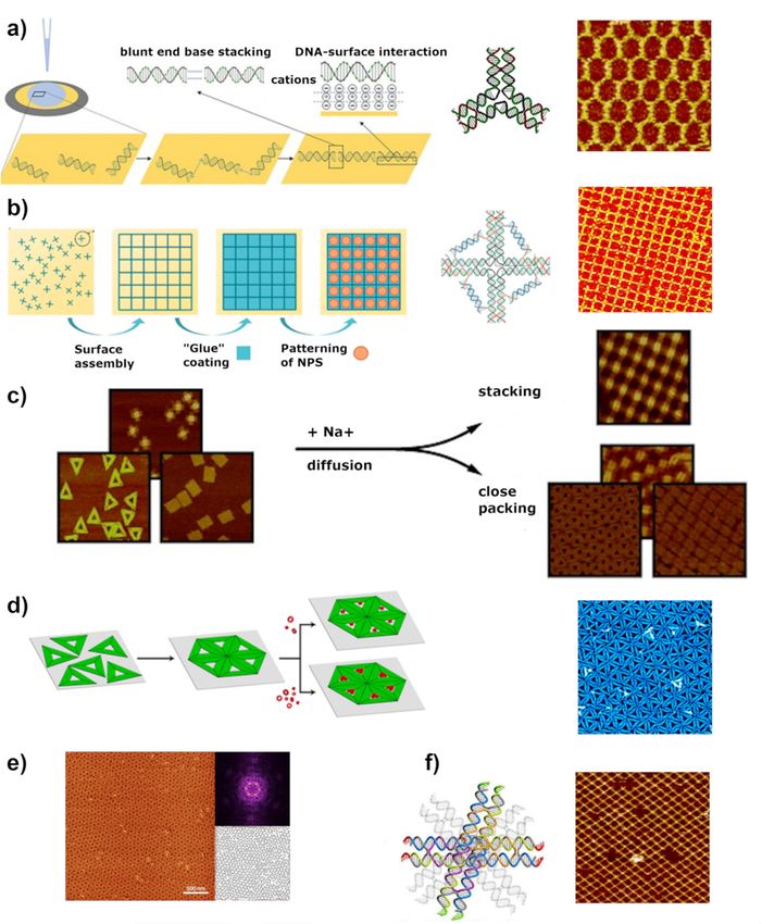
Molecules 2021, 26, 1502 2 of 28
structions [3,4]. Among these molecules, DNA remains the most attractive candidate for
nanoscale constructions due to its resolved sub-nanometer structural details and fairly
comprehensive understanding of the involved interaction forces, allowing precise engineer-
ing [5,6]. A single-stranded DNA (ssDNA) is a sequence of linked monomers, nucleotides.
When linked, they a sugar-phosphate backbone with one of the four possible organic
bases, adenine (A), cytosine (C), thymine (T), or guanine (G), attached to each sugar ring.
While all the bonds between the nucleotides in a single DNA strand are covalent and
thus very stable within the interplay of self-assembly, the formation of distinguished
double helices, governed by selective but easily detachable hydrogen bonds, leads to the
essential reversible assembly. The nature of these hydrogen bonds also makes the DNA
self-assembly predictable and specific, and consequently easy to program. Moreover, the
structure of DNA combines simultaneously stiffness and flexibility, making it an ideal
building block that ensures structural stability but still tolerates small strains within the
construction [7]. Besides, the fast and cost-effective synthesis of oligonucleotides allowed
the rapid development of structural DNA nanotechnology and the design of a variety of
DNA nanostructures with a nanoscale precision [8].
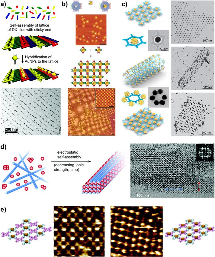
Seeman et al. [9] introduced the first concept of DNA nanotechnology in the 1980s by
constructing geometric objects and periodic 2D or 3D lattices based on multi-arm motifs
(Figure 1a). The idea comprised of single-stranded overhangs, or “sticky ends”, attached
at the ends of the branched DNA junctions serving as a connective glue to assemble the
structure. Since then, the field has flourished rapidly with new designs and concepts to
construct DNA-based systems [10]. A significant impact on the complexity of structural
DNA was introduced by an invention of the DNA origami technique by Rothemund in
2006 (Figure 1b) [11]. He reported a method where several kilobases long ssDNA, known as
a scaffold, was folded into different designed 2D shapes with help of hundreds of synthetic
oligonucleotides referred to as staple strands. This accomplishment truly changed the
scenery of structural DNA nanotechnology bringing new possibilities for forming complex
structures, especially after Shih’s research group extended the origami technique into 3D
structures as shown in Figure 1c [12]. Since then DNA origami has proven its versatility
as a building material in a nanometer-scale world by engineering custom structures with
control even at a single-molecule level [5,13–15]. DNA origami nanostructures have
been applied in variety of applications, such as weak and strong light-matter coupling
research [16,17], chiral objects formation [18,19], reconfigurable plasmonic devices [20],
drug delivery [21,22], Förster energy transfer research [23,24] and bio-applications [25,26].
Since the invention of the very first self-assembled DNA nanostructures, the aim has
been to achieve larger assemblies made up of individual DNA units. Even though the
origami structures are already about 100 nm in size what makes them much bigger than
the earlier DNA motifs of the size of ~10 nm or plain dsDNA with a thickness of ~2 nm,
the size of an individual origami structure is still limited by the length of the scaffold. To
scale up the structures up to micrometers, many attempts have been reported, including
extending the scaffold length with a variety of biotechnological methods, such as rolling
circle amplification (RCA) or polymerase chain reaction (PCR) [27–29]. Nevertheless,
these structures are still limited in size. An alternative method to overcome this problem
involves hierarchical self-assembly [30–37], where the individual self-assembled DNA
origami structures work as the construction units or tiles to form a larger organized
structure through higher-order self-assembly strategies.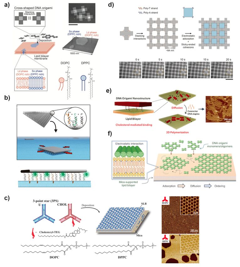
Molecules 2021, 26, 1502 3 of 28
Molecules 2021, 26, x FOR PEER REVIEW 3 of 28
Figure 1. (a) Fundamental structural motif of immobile branched DNA junction combined with sticky ends to self-assemble
Figure 1. (a) Fundamental structural motif of immobile branched DNA junction combined with sticky ends to self-assem-
intointo
ble 2D lattices [9,38].
2D lattices (b) DNA
[9,38]. origami
(b) DNA assembly
origami method.
assembly An example
method. of a DNA
An example of a origami scaffold
DNA origami (black line)
scaffold (blackand staples
line) and
(colored line) strand routing involving several crossover points where the scaffold or a staple strand crosses
staples (colored line) strand routing involving several crossover points where the scaffold or a staple strand crosses from from one helix
to another
one helix tothus connecting
another them together.
thus connecting themAdapted
together.by permission
Adapted from Springer:
by permission fromNature [11],Nature
Springer: copyright
[11],2006. Crossovers
copyright 2006.
Crossovers are topologically
are topologically similar to the similar to thejunction
branched branchedand junction and
hold the hold the
origami on origami on the
the desired desired
shape. The shape.
scaffoldThe
and scaffold and
the staples
the
are staples are mixed
mixed together together
resulting inresulting in the self-assembled
the self-assembled structure asstructure
shown inastheshown in the 3Dview.
3D schematic schematic view.
Adapted Adapted
with with
permission
permission from [39].2017
from [39]. Copyright Copyright
American 2017 American
Chemical Chemical
Society. Society.
Atomic Atomic force
force microscope microscope
(AFM) image of(AFM)
one of image of one of
the assembled the
DNA
assembled DNA origami structures. Adapted with permission from [11]. Copyright 2006 Springer: Nature.
origami structures. Adapted with permission from [11]. Copyright 2006 Springer: Nature. (c) Schematics of the design of a (c) Schematics
of the design of a three-dimensional origami structure. Adapted with permission from [12] copyright 2006 Springer: Na-
three-dimensional origami structure. Adapted with permission from [12] copyright 2006 Springer: Nature.
ture.
The driving forces contributing to the above-mentioned higher-order self-assembly are
The the
partially driving
sameforces contributing
as in the formation to of athe above-mentioned
single DNA construction, higher-order self-assembly
i.e., a sequence specific
are
Watson-Crick base pairing, but also unspecific base stacking forces can play aasignificant
partially the same as in the formation of a single DNA construction, i.e., sequence
specific Watson-Crick
role. Besides, base pairing,
such an assembly of DNAbut also unspecific base
nanostructures can bestacking
mediatedforces can play
in either a sig-
solution
nificant
or usingrole. Besides,
external supportsuchsurfaces,
an assembly
such as of mica,
DNA silicon,
nanostructures can be mediated
or lipid bilayer. Nowadaysinlatticeseither
solution or using external support surfaces, such as mica, silicon,
of even hundreds of micrometers in size have been constructed out of the self-assembled or lipid bilayer. Nowa-
days
DNAlattices of This
tiles [40]. evenishundreds
already much of micrometers
larger than most in size have
of the been constructed
biological organelles, out andof the
could
self-assembled DNA tiles [40]. This is already much larger than most
be utilized in biotechnology. This rapid development in the ability to build periodic large of the biological or-
ganelles, and could be utilized in biotechnology. This rapid development
domains has brought new opportunities to realize large-scale functional DNA structures. in the ability to
build
Thereby,periodic
DNAlargearraysdomains has brought
and lattices have been newincreasingly
opportunities to for
used realize
the large-scale
self-assembly func-
of
tional DNA structures.
nanometer-scale Thereby,
materials, rangingDNA from arrays and lattices
nanoparticles [41]have been increasingly
to proteins used for
[42,43]. In addition,
the self-assembly
ordered DNA arrays of nanometer-scale
could be further materials,
utilized inranging from nanoparticles
nanolithography to efficiently [41]fabricate
to pro-
teins [42,43]. In addition, ordered DNA arrays could be further utilized
extended surfaces filled with plasmonic nanostructures [44], which could further be utilized in nanolithogra-
phy to efficiently
for sensing fabricate
or to form optical extended surfacesHowever,
metamaterials. filled with thisplasmonic nanostructures
would require enlargement [44],
of
which could further
the structures to overbe utilized for
millimeter scale.sensing or to form optical metamaterials. However,
this would requireemphasizes
This review enlargement theofimportance
the structures to over
of DNA millimeter scale.
nanotechnology as a technique to
This review
construct extended,emphasizes
periodic the importance
patterns out ofof DNA nanotechnology
self-assembled DNA units, as ai.e.,
technique
tiles. We to
construct extended,and
describe methods periodic patterns
strategies out of self-assembled
for creating DNA units, i.e.,DNA
a variety of two-dimensional tiles. We de-
lattices.
In particular,
scribe methods weand
focus on the forces
strategies involved
for creating in the self-assembly
a variety of two-dimensionalprocess taking
DNA place In
lattices. in
solution aswe
particular, well as atonthe
focus the solid-liquid
forces involved interface,
in the and the delicate process
self-assembly balance taking
between them,
place in
solution as well as at the solid-liquid interface, and the delicate balance between them,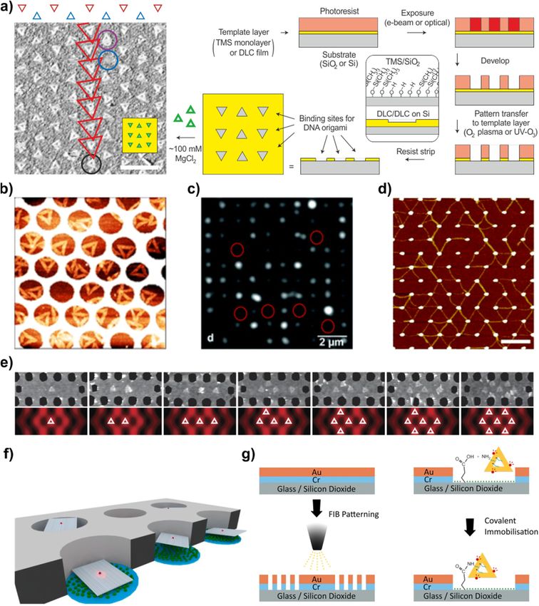
Molecules 2021, 26, 1502                                                                                            4 of 28

                           which one needs to optimize for successful self-assembly. We aim to provide a general
                           understanding of challenges and benefits in both, solution and surface assisted growth
                           techniques, including the use of lithographic patterning processes. Besides, essential
                           features for the generation of large-scale assemblies using tile-based assembly are discussed.
                           The last part of the review will briefly summarize the emerging applications of DNA
                           lattices to form novel materials by, e.g., special arrangements of gold nanoparticles (AuNP),
                           proteins, and other components.

                           2. Planks and Nails for DNA Lattices
                           2.1. The Planks
                                 To form a large-scale lattice of DNA structures one naturally needs suitable building
                           blocks, a.k.a. planks, to begin with. These individual self-assembled DNA structures form-
                           ing a variety of extended programmable DNA lattices through hierarchical self-assembly
                           are often called DNA-tiles, or for short just tiles. In general, for successful self-assembly of
                           a lattice, the structure of the tile should be more rigid than the bonding between them. As
                           mentioned, a single ssDNA is built by the formation of covalent bonds between nucleotides;
                           therefore, the structure remains stable during the self-assembly process. DNA-tiles, how-
                           ever, are bigger-self-assembled entities, usually formed by Watson-Crick base pairing
                           between complementary bases, i.e., held together by hydrogen bonds. In this case, the
                           connected complementary parts need to possess enough base pairs to have the melting
                           point well above the room temperature [45,46]. In addition, one naturally needs also a
                           clever way to organize the DNA strands into the desired form.
                                 There are numerous ways to fabricate DNA tiles suitable for constructing lattices.
                           Though, there are two distinguishable classes of DNA-tile structures: DNA motifs and
                           DNA origami units, which both can serve as building blocks, i.e., tiles [39]. DNA mo-
                           tifs are usually small constructions comprised of several different short oligonucleotides,
                           which corresponds to 100 to 350 base pairs (bp) per unit, and a maximum size of about
                           ten nanometers [47]. Good examples of DNA motifs are the first branched DNA junc-
                           tion by Ned Seeman [9] and the famous double crossover (DX) and triple crossover (TX)
                           tiles [48,49], which provided more rigidity compared to plain dsDNA. Using these struc-
                           tures for practical use, in particular for the formation of larger hierarchical patterns, has
                           been successfully demonstrated in many studies, but has still several limitations. In ad-
                           dition to their relatively small size, they require high control of stoichiometry and purity
                           of oligonucleotide strands, thus often resulting in a low yield, error-prone, and lengthy
                           synthetic process. Therefore, a new self-assembly route was quickly adapted where DNA
                           origami units were used as tiles forming even larger-scale assemblies.
                                 For the first time, Rothemund proposed a different route to create DNA origami units
                           in 2006 [11]. In his technique, a long single-stranded scaffold DNA is folded into predefined
                           structures through hundreds of complementary short oligonucleotide chains as shown
                           in Figure 1b. A variety of shapes can be created using the same scaffold but different
                           sets of staple strands, thus offering a broader set of possibilities and diverse self-assembly
                           strategies. One can easily form shapes like triangles, rectangles, or more complex objects
                           such as smiley faces and many others each of them consisting of around 7000 bp and having
                           the dimension of roughly 100 nm. Soon enough the method was adapted not only for 2D
                           arrays formation but also for building hollow and multilayered 3D origami structures [12].
                           It allowed packing the helices into triangular, square, or hexagonal arrangements, forming
                           bent and twisted structures [50]. The method is proven straightforward and efficient,
                           and at the same time offers an even broader set of possibilities in the formation of larger,
                           more complex shapes and structures. Consequently, DNA origami units were quickly
                           adapted as excellent components for studying self-assembly and the growth mechanism
                           of extended DNA lattices [39]. Further development of design strategies has led to new
                           DNA constructions, which are also suitable for tiles. These include for example DNA tube
                           circumferences [51] and ssDNA-based DNA-bricks [52], which both form lattices, as well
                           as a huge variety of different DNA wireframe constructions [53–55].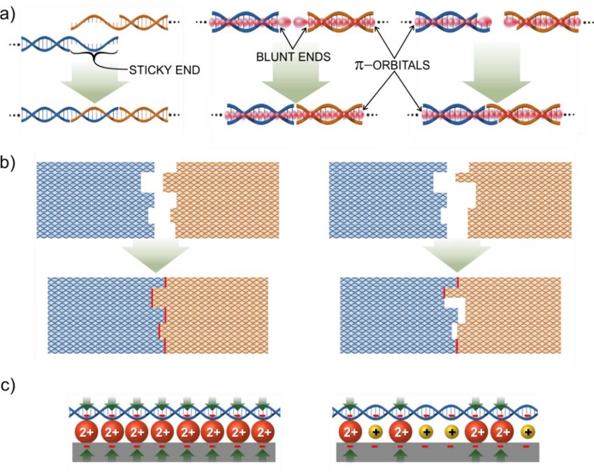
Molecules 2021, 26, x FOR PEER REVIEW 5 of 28
Molecules 2021, 26, 1502 5 of 28
example DNA tube circumferences [51] and ssDNA-based DNA-bricks [52], which both
form lattices, as well as a huge variety of different DNA wireframe constructions [53–55].
2.2.The
2.2. TheNails
Nails
To form any larger construction out of the building blocks, or planks, they need to be
To form any larger construction out of the building blocks, or planks, they need to be
joint together with some specific interactions, a.k.a. nails. The self-assembly of DNA tile
joint together with some specific interactions, a.k.a. nails. The self-assembly of DNA tile
can be achieved by following one of the two basic classic rules. While the first rule relies
can be achieved by following one of the two basic classic rules. While the first rule relies on
on the driving force of specific canonical, Watson-Crick base-pairing between comple-
the driving force of specific canonical, Watson-Crick base-pairing between complementary
mentary bases, i.e., A-T and G-C, the second is mediated by non-sequence-specific base
bases, i.e., A-T and G-C, the second is mediated by non-sequence-specific base stacking
stacking interactions. The early DNA motifs, like Seeman’s branched junctions [9] or DX
interactions. The early DNA motifs, like Seeman’s branched junctions [9] or DX and TX
and TX tiles [48,49], were assembled into lattices with help of so-called sticky ends, where
tiles [48,49], were assembled into lattices with help of so-called sticky ends, where a dsDNA
a dsDNA helix has an ssDNA overhang on its end, called sticky end. This overhang has a
helix hassequence
specific an ssDNA overhang
of bases that ison its end, calledtosticky
complementary end.
a sticky endThis overhang
protruding from hasthea other
specific
sequence of bases that is complementary to a sticky end protruding
DNA helix. When these two DNA helices meet, they can form a continuous helical struc- from the other DNA
helix. When these two DNA helices meet, they can form a continuous
ture by pairing their sticky ends as shown in Figure 2a (left). Since the method is the same helical structure
byaspairing
the one their
used sticky ends as
to assemble shown
a tile itself,inthe
Figure
binding2a (left).
energySince thekind
of this method is the
of joint same
is also
assimilar,
the one used to assemble a tile itself, the binding energy of this
i.e., roughly 10–20 kcal/mol per joint, depending on the number of base pairs and kind of joint is also
similar, i.e., roughly 10–20 kcal/mol per joint, depending on the
their type [45,46,56]. The DNA tiles usually carry several sticky ends with specific se- number of base pairs
and their matching
quences type [45,46,56]. The DNA
to the sticky ends on tiles usually
another tilecarry several
[57,58]. stickythe
In general, ends with specific
structure of a
sequences
tile shouldmatching to therelative
be more rigid sticky ends
to theon another tile
detachable [57,58]. between
connection In general,twothe structure
tiles facilitat-of a
tile
ingshould be more rigid
the self-assembly of relative to the detachable
larger constructs. Sticky endconnection between
hybridization cantwo tilesincorpo-
be also facilitating
the self-assembly
rated of largerdesigns.
in other geometric constructs.
For Sticky
example, endithybridization
can be used to canconnect
be alsoDNA
incorporated
tiles by in
other geometric
connecting designs.
adjacent Forside-by-side
helices example, itas can be used to connect
demonstrated DNA tiles
by the group by connecting
of Shalom Wind
adjacent
[59]. helices side-by-side as demonstrated by the group of Shalom Wind [59].
Figure 2. Principles of interactions between DNA-tiles: Single helix interactions are presented in (a) with a sticky end
Figure 2. Principles of interactions between DNA-tiles: Single helix interactions are presented in (a) with a sticky end
connection
connectionon on
thethe
left. This
left. interaction
This interaction is isbased
basedononbase
basepairing
pairingbetween
betweentwo twosingle-stranded
single-stranded overhangs
overhangs on on different
differentheli-
helices.
Theces.
middle one illustrates
The middle the blunt
one illustrates theend stacking,
blunt where π-orbitals
end stacking, of the last
where π-orbitals base
of the pairs
last baseform
pairsa form
π-π connection and complete
a π-π connection and
thecomplete
base pairthe base pair
stacking stacking
through the through
joint. The theright
joint. Theshows
part right part shows an
an example ofexample
how theof how ends
sticky the sticky ends and
and blunt end blunt endcan
stacking
stacking canAll
be combined. bethe
combined. All the
single helix single helix
interactions caninteractions
be used incan
the be usedcomplementary
shape in the shape complementary approach
approach illustrated in illustrated
(b). Bonding
in (b). Bonding energy between the tiles is directly proportional to the number of successful single helix joints marked as
energy between the tiles is directly proportional to the number of successful single helix joints marked as red. In (c) the
most common immobilization scheme of using negatively charged surface and divalent metal ions to glue the negatively
charged DNA on the surface, is illustrated on the left side. The right side shows the case of the addition of monovalent ions,
which occupy the positions of divalent ions and thus reduces the bonding between substrate and DNA as shown by the
lower number of green arrows.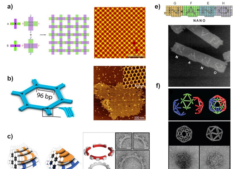
Molecules 2021, 26, 1502                                                                                               6 of 28

                                 The second driving force, i.e., blunt end stacking, uses the stacking interaction at
                           blunt ends of dsDNA helix structures. Inside a dsDNA helix, the base pairs are bound
                           not just by the backbone of the DNA, but also the connection is enhanced via stacking
                           interaction due to their overlapping π-orbitals, thus forming π-π interaction between the
                           bases. The base pairs at the ends of the helix have unpaired π-orbitals sticking out of the
                           helix. This can establish π-π interaction with the terminal bases of another dsDNA helix as
                           shown in Figure 2a (middle). The strength of the interaction between two DNA-tiles having
                           blunt ends can be controlled by the number of adjacent helices and the base sequence of
                           the end [11]. Today, base stacking interactions are widely used as directional forces in
                           hierarchical surface-mediated self-assembly of DNA origami units. The blunt end stacking
                           is much weaker than sticky ends and unspecific for a choice of bases. The binding energy
                           for a triple stacking has been measured to be about 8.6 kcal/mol [60].
                                 To fine-tune the binding energies between the helices and tiles, the above two rules
                           can be combined as shown in Figure 2a (right) [61]. Here, the helices have too short sticky
                           ends, i.e., one to three bases, to hold them together. However, when combined with the
                           blunt end stacking between the base pairs at the ends the total binding energy of the joint
                           becomes high enough to sustain the interaction, i.e., the intermediate force between the
                           plain blunt end and a proper sticky end.
                                 Shape complementarity can be additionally implemented in the design of the tiles
                           and thus combined with the blunt end stacking and/or sticky-end interactions. It enhances
                           the directional control of the self-assembly process because the connection between com-
                           plementary shapes is much stronger than for a non-matching pair. This is illustrated in
                           Figure 2b, where the formed blunt end π-π stackings are highlighted by red for matching
                           and non-matching tiles. Shape matching can also be used for arranging higher-hierarchy
                           systems as demonstrated by the research group of Endo [62]. Even so, often, the combina-
                           tion of inter-origami binding forces, i.e., sticky and blunt end interactions, in addition to
                           some more exotic bondings, such as Hoogsten bonding is needed to achieve directional
                           control of hierarchical origami assemblies. Combining different types of connections can
                           facilitate the fabrication of even kinematic DNA structures comprising multiple origami
                           units with well-defined moving ranges and routes [63]. This is, however, out of scope of
                           this review, where we concentrate on large-scale static lattices.

                           2.3. Assembly Protocols
                                 If the lattice out of DNA-tiles is first formed in a solution and only after that transferred
                           onto a substrate, the forming lattice is constantly subjected to the strong drag and shear
                           forces present in the solution. From the above-discussed forces between the tiles, the sticky
                           end hybridization is strong enough to govern the drag forces. However, plain blunt end
                           stacking is not usually strong enough and the feasibility of the combined connections
                           depends strongly on the total geometry, and number of the joins. Though, nowadays
                           people are keen on using surface-mediated self-assembly approaches, where the tiles
                           weakly bind to the substrate, and can still diffuse on it, rearrange, and consequently form a
                           lattice. In this case, all the above connection rules are feasible, since the substrate provides
                           additional support. However, it is very important to also control the binding energy
                           between the DNA-tile and the substrate. The most common way to attach DNA-structures
                           on a substrate is to use a freshly cleaved mica surface, in a presence of divalent metallic
                           ions, such as Mg2+ , which are already contained in the origami folding buffer, bridging
                           the DNA and substrate together as shown in Figure 2c. Even though the attraction forces
                           between DNA tiles and surface are rather weak, still they can partially impair the desired
                           diffusion on it. Yet, one can fine-tune this interaction by adding monovalent ions, like Na+ ,
                           which weaken the interaction by partially replacing divalent ions from the surface, forming
                           a more diffuse charge layer. As a result, DNA structures become more mobile and start
                           rearranging on the surface forming a lattice [64–66].
                                 The four main approaches can be distinguished in self-assembling DNA-tiles into
                           target structures. Among them is: one-pot “mix and go” -method, which is a key concept of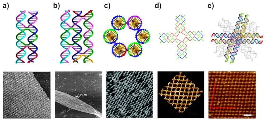
Molecules 2021, 26, 1502                                                                                             7 of 28

                           DNA nanotechnology, inspired by Seeman [49,67,68]. In this method, each tile of the set
                           contains unique connections, usually sticky ends, leading the tile into the desired position
                           during self-assembly, but above all the final large lattice is formed in the same process, and
                           thus within the same solution, in which the tiles are formed. The second method, hierarchical
                           self-assembly, is usually a two-step process where a specific group of tiles is pre-assembled
                           in test tubes and then afterwards combined to form a larger structure [12,69–72]. In this
                           approach, the combination of sticky and blunt end/shape complementary interaction
                           forces are often employed, as well as a supporting substrate. In the more sophisticated
                           algorithmic self-assembly, tiles are programmed with specific binding domains, which can
                           explicitly bind to another unit following an algorithmic rule. This often leads to a fully
                           programmed lattice whose size is also fixed [73–77]. The last distinguished method is called
                           scaffolded tile-assembly, where a longer strand is used as a scaffold upon which DNA-tiles
                           attach to form various patterns. The “scaffolded frames” procedure often takes advantage
                           of all the above-mentioned interaction forces, in addition to surface supports interaction
                           stabilizing the self-assembly process [78].

                           3. Assembly of DNA Lattices in Solution
                                 Many solution-based lattice assembly strategies have been reported in literature yield-
                           ing two- and three-dimensional DNA nanostructure lattices. Originally the solution-based
                           assembly was introduced by Seeman as a one-pot “mix and go” -procedure [9], which
                           remained the main method at the beginning of the structural DNA development, until
                           the more sophisticated ones like hierarchical self-assembly were introduced. Hierarchi-
                           cal self-assembly is a compelling approach to create structurally versatile, regular DNA
                           patterns with a dimension beyond a 1 µm limit. Many finite-two- and three-dimensional
                           DNA or infinite-crystal-like structures have been successfully assembled following this
                           approach [67,79–82]. Generally, in solution, the assembly is mostly driven by sticky end
                           associations connecting the structures, as the blunt end interaction is usually not strong
                           enough to hold the larger assemblies together. The selectivity of the tile-to-tile inter-
                           actions can be further enhanced by utilizing shape complementarity at the ends of the
                           nanostructures. Nevertheless, the challenge still remains in the optimization of assembly
                           parameters including annealing conditions to obtain structures at high yield. In addition,
                           the deposition of solution-assembled lattice onto a substrate without breaking is not a
                           trivial task.

                           3.1. Lattices by Early DNA Motif
                                 The essential foundation of DNA nanotechnology is Nadrian Seeman’s iconic design
                           of complementary sticky end and branched DNA junction to make geometric objects and
                           periodic lattices [9,83]. His design became the most common self-assembly method in the
                           DNA nanotechnology called, multi-arm approach. In this approach, DNA sequences are
                           designed to assemble into branch motifs with unique, complementary sticky ends. The
                           sticky ends precisely guide the inter-tile interaction and position of the tile in the assembly
                           (Figure 1a). Following this methodology, Seeman and coworkers created various DNA
                           motifs that could self-assemble into higher-order DNA arrays [84–86]. However, multi-arm
                           junction design suffered from geometric instability and flexibility, and that often resulted
                           in poor structural predictability. Hence, ultimately, they were not suitable for the assembly
                           of extended patterns.
                                 To overcome this problem, a new motif design was constructed by joining two
                           parallelly-oriented double helices by strands crossings between them at two crossover
                           points, which led to the formation of a branched complex called DNA double crossover
                           (DX) molecule (Figure 3a) [48]. The DX motif resulted in a more rigid nanostructure com-
                           pared to linear DNA, and for that reason, it was officially named as the “DNA tile” unit
                           for the first time. In Figure 3a, the successful construction of a first periodic 2D crystalline
                           lattice for the DX motif is presented [67]. The arrays were formed by mixing two to four
                           different DX tiles to display a striped pattern. Later the DX-tile was used for example
molecule (Figure 3a) [48]. The DX motif resulted in a more rigid nanostructure compared
to linear DNA, and for that reason, it was officially named as the “DNA tile” unit for the
Molecules 2021, 26, 1502 8 of 28
first time. In Figure 3a, the successful construction of a first periodic 2D crystalline lattice
for the DX motif is presented [67]. The arrays were formed by mixing two to four different
DX tiles to display a striped pattern. Later the DX-tile was used for example to produce
Sierpinski
to producetriangles
Sierpinskiviatriangles
algorithmic self-assembly
via algorithmic [74], and fully
self-assembly [74],addressable finite-size
and fully addressable
tile-blocks [87]. Soon after the invention of the DX-tile, the design was extended
finite-size tile-blocks [87]. Soon after the invention of the DX-tile, the design was extended to the
DNA
to thetriple
DNAcrossover complex
triple crossover (TX), (Figure
complex 3b) [49],3b)
(TX), (Figure which
[49], was
whichused
wastoused
produce a variety
to produce a
of self-assembled
variety linear arrays
of self-assembled linear[88], 2D lattices
arrays [88], 2D[49], and[49],
lattices DNAand tubes
DNA [89]. LaBean
tubes [89].etLaBean
al. [90]
increased the rigidity
et al. [90] increased theofrigidity
the TX-tile even
of the more
TX-tile by designing
even a circularaform
more by designing of itform
circular calledof
three-helix bundle. Almost simultaneously, Seeman et al. [91] also introduced
it called three-helix bundle. Almost simultaneously, Seeman et al. [91] also introduced another
tubular
another DNA-tile called six-helix
tubular DNA-tile bundle bundle
called six-helix shown shown
in Figure 3c, which
in Figure become
3c, which widely
become used
widely
later
used [92–95].
later [92–95].
Figure Theearly
3. The
Figure 3. earlyDNA DNAtiletilemotifs
motifs and
and AFM AFM images
images of their
of their corresponding
corresponding 2D 2D lattice
lattice assemblies.
assemblies. (a)DNA
(a) DX DX DNA tile
tile com-
comprises two parallel helices connected by the ssDNA strands (pink, green, and red) crossing to
prises two parallel helices connected by the ssDNA strands (pink, green, and red) crossing to the other helix at the cross- the other helix at the
crossover
over pointspoints [68,96].
[68,96]. (b) DNA
(b) TX TX DNAtile tile is formed
is formed similar
similar to to
thethe
DXDX tile,
tile, butbutnow
nowthe thessDNA
ssDNAstrands
strandsconnect
connect three
three helices
together.
together. The pink strand is even part of all the three helices [49,96]. (c) Six-helix bundle DNA tile, where all the six helices
are connected to their two neighbors by crossovers, finally forming a tube-like structure [91]. (d) Cross-shaped tile formed
from branched
from branched DX-tile
DX-tile oror four
four DX-tiles
DX-tiles joined
joined together
together atat one
one end
end [79].
[79]. Adapted
Adapted with
with permission
permission from
from AAAS.
AAAS. (e)(e) Layered
Layered
crossover DNA tile [97]. Adapted with permission from [68] copyright 1999, [49] copyright 2000,
crossover DNA tile [97]. Adapted with permission from [68] copyright 1999, [49] copyright 2000, [91] copyright 2005, [91] copyright 2005, [97]
[97]
copyright 2018 American Chemical Society.
copyright 2018 American Chemical Society.
Another
Another example
example of of tiles
tiles referred
referred toto as
as aa “cross
“cross tile”
tile” was
was introduced
introduced by by Yan et al.
Yan et al. [79]
[79]
in 2003, shown in Figure 3d. Thanks to its symmetric structure,
in 2003, shown in Figure 3d. Thanks to its symmetric structure, it assembles into well- it assembles into well-
formed
formed 2D-lattices.
2D-lattices. Consequently,
Consequently, aa variety
variety of of DNA
DNA tiles,tiles, including
including rigid rigid cross-shaped,
cross-shaped,
three
three point-star,
point-star, or or six
six point-star
point-star motifs
motifs tiles
tiles were
were designed
designed and and assembled
assembled into into large
large 2D
2D
arrays
arrays with square or hexagonal and triangular triangular cavities,
cavities, respectively
respectively [70,72,98–100].
[70,72,98–100]. In
addition, some
some finite
finitesize
sizeaddressable
addressablelattices
lattices were
were alsoalso designed
designed [69][69]
Most Most of these
of these de-
designs
signs followed a two-step hierarchical assembly, where each tile
followed a two-step hierarchical assembly, where each tile is formed separately and then is formed separately and
then
they they are combined
are combined together.
together. In recent
In recent studies,
studies, Yan Yanet al.etdeveloped
al. developed a derivative
a derivative fromfrom
the
the traditional
traditional double-crossover
double-crossover DNA DNA motif,
motif, named
named layered-crossover
layered-crossover tile tile to construct
to construct a seta
set of rhombus-like
of rhombus-like layered-crossover
layered-crossover DNA DNA lattices.
lattices. In the
In the motifmotif design,
design, twotwo or four-lay-
or four-layered
ered crossover
crossover tiles bridge
tiles bridge neighboring
neighboring layerslayers
withwith predetermined
predetermined orientation
orientation [97].[97]. Simi-
Similarly,
Qian and
larly, Qiancoworkers
and coworkerscreated a triangular
created tile thattile
a triangular could
thatform
couldlarge
form2D arrays
large 2D and 3Dand
arrays shapes
3D
approximating
shapes a rhombic
approximating triacontahedron
a rhombic [101]. [101].
triacontahedron
Although the DX and TX tiles tiles have
have proven
proven to to bebe fully
fully addressable
addressable assembly
assembly method,
method,
the number of required strands and the costs increase linearly with the number of unique
Further developments
tiles. Further developments of similar design strategies have been reported including the
paranemic crossover (PX) motif [102], which brought inspiration for nanoconstruction of 3D
objects [102], and multihelical bundles [90,91,103] or parallelogram DNA tiles [104] that self-
assemble into linear or 2D arrays and 2D lattices with diamond-shaped cavities [102]. Even
though a multi-arm approach is still a method of choice to efficiently construct crystalline-
like structures it requires high control of stoichiometry and purity of oligonucleotide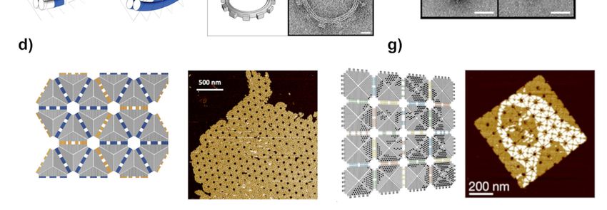
Molecules 2021, 26, 1502                                                                                            9 of 28

                           strands, thus often resulting in low yield, error-prone, and lengthy synthetic process.
                           Therefore, a new self-assembly route was quickly adapted where DNA origami units were
                           used as tiles forming even larger-scale assemblies.

                           3.2. DNA Origami-Tile Assembly
                                 An alternative method enabling the formation of larger, infinite crystal-like lattices or
                           more complex, finite structures is the hierarchical self-assembly of DNA origami. Similar
                           to the DNA motif, DNA origami can act as a tile unit and thereby self-assemble relying on
                           sticky ends or other interactions presented in Section 2. Correspondingly, these interactions
                           can be enhanced by applying the complementary matching shapes improving the tile-to-
                           tile interactions, especially their selectivity, as shown in Figure 2b. The first experimental
                           demonstrations of large hierarchical assemblies from DNA origami were a gear formed by
                           the association of four quarter circles shaped origami (Figure 4c) [50], an icosahedron made
                           from mixing three wireframe style monomers (Figure 4f) [12], and an infinite crystalline 2D
                           lattice of two origami tiles propagating into two independent directions (Figure 4a) [34,105].
                                 The large dimension and complex molecular dynamics of DNA origami have a pro-
                           found effect on the final design of a structure and dictate the interaction between the
                           origami units. While designing a 2D DNA origami structure, a global twist with respect to
                           a tile plane needs to be taken into account [106]. This structural distortion is a consequence
                           of the conformation design of DNA origami with an average twist of 10.67 bp per turn,
                           which slightly deviates from the natural B-type helical twist of 10.5 bp per turn. The small
                           increase accumulates when hundreds of units are assembled together. To overcome the
                           problem, Liu et al. [32] constructed a set of rectangular shape origami arranged in a zigzag
                           ribbon pattern to avoid distortions and investigated the self-assembly of higher-order struc-
                           tures. They also found that the position and strength of the connecting strands between
                           the linked units have a strong influence on the quality of the final assembly product. Even
                           in some cases, it led to the assembly of completely different structures [33].
                                 Large origami structures contain a significant number of DNA helixes and in many
                           cases, these have their ends side by side at the edges of the origami. Therefore, base
                           stacking interaction forces by many parallel blunt ends can easily hold the origami units
                           together [107]. Nevertheless, the solution-assembled larger structures are often fragile
                           and easy to break. Therefore, instead of base stacking interaction, sticky ends hybridiza-
                           tion is commonly used to ensure structural support and control over the assembly. The
                           studies have shown that the combination of both interactions improves control over the
                           various pattern assemblies. Qian and coworkers modified triangular DNA with both
                           interactions to form large 2D arrays and 3D shapes approximating a rhombic triaconta-
                           hedron (Figure 4d) [101]. Figure 4e represents a classic example of the formation of large
                           planar structures made of DNA jigsaw pieces where the combination of different types
                           of forces improves the control of a whole system [105]. The inter-unit hybridization and
                           stacking at the interfaces of shape-complementary edges allowed regioselective origami
                           pairing [107–109]. Wang et al. assembled a more complex, micron-scale honeycomb lattice
                           from a set of hexagonal DNA origami tiles as illustrated in Figure 4b. The hexagonal DNA
                           origami tiles were self-assembled through a combination of connector strand design, base
                           hybridization, and blunt end interaction. Later, they used a pre-assembled honeycomb
                           lattice as a platform for the positioning of nanoparticles (NPs) [61].
                                 The aforementioned approaches usually lead to the assembly of relatively simple
                           patterns. To increase complexity and range of the scale one needs to implement algorithmic
                           self-assembly [73–75]. Complex programmable features have been achieved using DNA
                           tiles leading to high yields of superstructures of micrometer dimensions [73–76]. The
                           fractal assembly allows scaling up the complexity of DNA nanostructures with arbitrary
                           patterns [77]. For example, Tikhomirov et al. [110] used this approach to generate patterns
                           in the shape of a famous painting by Leonardo da Vinci, Mona Lisa, as shown in Figure 4g.
                           Following the same approach, Gang et al. [111] designed a planar DNA origami frame and
                           used it to periodically organize gold nanoparticles, creating diverse 2D architectures.
Molecules 2021, 26, 1502 10 of 28
Molecules 2021, 26, x FOR PEER REVIEW 10 of 28
Figure 4. DNAFigure 4. DNA
structures structures
and lattices formedandfrom lattices formed
DNA origami tiles from
throughDNA origami
hierarchical tiles through
self-assembly. (a) 2D hierarchical
lattice self-
assembled from cross-shaped
assembly. (a) 2Dtileslattice
by sticky end hybridization
assembled [34]. (b) 2D hexagonal
from cross-shaped tiles by lattice
stickyassembled from hexagonal [34]. (b) 2D
end hybridization
DNA origami tiles [61]. (c) Design of a DNA bundle modified to bend into a quarter circle, which are subsequently joined
hexagonal
together to form lattice
a gear; scale barsassembled from hexagonal
20 nm [50]. Adapted DNA
with permission origami
from AAAS. tiles
(d) The[61]. (c) of
design Design of a DNA bundle
self-assembled
structure outmodified to bend into a quarter circle, which are subsequently joined together to form a gear; scale
of the asymmetric tiles and the corresponding AFM image [101]. (e) Jigsaw DNA origami units; although
sticky end hybridization controls the alignment of the pieces, base stacking and shape complementarity reinforce the
bars 20 nm [50]. Adapted with permission from AAAS. (d) The design of self-assembled structure
binding; image size 525 × 525 nm [105]. (f) Formation of large DNA cages “icosahedron” assembled three different
out subunits;
wireframe type of the asymmetric
scale bar 100 tiles and thewith
nm. Adapted corresponding
permission fromAFM [12].image [101].
Copyright 2006(e) JigsawNature.
Springer: DNA (g) origami units;
Design of DNA arrays with
although a Mona
sticky endLisa pattern and corresponding
hybridization controls the AFM image. Adapted
alignment of thewith permission
pieces, basefrom [110]. and shape
stacking
Copyright 2017 Springer: Nature. Adapted with permission from [61] copyright 2016, [101] copyright 2018, [91] copyright
complementarity
2005, [97] copyright 2018 Americanreinforce the binding; image size 525 × 525 nm [105]. (f) Formation of large DNA
Chemical Society.
cages “icosahedron” assembled three different wireframe type subunits; scale bar 100 nm. Adapted
The aforementioned
with permission from [12]. Copyright approaches usually
2006 Springer: lead to (g)
Nature. theDesign
assembly of relatively
of DNA arrayssimple
with a Mona
patterns. To increase complexity and range of the scale one needs to implement algorith-
Lisa pattern and corresponding AFM image. Adapted with permission from [110]. Copyright 2017
mic self-assembly [73–75]. Complex programmable features have been achieved using
Springer: Nature. Adapted
DNA tiles leadingwith permission
to high from [61] copyright
yields of superstructures 2016, [101]
of micrometer copyright
dimensions 2018, [91]
[73–76].
copyright 2005, [97] copyright 2018 American Chemical Society.
The fractal assembly allows scaling up the complexity of DNA nanostructures with arbi-
trary patterns [77]. For example, Tikhomirov et al. [110] used this approach to generate
4. Surface-Assisted Assembly
patterns in the shape of a famous painting by Leonardo da Vinci, Mona Lisa, as shown in
Figure 4g. Following the same approach, Gang et al. [111] designed a planar DNA origami
As mentioned in the previous chapter, the self-assembly of an extended lattice in the
frame and used it to periodically organize gold nanoparticles, creating diverse 2D archi-
solution is tectures.
limited, because the whole structure is exposed to the drag and shear forces
induced by the solvent. These forces increase with the dimensions of the lattice and thus
limit the conceivable size. Therefore, in almost all the lattices formed in a solution, sticky
ends have been utilized as the main driving force ensuring the connection between indi-
vidual tiles. Another challenge remains during deposition of the pre-assembled lattice
onto substrate surface. Many times, it results in structural deformations along with some
damages or torn structures—the larger the structure the lower the probability for a suc-
cessful and error-free deposition. Because of these reasons and with the emerging trend
towards larger origami-based lattices, researchers came up with a new strategy, where the
prefabricated tiles are adhered on a surface and the assembly of the lattice happens by
controlling the diffusion of the tiles on it [64,71]. In surface-assisted assembly, the substrateMolecules 2021, 26, 1502 11 of 28
provides needed support and route to more stable and reproducible patterning achieving
larger lattices.
The large surface area and thus highly negative charge of origami units promote the
electrostatic hierarchical self-assembly on external surfaces. In addition to the tile-to-tile
interactions, discussed above, organizing DNA nanostructures on a surface requires an
understanding of the role of the three main factors controlling the diffusions of origami tiles
on the substrate, namely DNA-surface interaction, DNA concentration, and the assembly
time. The type and number of components making up the system affect the type of acting
forces, their strength, and the energetic pathways towards the most stable structure. The
function of the substrate surface is also crucial, given the fact that the mobility of the DNA
units on the substrate is one of the key prerequisites for the lattice assembly. Therefore,
the assembly process typically requires weak adsorption conditions to ensure the DNA
surface mobility. The adsorption forces, however, should be tuned as such to still provide
required attraction between the surface and DNA tiles to keep the already attached units
together. Surface support is an important factor in obtaining large scale-assemblies because
it stabilizes the DNA-DNA interactions, and confines the structures within physical space,
thus boosting the probability for the DNA units to meet and initiate binding.
Even though the precise design of DNA structures and spatial organization of the
objects with nanoscale accuracy has been accomplished and monitored in real-time, it is
still challenging to obtain large periodic lattices. Many of the formed lattices still suffer
from assembly defects, low yield, and small dimensions. In the following section, we
go through feasible ways to create extended DNA patterns, particularly with the help of
cationic compounds through hierarchical assembly on mica and lipid bilayer membranes
used as surface supports. In addition, we discuss about template-based assembly on silicon
and other surfaces.
4.1. Mica
Mica surfaces are the most common solid support for DNA origami studies due to
their inherent advantages. Besides the low cost, mica features relatively large atomically
flat areas and requires simple sample treatment procedures. Mica has a mineral net-like
structure, containing an octahedral complex aluminum layer sandwiched between two
tetrahedral SiO-layers [112]. When it comes in contact with water, potassium ion, which
naturally bridge the ionic mineral sheet of mica, dissociates from the surface, leaving the
unbalanced negative charge. Like the mica surface, DNA origami structures are negatively
charged; hence, the adsorption on the mica surface is initiated by cations stabilizing
DNA-mica interaction as discussed above in Section 2. The buffer in which origami
is dissolved usually contains an Mg2+ counterion mediating the electrostatic attraction.
Surface-assisted self-assembly also requires a suitable interaction between DNA units to
form larger assemblies. On mica, the stacking interaction between blunt-ended origami is
already capable of holding structures together, forming 1D or 2D arrays.
One of the first attempts to arrange DNA tile structures into periodic patterns was
accomplished with pre-formed, branched star-shaped motifs. These relatively small struc-
tures were immobilized on a mica surface and incubated at 50 ◦ C [71]. Based on this
study, the DNA motifs-mica surface interaction confines the tiles, restricting their flex-
ibility, and increases the interaction between them. Additionally, similar studies have
revealed that the 2D assembly kinetics is relatively fast in contrast to the previously re-
ported sticky-end mediated-assembly [66]. The direct observation of the processes such as
the adsorption/desorption of the DNA and its diffusion on the surface would deepen the
understanding of kinetics at the solid-liquid interface. High-speed AFM is a method of
choice to investigate the morphology and dynamics of lattice formation [113,114]. Despite
relatively straightforward assembly protocol, the real challenge remains in understanding
the requirements and precise condition in regulating the self-assembly of the DNA-tiles
leading to the large single-layer patterns.Molecules 2021, 26, 1502 12 of 28
The large surface area and negative charge of DNA structures enable control of the
adsorption forces by exploiting the competition between different ions. The effects of
various ions allow tuning the forces and promote the formation of extended patterns.
Divalent cations promote the immobilization of DNA-tiles on the surface, by forming
salt bridges between the surface and structures, as discussed in Section 2 and shown in
Figure 2c. One of the advantages of surface-assisted methods is that by tuning the divalent
cation concentration, the integrity and homogeneity of the lattice can be achieved. Typically,
the immobilization of DNA origami requires magnesium concentration between 10 and
200 mM to overcome the repulsion barrier and bridge the surface and structures. Too low
concentration can lead to structural instability, whereas the addition of too high concentra-
tion results in too strong adsorption of DNA structures reducing surface mobility. Recently,
Liu and colleagues demonstrated that the homogeneity and integrity of a tetragonal array’s
formation
Molecules 2021, can
26, x FOR PEER be achieved only by tuning the concentration of Mg2+ ions and exploiting
REVIEW 13 of 28
blunt stacking interaction between blunt ends. (Figure 5b) [115].
Figure 5. Surface-assisted DNA origami self-assembly on mica. (a) DNA molecules loosely adsorb on the surface forming
Figure 5. Surface-assisted DNA origami self-assembly on mica. (a) DNA molecules loosely adsorb
salt bridges with the mica surface, after which they rearrange themselves to maximize DNA-DNA blunt end stacking
on the
interaction surface
[66]. forming process
(b) Self-assembly salt bridges with
of 4-point starthe mica
motifs intosurface, after
a 2D lattice, which
which they
is later usedrearrange themselves
for patterning NPs in to
the presence
maximizeof Ni2+ to glue the lattice
DNA-DNA bluntto mica
end[115]. (c) Theinteraction
stacking self-assembly [66].
of triangles and rectangles isprocess
(b) Self-assembly mediatedof by4-point
reach- star
ing an electrostatic balance between the DNA nanostructures and mica surface. Non-interacting triangular structures as-
motifs into a 2D lattice, which is later used for patterning NPs in the presence of Ni2+ to glue the lattice
semble into close-packed structures, while attractive blunt-end stacking interactions between the cross-tiles lead to the
to mica
extended [115].assembly
2D lattice (c) The[64].
self-assembly
(d) Triangularoforigami
triangles
tilesand rectangles
assembly at high is
Namediated by reaching
+ concentration an electrostatic
[116]. (e) AFM image
of a highly ordered lattice of triangular origami tiles as well as corresponding FFT and Delaunay triangulation. The lattice
was self-assembled within a buffer with Ca2+/Na+ ions during 90 min incubation [40]. (f) Design of layered-crossover tile
and a corresponding image of a self-assembled 2D crystal structure [97]. Adapted with permission from [115] copyright
2019, [116] copyright 2016, [97] copyright 2018 American Chemical Society.Molecules 2021, 26, 1502 13 of 28
balance between the DNA nanostructures and mica surface. Non-interacting triangular structures
assemble into close-packed structures, while attractive blunt-end stacking interactions between the
cross-tiles lead to the extended 2D lattice assembly [64]. (d) Triangular origami tiles assembly at
high Na+ concentration [116]. (e) AFM image of a highly ordered lattice of triangular origami tiles
as well as corresponding FFT and Delaunay triangulation. The lattice was self-assembled within
a buffer with Ca2+ /Na+ ions during 90 min incubation [40]. (f) Design of layered-crossover tile
and a corresponding image of a self-assembled 2D crystal structure [97]. Adapted with permission
from [115] copyright 2019, [116] copyright 2016, [97] copyright 2018 American Chemical Society.
Even though magnesium is the most commonly used divalent cation to bridge the
origami structures and mica surface together, other metals play a critical role in the origami
adsorption onto the surface. In particular, transition metals have been used as a form of
glue to preserve the structures on the surface. Mao group studied the influence of Ni2+
ions on 2D arrays formation, shown in Figure 5a [66]. The experiment showed that even a
small quantity of nickel cations is sufficient to stop the diffusion and “freeze” the origami
structures on the mica substrate [65]. Since Ni2+ ions bind generally stronger to DNA
structures, a high concentration of metal may lead to structural deformation.
Once DNA tiles are adsorbed onto the mica surface loosely enough, base stacking
interaction between blunt ends of DNA units enables the rearrangement of the structures
on the surface, to maximize the interactions between them. For successful self-assembly,
the strength of DNA-DNA and DNA-surface interactions need to be comparable [66].
The electrostatic balance between surface and DNA structures can be reached by the
addition of competing monovalent cations. For instance, the addition of sodium ion
reduces the number of salt bridges, displacing Mg2+ ions from the interface, directly
affecting the surface mobility of origami structures, as shown above in Figure 2c [114]. The
first successful assembly of large-scale ordered lattice on mica surface was accomplished by
Rafat et al., demonstrated in Figure 5c. In their work, the addition of sodium ion weakened
the interaction by partly replacing magnesium ion, thus forming a more diffusive charged
layer eventually leading to lattice self-assembly [64].
One of the assembly approaches is to only optimize the DNA tile concentration and
mobility on the mica surface and pack the surface so densely that it results in macroscopic
surface area, homogeneously covered with formed lattice [66]. This very straightforward
approach does not require any specific design and optimization of molecular forces. Ra-
makrishnan et al. [116] created a monolayer of densely packed triangular origami tiles
following the same, cation-mediated assembly (Figure 5d). At a Na+ concentration of
200 mM, that is, 20-fold excess over Mg2+ , a densely packed monolayer with hexago-
nal symmetry was formed. Keller group also conducted a comprehensive study on the
lattice formation by investigating the impact of sodium concentration in the 10 mM Mg2+ -
containing DNA origami buffer [114]. Notably, they suggested that the Na+ /Mg2+ ratio
has a similar effect on lattice assembly dynamics as a substrate temperature in thin-film
growth. The assembly of triangular DNA origami structures into highly ordered lattices
was best achieved at 75 mM concentration of sodium cations, i.e., 7.5-fold excess over Mg2+ .
Although the assembled lattices did not feature significant dislocation, some minor defects
were still observed. Longer incubation times increase lattice quality and order, as expected
because the triangular structures had more time to organize into a well-ordered structure.
Nonetheless, the assembled lattices are usually poly-crystalline with crystalline domains
having a random orientation to each other [117]. Lately, Hong et al. [97,118] studied the
assembly of layered-crossover tiles and found out that in the case of surface-mediated
assembly, the single-layer of DNA 2D crystal fully covered the mica surface even up to
several microns by only adjusting the origami concentration while keeping the monovalent
cation constant (Figure 5f).
The essence of the assembly process is to control the interplay between ionic strength
and electrostatic interaction at the liquid-surface interface. Ionic strength is determined
by the size and hydration energy of an ion [119]. Among alkali metals (1A group), K+ ,You can also read