The Molecular Mechanism of Vitamin E as a Bone-Protecting Agent: A Review on Current Evidence - MDPI
←
→
Page content transcription
If your browser does not render page correctly, please read the page content below
International Journal of
           Molecular Sciences

Review
The Molecular Mechanism of Vitamin E as a
Bone-Protecting Agent: A Review on
Current Evidence
Sok Kuan Wong , Nur-Vaizura Mohamad, Nurul ‘Izzah Ibrahim, Kok-Yong Chin ,
Ahmad Nazrun Shuid and Soelaiman Ima-Nirwana *
 Department of Pharmacology, Faculty of Medicine, Universiti Kebangsaan Malaysia, Jalan Yaacob Latif,
 Bandar Tun Razak, Cheras, Kuala Lumpur 56000, Malaysia; jocylnwsk@gmail.com (S.K.W.);
 vaizuramohd@gmail.com (N.-V.M.); nurulizzah88@gmail.com (N.I.I.);
 chinkokyong@ppukm.ukm.edu.my (K.-Y.C.); anazrun@yahoo.com (A.N.S.)
 * Correspondence: imasoel@ppukm.ukm.edu.my; Tel.: +603-9145-9579

 Received: 6 March 2019; Accepted: 20 March 2019; Published: 22 March 2019

 Abstract: Bone remodelling is a tightly-coordinated and lifelong process of replacing old damaged
 bone with newly-synthesized healthy bone. In the bone remodelling cycle, bone resorption is coupled
 with bone formation to maintain the bone volume and microarchitecture. This process is a result
 of communication between bone cells (osteoclasts, osteoblasts, and osteocytes) with paracrine and
 endocrine regulators, such as cytokines, reactive oxygen species, growth factors, and hormones.
 The essential signalling pathways responsible for osteoclastic bone resorption and osteoblastic bone
 formation include the receptor activator of nuclear factor kappa-B (RANK)/receptor activator of
 nuclear factor kappa-B ligand (RANKL)/osteoprotegerin (OPG), Wnt/β-catenin, and oxidative stress
 signalling. The imbalance between bone formation and degradation, in favour of resorption, leads
 to the occurrence of osteoporosis. Intriguingly, vitamin E has been extensively reported for its
 anti-osteoporotic properties using various male and female animal models. Thus, understanding the
 underlying cellular and molecular mechanisms contributing to the skeletal action of vitamin E is vital
 to promote its use as a potential bone-protecting agent. This review aims to summarize the current
 evidence elucidating the molecular actions of vitamin E in regulating the bone remodelling cycle.

 Keywords: inflammation; osteoblast; osteoclast; oxidative stress; tocopherol; tocotrienol

1. Introduction
     Bone is a dynamic connective tissue that continuously undergoes formation and resorption
processes throughout life. This is known as bone remodelling [1]. Bone resorption precedes bone
formation in the bone remodelling cycle. The former involves the removal of mineralized bone
by osteoclasts and the latter involves the formation of bone matrix by osteoblasts. The balance
between bone resorption and bone formation is crucial for sustaining bone mass and maintaining
systemic mineral homeostasis, thus preserving bone health. The direct interaction among osteoblasts,
osteoclasts, and osteocytes, organised by various systemic and local factors, facilitates precise bone
remodelling [2]. The activation of bone-forming or bone-resorbing activities is governed by the
receptor activator of nuclear factor kappa-B (RANK)/receptor activator of nuclear factor kappa-B
ligand (RANKL)/osteoprotegerin (OPG), canonical Wnt/β-catenin, and oxidative stress signalling
pathways [2]. The uncoupling of the bone remodelling process, explained by either an increase in
bone resorption or a decrease in bone formation, often results in the progression of pathological
bone diseases, like osteoporosis [3]. Due to the high prevalence and growing healthcare burden of

Int. J. Mol. Sci. 2019, 20, 1453; doi:10.3390/ijms20061453                       www.mdpi.com/journal/ijms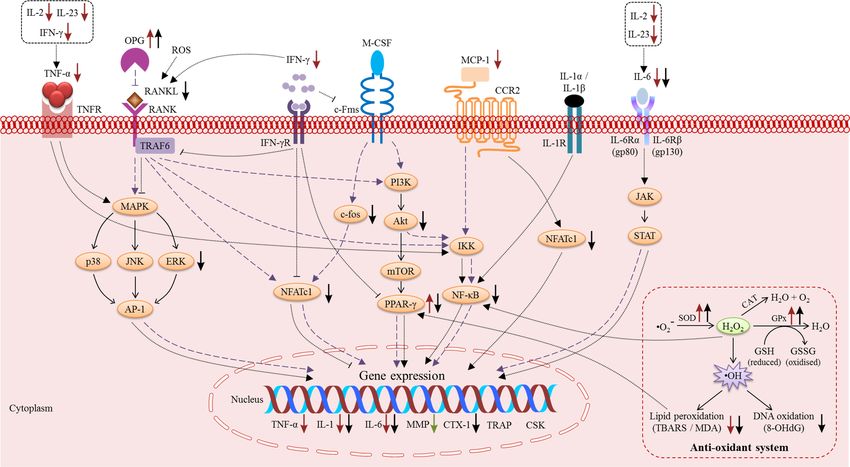
Int. J. Mol. Sci. 2019, 20, 1453                                                                     2 of 26

osteoporosis, much effort has been invested in the search for new anti-osteoporotic agents that can
mitigate the progression of osteoporosis.
       Vitamin E is an essential lipid-soluble vitamin that consists of two subgroups: tocopherol (TF)
and tocotrienol (T3). Both TF and T3 have a chromanol ring with a long carbon phytyl chain,
distinguishable by the presence of solely single bonds or three double bonds in the hydrocarbon
tail, respectively. Each isoform can be further divided into four distinct analogues, namely alpha (α),
beta (β), gamma (γ), and delta (δ), based on the location of the methyl groups in the chromanol nucleus.
The effects of vitamin E on bone in animal experimentations have been extensively reviewed [4,5].
Specifically, researchers revealed heterogeneous findings on the bone-protective properties of αTF [6–9].
High levels of αTF were associated with negligible effects on bone in young adult normal mice.
However, increased bone biomechanical strength was seen in old normal mice [10]. Meanwhile,
preclinical studies consistently showed that palm T3 (containing a mixture of TF and T3 isomers),
annatto T3 (containing a mixture of T3 isomers), and individual T3 isomers exhibited potential
skeletal-promoting effects in healthy animals [11,12] as well as in osteoporotic animals induced by
various stressors, such as ovariectomy [13–17], orchidectomy [18–21], nicotine [22–24], alcohol [25],
free radicals [26], glucocorticoid [27,28], buserelin [29,30], and metabolic syndrome (MetS) [31–33].
In comparison, between the two subgroups of vitamin E, some studies also reported that αTF exhibited
comparable [34,35] or less beneficial effects than T3 on the skeletal system [11,22,25]. Since the
bone-sparing effects of vitamin E in vivo are quite established, the molecular mechanisms of vitamin E
in achieving these actions are of major interest to scientists.
       This review highlights the recent advances in our understanding of the molecular aspects of
vitamin E as a bone-protecting agent, particularly through crosstalk between various signalling
pathways and signal transduction molecules. We hope to provide the readers with current updates on
the molecular machinery of vitamin E in promoting bone health and the research gap to be bridged by
investigators in order to promote vitamin E as an alternative therapeutic agent for osteoporosis.

2. The Effects of Vitamin E on Bone Cells
      The direct effects of vitamin E on bone cells have been previously investigated. In an earlier study,
Soeta et al. (2010) explored the effects of vitamin E (αTF and δTF) on osteoblast differentiation. It was
shown that TF inhibited osteoblast differentiation at the early stage, indicated by decreased alkaline
phosphatase (ALP) activity and expression of osteocalcin (OCN). Osteoblast differentiation returned
to normal at a later stage, whereby ALP activity and expression of OCN were comparable between the
TF-treated and normal control groups [36]. Another study by Ahn et al. (2011) found that proliferation
of human mesenchymal stem cells (MSCs) was enhanced after incubation with αTF. The expression of
ALP and runt-related transcription factor-2 (Runx-2) was significantly increased in the αTF-treated
cells [37]. On the other hand, Urban et al. (2012) reported that αTF could not improve proliferation
and differentiation of primary bovine osteoblasts as the elevation of collagen I, osteonectin, and OCN
was not detected [38]. For osteoclasts, Kim et al. (2012) revealed that α-tocopheryl succinate reduced
the expression of RANKL in osteoblasts as well as inhibited osteoclastogenesis in co-cultures of mouse
bone marrow cells and calvarial osteoblasts stimulated by interleukin-1 (IL-1) [39]. In line with previous
findings, an ex vivo experiment performed by Johnson et al. (2016) also showed that the osteoclast
number (Oc.N), and monocyte and lymphocyte production were suppressed in ovariectomized rats
fed a diet with supplemental D-α-tocopherol acetate (300 mg/kg diet) for 100 days. The results
reiterated that TF suppressed osteoclast formation, possibly by inhibiting monocyte and lymphocyte
production [40].
      Gamma-T3 was shown to improve proliferation (as shown by increased ALP, OCN, osteonectin,
and collagen I), differentiation (as shown by upregulation of Runx-2 expression), and mineralization
(as shown by increased bone nodules), and reduced apoptosis (as shown by low caspase-3 activity) of
osteoblastic cells [41,42]. In addition, Wan Hassan et al. (2018) demonstrated that annatto-derived T3
(consists of 10% γT3 and 90% δT3) enhanced osteogenic activity of murine osteoblasts by promoting
Int. J. Mol. Sci. 2019, 20, 1453 3 of 26
the expression of bone formation-related genes. Annatto T3 increased the expression of osterix (OSX),
collagen type 1 alpha 1 (COL1α1), ALP, and OCN, resulting in increased formation of collagen fibrils
and mineralization of the extracellular matrix [43]. The effects of T3 isomers on human osteoclasts were
investigated by Brooks et al. (2011). Both αT3 and γT3 isomers suppressed osteoclastogenesis, with
the latter exhibiting more potent inhibition on osteoclast formation and activity than the former [44].
Comparison between TF and T3, αT3 and δT3 exhibited more superior effects in promoting
osteoblast differentiation than αTF and δTF when the nano-emulsion mode of delivery was applied.
Higher expression of OSX, collagen I, and ALP activity were observed in primary human MSCs
after exposure of T3 relative to exposure of TF [45]. Potency between individual vitamin E isomers
were compared by using bone scaffolds cultured with osteoblasts or co-cultured with osteoblasts and
osteoclasts. These cultures and co-cultures were treated with 100 nM of αTF, αT3, βT3, γT3, and δT3
for 28 days. In the three-dimensional (3D) osteoblast culture model, scanning electron microscope
(SEM) analysis showed attachment and clumping of osteoblasts on the surface of αT3-, γT3-, and
δT3-treated bone scaffolds. Micro-computed tomography (micro-CT) analysis showed increased bone
volume (BV), trabecular thickness (Tb.Th), and trabecular number (Tb.N) in bone scaffolds treated
with γT3 and δT3. In the 3D osteoblast-osteoclast co-culture model, bone scaffolds incubated with γT3
had higher BV, Tb.Th, and Tb.N and lower bone porosity. A biomechanical strength test also showed
an increase in elasticity in the bone scaffolds treated with all types of vitamin E isomer, particularly γT3
and δT3. This study suggested that γT3 and δT3 might be the most active isomers acting on bone [46].
Overall, the existing studies indicated that the effects of αTF were heterogeneous whereas the
protective effects of T3 were consistent on osteoblastic cells. Meanwhile, the in vitro studies that
examine the effects of vitamin E (TF and T3) on osteoclasts are limited.
3. Molecular Actions of Vitamin E on Bone
3.1. The Macrophage-Colony Stimulating Factor (M-CSF) and RANK/RANKL/OPG Pathway
The M-CSF and RANK/RANKL/OPG trimolecular complex system play an important role in
osteoclast formation and the regulation of bone resorption. The expression of M-CSF by osteoblastic
stromal cells is required for the differentiation of osteoclast precursor cells into mature osteoclasts
in the presence of RANKL [47–49]. RANKL is a member of the tumour necrosis factor superfamily,
which is typically membrane bound on osteoblastic and activated T cells [47]. It is also commonly
referred to as an OPG ligand, osteoclast differentiation factor (ODF), or tumour necrosis factor
related activation-induced cytokine (TRANCE). The RANK messenger ribonucleic acid (mRNA)
was found to be highly expressed in bone marrow-derived osteoclast progenitors and mature
osteoclasts [50]. In vivo studies demonstrated that RANK-knockout mice experienced osteopetrosis or
bone hardening. This is due to an absence of osteoclasts as a result of an inability of osteoblasts
to support osteoclastogenesis [51,52]. In contrast, an activated mutation in exon 1 of RANK in
humans was reported to account for the increase in osteoclast formation, activities, and osteolysis.
This condition often occurs in some patients with familial Paget’s disease of the bone with an autosomal
recessive disorder [53].
The binding of RANKL to its receptor, RANK, on cells of the myeloid lineage activates a cascade of
intracellular signalling events, including interactions with tumour necrosis factor receptor-associated
factor 6 (TRAF6) adaptor molecules, thereby stimulating the activation of nuclear factor-kappa B
(NF-κB), nuclear factor of activated T-cells cytoplasmic 1 (NFATc1), mitogen-activated protein kinase
(MAPK), and phosphatidylinositol-3-kinase (PI3K), responsible for osteoclast formation, activation,
and survival [54–56]. NF-κB has been shown to mediate RANKL-induced osteoclast differentiation,
leading to activation of Fos proto-oncogene (c-Fos) prior to NFATc1 [57]. Described as the master
regulator of osteoclast formation, the presence NFATc1 is important in the differentiation of stem
cells into osteoclasts [58]. It is activated transiently in osteoclast precursors within an hour with the
presence of RANKL and NF-κB p65 interaction [59]. The proto-oncogene, c-Fos, also plays an essentialInt. J. Mol. Sci. 2019, 20, 1453 4 of 26
role in the osteoclastic differentiation of precursors generated by M-CSF and RANKL. A study found
that overexpression of c-Fos in mice lacking c-Fos macrophages resulted in the up-regulation of RANK
expression through induction of the RANKL signals [59].
OPG is a protein secreted mainly by cells of the osteoblast lineage and plays a role as a natural key
endogenous regulator of the RANK/RANKL/OPG pathway. Both OPG and RANK are receptors which
show affinity to the same ligand RANKL [60]. Acting as a decoy receptor by binding with RANKL, OPG
thereby inhibits osteoclastogenesis and the survival of pre-existing osteoclasts [48,61,62]. Generally,
RANKL expression is up-regulated when OPG expression is down-regulated or not induced to the same
degree as RANKL, suggesting that the ratio of RANKL/OPG represents an important determinant
of bone resorption [63,64]. OPG overexpression significantly increases the cortical and cancellous
bone mass in association with reduced osteoclast numbers [65]. In a study utilizing OPG-knockout
mice, OPG deficiency increased cortical porosity and reduced cortical and cancellous bone volume
and density [66]. Additionally, mice subjected to ovariectomy (animal model for postmenopausal
osteoporosis) followed by immediate treatment with adenoviral human OPG fusion protein were
found to have a higher bone volume with reduced osteoclast numbers in axial and appendicular bones
after 4 weeks [67]. Another study further demonstrated that a single intravenous OPG injection in
rats caused a significant increase in tibial cancellous bone volume and femoral bone mineral density
(BMD) with a significant reduction in osteoclast surface [68]. A single subcutaneous injection of OPG
was found to be effective in rapidly and profoundly reducing bone turnover for a sustained period in
postmenopausal women. Thus, elevating the level of OPG might be effective in the treatment of bone
diseases characterized by increased bone resorption, such as osteoporosis [69].
Therapeutic approaches in the treatment of pathological bone loss targeting this system have been
widely investigated. A growing body of literature delineated the biological properties of the natural
vitamin E and its various forms in the maintenance of bone metabolism. Interestingly, an in vitro
study by Lee et al. (2009) showed that Trolox (a hydrophilic derivative of TF with a carboxylic
group) inhibited osteoclast formation in bone marrow cell osteoblast co-culture by suppressing
the IL-1-induced RANKL expression in a dose-dependent manner (100, 200, and 500 µM) [70].
An in vivo study also demonstrated that OPG levels were increased in nicotine-induced osteoporotic
rats after three months treatment with 60 mg/kg αTF [24]. The anti-resorptive potential of αT3
was demonstrated using a co-culture of osteoblasts and bone marrow macrophages [71]. Treatment
with αT3 at 50 µM was discovered to suppress RANKL expression in osteoblast, RANKL-induced
expression of c-Fos, NFATc1, acute activation of extracellular signal-regulated kinase (ERK), NF-κB
activation, and pit formation (indicative of osteoclast resorptive activity) by mature osteoclasts [71].
Administration of γT3 at 100 mg/kg via subcutaneous injection once a month for three months has
been shown to significantly inhibit the increase of RANKL expression and block the decrease of
OPG expression in female ovariectomized mice [15]. A recent study also evaluated the effects of
annatto-derived T3 composed of 90% δT3 and 10% γT3 using a testosterone-deficient osteoporotic
rat model through orchidectomy. The results indicated that annatto T3 at 60 mg/kg significantly
decreased the osteoclast-related gene of RANKL expression when compared with the orchidectomized
group [19].
Apart from that, Shen et al. (2018) investigated the potential of annatto T3 containing 90% δT3
and 10% γT3 with 70% purity in protecting bone in postmenopausal osteopenic women who are
at increased fracture risk [72]. Clinical findings indicated that the level of the placebo group had
the highest soluble RANKL (sRANKL) and sRANKL/OPG ratio among the placebo (430 mg olive
oil/day), low T3 (430 mg T3/day), and high T3 (860 mg T3/day) groups, suggesting a higher rate
of bone resorption. The changes in the RANKL/OPG ratio at 6 and 12 weeks were observed and
results showed a significant reduction in both low (6.81% and 12.47%) and high levels of T3 (12.8%
and 23.78%), respectively. These findings further confirmed that the beneficial properties of dietary T3
supplementation on bone were mediated through suppression of osteoclastic activity [72].Int. J. Mol. Sci. 2019, 20, 1453 5 of 26
Overall, the RANK/RANKL/OPG system is an essential signalling pathway involved in bone
cell to cell communication, with ample evidence showing that modification of this signalling pathway
has major effects on bone remodelling. RANKL mediates osteoclastogenesis and activates mature
osteoclasts, whereas OPG negatively regulates the binding of RANKL to RANK and reduces the
half-life of membranous RANKL, thus inhibiting bone resorption induced by osteoclasts. Accumulated
evidence has reported that treatment with T3 shows promising bone protective effects by modulating
this pathway. However, further studies are needed to strengthen the findings.
3.2. Pro-Inflammatory Cytokines
Inflammation is the frontline defence against diseases characterised by the activation of immune
cells in the innate and adaptive immune system, leading to the production of inflammatory cytokines.
Inflammation plays an important role in eliminating damaged tissues and initiating tissue repair.
However, the perpetuation of the inflammatory response in turn inhibits bone formation and activates
bone resorption. Cytokines are the predominant players in regulating the inflammatory response
observed in metabolic bone diseases, including osteoporosis. These cytokines are, but not limited to,
tumour necrosis factor-alpha (TNF-α), interleukin (IL), and interferon (IFN). The relationship between
vitamin E (particularly αTF), inflammation, and bone health has been investigated in a large cohort
study of older women consisting of two visits (first visit: 1997–1999, age: 55 ± 2.2 years, n = 3883;
second visit: 2007–2011, age: 66 ± 2.2 years, n = 2130). In this study, Yang et al. (2016) found a
positive association between serum αTF and BMD at the femoral neck, but a negative association
between serum αTF and inflammation, evidenced by higher concentrations of IL-6 and high-sensitivity
C-reactive protein (hs-CRP), among participants not taking vitamin E supplements [73].
Pro-inflammatory mediators have been previously demonstrated to positively and negatively
regulate osteoclast and osteoblast functions, respectively. TNF-α, IL-1, and IL-6 are potent bone
resorption stimulators. The recognition of TNF-α and IL-6 by their respective receptors on bone marrow
stromal cells results in the downregulation of osteoblast gene products, partly via the inhibition of
MAPK, the activation of suppressor of mothers against decapentaplegic ubiquitylation regulatory
factor 1 (SMURF1) and SMURF2, as well as the activation of signal transducers and activators of
transcription (STAT) [74]. Pro-inflammatory cytokines also suppress the transcription process of
osteogenic factors through upregulation of Dickkopf-related protein 1 (DKK1) and sclerostin (SOST) to
inhibit the Wnt/β-catenin pathway [74]. In addition, the interaction between inflammatory cytokines
and their receptors increases the production of M-CSF and RANKL by osteoblasts, thus promoting
osteoclast differentiation, proliferation, and activation [75]. In osteoclasts or osteoclast precursors,
TNF-α, IL-1, and IL-6 signal via the NF-κB, MAPK, and Janus kinase (JAK)-STAT pathways to
upregulate osteoclast-related genes and further amplify osteoclastogenesis [74]. Both TNF-α and
IL-1 also have potent anti-apoptotic effects on osteoclasts, suggesting that IL-1 works intimately with
TNF-α to favour inflammatory osteoclastogenesis and bone destruction [75].
Other resorptive cytokines, such as IL-2 and IL-23, have been recently examined for their role
in the disease state of osteoporosis. They were initially recognised as inflammatory cytokines that
interconnect immune cells, osteoclasts, and synoviocytes, thus playing a crucial role in the pathogenesis
of rheumatoid arthritis [76]. The role of these cytokines in osteoporosis has been of particular interest
as they affect the differentiation and activation of pathogenic osteoclasts. Both IL-2 and IL-23 are
known as T cell growth factor to facilitate the expansion of T cell populations [76,77]. T cells are the
main source of other pro-inflammatory cytokines, such as TNF-α, IL-6, and IL-17 [75]. The interaction
between IL-23 and IL-23 receptors also transduces signal to induce the secretion of TNF-α, IL-1, and
IL-23 [76]. Thus, IL-2 and IL-23 contribute to the positive feedback to the inflammation cascade,
subsequently promoting the persistent release of RANKL by osteoblasts.
On the other hand, IFN-γ has biphasic effects on bone metabolism both in vitro and in vivo.
IFN-γ potentially promotes osteoblastogenesis through upregulation of various osteogenic factors,
such as Runx-2, OSX, ALP, and OCN, but inhibits adipogenesis through downregulation of peroxisomeInt. J. Mol. Sci. 2019, 20, 1453 6 of 26
proliferator-activated receptor-gamma (PPAR-γ) [78,79]. The underlying mechanisms of IFN-γ in
reducing osteoclast formation have been postulated through the downregulation of colony-stimulating
factor-1 receptor (c-Fms), NFATc1, and TRAF6, subsequently impairing the activation of downstream
targets, including NF-κB and c-Jun N-terminal kinase (JNK) [80,81]. IFN-γ also stimulates osteoblasts
to produce nitric oxide and enhances Fas–Fas ligand interaction to induce osteoclast precursor
apoptosis [82,83]. Conversely, IFN-γ indirectly promotes osteoclast differentiation by stimulating T cell
activation and T cell secretion of osteoclastogenic factors, including TNF-α and RANKL [84]. IFN-γ
also induces the expression of dendritic cell-specific transmembrane protein (DC-STAMP), which is
responsible for the fusion of mononucleated osteoclasts into functional mature osteoclasts [85]. Hence,
IFN-γ plays conflicting roles in osteoclastogenesis by inhibiting early stages, but promoting late stages
of osteoclast formation.
Monocyte chemoattractant protein-1 (MCP-1) is a chemokine that plays a role in the migration
and infiltration of monocytes or macrophages in response to inflammation [86]. MCP-1 is specifically
expressed in osteoclasts. A previous in vivo study reported that multinuclear osteoclast differentiation
was inhibited and NFATc1 was downregulated in osteoclasts derived from MCP-1-deficient mice [87].
Furthermore, MCP-1 did not induce cell–cell fusion in osteoclasts with a DC-STAMP deficiency
(DC-STAMP is a direct target of NFATc1, which is highly induced during osteoclast differentiation) [88].
These findings suggested that MCP-1 was implicated in the regulation of osteoclast differentiation and
fusion through upregulation of NFATc1 and in the presence of DC-STAMP.
A wide array of laboratory studies demonstrated the osteoprotective effects of vitamin E through
the reduction of systemic inflammation. Oestrogen deficiency caused by ovariectomy increased the
levels of IL-1 and IL-6 in animals. Treatment with 60 mg/kg palm-derived T3 (37.2% αT3, 39.1% γT3,
and 22.6% δT3) successfully prevented a rise in cytokine levels [89]. Metabolic syndrome, defined as
a medical condition with cardiovascular risk factors, such as obesity, hypertension, hyperglycaemia,
and hypercholesterolemia, was also associated with the occurrence of inflammation [31–33] and
skeletal impairment [90,91]. Palm-derived T3 (21.9% αTF, 24.7% αT3, 4.5% βT3, 36.9% γT3, and 12.0%
δT3) and annatto-derived T3 (16% γT3 and 84% δT3) reduced the levels of inflammatory markers
(IL-1α and IL-6), concurrent with improvements of bone microstructure in the MetS animals [31–33].
Recently, it was also found that high-fat diet-induced type 2 diabetes caused inflammation as well
as degeneration of bone microstructure in the femur and lumbar of male mice [92]. In this study,
the authors found that annatto T3 (10% γT3 and 90% δT3) improved glucose and insulin tolerance,
elevated serum procollagen I intact N-terminal propeptide (P1NP, a bone formation marker), decreased
serum carboxyl-terminal telopeptide of type I collagen (CTX, a bone resorption marker), improved
trabecular and cortical microstructure, as well as suppressed the expression of inflammation markers
(MCP-1, IL-2, IL-23, IFN-γ, and TNF-α) in the liver of high-fat diet-induced diabetic mice [92].
The distinct effects of T3 and αTF on inflammatory markers were also elucidated using free
radical- and nicotine-treated animals. Free radicals generated by ferric nitrilotriacetate elevated bone
resorbing cytokines (IL-1 and IL-6), reduced OCN levels, and impaired bone histomorphometry
(lowered osteoblast number (Ob.N), bone formation rate (BFR), BV, Tb.Th, raised eroded surface
(ES), and Oc.N). All these adverse changes were reversed by supplementation of 100 mg/kg of a
palm oil T3 mixture (30.7% αT3, 55.2% γT3, and 14.1% δT3). However, similar doses of αTF only
reduced ferric nitrilotriacetate-induced increase of Oc.N and IL-6 in the animals, suggesting that
the palm-derived T3 mixture was better than αTF [26]. Two months of nicotine treatment also
increased inflammatory cytokines (IL-1 and IL-6) and bone-resorption markers (pyridinoline, PYD)
while reducing a bone formation marker (OCN). Three types of vitamin E (αTF, T3-enhanced fraction,
(42.98% αT3, 30.98% γT3, 13.81% δT3, 12.23% palm oil devoid of αTF), and γT3) at a dose of 60 mg/kg
showed effectiveness in improving all these parameters compared to the negative control group.
In addition, only T3-enhanced fraction and γT3 showed higher levels of OCN compared to the normal
control animals. These results suggested that T3 was superior to αTF in reversing the adverse effects
of nicotine on the skeleton [23]. In another study, Norazlina et al. (2007) revealed that 60 mg/kg of aInt. J. Mol. Sci. 2019, 20, 1453 7 of 26
palm T3 mixture (composition not mentioned) was able to prevent increments of IL-1 and IL-6 due to
nicotine treatment. In contrast, treatment with αTF at a similar dose increased the levels of IL-1 and
IL-6 in the nicotine-treated animals [93].
Taken together, the data from these published papers revealed the skeletal-protecting properties
of vitamin E via its anti-inflammatory properties using various in vivo osteoporotic models.
The anti-inflammatory activities of T3 seem to be more effective than αTF in osteoporotic conditions.
Furthermore, TNF-α, IL-1, IL-2, IL-6, IL-23, and MCP-1 are well-known for their anti-osteogenic and
osteoclastogenic effects. However, most of these studies mainly assessed the effects of vitamin E on the
levels of IL-1 and IL-6. A paucity of data has shown the effects of vitamin E on other bone-resorbing
cytokines. IFN-γ has biphasic control of bone metabolism. We believe both the positive and negative
effects of IFN-γ on bone exist in parallel, but the net outcome of IFN-γ remains unresolved. Thus,
the role and exact mechanism of IFN-γ requires further clarification. There are other inflammatory
cytokines that have been recognised as osteoclastogenic (such as IL-7, IL-8, IL-11, IL-15, IL-17, and
IL-34) and anti-osteoclastogenic cytokines (such as IFN-α, IFN-β, IL-3, IL-4, IL-10, IL-12, IL-27, and
IL-33). The role of vitamin E on these cytokines in bone homeostasis awaits further investigation.
3.3. Reactive Oxygen Species (ROS) and the Anti-Oxidant System
Oxidative stress refers to a condition in which an imbalance occurs between ROS generated
from various oxidation pathways and the antioxidant defence system in the body, leading to
enhanced lipid peroxidation, protein modifications, and deoxyribonucleic acid (DNA) damage [94].
A certain physiological level of ROS is essential for cellular functions, but over-generation of ROS is
associated with the pathogenesis of multiple human diseases, such as cardiovascular disease, diabetes,
hypertension, cancer, and musculoskeletal disorders. In bone metabolism, oxidative stress positively
correlates with osteoclast differentiation and osteoblast apoptosis, but negatively correlates with
osteoblast activity and osteoclast apoptosis [95–97].
Mechanistically, ROS promotes osteoclastogenesis and inhibits osteoclast apoptosis via an
increase of RANKL production and activation of the NF-κB-mediated inflammatory response [97].
The inhibition of osteoblastogenesis and stimulation of osteoblast apoptosis by ROS are associated with
antagonisation of Wnt/β-catenin signalling through diversion of β-catenin from T cell factor-(Tcf-)
to Forkhead Box O-(FoxO-)mediated transcription and the direct activation of NF-κB [98,99].
Increased lipid peroxidation also activates PPAR-γ, thus favouring adipogenesis and inhibiting
osteoblastogenesis [100]. All these events contribute to increased bone turnover, reduced
mineralization, and, subsequently, bone loss. The natural enzymatic and non-enzymatic anti-oxidant
systems are essential in inactivating ROS and relieving oxidative stress. The enzymatic anti-oxidants
include superoxide dismutase (SOD), glutathione peroxidase (GPx), and catalase (CAT) whereas the
non-enzymatic antioxidants include reduced glutathione (GSH). ROS, such as superoxide radical anion
(O2 •− ), is the by-product generated by the oxidation of nicotinamide adenine dinucleotide phosphate
(NADPH) oxidase. Excess O2 •− is removed through SOD, which catalyses the disproportionation
of O2 •− to the less reactive hydrogen peroxide (H2 O2 ) and oxygen (O2 ) (Equation: 2O2 • + 2H+ →
H2 O2 + O2 ). H2 O2 , generated by SOD, can be further inactivated by CAT or GPx. CAT catalyses the
decomposition of H2 O2 to water (H2 O) and O2 (Equation: 2H2 O2 → 2H2 O and O2 ) whereas GPx
metabolizes H2 O2 to H2 O using GSH as a hydrogen donor, converting two GSH molecules to its
oxidised form (GSSG) (Equation: H2 O2 + 2GSH → 2H2 O + GSSG) [94,101].
Since the potential role of oxidative stress in osteoporosis is rapidly evolving, correcting the
oxidative stress using an anti-oxidative agent might be a viable method to prevent osteoporosis.
Vitamin E, a potent free radical scavenger, has been reported to possess bone-protecting properties
through the alleviation of oxidative stress. In a longitudinal study of 405 Swedish men, urinary
F2 -isoprostane (a biomarker of oxidative stress) and serum αTF were measured at the age of 77 years
whereas BMD was detected at the age of 82 years. In this study, Ostman et al. (2009) reported
that elderly men with low serum levels of αTF had high oxidative stress and reduced BMD [102].Int. J. Mol. Sci. 2019, 20, 1453 8 of 26
A double-blind, controlled clinical trial demonstrated a positive correlation between hip BMD and
anti-oxidant activities (SOD and GPx) in elderly subjects. Besides, there was a diminution in the
lipoperoxide level and less hip bone loss after the administration of ascorbic acid (1000 mg) and
αTF (400 IU) [103]. Findings from an earlier in vivo study using 32-day-old chicks indicated that
feeding the chicks with αTF (90 IU/kg diet) lowered the level of thiobarbituric acid-reactive substance
(TBARS, an index for lipid peroxidation) in the plasma and liver of the animals, resulting in improved
bone histomorphometric measurements in the trabecular bone [104]. Using a rat model of hindlimb
unloading, αTF in a 15, 75, and 500 IU/kg diet dose-dependently improved oxidation parameters,
evidenced by the increase in the ferric reducing ability of plasma (FRAP). Histological assessment also
showed that αTF preserved the bone microstructure in the distal femur of animals [105]. In addition,
αTF exerted beneficial effects on fracture healing. An animal study by Turk et al. (2004) found
that advanced fracture healing and decreased levels of malondialdehyde (MDA) were detected in
fractured male rats administered intraperitoneally with αTF (20 mg/kg) [106]. Another study using a
rat model with osteoporotic fracture also suggested that 60 mg/kg αTF supplementation improved
fracture healing by increasing the activities of anti-oxidant enzymes (SOD and GPx) in the bone of
ovariectomized rats with fractures [107].
For T3, a recent randomised double-blinded placebo-controlled trial evaluated the effects of
12-week annatto T3 (10% γT3 and 90% δT3) supplementation on the levels of RANKL, OPG, bone
markers (bone-specific ALP (BALP) and N-terminal telopeptide (NTX)) as well as oxidative stress
biomarkers (8-hydroxy-20 -deoxyguanosine (8-OHdG)) in postmenopausal women with osteopenia
(n = 87, aged 59.7 ± 6.8 years). There was a significant increase in the BALP/NTX ratio, but a
decrease in the RANKL/OPG ratio and 8-OHdG concentrations in postmenopausal women receiving
T3 supplementation relative to the placebo group [72]. In an in vitro study, Abd Manan et al. (2012)
evaluated the effects γT3 on lipid peroxidation, anti-oxidant enzymes activities, and apoptosis
of osteoblasts exposed to H2 O2 . Results indicated that γT3 at 1 µM prevented MDA elevation,
reduced osteoblast apoptosis, and increased SOD, GPx, and CAT activities [41]. An in vivo study by
Maniam et al. (2008) found that supplementation of 100 mg/kg palm-derived T3-rich fraction (14.62%
αT3, 32.45% γT3, 23.93% δT3, and 18.43% αTF) reduced TBARS and increased GPx in the femur of
normal adult male rats [108]. In contrast, these changes were not observed in the normal animals
treated with αTF at a similar dose [108]. Results from a study by Nazrun et al. (2008) revealed that
60 mg/kg of palm T3 (30.7% αT3, 55.2% γT3, and 14.1% δT3) raised GPx and SOD activities as well as
suppressing MDA levels in ovariectomized rats [109]. Comparatively, 60 mg/kg of αTF only raised
SOD and the increment was not as high as with palm T3 [109].
Overall, both αTF and T3 exhibit beneficial effects on bone and fracture healing by maintaining
a balanced profile between ROS production and levels of anti-oxidants. The current evidence also
implies the anti-oxidative effects of T3 outweigh αTF in osteoporotic conditions. Several research
gaps remain to be bridged by investigators. First, most of these studies focused on investigating the
effects of vitamin E on SOD and GPx activities. The evaluation on the effects of vitamin E on CAT
activity is indispensable since CAT and GPx work together to detoxify H2 O2 . Second, vitamin E
potentially reactivates the anti-oxidant system to prevent skeletal impairment caused by oxidative
stress. Therefore, the effects of vitamin E on the downstream targets of oxidative stress signalling,
including the expression of FoxO, β-catenin, PPAR-γ, and NF-κB, requires further elucidation.
3.4. Growth Factors
Growth factors are proteins that are usually stored in the extracellular matrix and are secreted for
critical functions, such as cell division, matrix synthesis, and tissue differentiation, on the appropriate
target cells [110,111]. They are also actively released after injury by the extracellular matrix, cells, and
platelets [110]. Several growth factors have been proven to act on the skeleton [110]. The potential of
vitamin E as a bone-protecting agent might be due to the ability of vitamin E to promote bone-related
growth factors, including transforming growth factor-beta (TGF-β) [37,112], bone morphogeneticInt. J. Mol. Sci. 2019, 20, 1453 9 of 26
protein (BMP) [16,37,112,113], vascular endothelial growth factor (VEGF) [112], fibroblast growth
factor (FGF) [112,114], and insulin-like growth factor-1 (IGF-1) [10].
The large superfamily of TGF-β consists of three mammalian isoforms of TGF-β (namely TGF-β1,
TGF-β2, and TGF-β3) and BMP [115]. Both TGF-β and BMP are abundantly found in bone and cartilage,
suggesting their crucial role in bone remodelling. Active TGF-β and BMP-2 bind to their respective
tetrameric transmembrane serine/threonine kinase receptors, TGF-βR and BMPR, expressed on the
surface of osteoblasts or osteoblast precursors to initiate intracellular signals via canonical suppressor
of mothers against decapentaplegic-(SMAD-)dependent and non-canonical SMAD-independent
pathways. In canonical SMAD-dependent signalling, the interactions of TGF-β/BMP-2 with their
receptors cause phosphorylation of their immediate downstream targets (SMAD proteins), which
then interact with SMAD4, forming SMAD complexes. Alternatively, TGF-β/BMP-2 activates the
MAPK signalling cascade and the downstream target, activator protein-1 (AP-1), in non-canonical
SMAD-independent signalling. The activated SMAD complexes and AP-1 proteins then translocate
into the nucleus to direct the transcription of osteoblast specific genes [116].
Ahn et al. (2011) analysed gene expression during differentiation of human MSCs into osteoblasts
after vitamin E stimulation. The expression levels of TGF-β1 and SMAD2 were upregulated >2-fold
compared to the control [37]. However, in the study by Ibrahim et al. (2015), the authors reported that
both the annatto-derived T3 alone group and the combined with lovastatin group showed increased
expression of TGF-β2 and TGF-β3, but did not reach statistical significance when compared to the
control group [112]. They concluded that TGF-β2 and TGF-β3 had maximal expressions during the
early phase of fracture healing, which might be within one week after the fracture occurred [117].
According to Mundy (1996), TGF-β2 and TGF-β3 were expressed early to initiate signalling for BMP
synthesis by osteoprogenitor cells, which is important during the early phase of fracture healing [118].
Thus, the TGF-β gene expressions may have been low when their levels were measured by the fourth
week of fracture healing in the study. In another study, nicotine was intraperitoneally injected to the
rats for two months to induce experimental osteoporosis and the expression of osteogenic genes was
downregulated [113]. Palm vitamin E (60 mg/kg) significantly increased expression of BMP-2, OSX,
and Runx-2 in these rats.
Annatto T3 was co-embedded with lovastatin (a hypolipidaemic agent) as particles for targeted
delivery and then injected into muscle near the fractured tibia of ovariectomized rats. This combination
was shown to upregulate BMP-2 expression. In this study, treatment with annatto T3 and lovastatin
also increased expression of OCN and Runx-2 [112]. Another in-vivo study further supported the fact
that co-supplementation of annatto T3 (60 mg/kg) and lovastatin for 2 months upregulated BMP-2 in
ovariectomized rats. The improvement was greater in the group treated with the combination of T3
and lovastatin compared to the T3 alone group, thus proving that both agents might have additive or
synergistic effects to improve the osteoporotic condition. It has been postulated that the upregulation
of BMP-2 might be influenced by the suppression of the mevalonate pathway [16]. Both agents (T3 and
lovastatin) suppress the mevalonate pathway by inhibiting isoprenoid intermediates, such as farnesyl
pyrophosphate and geranylgeranyl pyrophosphate, to attach and form cytosolic prenylated proteins
to small G proteins (Rac, Rho, Rap1, and Rab). The inhibited prenylation of RhoA, the first identified
Rho member, has been reported to promote BMP-2-induced osteoblastic differentiation processes [119].
The prenylation process is also important for Rac and Rho proteins to mediate cytoskeletal changes,
leading to membrane ruffling, lamellipodia formation, and stress fibres, eventually resulting in the
activation of osteoclasts [120–122]. Thus, reduced protein prenylation may contribute to the disruption
of the osteoclast cytoskeleton, inducing its apoptosis and reduced resorptive activity [123,124].
Interestingly, high intake of αTF and mixed T3/Tocomin (500 mg/kg diet) has no impact on bone
mass, density, or turnover in rats during skeletal maturation. Although bone morphogenic protein
receptor type 1B (BMPR1B) was found to be elevated in male rats, neither αTF nor mixed T3 had
significant effects on other key genes related to osteoblast differentiation, including Runx-2, OCN,
COL1α1, COL2α1, ALP, and osteopontin. More importantly, supplementation of αTF and mixed T3Int. J. Mol. Sci. 2019, 20, 1453 10 of 26
for 18 weeks upregulated noggin (a BMP inhibitor) as well as downregulated SMAD5 and growth
differentiation factor-10 (GDF-10, also known as BMP-3B) in the animals [114]. High levels of noggin
expression in mature osteoblasts of 20-month-old C57BL/6J mice were often associated with impaired
osteoblast formation, differentiation, and function [125]. These findings indicated that high doses of
αTF or mixed T3 suppressed BMP signalling through the SMAD-dependent pathway, thus overcoming
the other skeletal beneficial effects of vitamin E, and contributing to the negligible effect of high dietary
αTF or mixed T3 during skeletal maturation [114].
VEGF-α, the most abundant VEGF isoform, is commonly used in studies investigating the
biological effects of VEGF. Thus, VEGF-α is more commonly referred to as VEGF [126,127]. Deckers
et al. (2000) demonstrated that VEGF expression was strongly increased during terminal osteoblast
differentiation and reached maximum expression during the mineralization stage of bone repair [128].
VEGF has been reported to stimulate bone repair by promoting angiogenesis and bone turnover [129].
BMP-2 and VEGF-α are closely related in enhancing angiogenesis. It has been shown that BMP-2
stimulates the secretion of VEGF-α from osteoblasts [130]. Besides that, VEGF, which was given in
combination with local sustained BMP-2 release, had significantly enhanced ectopic bone formation
compared to BMP-2 alone [131]. Both the annatto T3 and combined (annatto T3 and lovastatin)
treatments significantly increased the expression of VEGF-α (a crucial gene for fracture healing) in
ovariectomized rats with fractures. The increase in VEGF expression was detected at week 4 after
fracture induction to the tibiae of the rats [112].
The role of FGF in bone development, maintenance, and fracture healing has been established.
FGF-2 is highly expressed in stromal cells and osteoblasts in bone [132]. FGF-2 exerts a differential
function: The low molecular weight isoform of FGF-2 (LMWFGF-2) enhances bone formation whereas
the high molecular weight isoform of FGF-2 (HMWFGF-2) suppresses bone mineralization [133].
Moreover, a study by Nakamura et al. (2005) reported that low doses of FGF-2 increased ectopic bone
formation and high doses of FGF-2 inhibited bone formation in vivo [134]. LMWFGF-2 stimulates
osteoblast differentiation and bone formation, which is in part mediated through modulation of the Wnt
signalling pathway [135]. Loss of endogenous FGF-2 reduces the expression of Wnt ligand, low-density
lipoprotein receptor-related protein 5 (LRP5) complex, β-catenin, and phosphorylation of glycogen
synthase kinase-3 beta (GSK3β), which results in inhibition of the canonical Wnt/β-catenin signalling
pathway [135]. On the other hand, HMWFGF-2 promoted the expression of SOST, a potent inhibitor
for the Wnt/β-catenin signalling cascade, thus possessing inhibitory effects on bone mineralization.
In short, the type of isoform and level of FGF-2 are the two important criteria for determining its net
outcome on bone, whether positive or negative. A study by Tennant et al. (2017) demonstrated that
administration of high dietary mixed T3 to 11-weeks male rats significantly increased the expression
of FGF-2, however, the type of isoform was not mentioned [114].
With advancing age, the rate of bone formation might gradually decline. Age-related decline
in IGF-1 levels was reported to be associated with bone loss in humans [136]. Bone cells respond
to IGF-1 as it is ubiquitously found within bone and serum. The binding of IGF-1 to its tyrosine
kinase receptor exerts anabolic effects on bone by transducing signals via the PI3K/protein kinase B
(Akt) and MAPK signalling pathways. Upon IGF-1 receptor (IGFR) activation, downstream protein
substrates are recruited, such as insulin receptor substrate 1 (IRS1) and Src homolog and collagen
protein (SHC). IRS1 interacts with PI3K to phosphorylate phosphatidylinositol-4,5-diphosphate (PIP2)
to phosphatidylinositol-3,4,5-trisphosaphate (PIP3). PIP3 further recruits phosphoinositide-dependent
kinase 1 (PDK1) to phosphorylate Akt, causing the activation of Akt. Meanwhile, the MAPK network
is activated through the formation of the SHC-Grb2-SOS complex. The role of PI3K/Akt and MAPK
activation on skeletal acquisition has been established in vitro and in vivo [137,138]. Beyond the
regulation of osteogenic differentiation factors through these pathways, IGF-1 secreted by osteoblasts
in the bone tissue markedly induce directional migration (chemotaxis) of osteoblasts to the site of
future bone formation [139]. In a study by Arjmandi et al. (2012), a diet containing high-doses ofInt. J. Mol. Sci. 2019, 20, 1453 11 of 26
vitamin E (500 mg/kg diet) enhanced material and structural bone quality, which was accompanied by
increased mRNA transcripts for OCN, COL1α1, and IGF-1 in old male mice [10].
Overall, BMP-2 is the most extensively studied growth factor by investigators for the effects of
vitamin E on bone healing. Vitamin E potentially increased the levels of BMP-2 expression during
the process of bone repair. Other growth factors, such as TGF-β, FGF-2, VEGF-α, and IGF-1, are also
promoted by vitamin E during skeletal impairment, although there is a paucity of studies regarding
these growth factors. Several limitations must be addressed by investigators. Firstly, a comparison
between the two types of vitamin E (TF and T3) in bone healing is lacking. Some articles did not
specify the type of vitamin E used in their experiments, making it difficult for a comparison to be
made between the two subtypes of vitamin E. Secondly, growth factors other than BMP-2 warrant
more studies to further investigate the mechanism of vitamin E as a bone-protecting agent.
3.5. Hormones
Parathyroid hormone (PTH), secreted by parathyroid glands, is a major hormone for the control
of extracellular calcium and phosphate levels [140]. PTH increases serum calcium levels via its targets
on the kidneys, intestines, and bone [141], thus suggesting its role in regulating bone remodelling.
In the kidneys, PTH inhibits the reabsorption of phosphate, thus increasing the amount of ionized
calcium in the blood. Besides, PTH stimulates the conversion of 25-hydroxyvitamin D (inactive form)
into 1,25-dihydroxyvitamin D (active form). The production of activated vitamin D further enhances
calcium uptake from the intestines [141]. PTH also breaks down the bone (the largest reservoir of
calcium in the body) to release calcium.
PTH exerts both catabolic and anabolic skeletal effects. Chronic hyperthyroidism is associated
with bone loss whereby sustained PTH elevation stimulates excessive bone turnover [142].
The osteoclastogenic activity of PTH is mainly through its indirect action on osteoblasts due to
the absence of functional PTH receptors on osteoclasts. PTH downregulates the expression of OPG
and upregulates the expression of RANKL in osteoblastic cells via protein kinase A-(PKA)-induced
activation of cAMP-response element-binding (CREB) protein [143,144]. PTH also stimulates
the osteoblastic expression of MCP-1, which has a role in the recruitment, differentiation, and
multinucleation of osteoclast precursors [145]. On the contrary, intermittent administration of PTH
has been reported to increase bone mass [146]. The bone-building mechanism of PTH involves the
activation of the canonical Wnt/β-catenin signalling pathway. An in vitro study by Tian et al. (2011)
demonstrated that intermittent administration of PTH stimulated the gene expression of β-catenin,
Runx-2, OCN, ALP, and bone sialoprotein (BSP) in osteoblastic MC3T3-E1 cells [147]. Another in vivo
study also showed that the interaction between PTH and its receptors suppressed the synthesis of the
osteocyte-derived Wnt antagonist, SOST, to further intensify the pro-osteoblastogenic signal [148].
The association between vitamin E, PTH levels, and bone health was previously investigated in an
in vivo study. Norazlina et al. (2004) showed that Sprague-Dawley rats fed with a vitamin E-deficient
diet had higher PTH levels as well as lower serum calcium levels and bone calcium contents in the
lumbar bone as compared to control animals fed with standard rat chow [149]. However, vitamin E
supplementation (αTF and αT3) at 60 mg/kg failed to reverse all these changes caused by a vitamin E
deficiency. These findings reiterated the close relationship between vitamin E deficiency, secondary
hyperparathyroidism, and vertebral bone loss even though replacing the vitamin E did not have any
effects on PTH levels [149].
Leptin is initially a hormone predominantly produced by adipose tissue that acts on the
hypothalamus and functions to regulate satiety and energy expenditure. The level of leptin is directly
proportional to body fat content. Subsequently, leptin has emerged as a factor in the regulation of bone
mass attributed to its diverse functions, including the promotion of haemopoietic and osteoblastic
differentiation [150]. Adiponectin is another regulator that co-regulates energy homeostasis and
bone metabolism. Similar to leptin, adiponectin is an adipocyte-derived hormone, but the level of
adiponectin is inversely correlated with obesity. The presence of adiponectin receptors on osteoblastsInt. J. Mol. Sci. 2019, 20, 1453 12 of 26
suggests the role of adiponectin in bone homeostasis. Ample clinical evidence has indicated the
relationship between these hormones (leptin and adiponectin) and BMD in osteoporotic patients of
various causes [151–153].
Previous studies have disclosed the distinct effects of leptin on bone metabolism through central
and peripheral pathways. Pioneering in vivo studies demonstrated that the activation of hypothalamic
receptors of leptin in the brain suppressed osteoblastogenesis and increased osteoclast activity [154,155].
Two downstream molecular cascades involved are: (a) The downregulation of c-myc expression that
increases production of cyclin D to inhibit osteoblast proliferation; and (b) the upregulation of RANKL
via the PKA signalling pathway to promote resorption effects of osteoclasts [156]. Paradoxically, leptin
binds to its receptors on bone marrow MSCs to enhance proliferation and differentiation of MSCs into
an osteoblastic lineage via the direct peripheral pathway [157]. Leptin also binds with its receptor on
osteoblasts to increase expression of OPG and inhibit secretion of RANKL [150]. Hence, the important
question that remains unresolved for leptin signalling is the absolute dependency is on central or
peripheral pathways as the nature of bone remodelling. On the other hand, adiponectin is known for its
anti-atherogenic and insulin-sensitizing properties as well as its positive effects on bone development.
In vitro studies showed that adiponectin promoted chondrocyte proliferation, proteoglycan synthesis,
and matrix mineralization via an enhancement of the expression of type II collagen, Runx-2, BMP-2,
and ALP activity [158,159]. The underlying mechanisms of adiponectin-mediated BMP-2 expression
in osteoblastic cells involved the activation of the adenosine monophosphate-activated protein kinase
(AMPK), p38 MAPK, and NF-κB pathways [158].
Since there is a link between these hormones and bone health, the evaluation of the effects of
vitamin E (a bone-protecting agent) on the levels of leptin and adiponectin is necessary. Previous studies
revealed that the rats fed with a high-carbohydrate high-fat diet experienced MetS, hyperleptinemia,
and hypoadiponectinemia along with the impairment of trabecular microarchitecture as compared
to normal control animals fed with standard chow diet. The intervention with palm T3 (21.9% αTF,
24.7% αT3, 4.5% βT3, 36.9% γT3, and 12.0% δT3) and annatto T3 (16% γT3 and 84% δT3) reduced the
elevated leptin whereas only annatto T3 improved the level of adiponectin in the animals with bone
loss due to MetS [31,32].
Based on the current state of knowledge, hormones seem to have a crucial role in both
bone anabolism and catabolism. The underlying signalling of these hormones aforementioned in
regulating bone metabolism is complex, thus meriting further investigation. In addition, the current
available literature on the relationship between vitamin E and hormones using osteoporotic models is
still lacking.
3.6. MicroRNA
MicroRNA (miRNA) is a small and highly conserved non-coding endogeneous RNA molecule
containing 20–22 bases that regulates protein expression [160]. The role of miRNA has been widely
established using in vitro studies. A panel of miRNAs targets transcription factor Runx-2 to control
osteogenic and osteoblast differentiation [161]. Li et al. (2015) reported that the overexpression of
miR-194 downregulated STAT1 and promoted nuclear translocation of Runx-2, leading to enhanced
osteoblastogenesis [162]. In addition to the regulation of osteoblast differentiation, the expression
of miRNA is also closely associated with osteoclast differentiation. Several miRNAs targeted the
RANK/RANKL/OPG system to attenuate osteoclast formation and function. MiR-17/20a and miR-26a
inhibited the RANKL expression [163,164]. Other miRNAs (e.g., miR-155) exhibited inhibitory effects
on osteoclast differentiation via the TGF-β/SMAD signalling pathway in bone marrow-derived
macrophages [165].
The role of vitamin E on miRNA expression has been investigated. Vitamin E deficiency resulted in
a reduced concentration of miRNA-122a and miRNA-125b in the rat liver [166]. Similar outcomes were
reported using Nile tilapia, whereby a diet deficient of DL-α-tocopherol acetate caused oxidative stress
as well as downregulated miR-223, miR-146a, miR-16, and miR122 in the liver [167]. The evidenceInt. J. Mol. Sci. 2019, 20, 1453 13 of 26
suggests that vitamin E potentially regulates the expression of miRNA, which might have a crucial
role in the coordination of bone homeostasis. Thus, the role of vitamin E in modulating the expression
of miRNA should be further evaluated in vivo using osteoporotic animal models and in vitro using
various types of bone cells.
4. Future Outlook and Conclusion
The underlying mechanism of the beneficial effects of vitamin E on bone is currently a topic of
investigation. The endocrine and paracrine regulators direct the activation or inhibition of various
downstream signalling pathways that contribute to the close coordination between bone resorption
and formation. An increasing number of scientific studies have identified the molecular targets of
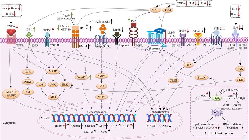
vitamin E in regulating bone metabolism (Table 1). Two models summarizing the actions of vitamin
E on osteoblasts (Figure 1) and osteoclasts are provided (Figure 2). Vitamin E modulated the levels
of inflammatory mediators, ROS, growth factors, and hormones, which are locally and systemically
released factors with a potent regulatory role in bone metabolism. Accordingly, vitamin E also plays a
pivotal role in the modulation of RANK/RANKL/OPG, NF-κB, MAPK, and oxidative stress signalling.
The process of osteogenesis involves preosteoblast proliferation, osteoblast differentiation,
and collageneous extracellular matrix formation [43]. Runx-2 is important for the differentiation
of mesenchymal progenitor cells into osteoblasts whereas osterix has a role in both osteoblast
differentiation and bone formation. Osterix is downstream of Runx-2 in osteoblast differentiation, this
was supported by a study showing that Runx-2 was expressed in osterix-knockout mice, but osterix
was not expressed in Runx-2 knockout mice [168]. Next, osterix regulates the downstream transcription
of special AT-rich sequence-binding protein (Satb2) [169]. Satb2 functions to increase the expression of
bone matrix proteins and osteogenic transcription factors during bone formation [170]. Firstly, the early
osteoblast differentiation marker genes (such as COL1α1 and ALP) will be expressed. Subsequently,
osteocalcin and BSP will be activated [43]. The current available evidence indicates that vitamin E
targeted the Runx-2 mediated mechanism to increase the expression of bone formation-related genes.
Table 1. The targets for vitamin E in the regulation of bone metabolism.
Targets References Targets References
Cytokines Kinases
IFN-γ ↓ [92] Akt ↓ [171]
IL-1 ↓ ↓ [23,26,32,33,89,93] ERK ↓ [71]
IL-2 ↓ [92] Enzymes
IL-6 ↓ ↓ [23,26,32,33,89,93] GPx ↑ ↑ [107–109]
IL-23 ↓ [92] SOD ↑ ↑ [107,109]
TNF-α ↓ [92] Chemokines
RANKL ↓ [15,19,70,72] MCP-1 ↓ [92]
Growth factors Hormones
TGF-β1 ↑ [37] Leptin ↓ [31,32]
FGF-2 ↑ [114] Adiponectin ↑ [32]
VEGF-α ↑ [112] Receptors
GDF-10 / BMP-3B ↓ [114] OPG ↑ ↑ [15,24]
IGF-1 ↑ [10] BMPR1B ↑ [114]
Oxidative stress markers Bone markers
8-OHdG ↓ [72] NTX ↓ [72]
TBARS/MDA ↓ ↓ [104,106,108,109] ALP ↑ [43]
FRAP ↑ [105] OCN ↑ [15,43]
Transcription factors CTX-1 ↓ [15,17]
Osterix ↑ [15,43,113] Genes
Runx-2 ↑ ↑ [15,37,112,113] BMP-2 ↑ [16,112,113]
PPAR-γ ↑ ↓ [19,160] ALPL ↑ [19]
NFAT2 ↓ [70] SPP1 (osteopontin) ↑ [19]
NFATc1 ↓ [71] Noggin ↑ [114]
NF-κB ↓ [71] SMAD5 ↓ [114]
Protein c-Fos ↓ [70,71]
COL1α1 ↑ [19,43]
1 The effects of TF (red arrows), T3 (black arrows) as well as high doses of TF and T3 (green arrows) are indicated.You can also read