Virus-Incorporated Biomimetic Nanocomposites for Tissue Regeneration - MDPI
←
→
Page content transcription
If your browser does not render page correctly, please read the page content below
nanomaterials

Review
Virus-Incorporated Biomimetic Nanocomposites for
Tissue Regeneration
Iruthayapandi Selestin Raja 1,† , Chuntae Kim 2,† , Su-Jin Song 3 , Yong Cheol Shin 4 ,
Moon Sung Kang 3 , Suong-Hyu Hyon 5 , Jin-Woo Oh 2, * and Dong-Wook Han 3, *
 1   Monocrystalline Bank Research Institute, Pusan National University, Busan 46241, Korea
 2   Department of Nanofusion Technology, College of Nanoscience & Nanotechnology,
     Pusan National University, Busan 46241, Korea
 3   Department of Cogno-Mechatronics Engineering, College of Nanoscience & Nanotechnology,
     Pusan National University, Busan 46241, Korea
 4   Department of Medical Engineering, Yonsei University, College of Medicine, Seoul 03722, Korea
 5   Joint Faculty of Veterinary Medicine, Kagoshima University, Kagoshima 890-8580, Japan
 *   Correspondence: ojw@pusan.ac.kr (J.-W.O.); nanohan@pusan.ac.kr (D.-W.H.)
 †   These authors contributed equally to this work.

 Received: 31 May 2019; Accepted: 8 July 2019; Published: 15 July 2019

 Abstract: Owing to the astonishing properties of non-harmful viruses, tissue regeneration using
 virus-based biomimetic materials has been an emerging trend recently. The selective peptide
 expression and enrichment of the desired peptide on the surface, monodispersion, self-assembly,
 and ease of genetic and chemical modification properties have allowed viruses to take a long
 stride in biomedical applications. Researchers have published many reviews so far describing
 unusual properties of virus-based nanoparticles, phage display, modification, and possible biomedical
 applications, including biosensors, bioimaging, tissue regeneration, and drug delivery, however the
 integration of the virus into different biomaterials for the application of tissue regeneration is not yet
 discussed in detail. This review will focus on various morphologies of virus-incorporated biomimetic
 nanocomposites in tissue regeneration and highlight the progress, challenges, and future directions
 in this area.

 Keywords: virus; tissue regeneration; biomimetic nanocomposites; phage display

1. Introduction

1.1. Emerging Trends in Tissue Regeneration
     Tissue engineering is a part of the regenerative medicine field, which emphasizes the fabrication
of various functional biological constructs to reduce the increased demand for donor organs [1].
The shortage of organ donors and the increased number of people undergoing transplantation have
necessitated the development of effective biomimetic materials adopting advanced technologies [2].
The aim of the field is to harness nature’s ability to treat the damaged tissues., ensuring biocompatibility
and supporting cellular biological events. When muscles are damaged by incidents such as illness,
accidents, and microbial invasion, they lose integrity for healthy functioning at the cellular level and
subsequently follow a cascade of biochemical events, including hemostasis, inflammation, proliferation,
and maturation, to restore integrity [3]. However, the first ever immediate response after an injury
is the production of reactive oxygen species by NADPH oxidase before inflammatory reaction [4].
Moreover, the time consumption to retain normal function in the dysfunctional organ is dependent
on one’s age and the seriousness of the damage. When tissue fails in the ability for self-regeneration,
especially in a pathological condition, the external application of a scaffold becomes inevitable [5].

Nanomaterials 2019, 9, 1014; doi:10.3390/nano9071014                      www.mdpi.com/journal/nanomaterials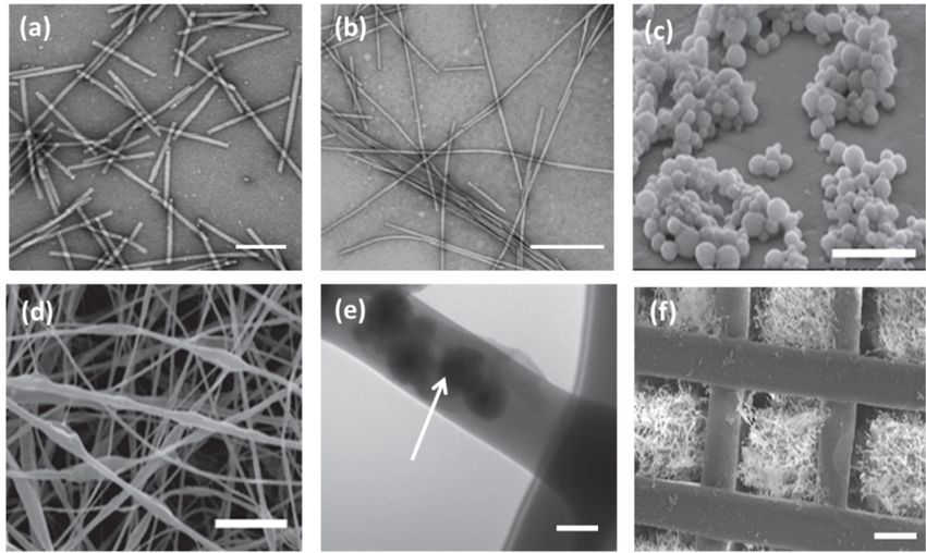
Nanomaterials 2019, 9, 1014                                                                            2 of 18

      Stainless steel was first implanted as an artificial hip in 1929, which paved the way for the
researchers to design biomimetic materials to replace body organs [6]. However, tissue and organ
failure were considered as major medical complications until 1954, as there were not enough remedies
to treat dysfunctional organs. The Nobel Laureate in medicine, Joseph Murray, transplanted a healthy
kidney to a genetically identical twin brother successfully [1]. Following this historical incident,
the researchers began to utilize cell structures to treat damaged tissues. These structures are autografts,
isografts, allografts, and xenografts, which are biological tissues harvested from a patient’s organ,
a genetically identical twin, a genetically non-identical individual of the same species, and a donor
of a different species, respectively [7]. The autograft has some advantages over other forms of grafts,
with no rejection concerns. However, it requires a secondary procedure for the reconstructive surgery
and faces donor site complication.
      Meanwhile, isografts, allografts, and xenografts express slight or significant degrees of rejection as
the Major Histocompatibility Complex (MHC) recognizes them as foreign antigens causing cytotoxic
T-cell-mediated demolition. Unlike autografts, other forms of grafts eliminate the need for the
secondary procedure without the donor site complications [8]. Though researchers are involved
in finding therapeutic techniques, such as immunosuppressive medications, infectious prophylaxis,
and DNA-based tissue typing, graft-versus-host disease (GVHD) remains a hurdle limiting the use of
grafts in tissue regeneration [9].
      The extracellular matrix (ECM) is an important component in tissues, which has the intrinsic
cues to provide sites with all their cellular activities, including migration, orientation, shape, plasticity,
and cell–matrix and cell–cell interactions [10]. The ECM is a complex three-dimensional network
comprising proteins, such as collagen, elastin, and laminin, proteoglycans, and glycoproteins [11].
It acts as a glue and platform to bind cells together with the connective tissues. The decellularized
extracellular matrix (DECM), a water-insoluble matrix, can be prepared from any organ or tissue by
removing cellular components from ECM. In recent years, DECM has been used as a scaffold to hold
the cells together and maintain integrity during wound healing processes [12]. Over the past two
decades, the researchers have been inclined towards the preparation of artificial biomimetic grafts from
the naturally available biomacromolecules, synthetic polymers, and ceramic materials, to replace tissue
grafts and DECM. The remarkable features of artificial grafts are that they are economically inexpensive,
their ease of preparation and storage, biocompatibility, and the tuning of the physicochemical and
biological properties [13]. The most widely studied natural polymers include collagen, chitosan,
hyaluronic acid, carboxymethyl cellulose, alginate, and silk fibroin, which have been reported to trigger
a less immunogenic reaction in the human body, while the synthetic polymers, such as polycaprolactone,
polyvinyl alcohol, nylon, and polyphosphazene, provide longer shelf life. To obtain combinatorial
properties, nowadays researchers utilize a blend of polymers and their derivatives [14,15]. Ceramic and
bioglass materials exhibit osteoconductive properties, showing remarkable mechanical strength and
resistance to deformation, and hence, they have been widely used for bone tissue regeneration [16].
While designing a synthetic biomimetic scaffold, it is a requisite to create a benign microenvironment
in a way that mimics the ECM niche for the application of effective tissue regeneration.
      In the past ten years, researchers have explored a variety of biomimetic nanocomposites
by incorporating bioactive molecules, such as growth factors, cytokines, genes, antibiotics,
and anti-inflammatory drugs within the scaffold to increase tissue regeneration potential [17,18].
As the direct administration of therapeutic medications is not effective, leading to overdose toxicity
and short time exposure to biological tissue, these biomimetic nanocomposites were much appreciated
by chemists, biologists, and nanotechnologists [19]. Mostly, the bioactive molecules are cross-linked
with the primary polymeric component by physical, chemical, and enzymatical treatment with or
without the aid of cross-linkers and their release from the matrix is adjusted for either immediate,
sustained, or slow release, according to the desired site of the target tissue. The drug release rate from
the biomaterial majorly involves simple diffusion, erosion, or degradation of the matrix and swelling
of the polymeric scaffold [5]. After the advent of nanotechnology, the nanoparticles were entrapped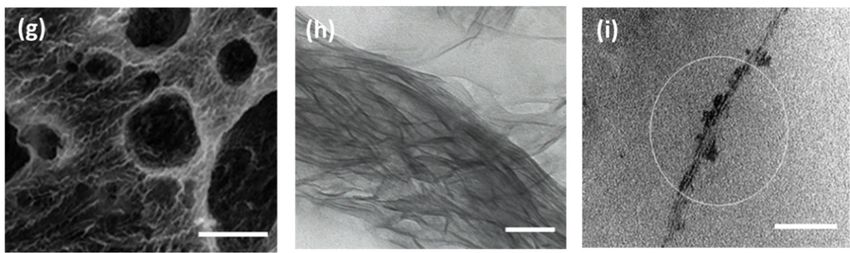
Nanomaterials 2019, 9, 1014                                                                        3 of 18

into the scaffold to exploit their medicinal properties. A composite of collagen and silver nanoparticles
has been used to augment the burn tissue repair process [20]. Mieszawska et al. studied a composite
film composed of silk and nanoclay to serve as a supportive biomaterial to improve bone tissue
regeneration [21]. The in vivo effects of reduced graphene oxide and hydroxyapatite nanocomposite
powders were investigated on critical-sized calvarial defects in a rabbit model and it was reported
that the nanocomposite stimulated osteogenesis and enhanced bone formation without inflammatory
responses [22]. Our research group also studied the influence of graphene oxide dispersed into
a polylactic-co-glycolic acid (PLGA) electrospun nanofiber towards stimulation of myogenesis and
enhanced vascular tissue regeneration [23,24]. The nanoparticles with diameters in the range of
50–700 nm acted as therapeutic drug carriers to pass through the capillaries into cells, facilitating the
regeneration of new tissues [5,25].
      Cell-laden scaffolds have also received attention among researchers aiming to achieve
a tissue-imitating engineered graft. Kizilel et al. encapsulated pancreatic islets into nano-thin
polyethylene glycol coating for enhanced insulin secretion [26].              Yoon et al.     fabricated
a three-dimensional layered structure using the blend of collagen epidermal keratinocytes and
dermal fibroblasts to progress migration and proliferation of keratinocytes and fibroblasts during
the skin repair process [27]. Within this context, microbe-based biomimetic materials have appeared
as an emerging trend in tissue regeneration in recent times. Virus-based biomaterials have many
biomedical applications, including cancer markers, antibacterial materials, drug carriers, and tissue
regeneration [28]. Not only do they encapsulate and release the therapeutic agents to the target site,
but the morphology of biomaterials also plays a pivotal role in altering physicochemical and biological
properties. In this review, we have focused on summarizing the impacts of various virus-incorporated
biomimetic nanocomposites with different morphologies, such as nanoparticles, nanofibers, hydrogels,
and organic-inorganic hybrids, in the field of tissue regeneration. The same has been schematically
shown in Figure 1. The nanoparticles that have a large surface area to volume have proven their
effectiveness with long-term functionality and stability in the biological milieu. The nanoparticles can
diffuse across the cell membrane and interact with cellular biomacromolecules residing inside the
cell [20]. Remarkably, the hydrogel provides wettability and cell migration, while the nanofibrous
matrix ensures air permeability and mechanical properties in tissue regeneration [29,30].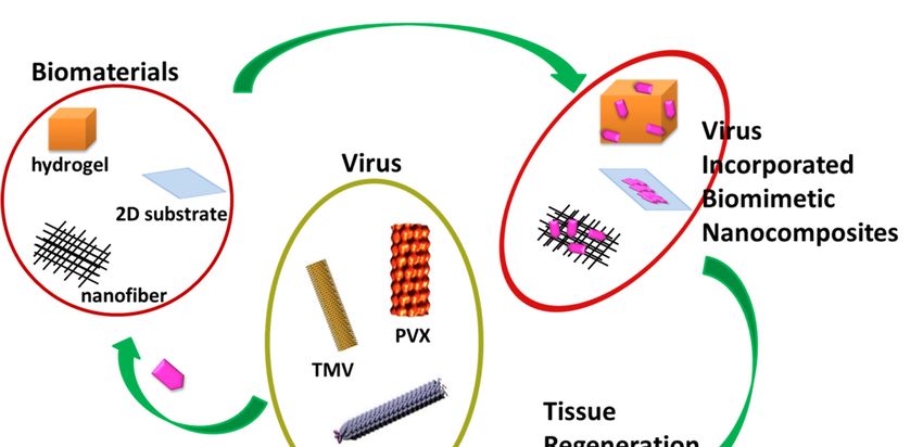
Nanomaterials
 Nanomaterials2019,
               2019,9,9,
                      1014
                         x FOR PEER REVIEW                                                                 4 4ofof1818

     Figure    A compendium
      Figure1. 1. A compendiumof tissue regeneration
                                    of tissue        functionality
                                               regeneration        of variousof
                                                             functionality    virus incorporated
                                                                                 various         biomimetic
                                                                                          virus incorporated
     nanocomposites.  Plant-based    viruses Tobacco  Mosaic Virus   (TMV)   and  Potato Virus
      biomimetic nanocomposites. Plant-based viruses Tobacco Mosaic Virus (TMV) and Potato     X  (PVX)  andX
                                                                                                       Virus
     bacteriophage M13 were  shown.    Reproduced  with permission    from [31]. Copyright  Elsevier,
      (PVX) and bacteriophage M13 were shown. Reproduced with permission from [31]. Copyright         2010.
       Elsevier, 2010.
1.2. Remarkable   Properties of Medicinally Valuable Viruses

  1.2. Not all viruses
       Remarkable      cause of
                   Properties infectious diseases
                                Medicinally        in Viruses
                                            Valuable  the human body. Viruses can be classified as lytic,
temperate, or lysogenic based on the level of adverse effects produced in its host [32]. During infection,
        Not all viruses cause infectious diseases in the human body. Viruses can be classified as lytic,
lytic phages kill the host bacteria, triggering the release of progeny. Lysogenic phages do not affect
  temperate, or lysogenic based on the level of adverse effects produced in its host [32]. During
the host cell and infection occurs with replication of the phage genome but not the host bacterial
  infection, lytic phages kill the host bacteria, triggering the release of progeny. Lysogenic phages do
genome. Temperate phages reside in host bacteria for amplification with no lysis, however some
  not affect the host cell and infection occurs with replication of the phage genome but not the host
phages, including the λ phage, exceptionally, have both categories and thrive following either lytic
  bacterial genome. Temperate phages reside in host bacteria for amplification with no lysis, however
or lysogenic cycles. The lytic phages, including T1–T7, contain a head and flexible tail but lack
  some phages, including the λ phage, exceptionally, have both categories and thrive following either
filaments. The T7 phage belongs to the Podoviridae family and structurally has an icosahedral head
  lytic or lysogenic cycles. The lytic phages, including T1–T7, contain a head and flexible tail but lack
and a short tail. They were reported to lyse the host cell within a minute by secreting the lysozyme
  filaments. The T7 phage belongs to the Podoviridae family and structurally has an icosahedral head
enzyme [33]. Professionally, T4 phages have found applications in food preservation, antibiotics,
  and a short tail. They were reported to lyse the host cell within a minute by secreting the lysozyme
detection of bacteria, DNA and protein packing systems, and DNA-based vaccines. The literature
  enzyme [33]. Professionally, T4 phages have found applications in food preservation, antibiotics,
reports revealed that Podovirus P22 assisted the assembly of cadmium sulfide nanocrystals to improve
  detection of bacteria, DNA and protein packing systems, and DNA-based vaccines. The literature
photosensitization in tissue imaging [34]. The filamentous phages, including Ff, f1, M13, N1, and Ike,
  reports revealed that Podovirus P22 assisted the assembly of cadmium sulfide nanocrystals to
are the examples of temperate phages. As they can act as a template for the synthesis of nanomaterials,
  improve photosensitization in tissue imaging [34]. The filamentous phages, including Ff, f1, M13, N1,
the general applications of temperate phages are huge compared to lytic phages [33].
  and Ike, are the examples of temperate phages. As they can act as a template for the synthesis of
       Virus-based biomimetic materials are generally derived from plant viruses and bacteriophages,
  nanomaterials, the general applications of temperate phages are huge compared to lytic phages [33].
as they rarely generate harmful side effects in human beings. Generally, bacteriophages (excluding
        Virus-based biomimetic materials are generally derived from plant viruses and bacteriophages,
fd and M13) are categorized into filamentous type of viruses, follow a non-lytic mode to infect and
  as they rarely generate harmful side effects in human beings. Generally, bacteriophages (excluding
thrive in bacteria. It has been reported that these phages do not consist of mammalian promoter
  fd and M13) are categorized into filamentous type of viruses, follow a non-lytic mode to infect and
sequences in their genome, and hence, do not instigate dreadful human diseases [35]. In the human
  thrive in bacteria. It has been reported that these phages do not consist of mammalian promoter
body, bacteriophages are present abundantly in the gut, bladder, and oral cavity, functioning to shape
  sequences in their genome, and hence, do not instigate dreadful human diseases [35]. In the human
bacterial metabolisms and populations of microbial communities. It has been described that the
  body, bacteriophages are present abundantly in the gut, bladder, and oral cavity, functioning to shape
potential role of phages increases from childhood to adulthood [36]. The monodispersed phages
  bacterial metabolisms and populations of microbial communities. It has been described that the
can self-assemble themselves into hierarchically ordered structures, such as rope-like bundles and
  potential role of phages increases from childhood to adulthood [36]. The monodispersed phages can
  self-assemble themselves into hierarchically ordered structures, such as rope-like bundles and liquid
Nanomaterials 2019, 9, 1014                                                                          5 of 18

liquid crystals. The protein surface can be modified either by covalent and non-covalent interactions
or genetic alterations [35]. These unique properties have prompted the researchers to take a long
stride in utilizing the phage-based biomaterials towards a wide range of biomedical applications,
including biomedical imaging, drug delivery, biosensors, tissue regeneration, energy, and catalysis [37].
      Owing to economically inexpensive, large scale production, ease of manipulation, and stability
against a wide range of pH and temperature, a variety of phage-based biomimetic nanocomposites
have been constructed for the application of effective tissue regeneration [38]. As far as morphology is
concerned, a typical phage has a diameter of 68 A◦ and a length in the range of 800–2000 nm. The circular
single-stranded DNA (ssDNA) of the phage encodes 10 genes containing 5000–8000 nucleotides,
which encode a highly ordered major coat protein (p8) located around the center of phage, two minor
coat proteins (p7 and p9) at one end, and two others (p3 and p6) at another terminal portion of the phage.
The helical arrays of major coat proteins assemble to form the capsid shell. Generally, minor coat proteins
display larger sized peptides than the major coat protein (p8) [39]. The major coat protein, p8, of M13
phage, has different segments, such as the N-terminal amphipathic, hydrophobic transmembrane (TM),
and DNA binding segments. The small residues (Gly, Ala, and Ser) present on these segments have
been reported to be involved in helix–helix axial and lateral interactions, which facilitate extrusion of
the virion from the membrane during assembly, and hence have been known as conserved regions
in the DNA sequence. Fiber diffraction and spectroscopic data show that M13 differs from fd at
the 12th residue, where M13 replaces Asp of fd with Asn [40]. Filamentous phages are defined as
non-enveloped bacterial viruses, having some properties in common, namely, life cycles, organization,
and morphology.
      The ssDNA has a left-handed helix structure possessing strong interactions with the positively
charged inner surface of the capsid shell. The diffraction pattern studies classified filamentous phages
into two distinct groups. Class I symmetry group consists of fd, M13, If1, and IKe, which are consistent
with 5-fold symmetry. Class II symmetry group includes Pf3, Pf1, and Xf, wherein the helices are
arranged with a rising per monomer of about 3.0 A◦ [39]. The aligned solid-state NMR studies
proved that fd has O-P-O phosphate linkages in an ordered manner, whereas Pf1 did not possess such
linkages [41]. According to NMR studies, phage fd has strong electrostatic interactions between the
negatively charged phosphate backbone of the ssDNA nucleotide and two of four positively charged
amino acid residues present at the C-terminal portion of the major coat protein, which is attributed
to stabilization of the DNA core structure. The literature reports revealed that M13 and IKe showed
similarity in π–π interactions between the residues of Tyr9 of one p8 and Tyr29 of an adjacent p8 [42].
      Infection of E. coli by phage is initiated by the attachment of N-terminal amino acids of p3,
which is present in the specialized threadlike appendage, F pilus. Subsequently, the coat protein of
the phage dissolves onto the envelope of the host, which allows the only ssDNA into the cytoplasm.
The host machinery synthesizes a complementary DNA strand with the involvement of two virally
encoded proteins, p2 and p10, which leads to the formation of a double-stranded replicative form.
The replicative form acts as a template to transcript phage genes for the synthesis of progeny ssDNAs.
These progeny phage particles discharge from the bacterial cell envelope through the membrane pore
complex, acquire coat proteins from the membrane, and appear as mature virions. The fact is that the
infected cells undergo division at a slower rate than the uninfected cells [39].
      In recent times, the researchers have sought to explore multifunctional phage-based biomaterials
by precisely adjusting the surface chemistry of phage nanofibers. Covalent, non-covalent, and genetic
modifications of phage coat proteins have been described comprehensively by the researchers.
The genetic modification of phage coat proteins would display various foreign peptides with different
functional groups at the side wall and the two termini of the phage. The endogenous amino acids
of phage coat proteins are genetically combined with the foreign amino acid sequence in order to
form a hybrid fusion protein, which is incorporated into phage particles and released from the cell
subsequently. As a result, the foreign peptide is displayed on the surface of the phage coat protein [35].
The phage display is generally specified after N-terminal modification in its respective coat proteins. For
Nanomaterials 2019, 9, 1014 6 of 18
example, if the N-terminus of p3 of the phage undergoes modification, the resulting phage is designated
as a p3 display. When two or more coat proteins are controlled for modification in the same phage,
then they can be known as double display, and so on [43]. In the phage coat protein, the carboxylates
of aspartic and glutamic acid residues, the amine of lysine, and the phenol of tyrosine are the majorly
available functional entities for the chemical modification. Introducing aldehyde into the reactive
amine group has been involved in a wide range of bio-conjugation reactions, whereas the cross-linkage
of p-azidophenylalanine has provided an azide handle on the phage surface, which can be easily
modified for further reactions [44,45]. The EDC treatment has been helpful in cross-linking the reactive
carboxylate groups with amine-functionalized moieties in phage proteins [46]. Strong nucleophile
selenocysteine has been successfully genetically incorporated into phage protein using an opal stop,
codon suppressing mRNA [47].
The phage-display library, with a heterogeneous mixture of phages carrying different foreign DNA
insert, was created for selective binding of phage proteins with the target ligands, such as polymers,
proteins, organic and inorganic crystals, small molecules, such as trinitrotoluene, and cells [48–51].
Among the phage-display libraries, the reports of p3 and p6 libraries have been well documented
in research publications. Conventionally, research studies have adopted the biopanning method to
find extensive use of phage particles in tissue regeneration. Biopanning is a typical technique to
form a population of enriched phage-displayed peptides and specifically identify a target binding
peptide [52]. According to this selection procedure, initially, a phage-display random library is
incubated with the targets. Subsequently, the non-bound phage particles are eliminated with the help
of detergent solubilized buffer. The target-bound phage particles are then eluted using a specialized
buffer maintaining acidic pH around 2.2, and the amplification process is followed by infection
of host bacteria. The resulting amplified phages form a newly enriched sub-library with more
specificity to interact with the targets. The procedure is repeated several times until a only few
desired peptides are predominantly available in the sub-library [53]. In the subsequent section, we will
investigate the contribution of plant virus and phage-based biomimetic nanocomposites in the field of
tissue regeneration.
2. Different Morphologies of Virus-Incorporated Biomimetic Nanocomposites in
Tissue Regeneration
2.1. Virus-Based Nanoparticles
Many plants and phage-based viral nanoparticles have been employed so far for tissue regeneration.
Plant viral nanoparticles are mono-dispersed, meta-stable, and structurally uniformed [54]. Li revealed
that when the virus-based nanoparticle is more robust, the functional nanostructure is more stable,
but at the same time they might be harmful to the encapsulated cargo [55].
Though the unmodified TMV nanoparticles have the potential to accelerate osteogenic
differentiation in adult stem cells, the lack of affinity to the mammalian cell surface diminishes
the cell adhesion property. Hence, the researchers opt for either genetic or chemical modification in
viral nanoparticles in order to increase the cell binding capacity and find versatile biomedical
applications. Sitasuwan et al. [62] modified the surface of a TMV nanoparticle by coupling
azide-derivatized Arg-Gly-Asp-(RGD) tripeptide with tyrosine residues through Cu (I) catalyzed
azide-alkyne cycloaddition reaction. When incorporated into the artificial scaffold, the RGD peptides
overexpressed on ECM increase initial cell attachment by binding integrin receptors. The spacing
between RGD motifs alter biological events, such as fibroblast adhesion and spreading (Nanomaterials 2019, 9, 1014 7 of 18
TMV is a polyvalent nanoparticle. The TMV can withstand temperatures up to 60 ◦ C and can be stable
in a pH range
Nanomaterials 2019,of
9, 2–10. The TEM
x FOR PEER micrograph of wild type TMV is shown in Figure 2a [56].
REVIEW 7 of 18
Figure 2.
Figure TEMmicrographs
2. TEM micrographsof ofwild
wildtype
type Tobacco
Tobacco Mosaic
Mosaic VirusVirus (TMV)
(TMV) nanoparticles,
nanoparticles, scale
scale bar
bar 200
200
nm (a)
nm (a) and
and TMV/PANI/PSS
TMV/PANI/PSS nanofiber
nanofiber (b)(b) generated
generated by by flow
flow assembly
assembly method,
method, scale
scalebar
bar500 500nm.nm.
Reproduced with
Reproduced with permission
permission from [56]. CopyrightCopyright American
American Chemical
Chemical Society, 2015. (c) (c) SEM
SEM
micrograph of
micrograph offreeze-dried
freeze-dried capsules
capsulesof ofT4T4bacteriophage/alginate
bacteriophage/alginatewater waterin inoil
oilemulsion
emulsionin inchloroform,
chloroform,
scale bar
scale bar 55 μm.
µm. (d)(d) SEM
SEM andand TEM
TEM micrographs
micrographs (e) (e) of
of PEO
PEO electrospun
electrospun nanofiber
nanofiber containing
containing T4 T4
bacteriophage/alginatehave
bacteriophage/alginate havebeen
beenshown
shownscalescale bars
bars ofof 10
10 μm and 100
µm and 100 nm,
nm, respectively.
respectively. The The arrow
arrow
indicates the
indicates the presence
presence of of T4/alginate
T4/alginate into
into the
the nanofiber. Reproduced
Reproducedwith withpermission
permissionfrom from[57].
[57].
Copyright John Wiley and Sons, 2013. (f) SEM image of the 3D printed
Copyright John Wiley and Sons, 2013. (f) SEM image of the 3D printed bioceramic bone scaffold bioceramic bone scaffold
consisting of
consisting ofbiphasic
biphasiccalcium
calciumphosphate
phosphatewith withpores
poresfilled
filledwith
withaamatrix
matrixof ofchitosan
chitosanand
andRGDRGDphage,
phage,
scale bar
scale bar 200 μm. Reproduced
200 µm. Reproducedwith withpermission
permission from
from[58]. Copyright
[58]. Copyright John Wiley
John andand
Wiley Sons,Sons,
2014.2014.
(g) SEM
(g)
micrograph of porous alginate hydrogel containing TMV particles displaying
SEM micrograph of porous alginate hydrogel containing TMV particles displaying interconnected interconnected channels
and macropores,
channels scale bar scale
and macropores, 100 µm.bar Reproduced with permission
100 μm. Reproduced from [59]. from
with permission Copyright American
[59]. Copyright
Chemical Society, 2012. (h) The TEM image of a mineralized E8-displaying
American Chemical Society, 2012. (h) The TEM image of a mineralized E8-displaying phage bundle phage bundle formed after
90 h incubation
formed after 90 hinincubation
the solution in containing
the solutioncalcium and calcium
containing phosphate andions, scale bar
phosphate 100scale
ions, nm. Reproduced
bar 100 nm.
with permission from [60]. Copyright John Wiley and Sons, 2010.
Reproduced with permission from [60]. Copyright John Wiley and Sons, 2010. (i) The TEM (i) The TEM image of theimage
HAP-fd of
phage bundle with scale bar 100 nm. The circle indicates the presence of hydroxyapatite
the HAP-fd phage bundle with scale bar 100 nm. The circle indicates the presence of hydroxyapatite nanoparticles
in the fibrousinstructure.
nanoparticles the fibrous Reproduced with permission
structure. Reproduced from [61]. from
with permission Copyright American American
[61]. Copyright Chemical
Society, 2010.
Chemical Society, 2010.
In the human body, bone tissue regenerates to a greater extent when compared to other types of
Though the unmodified TMV nanoparticles have the potential to accelerate osteogenic
tissues. However, the regeneration process is complicated in the case of tumor resection, hip implant
differentiation in adult stem cells, the lack of affinity to the mammalian cell surface diminishes the
revision, and major fractures [63]. Pi et al. constructed a cartilage targeting gene delivery nanocomposite
cell adhesion property. Hence, the researchers opt for either genetic or chemical modification in viral
system by conjugating polyethylenimine (PEI) with M13 phage-displayed chondrocyte-affinity peptide
nanoparticles in order to increase the cell binding capacity and find versatile biomedical applications.
Sitasuwan et al. [62] modified the surface of a TMV nanoparticle by coupling azide-derivatized Arg-
Gly-Asp-(RGD) tripeptide with tyrosine residues through Cu (I) catalyzed azide-alkyne
cycloaddition reaction. When incorporated into the artificial scaffold, the RGD peptides
overexpressed on ECM increase initial cell attachment by binding integrin receptors. The spacingNanomaterials 2019, 9, 1014 8 of 18
(CAP), DWRVIIPPRPSA, which was isolated after two rounds of biopanning. During incubation,
the phages expressing CAP showed higher affinity towards rabbit chondrocytes at 265.5-fold when
compared to unmodified phages. They reported that the CAP-conjugated PEI particles had no
species specificity in binding chondrocytes of rabbit and humans. Furthermore, most of the particles
were found to enter chondrocytes without being trapped in ECM, which acknowledges their larger
transfection efficiency [64].
T7 viral nanoparticles were explored to display two different functional peptides CARSKNKDC
(CAR) and CRKDKC (CRK) to target the microvasculature of regenerating wound tissue, including
skin and tendon [25]. Skin disintegration may occur in many ways, such as bruising, abrasion,
hacking, burning, stabbing, and laceration. It was observed that CAR was similar to heparin-binding
sites, whereas CRK was homologous to a segment of thrombospondin type I repeat. Interestingly,
CAR displayed a dominant function in the early stages of skin wound healing, while CRK showed
preferences in the later stages of the same process. As the terminal residues contain cysteine,
the screened peptides had more feasibility to be involved in disulfide bond formation to form
a molecular cycle structure. The CAR-expressing T7 phage nanoparticles had been found to appear
in wound sites 100–140-fold more efficiently than the non-recombinant phage nanoparticle [65].
The biomedical application of siRNAs is minimal owing to their low absorption across the stratum
corneum, a horny outer layer of skin. Hsu et al. [66] explored M13 phage (from Ph.D-C7C library)
viral nanoparticle-expressing skin penetrating and cell entering (SPACE) peptide with the sequence
of AC-KTGSHNQ-CG in order to reach therapeutic macromolecules, including siRNAs, into the
skin-associated cells. The in vitro physicochemical studies explored that the various macromolecules,
including siRNA, penetrated across the stratum corneum into the epidermis layer of skin through the
macropinocytosis pathway when the molecules were conjugated with SPACE.
A muscle binding M13 phage nanoparticle with peptide sequence ASSLNIA was identified to
possess more excellent selectivity (at least five-times more) compared to the control phage nanoparticle.
While investigating overall muscle selectivity on different organs, the muscle binding affinity was found
to be 9–20-fold for the skeletal and 5–9-fold for cardiac muscle [67]. Sun et al. synthesized functional
multivalent M13 phage (Ph.D.-7TM display library) nanoparticles to express RIYKGVIQA and SEEL
sequences, which are found in Nogo-66, a neurite outgrowth inhibitory protein. They selectively bound
negative growth regulatory protein 1 (NgR1) with electrostatic forces of repeated leucine residues,
enhancing neural differentiation of pc12 cells. Hence, this specific engineered viral nanoparticle has
been appreciated for its potential use in neurite tissue regeneration, including spinal cord injury,
optic nerve injury, ischemic stroke, and neurodegenerative diseases [68]. Collett et al. suggested
that hepatitis C virus-based nanoparticles could act as a quadrivalent vaccine to trigger humoral and
cellular immune responses. They explored biophysical, biochemical, and biomechanical properties
of nanoparticles using Atomic Force Microscopy and observed that glycosylation occurred on the
surface of the nanoparticle with ordered packing of the core [69]. The literature reports revealed that
Sendai virus vectors displaying cardiac transcription factors could efficiently reprogram both mouse
and human fibroblasts into induced cardiomyocyte-like cells in vitro. In addition, they could reduce
scar formation, maintaining cardiac function in myocardial infarction affected animals [70].
The phosphate tailored TMV nanoparticle was demonstrated to induce expression of osteospecific
genes of rat bone marrow stem cells (BMSCs), including osteocalcin and osteopontin, when compared
to unmodified TMV nanoparticles. As shown in Figure 3d–f, the enhanced cell attachment and
spreading of BMSCs were observed in phosphate grafted TMV (TMV-Phos) coated Ti substrates more
than TMV coated substrates after 14 days of incubation in cell culture [73].Nanomaterials 2019, 9, x FOR PEER REVIEW 9 of 18
vitro. In addition, they could reduce scar formation, maintaining cardiac function in myocardial
Nanomaterials 2019, 9, 1014 9 of 18
infarction affected animals [70].
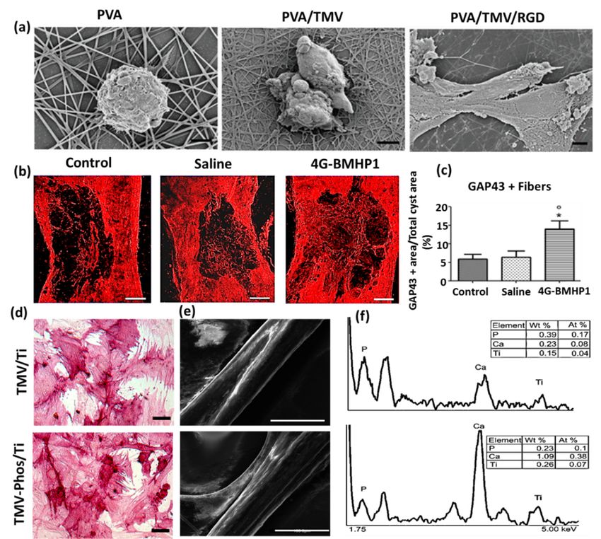
Figure
Figure 3.3. (a)
(a) Field
Field emission
emission scanning
scanning electron
electron micrographs
micrographs of of Baby
Baby Hamster
Hamster Kidney
Kidney cells
cells after
after 11 hh
incubation
incubation on electrospun nanofibrous substrates PVA, PVA-TMV, and PVA-TMV/RGD. Scale bar bar
electrospun nanofibrous substrates PVA, PVA-TMV, and PVA-TMV/RGD. Scale 2 µm. 2
μm. Reproduced
Reproduced with permission
with permission from [71]. from [71].John
Copyright Copyright
Wiley andJohn Sons,Wiley
2014. (b)and Sons, 2014. (b)
Immunofluorescence
Immunofluorescence
staining of longitudinal staining of longitudinal
sections of total cystsections
area ofin total cystcord
spinal areainjury
in spinal cord injuryfemale
(SCI)-treated (SCI)-
treated female Sprague-Dawley rats during the chronic phase.
Sprague-Dawley rats during the chronic phase. The synthesis of GAP-43 immunopositive fibersThe synthesis of GAP-43
immunopositive
was significantlyfibers
greater wasinsignificantly
bone marrow greater
homing in bone marrow homing
peptide-expressed peptide-expressed
phages (4G-BMHP1) phagestreated
(4G-BMHP1) treated group
group when compared when groups
to control compared (SCItocontrol
controlandgroups (SCIScale
saline). control
barand
400 saline).
mm. The Scale bar 400
percentage
mm. The percentagefibers
of immunopositive of immunopositive fibers
into the cyst area perinto thecyst
total cystarea
areaisper total cyst area
represented is represented
in a bar in a
graph (c) from
bar graph (c) from
six independent six independent
experiment results,experiment
and the valuesresults,
areand the values
reported with are reported
± SEM. with ± factor
Significant SEM.
* p < 0.05, 4G-BMHP1
Significant factor * p ˂vs.0.05,
SCI 4G-BMHP1 vs. SCI control;
control; 4G-BMHP1 vs. saline. 4G-BMHP1
Reproduced vs.with
saline. Reproduced
permission fromwith
[72].
permission
Copyright PLOS, from 2011.
[72]. (d)
Copyright PLOS,
Bright-field optical2011. (d) Bright-field
microscope optical microscope
images of histochemical stainingimages of
of alkaline
phosphatase (ALPL)
histochemical stainingin of
bone marrow-derived
alkaline phosphatase stem cells (BMSCs)
(ALPL) in TMV and phosphate
in bone marrow-derived grafted
stem cells TMV
(BMSCs)
(TMV-Phos)-coated
in TMV and phosphate Ti substrates
grafted TMVafter(TMV-Phos)-coated
14 days of culture. BMSCs have been
Ti substrates aftershown
14 daystoofform neighboring
culture. BMSCs
well-spread
have cells under
been shown osteogenic
to form conditions,
neighboring which are
well-spread stained
cells under highly positiveconditions,
osteogenic for ALPL. Scale
whichbar is
are
100 µm.highly
stained (e,f) SEM micrographs
positive for ALPL.and EDX
Scale baranalyses
is 100 μm.of TMV
(e,f) and
SEMTMV-Phos
micrographs coating on Tianalyses
and EDX substrates.
of
Scale bar
TMV andfor SEM is 100coating
TMV-Phos µm. Reproduced with permission
on Ti substrates. Scale barfrom for [73].
SEM Copyright
is 100 μm. Elsevier, 2010. with
Reproduced
permission from [73]. Copyright Elsevier, 2010.
2.2. Virus-Incorporated 2D Films and Nanofibers
The phosphate biomaterial
A combinatorial tailored TMV nanoparticle
consisting was cyclic
of PVX-based demonstrated to induce
RGD, containing expression
filament (RGD-PVX)of
osteospecific genes of
and polyethylene rat bone
glycol marrow stealth
conjugated stem cells (BMSCs),
filament including osteocalcin
(PEG-PVX), and osteopontin,
was developed to analyze
when comparedintomice
biodistribution unmodified
xenograftTMV nanoparticles.
models. As shown
The comparative in Figure
studies 3d–f, the that
demonstrated enhanced cell
PEG-PVX
was preferentially accumulated into tumor cells, while RGD-PVX was trapped into the lung site in
a large quantity. It has been reported that the filamentous and elongated nanoparticles are moreNanomaterials 2019, 9, 1014 10 of 18
advantageous in drug targeting than the spherical counterparts. Non-spherical nanoparticles present
more ligands on their surfaces and show significant accumulation towards the vessel wall, improving
the efficiency of tumor homing. Owing to the flexible nature of viral capsid, PVX-based nanoparticles
could pass through restrictions in the complex biological environments and permeate into tissue cells
without difficulty [74]. Wu et al. [56] successfully synthesized TMV-based electroactive nanofibers for
neural tissue regeneration from the blend of polyaniline (PANI) and sodium polystyrene sulfonate
(PSS). The morphology of the TMV/PANI/PSS nanofiber has been shown through TEM micrograph in
Figure 2b.
An electrospun nanofiber of blends of polyvinyl alcohol (PVA) and TMV/RGD afforded higher cell
density of baby hamster kidney (BHK) cells in culture. The enhanced cell adhesion and spreading and
the formation of F-actin filaments were observed more on PVA/TMV/RGD nanofiber than PVA and
PVA/TMV nanofibrous substrates, which were noticed in SEM micrographs (Figure 3a). The resulting
nanofiber provided electroactivity and topographical cues to the neural cells and was reported
to augment the length of neurites, increase the population of cells, and lead the cellular bipolar
morphology more than TMV-based non-conductive nanofibers [71]. Korehei et al. [57] produced
a virus incorporated nanofiber by electrospinning the blends of polyethylene oxide (PEO) and T4
bacteriophage suspension. The SEM measurement showed that T4 bacteriophages were protected
from severe electrospinning conditions, as they were concentrated within the alginate capsule, as can
be seen in Figure 2c. The alginate beads containing phages were found to exhibit smooth rounded
surfaces. The size of the electrospun nanofiber of PEO/alginate/T4 bacteriophages had an average
diameter 500 ± 100 nm (Figure 2d). According to TEM measurement, the capsules of T4 bacteriophages
were distributed without uniformity throughout the fiber matrix (Figure 2e).
The induced pluripotent stem cells (iPSCs) are a promising cell source, which can rise to different
cell lineages and construct a well-developed functional bone substitute. However, there is a challenge
in osteoblastic differentiation of iPSCs by a conventional biomaterial, as it may form teratoma,
raising health risks. Wang et al. [75] demonstrated that a phage (M13)-based nanofiber with four
different signal peptides aiming to influence stem cell fate could be potentially utilized for bone
tissue regeneration. The aligned nanofibrous matrix provided biochemical and biophysical cues to
the cells promoting differentiation of iPSCs into osteoblasts. Among the signal peptides investigated
by them were two adhesive-directing peptides RGD and RGD/PHSRN from fibronectin, and the
remaining two included ALKRQGRTLYGFGG and KIPKASSVPTELSAISTLYL sequences, which are
the growth regulating peptides from osteogenic growth factor and bone morphogenetic protein 2
(BMP2), respectively. The layer-by-layer technique produced a phage-assembled nanofiber assuming
nanotopography of the ridge-groove structure, wherein the phage strands were parallel to each other but
separated by grooves. Due to this specialized nanotopography of material, the occurrence of controlled
osteoblastic differentiation was observed, even in the absence of osteogenic supplements. The research
group reported that the phages displaying growth factor signal peptides could express a higher
level of alkaline phosphatase (ALP) than the phages having adhesive signal peptides on the surface.
The in vivo animal studies disclosed that iPSCs alone caused teratoma after one month of cells injection
into nude mice, whereas the group of iPSC-derived osteoblasts did not. Cigognini and co-workers
engineered an electrospun nanofibrous scaffold dispersing phage-displayed bone marrow homing
peptide (BMHP (1) with sequence PFSSTKT and investigated its potential use in a chronically damaged
spinal cord, which was caused by the degeneration of the central nervous system [72]. The clinical
data showed that the biomimetic material enhanced nervous tissue regeneration, owing to porosity
and nanostructure at the microscopic level, and improved the locomotor recovery of experimental
rats. From Figure 3b,c, the histological analyses revealed that the scaffold affected increased cellular
infiltration and axonal regeneration after eight weeks of experimental investigation in rats. They found
a higher synthesis of growth-associated protein 43 (GAP-43) in engineered scaffold-treated animal
when compared to saline and control groups with spinal cord defects. Our research group has explored
electrospun nanofibrous matrices of PLGA containing self-assembled M13 bacteriophages along withNanomaterials 2019, 9, 1014 11 of 18
additives RGD and graphene oxide to show enhanced differentiation of fibroblasts, smooth muscle
cells, and myoblasts [76–80].
2.3. Virus-Incorporated 3D Hydrogel Scaffolds
Cell-laden-agarose hydrogel was prepared by dispersing genetically engineered rod-shaped PVX
nanoparticles, which present functional RGD peptides and mineralization inducing peptides (MIP) on
its surface, into agarose polymeric components [81]. Luckanagul et al. [59] prepared freeze-dried solid
foam of a porous alginate hydrogel (PAH) comprising TMV. The incorporation of TMV nanoparticles
resulted in large sized and well-defined spherical pores (100–500 µm) in TMV/PAH, analyzed by Field
Emission Scanning Electron Microscope image (Figure 2g).
The PVX nanoparticles adopted a nano-filamentous structural network on coated surfaces.
Exploiting the synergistic effect of both peptides, the PVX nanoparticles in hydrogel expressed
significant cell adhesion as well as hydroxyapatite nucleation. Confirmed by SEM and immunostaining
characterizations, it was further reported that the viral nanoparticles could be preserved over 14 days in
hydrogel and the whole biomaterial could act as a promising bone substitute. Maturavongsadit et al. [82]
developed an injectable TMV based hydrogel under physiological conditions to imitate a cartilage
microenvironment. The hydrogel was prepared by cross-linking methacrylate hyaluronic acid polymers
by cysteine inserted TMV mutants involving in situ Michael addition reaction. The hydrogel was
reported to influence enhancement of cartilage tissue regeneration by promoting chondrogenesis via
up-regulation of BMP-2. The interaction of TMV nanoparticles with the cells assisted the high-level
expression of BMP-2, an effective inducer of differentiation of mesenchymal stem cells into chondrocytes.
Luckanagul et al. [58] investigated the performance of functional TMV-RGD-blended
alginate hydrogel nanocomposites to treat in vivo cranial bone defects in Sprague-Dawley rats.
The TMV-functionalized sponge-like hydrogel supported cell localization without triggering any
systemic toxicity in the defect area, and hence was envisaged as an active bone replacement biomimetic
material in the future direction of reconstructive orthopedic surgery. Shah et al. [83] studied an integrated
co-assembled hydrogel system of peptide amphiphiles, in which M13 phage coat protein was modified
to express a high density of binding peptide HSNGLPL to combine with transforming growth factor
β1 (TGF-β1). The research group found an enhancement in articular cartilage tissue regeneration in
a rabbit model with a full-thickness chondral defect because of the slower release of growth factor
from the hydrogel, with approximately 60% of cumulative drug release at 72 h, which supported the
viability and chondrogenic differentiation of mesenchymal stem cells in the defective site. The in vivo
evaluation of the rabbit model showed that the hydrogel treated animal group had no apparent
symptoms of chronic inflammatory responses after four weeks. All of the rabbits appeared with
a full range of motion in their knees at the end of the investigation. Caprini et al. [84] isolated M13
phage-displayed peptide, KLPGWSG, which could adhere on the surface of murine neural stem cells.
Subsequently, the research group designed a self-assembled KLPGWSG-based biomimetic hydrogel
with tunable visco-elastic properties for the regeneration of the degenerated nervous system. It was
discovered that the phage-based hydrogel favored cell adherence and differentiation in the range of
100–1000 Pa, suggesting that the elastic property of the matrix is a crucial factor in tissue regeneration.
2.4. Virus-Incorporated Organic-Inorganic Hybrid Nanocomposites
The interaction of organic and inorganic biocompatible materials in scaffolds bring about
significant impacts in biomedical applications. Cementum, classified as a hard mineralized tissue,
surrounds tooth root and has been a part of periodontal tissue that connects the tooth to the bone.
When an infectious biofilm adheres to tooth root, triggering periodontal disease, the tooth loss is
more enhanced. Gungormus et al. [85] demonstrated amelogenin-derived M13 phage-displayed
peptide controlled hydroxyapatite biomineralization for dental tissue regeneration. It was reported that
Amelogenin directed hydroxyapatite to form a protein matrix during the formation of enamel. Hence,
the research group synthesized the cementomimetic material by applying an aqueous solution of theNanomaterials 2019, 9, 1014 12 of 18
amelogenin-displayed peptide on the human demineralized root surface to form a layer, which was
subsequently immersed into the solution of calcium and phosphate ions. Ramaraju et al. [86] isolated
M13 phage-displayed peptides to design a dual functional apatite-coated film for effective bone tissue
regeneration. They reported that one peptide sequence of the phage, VTKHLNQISQSY, had mineral
(apatite) binding affinity with 25% hydrophobicity, whereas another peptide, DPIYALSWSGMA,
had cell binding affinity with 50% hydrophobicity. Also, they discovered that the dual functional
apatite-based biomaterial could stimulate the adhesion strength of human bone marrow stromal cells
(hMSC) and subsequently increase cell proliferation and differentiation. Due to the mineral binding
affinity, the film provided a platform for the adherence of osteogenic cells with osteoconductive and
osteoinductive signals. Further, the biomimetic nanocomposite showed a greater extent of proliferation
of hMSCs with an elevated level of Runx2 expression when compared to biomimetic apatite without
functional peptides.
Wang et al. [58] prepared a 3D-printed biomimetic nanofiber with M13 phage-displayed RGD
peptides residing in the pores of the scaffold to enhance bone tissue regeneration. The nanocomposite
consisted of hydroxyapatite and tri-calcium phosphate showing an ordered pattern with interconnected
micro and macro scale pores, which are shown in the TEM micrograph (Figure 2f). The research group
implanted a MSC-seeded biomimetic scaffold into a rat radial bone defect and discovered that the
order of regeneration was found as follows: scaffold filled with modified phages > scaffolds filled with
wild-type phages > pure scaffold. He et al. [60] carried out a similar kind of research work, genetically
modifying M13 phage to express oligonucleotide encoding E8 and inducing self-assembly followed by
oriental mineralization to synthesize nanofibrous biomimetic materials under the influence of divalent
calcium ions. The resulting mineralized phage bundle has been shown in TEM micrography (Figure 2h).
Wang et al. [61] used Ca2+ ions to prompt self-assembly of fd phage-based anionic nanofibers and
transform them into a bundle sheet (Figure 2i), which provided insights into biomineralization and
fabrication of organic–inorganic hybrid nanocomposites. The divalent ion-triggered bundle not only
acted as a biotemplate but also served as a Ca source to initiate the ordered nucleation and growth of
crystalline hydroxyapatite in the biological fluid.
3. Other Formulations of Virus-Based Nanocomposites with Different Biomedical Applications
Apart from tissue regeneration, virus-based biomimetic nanocomposites have traced their steps in
different biomedical applications, such as drug delivery, bioimaging, and biosensing. Wang et al. [87]
studied f8/8 phage-based polymeric micelles from the self-assembly of polymeric PEG- diacyl lipid
conjugates. These polymeric micelles were reported to have cell-targeting ability to release less
water-soluble drugs with more specificity towards breast cancer Michigan Cancer Foundation-7 (MCF-7)
cells. The non-toxic filamentous f88.4 bacteriophage viral nanoparticle, which was designed to display
a single chain antibody, delivered the vectors to the different regions of the brain in albino, laboratory-bred
nude mice (BALB/c), and hence was proposed for treating Alzheimer’s disease with early diagnosis [88,89].
Wang et al. [90] studied a M13 phage-displayed peptide with the sequence HSQAAVP to target fibroblast
growth factor 8b (FGF8b) to treat prostate cancer. The genetic level disturbances in homeostasis between
prostate epithelial and stromal cells cause prostate cancer. The major isotherm of fibroblast growth factor
8 is FGF8b, which is associated with the stages of prostate cancer and has been a potential target for
appropriate therapies. In this study, the research group revealed that the biomimetic material interrupted
FGF8b binding to its receptors, and thereby prevented FGF8b-induced cell proliferation. Furthermore,
they reported that the biomaterial had the potential to arrest the cell cycle at the phase G0/G1 by suppressing
cyclin D1 and proliferating cell nuclear antigens (PCNA).
Carrico et al. [43] chemically modified the amino acid residues present on the surface of filamentous
fd phage coat protein following a two-step transamination/oxime reaction for its potential use in
characterizing breast cancer cells. The research group discovered that the chemical reaction selectively
targets N-terminal groups but is not involved in transamination of lysine ε-amines. They conjugated
PEG polymeric chains to the phage protein in order to reduce immunogenicity, decrease non-specificNanomaterials 2019, 9, 1014 13 of 18
binding, and increase solubility in the aqueous environment. They observed that there were no
significant differences in either absorption or emission properties after fluorophores were labeled
with polymer conjugated phages. Fan et al. [91] isolated cyclic peptide CAGALCY from T7 phage
nanoparticles in order to target the pial microvasculature of the brain and inhibit platelet adhesion.
The presence of the bulky hydrophobic core, two cysteine residues at each end, and the tyrosine
residue at the carboxy terminus are considered as remarkable features for selectively binding the brain
microvasculature. When pharmacokinetic properties were assessed, the non-filamentous phage, T7,
showed a fast clearance rate from the blood with a half-life of 12 min, whereas the filamentous phages
M13 and fUSE5 had longer half-lives of 7 h and 9 h, respectively. To identify the specificity of the T7
phage-displayed peptide, they determined selectivity indices using plaque assay for various organs of
mice, including lung, liver, brain, kidney, colon, small intestine, and large intestine. The characterization
results exposed that T7 displayed peptide resided (accumulated) in the brain, with a selectivity index
of 1000, whereas other organs possessed low specificity for the peptide, with selectivity indices less
than 50.
Bean et al. [92] prepared a bacteriophage K (ΦK) by incorporating the virus into a photo cross-linked
hyaluronic acid methacrylate (HAMA)-based hydrogel that resulted in a material with antimicrobial
properties. The presence of two zinc finger genomes (CX2 CX22 CX2 C and CX2 CX23 CX2 C) in the virus
caused it to be virulent against a wide range of infective Staphylococci. The secretion of hyaluronidase
enzyme-mediated S. aureus sensitizes HAMA and triggered degradation of the hydrogel, facilitating the
release of ΦK at a sustained level to inhibit bacterial growth effectively. This stimuli-responsive hydrogel
was shown to reduce pain, promote cell migration and tissue hydration in the wound site, and was
suggested for the application of dermal tissue regeneration. Schmidt et al. [93] identified two different
adenovirus phage-displayed peptides QTRFLLH and VPTQSSG to target neural precursor cells
(NPC) in the hippocampal dentate gyrus of adult mice through adenovirus-mediated gene transfer.
The peptides were found to be strongly internalized into NPCs when the investigated material was
added to neurosphere culture containing clusters of neural stem cells.
Kelly KA et al. [94] isolated high-throughput fluorochrome-labeled M13 phage particles (Ph.D. C7C
library) to rapidly identify ligands of biological interest in vivo using secreted protein acidic and rich in
cysteine (SPARC) molecules and vascular cell adhesion molecules-1 (VCAM-1). The engineered phage
particles led to higher sensitivity with an attachment of 800 fluorophores per phage. Wan et al. [37]
developed an f8/8 phage-based biosensor exploiting magnetoelastic wireless detection system.
The genetically modified phage-expressed peptide sequence EPRLSPHS on the surface of the target
biological agent, Bacillus anthracis spore. The resonance frequency of the sensor decreased gradually
depending on the binding agent on the surface. They reported that this affinity-based phage-displayed
biosensor exhibited more longevity activity as a diagnostic probe to target numerous agents with more
efficiency than antibody-based biosensors.
4. Conclusions and Perspectives
The potential application of virus-incorporated biomimetic nanocomposites in the form of
self-assembled nanoparticles, nanofibers, hydrogels, and organic–inorganic hybrids in the field of
tissue regeneration has been elucidated in this review. Though virus-based biomaterial has displayed
many beneficial properties, there are some issues to be addressed. (1) Many research groups have
expressed desired peptides on the surface of phage-based viral nanoparticles exploiting phage libraries.
However, whether the number of peptides exhibited by each nanoparticle is the same is questionable.
(2) Biodistribution of viral nanoparticles in different organs of animal tissues has been studied by
some researchers. Still, a comprehensive study to describe bioavailability must be demonstrated.
(3) It has been well documented that viral nanoparticles contribute to the enhancement in tissue
regeneration. However, a systematic study is required to explain the phases of tissue regeneration,
in which viral nanocomposites contribute more. (4) The viral nanocomposites in the form of polymeric
micelles, vesicles, and dendrimers are less formulated and have not been explored enough forYou can also read